1. Introduction
Research in pharmaceutical and biomedicine faces the great challenge of increasing drug resistance, which endlessly forces the research community to implement new therapies [
1]. In fact, resistant microbes with present medicines are weakly managed and constitute a major public health problem [
2].
The oral cavity contains the habitat of numerous bacteria, whose synergy and interaction contributes to combat the invasion of external pathogens [
3]. On the other hand, the human oral microbiome database (HOMD) reports more than 700 prokaryotic different species that coexist in the oral cavity that could negatively influence the health of human body and could also be involved in the progression of many infections and systemic pathologies [
4].
Antibiotics are generally used to tackle the infectious states of the oral cavity, but recently the use of antimicrobial peptides (AMPs) is being considered. AMPs are a wide and diverse family of small amino acid peptides produced by the immune system of several species ranging from plant and animal kingdoms to all orders of life. AMPs play a protective function by establishing a defense system that may react in a fast and efficient manner [
5]. They have a broad-spectrum activity against microorganisms, a low aptitude to induce resistance [
6,
7], and are excellent candidates for clinical exploitation [
8,
9]. However, only few AMPs have to date received the certification for entry into the market and for clinical use. Two lead examples are the magainin derivative (MSI-78), used as a topical cream for the treatment of foot infections in diabetes [
10], and indolicidin (CP-226), used against severe acne and skin infections with
Staphylococcus aureus (
S. aureus) resistant to methicillin [
11]. AMPs mechanism of action is the disruption of the lipid bilayer integrity, leading to leakage of the intracellular content and death of the bacterium.
AMPs are more specific and efficient than common antibiotics and are related to lower cell and tissue toxicity [
12]. Nowadays, we know of more than 40 antimicrobial peptides in the oral cavity and many of these play a pivotal role in periodontal diseases as well as in orthodontics [
13,
14].
On the other hand, thanks to their powerful antimicrobial activity, metal nanoparticles (NPs) have gained growing interest in the treatment of resistant pathogens. In this context, the combined use of AMPs and metal NPs seem to be very versatile since they hold the promise of interacting with different targets, therefore becoming of huge interest as an attractive alternative to antibiotics [
15]. Moreover, microorganisms are unlikely to develop resistance against nanosystems, which attack multiple targets in the microbe.
Together with AMPs, several metal NPs that represent a possible solution for the control of drug-resistant bacterial infections has also evolved [
16,
17]. Among metal NPs, silver NPs (AgNPs) have received attention thanks to their potent broad spectrum antimicrobial activity. Since ancient times, the antimicrobial characteristics of silver have been studied and used for various purposes, primarily to improve tissue repair, fight infections, preserve drinking water, and prevent food spoilage. Silver use became less pronounced since the introduction of antibiotics. However, the recent influx of multidrug resistance it has become so potentially fatal that silver NPs have produced an astonishing return as an alternative or supplementary medication [
18], opening up a whole pathway to new tools to combat a wide range of microorganisms [
19,
20,
21,
22,
23].
The key to silver NPs broad and potent antibacterial activity is the multifaceted mechanism by which they have an effect on microbes. These mechanisms involve the disruption of ATP production and DNA replication due to gradual release of silver ions, the direct damage of cell membranes, and the generation of reactive oxygen species [
24,
25,
26,
27,
28]. The mechanisms are likely to participate in their reported wide range of antimicrobial activity.
In order to enhance their antimicrobial effects, AgNPs were used together with some antibiotics such as penicillin G, amoxicillin, erythromycin, and vancomycin [
29,
30]. Gade et al. [
31] demonstrated that this combined use was able to strengthen the nanoparticles’ antibacterial activity. The bactericidal potential of NPs synthesized from the leaf extract of
Murrayakoenigii, an Indian curry leaf tree, alone and in combination with commercial antibiotics (gentamycin, ampicillin, tetracycline, and streptomycin) was investigated against
Escherichia coli,
S. aureus and
Pseudomonas aeruginosa. The antibiotics’ potential was effectively increased by combining NPs, making their use possible also against antibiotic-resistant pathogens [
32].
Another possible approach to the effective treatment of microbial infections may be represented by the combination of drugs with different mechanism of action [
33,
34]. The combination of AMPs and AgNPs can aid in achieving enhanced antimicrobial activity specificities, efficiencies, and strengths with reduced toxicity, and may be an effective alternative to antibiotics. The aim is not only to promote the synergy of AMP and AgNPs in the bactericidal action, but also to overcome the eventual solubility problems of the peptide and to decrease NPs toxicity on healthy cells. The antimicrobial peptide selected for the present study is indolicidin (Ile-Leu-Pro-Trp-Lys-Trp-Pro-Trp-Trp-Pro-Trp-Arg-Arg-NH2), a 13-residue cationic peptide with extremely high tryptophan content. This short peptide was isolated from cytoplasmic granules of bovine neutrophils [
35] and has been shown to possess a broad-spectrum activity against both Gram-positive and Gram-negative bacteria [
36], and also against fungi [
37,
38] and HIV-1 [
39]. Nevertheless, this peptide is endowed of residual cytotoxicity and haemolytic activity. Here, we report on the preparation of AgNPs coated with the indolicidin peptide and antibacterial experiments performed on the Gram-positive bacterium
S. aureus and the Gram-negative bacteria
Escherichia coli (
E. coli) and
Pseudomonas aeruginosa (
P. aeruginosa). It has been widely reported that Gram-negative bacteria, such as
P. aeruginosa, seem to be responsible for the Oral squamous cell carcinoma progression [
40], whereas
S. aureus bacteria are one of the major species that can be related to denture stomatitis [
41]. Other pathogens such as
Chlamydia trachomat is induce the production of inflammatory cytokines in association with growth factors involved in epithelial mesenchymal transition, a potential mechanism correlated with cancer progression [
42,
43,
44]. The interplay between the pathogens’ steroid receptors and neoplastic progression is a crucial node in the development on inflammatory pathologies, which represent some of main focus in every day dental practice [
45,
46,
47,
48,
49,
50].
2. Materials and Methods
2.1. Materials
Rink amide p-methylbenzhydrylamine (MBHA) resin, connection reagents, and Fmoc-protected amino acid derivatives were acquired from Calbiochem-Novabiochem (Laufelfingen, Switzerland). The rest of chemicals were provided by Sigma–Aldrich, Fluka (Buchs, Switzerland).
2.2. Peptide Synthesis
Indolicidin was synthesized using the standard solid-phase-9-fluorenylmethoxycarbonyl (Fmoc) method as previously reported [
51].
2.3. Preparation of Silver Colloids Using Hydrazine
150 μL and 10 μL hydrazine monohydrate (N
2H
4∙H
2O, Sigma Aldrich) were added to 1 mL AgNO
3 solution (1 mM). Subsequently, the solution was topped up to 2 mL with deionized water, thoroughly mixed for 1 min and allowed to settle at room temperature for 4 h. Hereafter, silver colloids were prepared in the presence of indolicidin. For this purpose, 150 μL and 10 μL N
2H
4∙H
2O were added to 1 mL AgNO
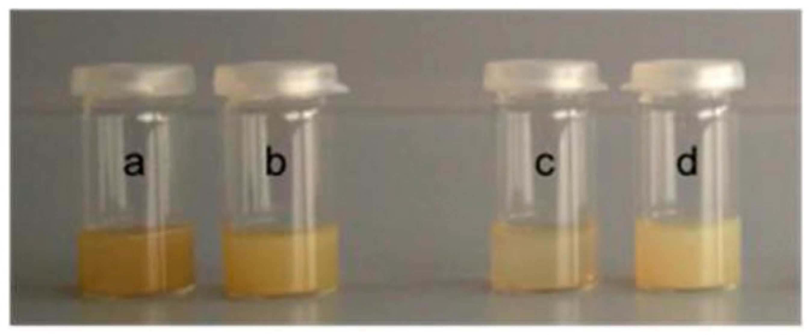
3 solution (1 mM). The solution was topped up to 2 mL with indolicidin solution (concentration of indolicidin in deionized water = 560 μg/mL), mixed, and allowed to settle at room temperature as outlined above. The obtained silver NPs were named as follow (
Table 1): AgNP1 or AgNP2 for naked NPs, where 1 stands for the preparation with 150 μL of N
2H
4∙H
2O and 2 stands for the preparation with 10 μL of N
2H
4∙H
2O, and IndAgNP1 or IndAgNP2 for silver colloids incorporating the peptide indolicidin and 1 or 2, as stated above (
Figure 1).
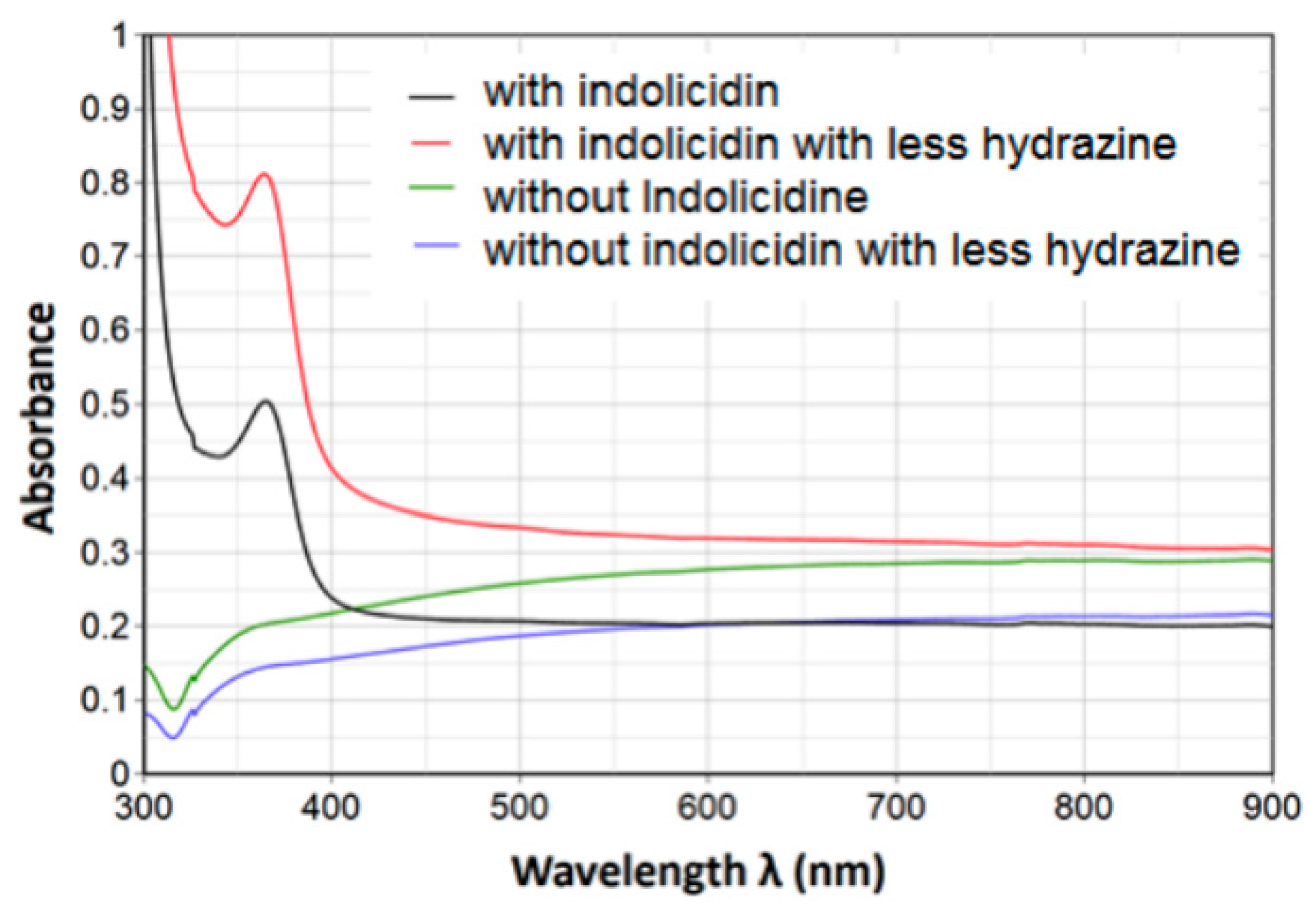
2.4. Characterization UV-VIS
UV-visible spectra of the resulting nanoparticle solutions were recorded at room temperature using a Lambda 25 UV-Vis spectrophotometer (Perkin Elmer, Milan, Italy). The monochromator slit width was 10 nm.
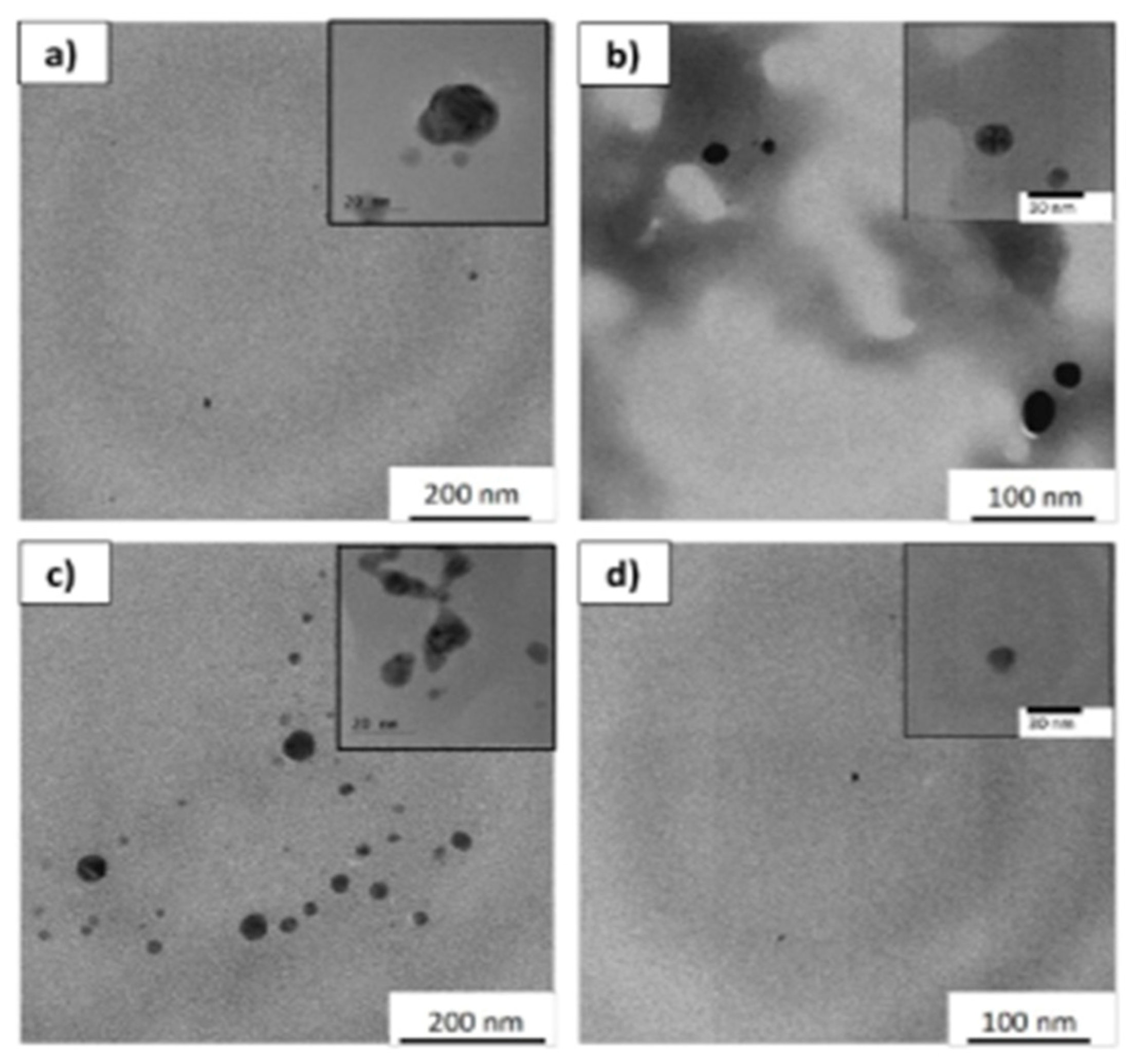
2.5. Characterization TEM
Silver NPs’ morphology was analyzed using a Transmission Electron Microscope (TEM Joel 2200 fs) operated at 200 keV. Prior to TEM measurements, 0.3 mL of each sample were spotted onto a carbon-coated copper grid (300 mesh, Science Services, München, Germany). The primary particle size was measured using the ImageJ software package (Version 1.51p).
2.6. Microorganisms
For the antimicrobial assays, E. coli ATCC 11219, P. aeruginosa ATCC 13388, and S. aureus ATCC 6538 were used. Fresh colonies of each strain were cultured on Mueller Hinton Agar (MHA, Oxoid) and grown overnight. Then, they were inoculated in MH Broth (MHB) for another night. Subsequently, hundred-fold dilutions of the bacterial suspension were resuspended in fresh medium and further incubated at 37 °C. When the inoculum was turbid, it was resuspended in 0.9% sterile saline solution to reach an appropriate OD600 value (with a Bio-Rad Microplate Reader—Bio-Rad Laboratories, Hercules, CA, USA) corresponding to a concentration of about 1 × 10⁸ CFU/mL. This standardized inoculum was diluted 1:10 in MHB and the inoculum size was confirmed by colony counting.
2.7. Antimicrobial Activity Assays
Susceptibility testing was performed using the broth microdilution method outlined by the Clinical and Laboratory Standards Institute using sterile 96-well microtiter plates (Falcon, NJ, USA). Starting from the concentration of 1014 NPs/L, two-fold serial dilutions of NPs were prepared together with the peptide indolicidin (IndAgNP1, IndAgNP2, AgNP1, and AgNP2) in Cation-Adjusted MHB (CAMHB) at a volume of 100 µL/well. Each well was inoculated with 5 µL of the standardized bacterial inoculum, corresponding to the final concentration of about 5 × 10⁵ CFU/mL. Antimicrobial effect was expressed as the Minimum Inhibitory Concentration (MIC), which is the lowest concentration of compound that completely inhibited visible growth after 24 h of incubation at 37 °C. All experiments were carried out in triplicate and standard deviations are reported.
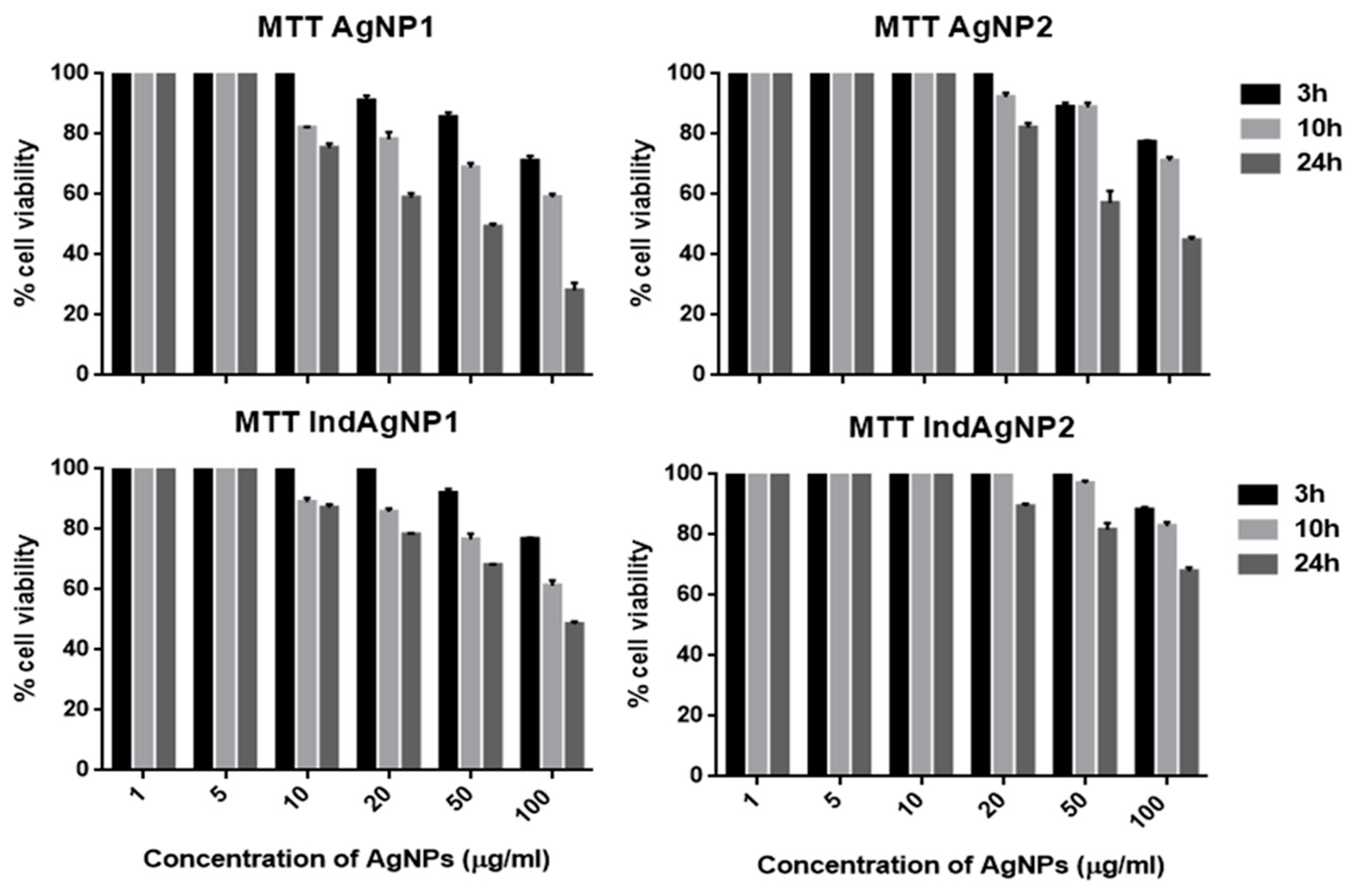
2.8. Cytoxicity
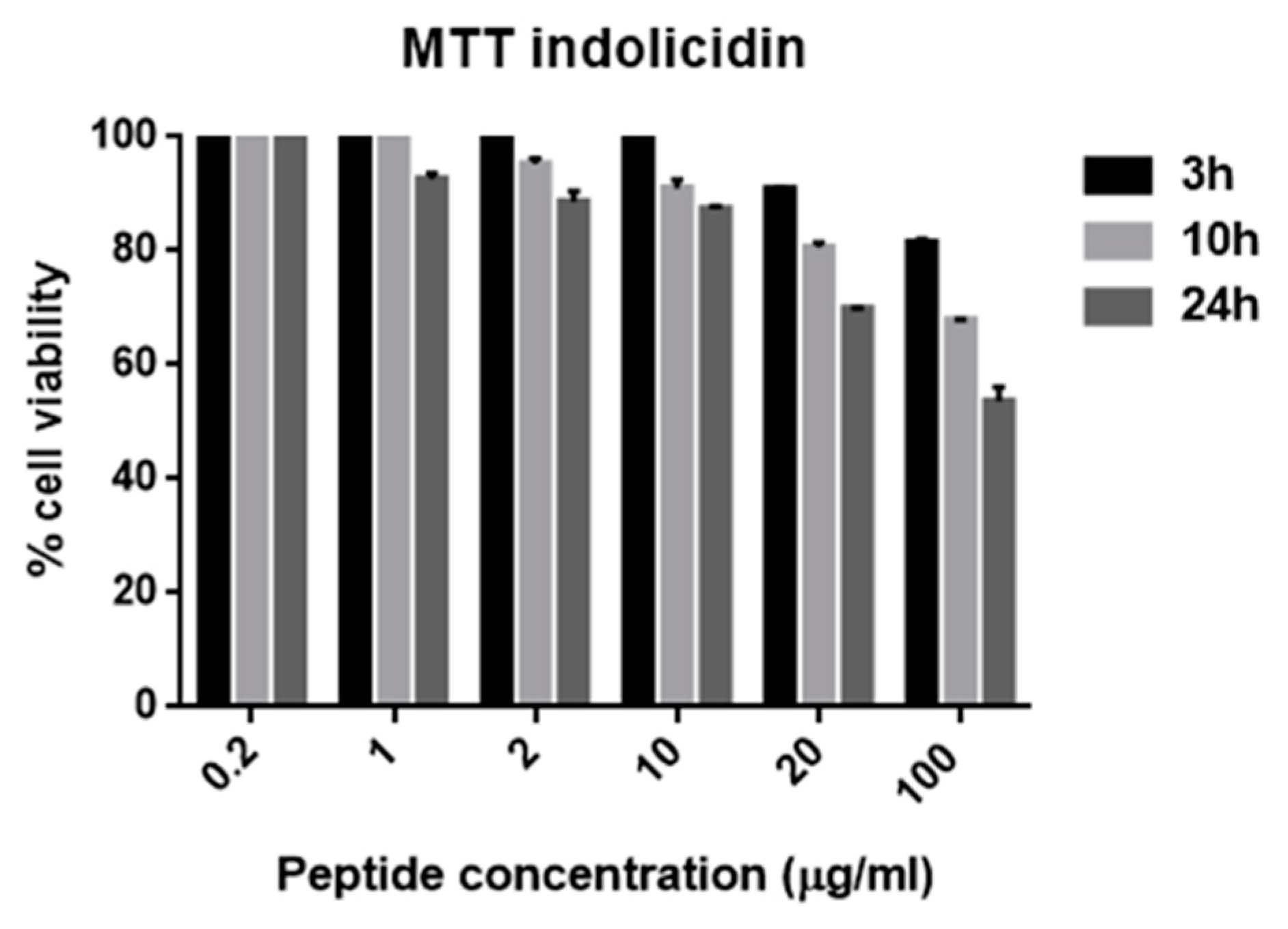
Vero cells were treated with increasing concentrations of compounds, and the cell viability was analyzed through the 3-(4,5-dimethylthiazol-2-yl)-2,5-diphenyltetrazolium bromide (MTT) assay. Vero cells were cultured in 96-well plates (2 × 10⁴ cells/well), and after 24 h were treated with a series of concentrations (from 1012 to 1014 NPs/L) of filter sterilized NPs (IndAgNP1, IndAgNP2, AgNP1, and AgNP2) and peptide indolicidin for 3, 10, and 24 h. The medium was then removed, MTT solution was added, and cells were incubated for further 3 h at 37 °C. Formazan crystals were dissolved with dimethyl sulfoxide and the absorbance was measured at 570 nm using a Bio-Rad Microplate Reader. All experiments were repeated three times and standard deviations are reported.
2.9. Hemolytic Assay
The hemolytic activity of filter sterilized NPs (IndAgNP1, IndAgNP2, AgNP1, and AgNP2) and peptide indolicidin were determined using fresh human erythrocytes from healthy donors. The blood was centrifuged, and the erythrocytes were washed three times with 0.9% NaCl. The NPS and peptide were applied to the suspension of the erythrocyte (5% [vol/vol]), at the concentrations used for the MTT and for the volume of 100μL. The samples were incubated with agitation at 37 °C for 60 min. The release of hemoglobin was monitored by measuring the absorbance (Abs) of the supernatant at 540 nm. The percentage of hemolysis was calculated using the equation % hemolysis = [(Abssample − Absblank)/(Abstotal lysis − Absblank)] × 100. The control for zero hemolysis (blank) consisted of erythrocytes suspended in the presence 1 × PBS, while the positive control (0.1% Triton) showed a 100% hemolytic effect. For a good statistical analysis and reproducibility the experiments were performed in triplicate. Results are integrated with standard deviations.
4. Conclusions
Nowadays, the growing threat of microbial resistance against traditional antibiotics in oral disease has prompted the mandatory development of alternative therapies, especially against bacterial strains showing resistance to one or more antibiotics [
60,
61]. In this article, silver NPs (AgNPs) were successfully prepared using hydrazine and were further modified by a coating of indolicidin, a well-known AMP.
The prepared AgNPs possessed good, dose-dependent in vitro antimicrobial activity against Gram-positive and Gram-negative bacteria. The better results were achieved with indolicin-coated AgNps, regardless having used either 10 or 150 µL for nanoparticles production. Notwithstanding the moderate antibacterial increase obtained with the coated NPs versus the naked ones, the importance of our results resides in the fact that the amount of indolicidin attached to the NPs was in a range well below the reported concentrations normally used for achieving antibacterial activity. Therefore, the major interest in attaching low concentration AMPs on AgNPs is to reduce the overall toxicity shown by both the naked AgNPs and indolicidin on their own.
The bactericidal effect of silver NPs may be translated into important therapeutic and clinical opportunities in the future, principally in view of the dearth of new antibiotics against the emerging antimicrobial resistant microorganisms, in particular against Gram-negative bacteria.


 ,
, 


