Article

Antifungal and Antioxidant Potential of Methanolic Extracts from Acorus calamus L., Chlorella vulgaris Beijerinck, Lemna minuta Kunth and Scenedesmus dimorphus (Turpin) Kützing

1
Department of Biochemistry, Microbiology and Physics, Faculty of Agriculture, Trakia University, Stara Zagora 6000, Bulgaria
2
Department of Biology and Aquaculture, Faculty of Agriculture, Trakia University, Stara Zagora 6000, Bulgaria
3
Department of Applied Ecology and Animal Hygiene, Faculty of Agriculture, Trakia University, Stara Zagora 6000, Bulgaria
*
Author to whom correspondence should be addressed.
Appl. Sci. 2021, 11(11), 4745; https://doi.org/10.3390/app11114745
Submission received: 29 March 2021 / Revised: 29 April 2021 / Accepted: 17 May 2021 / Published: 21 May 2021
(This article belongs to the Section Applied Microbiology)

Abstract

:
Plant extracts are an important alternative to antibiotics, which are ever more restricted because of their developing microbial resistance and some adverse effects that have been observed following frequent application. The aim of the present study was to determine the antifungal and antioxidant activity of the methanolic extracts of Acorus calamus, Chlorella vulgaris, Lemna minuta and Scenedesmus dimorphus. The antifungal activity of the extracts against strains of Aspergillus flavus, Aspergillus parasiticus, Aspergillus ochraceus, Aspergillus niger, Aspergillus carbonarius, Fusarium graminearum, Fusarium oxysporum, Penicillium chrysogenum and Alternaria alternata was evaluated via the agar well diffusion method. The antioxidant activity of the extracts was measured through the determination of three parameters—total phenolic content, total flavonoid content and radical scavenging potential (determined through UV/Vis analysis). A. calamus extracts had the highest antimicrobial activity against eight fungal strains, followed by the C. vulgaris, L. minuta and S. dimorphus extracts, which were inhibitory against two to three strains. Among the extracts from the species studied, the extract from S. dimorphus showed the highest antioxidant potential, as determined via the DPPH (1,1’-diphenyl-2-picrylhydrazil-radical) method. This correlated to its high total phenolic and flavonoid content. From A. calamus and L. minuta, methanolic extracts were obtained that exhibited similar values of the aforementioned parameters, followed by C. vulgaris extracts, which showed the lowest antioxidant activity. Based on the Pearson correlation coefficients, the impacts of the total phenolic content and the total flavonoid content on radical scavenging capacity are similar, and flavonoids were a significant part of the total phenolic compounds extracted from the plant materials studied.

1. Introduction

Plants are important source of bioactive substances with medicinal significance. These substances are the secondary metabolites of medicinal plants, which are some of the main sources of drugs for the pharmaceutical industry and traditional medicine [1,2]. Such bioactive compounds could also serve as lead molecules for the development of many synthetic drugs [3]. Currently, plants are viewed as the main source for the discovery of new therapeutic compounds [4].
Among the most important medicinal properties of plants is their antimicrobial activity, which serves to protect the plant from existing pathogenic microorganisms. This important property, which is used in medicine, agriculture and the food industry, is due to various substances produced and exuded by the plants—essential oils, alkaloids, phenols, tannins, flavonoids, terpenes, glycosides, saponins, etc. [5,6]. A number of studies have shown that the synergistic effect of phytochemicals plays an important role in the use of plant extracts as antimicrobial agents [7]. Phytochemical agents can act alone or in combination with antibiotics to enhance the antimicrobial activity against a wide range of microorganisms [6]. The concentration of bioactive compounds in each plant species largely depends on the environmental conditions and the pathosystem. The presence of various pathogens induces the production and exudation of the relevant antimicrobial substances [5]. Because of the ongoing development of resistance to existing antibiotics, there is a continuous need for new antimicrobial agents [8]. Moreover, the products of beneficial medicinal plants could be a more natural and safer alternative to synthesized antibiotics and other chemotherapeutics [4,9].
Another important property of medicinal plants is their antioxidant activity, which is due to the high content of natural antioxidants in a plant. Antioxidants are used to preserve food quality, mainly through the prevention of the oxidative deterioration of lipid constituents. Nowadays, the most commonly used antioxidants are butylated hydroxyanisol (BHA), butylated hydroxytoluene (BHT), propyl gallate (PG) and tetra-butylhydroquinone (TBHQ) [2,10]. However, there is a tendency to restrict the use of butylated derivates because of the potentially resultant liver damage and carcinogenesis [11]. Therefore, the search for effective antioxidants of natural origin is a priority nowadays. These antioxidants could protect the human and animal body by scavenging free radicals and preventing the subsequent oxidative damage that induces degenerative and pathological processes, including aging and cancer [12]. Phenols and flavonoids are among the main antioxidant compounds of the plants that are useful not only as pharmaceutical ingredients but also as food preservatives [10].
Acorus calamus L. (Family Acoraceae), commonly known as “sweet flag”, is a renowned semiaquatic medicinal plant native to temperate to subtemperate regions of Europe, India, The Himalayas and Southern Asia. The plant rhizomes of A. calamus are considered to possess a great deal of beneficial properties—they are known to be spasmolytic, carminative, antimicrobial, antihelmintic, insecticidal, vascular modulators, anticancer, antioxidant, memory enhancing, etc. [13,14]. Green microalgae (such as Chlorella vulgaris Beijerinck (family Chlorellaceae) and Scenedesmus dimorphus (Turpin) Kützing (family Scenedesmaceae)) are a diverse group of photosynthetic microorganisms found in soil and fresh water environments. They have the ability to produce a wide range of active substances with antimicrobial, immunostimulant, cytotoxic, antioxidant, anticoagulant, anti-inflammatory, antiallergenic and enzyme-inhibiting activities [15,16,17]. Lemna minuta Kunth (family Araceae) is an aquatic plant with a small size commonly known as “duckweed”. It is native to parts of the Americas, and because of its ever-expanding distribution, it is found on other continents, including Europe. Although the plants of Lemna spp. are not extensively studied, a number of beneficial properties are still known—it is antimicrobial, antioxidant, anti-inflammatory and immunomodulatory [18,19,20].
In the available literature, there are some studies on the antioxidant activity of A. calamus and C. vulgaris, but antifungal experiments on these plants are not abundant. Moreover, the antioxidant and antifungal properties of L. minuta and S. dimorphus are not studied, with the exception of the experiment of Velichkova et al. [19], who established the activity of the ethanolic and methanolic extracts of L. minuta on four mold strains. The aim of the present study is to determine the antifungal and antioxidant activity of the methanolic extracts of A. calamus, C. vulgaris, L. minuta and S. dimorphus.

2. Materials and Methods

2.1. Plant Material and Extract Preparation

Plant materials of L. minuta were collected from the town of Banya located in the Plovdiv region (42°32′226″ N 24°50′213″ E). The plant had been developing in a small (about 30 m2) warm swamp formed by a hot mineral water flowing out from a “Bancheto” bath. The voucher specimens of L. minuta were kept in the herbarium of Agricultural University in Plovdiv, Bulgaria (SOA 061400). C. vulgaris (SKU: 100-CVC00-50), and S. dimorphus (SKU: AC-1002) were supplied from the Algae depot—USA (www.algaedepot.com accessed on 21 May 2021) in 2013. Algae cultivation was maintained in a laboratory bioreactor of the Department of Biology and Aquaculture of Faculty of Agriculture, Trakia University, in Bold Basal Medium (BBM) [21]. Dried rhizomes of A. calamus were purchased from an herbal and medicinal pharmacy, Sofia (Bilki EOOD), in a paper package of 50 g. The climate of Bulgaria (where L. minuta and A. calamus were harvested from) is transitional continental, with influence from the Mediterranean Sea.
Plant material was airdried in shade at room temperature and ground in a mechanical grinder (with a final powder size of less than 400 μm). The samples were stored in dark and cool rooms at 16–18 °C prior to analysis.
The extracts were prepared via ultrasonication of the powdered plant material in methanol (in a ratio of 1:10) for 30 min at 40 °C in triplicate. Ultrasonic extraction is convenient and straightforward, and it was selected because of the high rate extraction of flavonoids and polar bioactive compounds [22].

2.2. Microorganisms Studied

In this study, reference fungal strains were included—Aspergillus flavus NBIMCC 916, Aspergillus niger NBIMCC 3252, Aspergillus parasiticus NBIMCC 2001, Aspergillus carbonarius NBIMCC 3391, Aspergillus ochraceus NBIMCC 2002, Fusarium oxysporum NBIMCC 125, Fusarium graminearum NBIMCC 2294, Penicillium chrysogenum NBIMCC 129 and Alternaria alternata NBIMCC 109, purchased from National Bank for Industrial Microorganisms and Cell Cultures (NBIMCC), Bulgaria. The fungal strains were stored at 0–4 °C. Prior to use, they were grown on Potato dextrose agar (Biolife, Milano, Italy).

2.3. Antimicrobial Activity

The antifungal activity of the extracts was evaluated via the agar well diffusion method described by Velichkova et al. [19]. In brief, 72 h old fungal cultures were grown on Potato dextrose agar (PDA). 20 mL of PDA was poured in every Petri dish. After solidification, 0.1 mL inoculum of the fungal strains (1–2 × 10⁴ CFU/mL) was introduced on the agar plate surface and the wells were created using a sterile cork borer 6.0 mm in size. The wells were filled with 0.1 mL of the methanolic extract at a concentration of 64 mg/mL. A positive control with amphotericin B (Sigma-Aldrich, St. Louis, MO, USA) at a concentration of 25 μg/mL and a negative control with methanol were performed. An incubation period of 3–5 days at 26–28 °C was maintained.
Antifungal activity was evaluated by measuring the zones of inhibition of microbial growth surrounding the plant extracts in the wells. The zones of inhibition were measured in millimeters. Antifungal activity was assumed in the presence of a growth inhibition zone ≥ 7.0 mm. The tests were performed in triplicate to determine the reproducibility of the results. The complete experiment was carried out under strict aseptic conditions.

2.4. Determination of Total Phenolic Content (TPC)

The collected methanolic extracts were concentrated to a final volume of ca 7 mL through the use of a rotary evaporator under vacuum at 30 °C and transferred into 10 mL volumetric flasks. The dry matter of these methanolic extracts was determined gravimetrically by drying 1 mL of each extract at 120 °C for 6 h. The experimental procedure described by Tzanova et al. [23] was applied for the determination of TPC. In brief, 1 mL of the methanolic plant extract with a concentration of 1 mg/mL (or 1 mL standard solution) was mixed in separate tubes with 5.0 mL of Folin–Ciocalteu’s reagent (1/10 dilution of the commercial reagent using water). Following this, 4 mL of Na2CO3 in water (7.5% w/v) was added and the tubes were left at room temperature for 1 hour. The absorbance at 765 nm was measured against a prepared reagent blank on a Thermo Scientific Evolution 300 spectrophotometer (Thermo Electron Corporation, Madison, WI, USA). Each sample was analyzed in triplicate. Gallic acid (Sigma-Aldrich, St. Louis, MO, USA) solutions in methanol ranging from 0.1 to 10 μg/mL were used to build the calibration curve (R2 = 0.998). The TPC of each sample was expressed as g gallic acid equivalent (GAE) in 1 kg dry matter (dm) of the extract prepared.

2.5. Determination of Total Flavonoid Content (TFC)

TFC was determined via the aluminum trichloride method, using catechin as reference material [24]. In brief, 1.0 mL extract with concentration of 1 mg/mL, 0.3 mL 5% NaNO3 (pure for analysis), and after 5 min, 0.3 mL 10% AlCl3 (pure for analysis) were added in a 10 mL volumetric flask containing 4.0 mL deionized water in this order. After 6 min, 2.0 mL of 1 M NaOH solution was added and the total volume was adjusted up to 10 mL using deionized water. The solution was homogenized and the absorbance was measured against a prepared reagent blank at 510 nm on a Thermo Scientific Evolution 300 spectrophotometer. Standard solutions of (+)-catechin hydrate (Sigma Aldrich, St. Louis, MO, USA) in the concentration range from 10 to 100 mg/L were used to plot the calibration curve. The total flavonoid content was expressed as g catechin equivalent (CE) in 1 kg dm. Each sample was analyzed in triplicate.

2.6. Determination of Radical Scavenging Activity by DPPH Method

DPPH (1,1′-diphenyl-2-picrylhydrazil-radical) was purchased from Sigma-Aldrich (St. Louis, MO, USA). This substance has a single electron on the nitrogen atom and its solution in methanol has an absorption maximum at λ = 517 nm. The mechanism of the DPPH method is based on the reaction between the test compound and DPPH-radical, wherein the potential free radical scavengers reduce DPPH-radical (violet solution) to a yellow colored 1,1′-diphenyl-2- (2,4,6-trinitrorhenyl) hydrazine by donating a hydrogen atom.
The method described by Tzanova et al. [23] was applied to measure the radical-scavenging potential of methanolic extracts from A. calamus, C. vulgaris, L. minuta and S. dimorphus. In brief, 100 µL of methanolic extract (1 mg/mL) was added to 3.9 mL of 100 µ M solution of DPPH in methanol. The absorption at 517 nm was measured 30 min later. Since the composition of the extracts is complex, the results for their radical-binding capacity were compared with those of Trolox (a water-soluble analogue of Vitamin E) and calculated using regression analysis from the linear dependence between the concentration of Trolox and the absorption at 517 nm on a Thermo Scientific Evolution 300 spectrophotometer. The results were expressed as mmol Trolox equivalent (TE) in 1 kg dm of the extract prepared. Each sample was analyzed in triplicate. The radical scavenging capacity was also calculated according to the following equation: DPPH radical scavenging (%) = [(A0 − A)/A0] × 100, in which A0 and A are blank absorption and sample absorption, respectively. The values obtained are expressed as percentages in order to compare to the data published by other researchers.

2.7. Statistical Analysis

All analytical assays were carried out in triplicate and expressed as mean values ± standard deviation (SD). The data were evaluated statistically using analysis of variance (one-way ANOVA), and a Fisher’s Least Significant Difference (LSD) test using Statistica 10 (Statistica for Windows, StatSoft. Inc., Tulsa, OK, USA, 2010).

3. Results and Discussion

A. calamus and C. vulgaris are well known medicinal plants, extensively used and studied nowadays [14,25]. L. minuta and S. dimorphus are promising (albeit less popular) plants with medicinal properties [18,19,26]. As a whole, studies of plant extracts with antimicrobial and antioxidant activities have been increasing over the last decades. Nevertheless, while there are some studies on the antioxidant activity of A. calamus and C. vulgaris, the antifungal effects of these plants are not sufficiently described. Moreover, the antioxidant and antifungal properties of L. minuta and S. dimorphus are not studied, with the only exception being the experiment of Velichkova et al. [19], which evaluated the activities of the ethanolic and methanolic extracts of L. minuta on several mold strains. Because of the major differences of the type of solvents, concentrations of the extracts and methods of extraction reported by various authors, few studies could be used for comparison with our experimental results.

3.1. Antifungal Potential of Methanolic Extracts from Acorus calamus, Chlorella vulgaris, Lemna minuta and Scenedesmus dimorphus

Mycotoxigenic fungi are perhaps the most important pathogens of global significance in the context of food security and safety. They can reduce the quality and quantity of production (e.g., corn, rice, and peanuts) while producing mycotoxin metabolites that could be carcinogenic in both damaged and apparently healthy products [27]. Therefore, strategies to eliminate and inactivate mycotoxins or prevent them from contaminating food and feed are urgently needed. One such strategy is the extermination of mycotoxigenic fungi with the help of antifungal agents, which prevents the appearance of mycotoxins [28,29]. According to the experimental data (Table 1), A. calamus methanolic extracts exhibited antifungal activity against eight of the nine strains of mycotoxigenic fungal species examined, measured in terms of the diameter of inhibition zones (mm, mean ± SD), as follows: F. oxysporum (10.3 ± 0.6), A. flavus (10 ± 0), A. niger (9.3 ± 0.3), F. graminearum (9.2 ± 0.3), A. ochraceus (8.7 ± 0.3), Alt. alternata (8.2 ± 0.3), A. carbonarius (7.8 ± 0.3) and P. chrysogenum (7.7 ± 0.3). Most of the inhibition zones diameters of rhizome extracts are significantly different from the negative control (methanol) values (p ≤ 0.05).
The activity of rhizome extracts was lower than the activity reported by Devi and Ganjevala [3], who found that 200 µL 10% ethyl acetate extract of A. calamus was active against P. chrysogenum, A. niger and A. flavus with inhibition zones of 22 mm, 25 mm and 28 mm, respectively. Kumar et al. [30] found that 10% methanolic extracts of A. calamus inhibited the growth of A. niger with an inhibition zone of 13 mm. Rawal et al. [1] established that very high concentrations of ethanolic extract (250 mg/mL) of A. calamus were active against F. oxysporum with a zone of inhibition of 11.8 mm. Supercritical extracted A. calamus rhizome oil demonstrated inhibitory activity against Aspergillus and Penicillium species, with inhibition zones of 11.5 mm and 10.3 mm, respectively [8]. The available literature is missing data about the activity of A. calamus rhizome extracts against F. graminearum, A. ochraceus, A. carbonarius, A. parasiticus and Alt. alternata.
Methanolic extracts of C. vulgaris demonstrated antifungal activity against A. niger, P. chrysogenum and Alt. alternata (although the levels of antifungal activity were not high), with the diameters of the inhibition zones of 9.2 ± 0.3 mm, 8.3 ± 0.3 mm and 7 ± 0 mm, respectively (Table 1). Velichkova et al. [19] also reported a lack of activity of the methanolic extracts of C. vulgaris against A. ochraceus and F. graminearum. Abedin and Taha [31] found some inhibitory activity of 5% methanolic extracts of Chlorella pyrenoidosa against A. niger and A. flavus, with the diameters of the inhibition zones of 10 mm and 15 mm, respectively, which were higher than those obtained in this study. However, according to Dantas et al. [32], the methanolic extracts of C. vulgaris did not suppress the growth of A. niger and A. flavus. Ghasemi et al. [15] found that the supernatant of C. vulgaris 025 and C. vulgaris 012 inhibited the growth of A. niger, with the diameters of the inhibition zones of 11 mm and 14 mm, respectively, which are consistent with this experiment. However, the supernatant of another strain of this species (C. vulgaris 030) did not inhibit A. niger, thereby showing strain-specific activity. Some authors described a time- and concentration-dependent inhibitory activity of the C. vulgaris supernatant on the mycelial growths of A. niger and F. oxysporum. C. vulgaris supernatant inhibited the growth of Alt. alternata as well, but with a less pronounced time- and concentration-dependent effect [17,33]. Zielinski et al. [34] also found antifungal activity of the aqueous extract of C. vulgaris biomass on A. niger with a very large diameter of the inhibition zone—51 mm. There are no literature data about the activity of C. vulgaris extracts against P. chrysogenum, A. carbonarius and A. parasiticus. The above-mentioned methods of measuring the antifungal activity of C. vulgaris are very diverse, which could explain the differences found with this experiment.
Methanolic extracts of S. dimorphus were inhibitory only against A. niger and P. chrysogenum, with small zones of inhibition—8.2 ± 0.6 mm and 8 ± 0 mm, respectively. The differences with negative control (methanol) values were not statistically significant (Table 1). Ghasemi et al. [15] also reported that the supernatant of Scenedesmus obliquus 019 was active against A. niger, with a zone of inhibition of 8 mm. Abbassy et al. [35] found that the ethanolic and hexane extracts of Scenedesmus sp. showed a large difference between their antifungal activity against F. oxysporum and A. niger, although both extracts were inhibitory towards these fungi. Contrary to the aforementioned results, Abedin and Taha [31] established a lack of activity of 5% methanolic extracts of Scenedesmus quadricauda against A. niger, as well as a good effect against A. flavus with a large inhibition zone diameter—20 mm. Marrez et al. [16] found the antifungal activity of Scenedesmus obliquus methanolic extracts against A. flavus and A. parasiticus to exhibit inhibition zone diameters of 9.7 and 10.7 mm, respectively, as well as a lack of effect on A. ochraceus and A. carbonarius. There are some discrepancies regarding the antifungal potential of S. dimorphus and the literature data, probably due to the different species of Scenedesmus examined, as well as the various methods used for measuring antifungal activity.
The methanolic extracts of L. minuta showed minimal activity against A. ochraceus, P. chrysogenum and Alt. alternata, with inhibition zone diameters of 7 ± 0 mm, 7 ± 0 mm and 7.7 ± 0.3 mm, respectively, and an absence of statistically significant difference from the negative control values (Table 1). Velichkova et al. [19] also found a lack of activity of the methanolic extract of L. minuta against F. graminearum, but a higher effect on A. ochraceus, with an inhibition zone diameter of 9.0 mm. According to Effiong and Sanni [36], the ethanolic extract of Lemna pauciscostata inhibited the mycelial growth of F. oxysporum, A. niger and A. flavus, and the inhibition effect was concentration-dependent. As a whole, the antifungal activity of Lemna spp. (and L. minuta in particular) is not well studied, which hinders any comparison with literature data.
The diameters of the inhibition zones of the positive control (25 μg/mL amphotericin B) for A. carbonarius, A. flavus, A.parasiticus, A. niger and Alt. alternata were 13.8 ± 0.3 mm, 11.5 ± 0.3 mm, 11 ± 0 mm, 9 ± 0 mm and 11.5 ± 0.3 mm, respectively, which were statistically different from the negative control values (ranging from p ≤ 0.05 to p ≤ 0.01 (Table 1)). Amphotericin B was not active against A. ochraceus, F. graminearum, F. oxysporum and P. chrysogenum. However, according to literature data, amphotericin B demonstrated inhibitory effects against some species from all fungi genera studied—Aspergillus, Fusarium, Penicillium and Alternaria [37,38,39,40,41]. Because of this, the low activity (or lack of such) of amphotericin B against most of the filamentous fungi implies the existence of an intrinsic or acquired resistance to this drug among the fungal species studied.
The differences in the antifungal activity of the methanolic extracts of A. calamus, C. vulgaris, S. dimorphus and L. minuta between this study and the other experiments could be explained by various reasons—cultivation area, climatic conditions, fungal strains, plant species, method of extract preparation, etc. [42,43]. For example, Abbassy et al. [35] and Effiong and Sanni [36] found that the antifungal activity of Lemna pauciscostata and Scenedesmus sp. extracts is very strongly dependent on the extractant used. According to Dimitrova–Dyulgerova et al. [42], climatic and geographical area differences may change the amount and types of secondary metabolites of plant species, which could lead to variations in antimicrobial activity. Because of the above-mentioned differences, only the general trends of the antifungal activity of A. calamus, C. vulgaris, S. dimorphus and L. minuta methanolic extracts could be found.

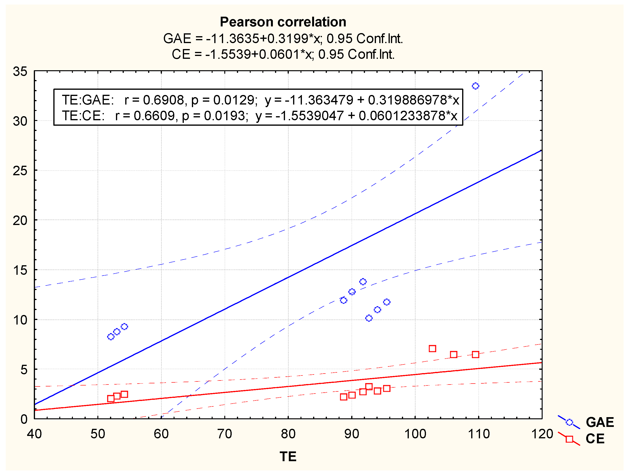
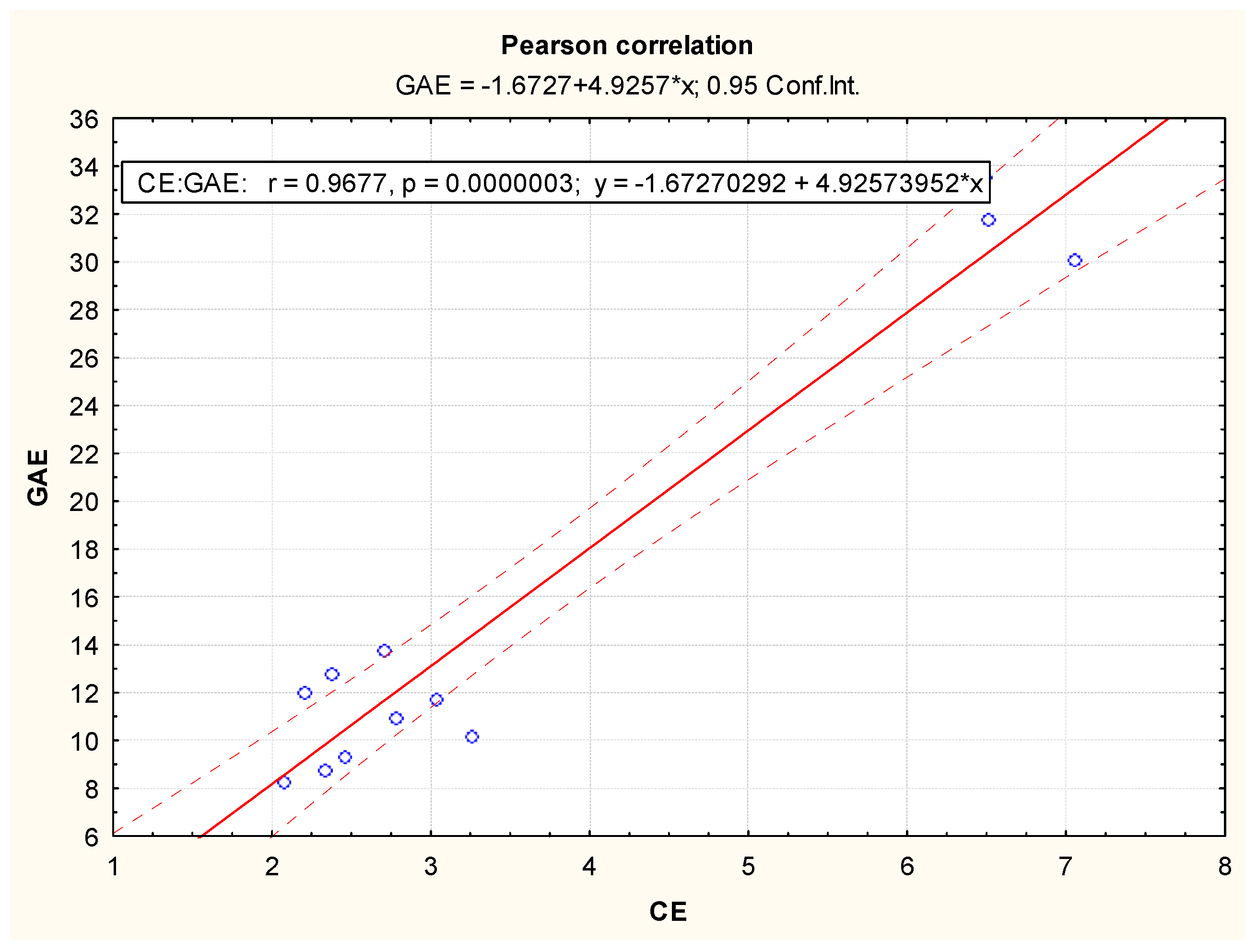
3.2. Antioxidant Potential of Methanolic Extracts from Acorus calamus, Chlorella vulgaris, Lemna minuta and Scenedesmus dimorphus

Phenolic compounds are one of the most frequent and widespread group of substances in the world of plants, with more than 8000 identified phenolic structures, which include a variety of patterns, from rather simple structures (e.g., phenolic acids) through polyphenols such as flavonoids to polymeric compounds based on the different classes [44]. Polyphenols are secondary metabolites of plants, essential for their growth and development. As natural antioxidants, these compounds have important functions, which include the inhibition of lipid peroxidation, antimicrobial and anticancer activities, stabilization of ascorbic acid, a direct constrictive effect on the capillaries and an antiaging and anti-inflammatory effect [45]. According to the experimental results, the highest concentration (mean ± SD) of TPC (g GAE/kg) was found in the methanolic extracts of S. dimorphus (31.78 ± 1.7), followed in descending gradation by A. calamus (12.83 ± 0.91), L. minuta (10.94 ± 0.78) and C. vulgaris (8.79 ± 0.5) (Figure 1).
The TPC of the methanolic extracts of S. dimorphus (31.78 g GAE/kg) was much higher than the values obtained by Bulut et al. [26] (5.4 g GAE/kg) and El-Chaghaby et al. [46] (0.511 g GAE/kg) from studies of other Scenedesmus spp. In this way, the differences in the species examined could explain to some extent the above-mentioned variations in the experimental results. Moreover, Bulut et al. [26] used ethanol/water as extractant, while El-Chaghaby et al. [46] utilized ethanol. In this respect, Bulut et al. [26] found a nearly 5-fold difference between the yield of TPC from different extractants—water, ethanol/water, ethyl acetate and hexane. Furthermore, Bulut et al. [26] and El-Chaghaby et al. [46] applied ultrasonication as a method of extract preparation, which corresponds to this study. The TPC of C. vulgaris (8.79 g GAE/kg) is consistent with the results of Mtaki et al. [25]—8.53 g GAE/kg, but is higher than the values obtained by Abdel–Karim et al. [47] (3.17 g GAE/kg), Bhuvana et al. [48] (2.13 g GAE/kg) and El-Chaghaby et al. [46] (0.71 g GAE/kg), and much lower compared to the findings of Gürlek et al. [49] (78 g GAE/kg). It should be noted that Mtaki et al. [25] and El-Chaghaby et al. [46] used ethanol as an extractant, whereas Bhuvana et al. [48] and Gürlek et al. [49] utilized methanol. The results of Abdel–Karim et al. [47] (3.17 g GAE/kg) were obtained by testing acetone extracts, which have a higher TPC yield than do methanolic (2.7–3 g GAE/kg) and ethanolic extracts (0.65 g GAE/kg). These findings again emphasize the importance of the extractant type on the yield of polyphenols. The TPC of A. calamus methanolic extracts (12.83 g GAE/kg) was very similar to the findings of Funde [50] (12.1 g GAE/kg) but higher than the results of Parki et al. [51] (3.41–4.8 g GAE/kg) and Chaubey et al. [52] (1.67–10.42 g GAE/kg) and lower than the data of Devi and Ganjewala [53] (27 g GAE/kg), Li and Wah [11] (36 g GAE/kg), and (especially) of Djarkasi et al. [10] (240.32 g GAE/kg). The aforementioned results were obtained using methanol as an extractant [10,11,50,51,52,53]. Therefore, the major differences above should be explained by factors different from the type of solvent, such as extraction method, preparation of the sample, method of TPC determination, geographic and climatic conditions, etc. In this respect, Li and Wah [11] performed the extraction of polyphenols at 60 °C in a horizontal water bath shaker and Funde [50] likewise used maceration with a shaking condition, whereas Parki et al. [51] utilized cold percolation, Chaubey et al. [52] applied the Soxhlet method and Devi and Ganjewala [53] and Djarkasi et al. [10] used maceration. It should be noted that the major differences in the aforementioned study of Chaubey et al. [52] were due to the various geographic and climatic conditions of 20 ecological niches in India from which A. calamus was harvested. Based on the statistical data processing in this study, the altitudes were positively correlated with TPC in plants [52]. However, this conclusion was not supported by the work of Parki et al. [51], who reported low TPC (3.41–4.8 g GAE/kg) from rhizome extracts from different altitudes of Uttarakhand Himalayas, India. Moreover, the TPC from the methanolic extracts of A. calamus harvested from the highest altitude (1514 m) was the lowest (3.41 g GAE/kg) compared to the polyphenolic yield of A. calamus methanolic extracts from lower altitudes of 344 m and 1370 m (4.1 and 4.8 g GAE/kg, respectively). In the study of Djarkasi et al. [10], methanol showed a higher polyphenol extraction potential for A. calamus rhizomes and leaves compared to acetone, ethyl acetate and hexane, with up to 2-fold difference in the yield of polyphenols. Gülçin et al. [18] found that the TPC of ethanolic extracts of Lemna minor was 16.7 g GAE/kg, which was higher than the values from L. minuta methanolic extracts (10.94 g GAE/kg) obtained in this study. The lack of experimental data for L. minuta hindered the further comparison of TPC.
Flavonoids are phenolic compounds composed of two aromatic rings linked by a unit of three carbon atoms (C6–C3–C6). This group includes flavonols, flavones, isoflavones, flavanones, flavanonols, anthocyanidins and tannins [45]. Flavonoids have a number of medicinal benefits, including anticancer, antioxidant, anti-inflammatory, and antiviral properties [54]. Their regular consumption is associated with a diminished risk of some chronic diseases, including cancer, cardiovascular disease and neurodegenerative disorders [55]. The TFC of methanolic extracts (g CE/kg) corresponds to TPC (g GAE/kg) and the highest concentrations (mean ± SD) were found in S. dimorphus (6.68 ± 0.32), followed in descending order by A. calamus (3.42 ± 0.26), L. minuta (3.02 ± 0.24) and C. vulgaris (2.29 ± 0.19) (Figure 1).
The TFC of A. calamus (3.42 g CE/kg) was lower than the results of Devi and Ganjewala [53] (11.1 g CE/kg) and especially of Li and Wah [11] (74.11 g CE/kg). Both authors used methanol as an extractant [11,53]. One of the possible explanations for the substantial discrepancies of the experimental data are the findings of Chaubey et al. [52], who reported some great differences between the TFC of methanolic extracts of A. calamus harvested from 20 ecological niches in India. The various sample values ranged from 0.5 g CE/kg to 24.5 g CE/kg, which further emphasizes the role of geographic and climatic conditions in the antioxidant potential of different plants. After statistical data processing, the authors concluded that altitudes are positively associated with TFC in plants [52]. It should be noted that all aforementioned authors applied different methods for extract preparation—Li and Wah [11] performed extraction at 60 °C in a horizontal water bath shaker, whereas Chaubey et al. [52] used the Soxhlet method and Devi and Ganjewala [53] utilized maceration, which further increased the differences in the yield of total flavonoids. The lack of literature data or major methodological differences prevents the comparison of TFC of S. dimorphus, L. minuta and C. vulgaris methanolic extracts.
There are many different antioxidants presented in plants. Therefore it is very difficult to measure each antioxidant component separately. A DPPH assay tests the ability of a substance to quench a DPPH radical through the donation of an electron [56]. In this way, DPPH radicals are widely used as a model system to evaluate the scavenging activities of natural antioxidant compounds [57]. The radical scavenging potentials of S. dimorphus, L. minuta, A. calamus and C. vulgaris corresponded to TPC and TFC and were 106 ± 10 mmol TE/kg (26.86 ± 1.94%), 94 ± 9 mmol TE/kg (25.17 ± 1.93%), 90 ± 9 mmol TE/kg (24.56 ± 1.87%), 53 ± 5 mmol TE/kg (19.5 ± 1.75%), respectively (mean ± SD, Figure 1).
The DPPH of S. dimorphus methanolic extracts (26.86%) corresponds to the DPPH of ethanol/water extracts of Scenedesmus sp.—25.65% [26]. Bulut et al. [26] reported the highest radical scavenging effect from ethyl acetate solvent (52.02%), followed by ethanol/water (25.65%), hexane (12.99%) and water (8.40%). These experimental results showed the impact of extractant type on radical scavenging activity. As mentioned above, Bulut et al. [26] used ultrasonication as a method of extract preparation. The DPPH of C. vulgaris extracts (19.5%) is lower than the radical scavenging activity obtained by Vehapi et al. [17] (35.81–37.67%) and Abdel–Karim et al. [47] (39.4%), but is higher than the results of Bhuvana et al. [48] (16.87%). All of the aforementioned authors used methanol as a solvent [17,47,48]. It is important to emphasize that methanolic extracts of C. vulgaris tested by Abdel–Karim et al. [47] showed higher radical scavenging activity than did ethanolic extracts (20.76%) but showed lower radical scavenging activity than did acetone extracts (50.81%). Vehapi et al. [17] applied the Soxhlet method for extract preparation, while Abdel–Karim et al. [47] performed extraction in a shaking incubator and Bhuvana et al. [48] utilized maceration. The diverse methods of extraction could explain to a certain extent the differences in radical scavenging activity of the methanolic plant extracts. Djarkasi et al. [10] found a very high radical scavenging activity of A. calamus methanolic extracts (90.29%), which is much higher than the scavenging potential of the plant extracts recorded in this experiment (24.56%). Li and Wah [11] obtained data that corresponded to Djarkasi et al. [10]—over 90% radical scavenging activity of A. calamus methanolic extracts, which was nearly nine times higher than the scavenging activity of A. calamus hexane extracts. These great differences further emphasize the major impact of solvent type on the antioxidant activity of plant extracts. Djarkasi et al. [10] used maceration as an extraction method, whereas Li and Wah [11] performed extraction at 60 °C in a horizontal water bath shaker. Generally, the discrepancies in the radical scavenging potential (expressed as DPPH inhibition in percentage) obtained in this work and described by the authors cited could be mainly explained by the different concentrations of DPPH solution (e.g., 0.1 mM [10], 3 mM [11], 0.2 mM [26] and 0.16 mM [48]) and by the application of extract solutions in different concentration levels (e.g., 20 µg/mL [17] and 200 mg/mL [11]). Additionally, there are differences in the calculation of DPPH inhibition (%) (e.g., [(A0 − A)/A0] × 100 [17,47,48] vs. [1 − (A − A0)/A0] × 100 [26]). There are no literature data concerning the radical scavenging potential of L. minuta.
Based on the Pearson correlation coefficients, both total phenolic content and total flavonoid content have similar impacts on radical scavenging capacity (correlation coefficients r = 0.6808 and r = 0.6609, respectively) (Figure 2). The Pearson correlation between the total phenolic content and the total flavonoid content of the methanolic extracts was characterized by a high and positive correlation coefficient (r = 0.9677), which means that flavonoids were a significant part of the total phenolic compounds extracted from the plant materials studied (Figure 3).

4. Conclusions

A. calamus methanolic extracts exhibited the highest antimicrobial activity against eight fungal strains (F. oxysporum, A. flavus, A. niger, F. graminearum, A. ochraceus, Alt. alternata, A. carbonarius and P. chrysogenum), followed by C. vulgaris, L. minuta and S. dimorphus methanolic extracts, which were inhibitory against two to three strains. As a whole, the antifungal activity of these plant extracts was comparatively low or completely lacking (zones of inhibition ≤ 10.3 mm), and the same could be said for the antifungal activity of amphotericin B (zones of inhibition ≤ 13.8 mm). Among the plant species studied, the extract from S. dimorphus showed the highest antioxidant potential, followed by the A. calamus and L. minuta extracts. C. vulgaris methanolic extracts demonstrated the lowest antioxidant activity. Based on the Pearson correlation coefficients, total phenolic content and total flavonoid content both had similar impacts on radical scavenging capacity and flavonoids were a significant part of the total phenolic compounds extracted from the plant materials studied.

Author Contributions

Conceptualization, T.D., M.T. and K.V.; methodology, T.D. and M.T.; software, M.T. and K.V.; validation, T.D. and M.T.; formal analysis, T.D., M.T. and K.V.; investigation, T.D., K.V. and D.D.; resources, K.V.; data curation, M.T.; writing—original draft preparation, T.D.; writing—review and editing, M.T. and K.V.; visualization, D.D.; supervision, G.B.; project administration, G.B.; funding acquisition, D.D. and G.B. All authors have read and agreed to the published version of the manuscript.

Funding

This work was supported by the Bulgarian Ministry of Education and Science under the National Research Programme “Healthy Foods for a Strong Bio-Economy and Quality of Life” approved by DCM #577/17.08.2018.

Data Availability Statement

All data is contained within the article.

Conflicts of Interest

The authors declare no conflict of interest.

References

  1. Rawal, P.; Adhikari, R.S.; Danu, K.; Tiwari, A. Antifungal activity of Acorus calamus against Fusarium oxysporum f. sp. lycopersici. Int. J. Curr. Microbiol. App. Sci. 2015, 4, 710–715. [Google Scholar]
  2. Mahdavi, B.; Yaacob, W.A.; Din, L.B. Antioxidant and antimicrobial activity of the extracts from different parts of Etlingera sayapensis (Zingiberaceae). Sains Malays. 2017, 46, 1565–1571. [Google Scholar] [CrossRef]
  3. Devi, S.A.; Ganjewala, D. Antimicrobial activity of Acorus calamus (L.) rhizome and leaf extract. Acta Biol. Szeged. 2009, 53, 45–49. [Google Scholar]
  4. Bonifácio, B.V.; dos Santos Ramos, M.A.; da Silva, P.B.; Bauab, T.M. Antimicrobial activity of natural products against Helicobacter pylori: A review. Ann. Clin. Microbiol. Antimicrob. 2014, 13, 54. [Google Scholar] [PubMed] [Green Version]
  5. Sales, M.D.C.; Costa, H.B.; Fernandes, P.M.B.; Ventura, J.A.; Meira, D.D. Antifungal activity of plant extracts with potential to control plant pathogens in pineapple. Asian Pac. J. Trop. Biomed. 2016, 6, 26–31. [Google Scholar] [CrossRef]
  6. Khameneh, B.; Iranshahy, M.; Soheili, V.; Bazzaz, B.S.F. Review on plant antimicrobials: A mechanistic viewpoint. Antimicrob. Resist. Infect. Control 2019, 8, 118. [Google Scholar] [CrossRef] [PubMed] [Green Version]
  7. Mickymaray, S.; Al Aboody, M.S. In vitro antioxidant and bactericidal efficacy of 15 common spices: Novel therapeutics for urinary tract infections? Medicina 2019, 55, 289. [Google Scholar] [CrossRef] [Green Version]
  8. Shreelaxmi, S.H.; Ramachandra, C.T.; Roopa, R.S.; Hanchinal, S.G. Antimicrobial activity of supercritical fluid extracted Acorus calamus oil against different microbes. J. Pharmacogn. Phytochem. 2018, 7, 2836–2840. [Google Scholar]
  9. Hasan, N.A.; Nawahwi, M.Z.; Malek, H.A. Antimicrobial activity of Nigella sativa seed extract. Sains Malays. 2013, 42, 143–147. [Google Scholar]
  10. Djarkasi, G.S.S.; Lalujan, L.E.; Nurali, E.; Sumual, M.F. Antioxidant activity of karimenga (Acorus calamus). AIP Conf. Proc. 2019, 2155, 020051. [Google Scholar]
  11. Li, K.S.; Wah, C.S. Antioxidant and antibacterial activity of Acorus calamus L leaf and rhizome extracts. J. Gizi Klinik Indones. 2017, 13, 144–158. [Google Scholar] [CrossRef] [Green Version]
  12. Tadhani, M.; Patel, V.; Subhash, V. In vitro antioxidant activities of Stevia rebaudiana leaves and callus. J. Food Compos. Anal. 2007, 20, 323–329. [Google Scholar] [CrossRef]
  13. Singh, S.; Srivastava, R.; Choudhary, S. Antifungal and HPLC analysis of the crude extracts of Acorus calamus, Tinospora cordifolia and Celestrus paniculatus. J. Agric. Technol. 2010, 6, 149–158. [Google Scholar]
  14. Dissanayake, K.G.C.; Perera, B.T.; Perera, W.P.R.T. Potential of the pathogenic microorganisms mitigation using rhizome extract of Acorus calamus as a medicinal herb. World J. Pharm. Pharm. Sci. 2020, 9, 85–99. [Google Scholar]
  15. Ghasemi, Y.; Moradian, A.; Mohagheghzadeh, A.; Shokravi, S.; Morowvat, M.H. Antifungal and antibacterial activity of the microalgae collected from paddy fields of Iran: Characterization of antimicrobial activity of Chroococcus dispersus. J. Biol. Sci. 2007, 7, 904–910. [Google Scholar] [CrossRef] [Green Version]
  16. Marrez, D.A.; Naguib, M.M.; Sultan, Y.Y.; Higazy, A.M. Antimicrobial and anticancer activities of Scenedesmus obliquus metabolites. Heliyon 2019, 5, e01401. [Google Scholar] [CrossRef] [Green Version]
  17. Vehapi, M.; Koçer, A.T.; Yilmaz, A.; Özçimen, D. Investigation of the antifungal effects of algal extracts on apple-infecting fungi. Arch. Microbiol. 2020, 202, 455–471. [Google Scholar] [CrossRef]
  18. Gülçin, I.; Kireçci, E.; Akkemik, E.; Topal, F.; Hisar, O. Antioxidant, antibacterial, and anticandidal activities of an aquatic plant: Duckweed (Lemna minor L. Lemnaceae). Turk. J. Biol. 2010, 34, 175–188. [Google Scholar]
  19. Velichkova, K.; Sirakov, I.; Rusenova, N.; Beev, G.; Denev, S.; Valcheva, N.; Dinev, T. In Vitro antimicrobial activity on Lemna minuta, Chlorella vulgaris and Spirulina sp. extracts. Fresenius Environ. Bull. 2018, 27, 5736–5741. [Google Scholar]
  20. Petrova-Tacheva, V.; Ivanov, V.; Atanasov, A. Lemna minor L. as a source of antioxidants. Trakia J. Sci. 2020, 18 (Suppl. 1), 157–162. [Google Scholar]
  21. Culture Collection of Algae and Protozoa. Available online: http://www.ccap.ac.uk/media/documents/BB_000.pdf (accessed on 20 April 2021).
  22. Hajimehdipoor, H.; Kondori, B.M.; Amin, G.R.; Adib, N.; Rastegar, H.; Shekarchi, M. Development of a validated HPLC method for the simultaneous determination of flavonoids in Cuscuta chinensis Lam. by ultra-violet detection. DARU J. Pharm. Sci. 2012, 20, 57. [Google Scholar] [CrossRef] [Green Version]
  23. Tzanova, M.; Grozeva, N.; Gerdzhikova, M.; Argirova, M.; Pavlov, D.; Terzieva, S. Flavonoid content and antioxidant activity of Betonica bulgarica Degen et Neič. Bulg. Chem. Commun. 2018, 50, 90–97. [Google Scholar]
  24. Rebaya, A.; Belghith, S.I.; Baghdikian, B.; Leddet, V.M.; Mabrouki, F.; Olivier, E.; Cherif, J.K.; Ayadi, M.T. Total phenolic, total flavonoid, tannin content, and antioxidant capacity of Halimium halimifolium (Cistaceae). J. Appl. Pharm. Sci. 2015, 5, 52–57. [Google Scholar]
  25. Mtaki, K.; Kyewalyanga, M.S.; Mtolera, M.S.P. Assessment of antioxidant contents and free radical-scavenging capacity of Chlorella vulgaris cultivated in low cost media. Appl. Sci. 2020, 10, 8611. [Google Scholar] [CrossRef]
  26. Bulut, O.; Akin, D.; Sönmez, Ģ.; Öktem, A.; Yücel, M.; Öktem, H.A. Phenolic compounds, carotenoids, and antioxidant capacities of a thermo-tolerant Scenedesmus sp. (Chlorophyta) extracted with different solvents. J. Appl. Phycol. 2019, 31, 1675–1683. [Google Scholar] [CrossRef]
  27. Balendres, M.A.O.; Karlovsky, P.; Cumagun, C.J.R. Mycotoxigenic fungi and mycotoxins in agricultural crop commodities in the Philippines: A review. Foods 2019, 8, 249. [Google Scholar] [CrossRef] [Green Version]
  28. Ji, C.; Fan, Y.; Zhao, L. Review on biological degradation of mycotoxins. Anim. Nutr. 2016, 2, 127–133. [Google Scholar] [CrossRef]
  29. Sadhasivam, S.; Shapiro, O.H.; Ziv, C.; Barda, O.; Zakin, V.; Sionov, E. Synergistic inhibition of mycotoxigenic fungi and mycotoxin production by combination of pomegranate peel extract and azole fungicide. Front. Microbiol. 2019, 10, 1919. [Google Scholar] [CrossRef] [Green Version]
  30. Kumar, V.; Singh, R.; Joshi, V. Antimicrobial activity of rhizome extract of Acorus calamus against different microorganisms. Octa J. Biosci. 2014, 2, 59–63. [Google Scholar]
  31. Abedin, R.M.A.; Taha, H.M. Antibacterial and antifungal activity of cyanobacteria and green microalgae. Evaluation of medium components by Plackett-Burman design for antimicrobial activity of Spirulina platensis. GJBB 2008, 3, 22–31. [Google Scholar]
  32. Dantas, D.M.M.; Costa, R.M.P.B.; Carneiro-da-Cunha, M.G.; Galvez, A.O.; Drummond, A.R.; Bezerra, R.S. Bioproduction, antimicrobial and antioxidant activities of compounds from Chlorella vulgaris. Res. Rev. J. Bot. Sci. 2015, 4, 12–18. [Google Scholar]
  33. Vehapi, M.; Yilmaz, A.; Özçimen, D. Antifungal activities of Chlorella vulgaris and Chlorella minutissima microalgae cultivated in bold basal medium, wastewater and tree extract water against Aspergillus niger and Fusarium oxysporum. Rom. Biotechnol. Lett. 2018, 1, 1–8. [Google Scholar]
  34. Zielinski, D.; Fraczyk, J.; Debowsky, M.; Zielinski, M.; Kaminski, Z.J.; Kregiel, D.; Jacob, C.; Kolesinska, B. Biological activity of hydrophilic extract of Chlorella vulgaris grown on post-fermentation leachate from a biogas plant supplied with stillage and maize silage. Molecules 2020, 25, 1790. [Google Scholar] [CrossRef] [Green Version]
  35. Abbassy, M.A.; Marei, G.I.K.; Rabia, S.M.H. Antimicrobial activity of some plant and algal extracts. Int. J. Plant Soil Sci. 2014, 3, 1366–1373. [Google Scholar] [CrossRef]
  36. Effiong, B.N.; Sanni, A. Antifungal properties and phytochemical screening of crude extract of Lemna pauciscostata (Helgelm) against fish feed spoilage fungi. Life Sci. J. 2009, 6, 19–22. [Google Scholar]
  37. Sekhon, A.S.; Padhye, A.A.; Garg, A.K. In vitro sensitivity of Penicillium marneffei and Pythium insidiosum to various antifungal agents. Eur. J. Epidemiol. 1992, 8, 427–432. [Google Scholar] [CrossRef]
  38. Lalitha, P.; Shapiro, B.L.; Srinivasan, M. Antimicrobial susceptibility of Fusarium, Aspergillus, and other filamentous fungi isolated from keratitis. Arch. Ophthalmol. 2007, 125, 789–793. [Google Scholar] [CrossRef]
  39. Alastruey-Izquierdo, A.; Cuenca-Estrella, M.; Monzón, A.; Mellado, E.; Rodríguez-Tudela, J.L. Antifungal susceptibility profile of clinical Fusarium spp. isolates identified by molecular methods. J. Antimicrob. Chemother. 2008, 61, 805–809. [Google Scholar] [CrossRef] [Green Version]
  40. Alastruey-Izquierdo, A.; Cuesta, I.; Ros, L.; Mellado, E.; Rodríguez-Tudela, J.L. Antifungal susceptibility profile of clinical Alternaria spp. identified by molecular methods. J. Antimicrob. Chemother. 2011, 66, 2585–2587. [Google Scholar] [CrossRef] [Green Version]
  41. Kawakami, H.; Inuzuka, H.; Hori, N.; Takahashi, N.; Ishida, K.; Mochizuki, K.; Ohkusu, K.; Muraosa, Y.; Watanabe, A.; Kamei, K. Inhibitory effects of antimicrobial agents against Fusarium species. Med. Mycol. 2015, 53, 603–611. [Google Scholar] [CrossRef] [Green Version]
  42. Dimitrova-Dyulgerova, I.; Merdzhanov, P.; Todorov, K.; Seymenska, D.; Stoyanov, P.; Mladenov, R.; Stoyanova, A. Essential oils composition of Betonica officinalis L. and Stachys sylvatica L. (Lamiaceae) from Bulgaria. Comptes Rendus Acad. Bulg. Sci. 2015, 68, 991–998. [Google Scholar]
  43. Dinev, T.G.; Rusenova, N.V.; Tzanova, M.T.; Grozeva, N.H.; Gerdzhikova, M.A.; Stoyanov, P.S.; Mladenova, T.R.; Beev, G.G. Antimicrobial potential of methanolic extracts from Betonica bulgarica Degen et Neič. (Lamiaceae). Ecol. Balk. 2020, 12, 165–174. [Google Scholar]
  44. Cheynier, V. Phenolic compounds: From plants to foods. Phytochem. Rev. 2012, 11, 153–177. [Google Scholar] [CrossRef]
  45. Tanase, C.; Coşarcă, S.; Muntean, D.-L. A critical review of phenolic compounds extracted from the bark of woody vascular plants and their potential biological activity. Molecules 2019, 24, 1182. [Google Scholar] [CrossRef] [Green Version]
  46. El-Chaghaby, G.A.; Rashad, S.; Abdel-Kader, S.F.; Rawash, E.-S.A.; Moneem, M.A. Assessment of phytochemical components, proximate composition and antioxidant properties of Scenedesmus obliquus, Chlorella vulgaris and Spirulina platensis algae extracts. Egypt. J. Aquat. Biol. Fish. 2019, 23, 521–526. [Google Scholar] [CrossRef] [Green Version]
  47. Abdel-Karim, O.H.; Gheda, S.F.; Ismail, G.A.; Abo-Shady, A.M. Phytochemical screening and antioxidant activity of Chlorella vulgaris. Sci. JBAS 2020, 41, 76–86. [Google Scholar]
  48. Bhuvana, P.; Anuradha, V.; Syed, A.M.; Suganya, V.; Sangeetha, P. In vitro antioxidant activity of methanolic extracts of Chlorella vulgaris. IJAR 2017, 5, 1465–1474. [Google Scholar]
  49. Gürlek, C.; Yarkent, Ç.; Köse, A.; Tuğcu, B.; Gebeloğlu, I.K.; Öncel, S.Ş.; Elibol, M. Screening of antioxidant and cytotoxic activities of several microalgal extracts with pharmaceutical potential. Health Technol. 2020, 10, 111–117. [Google Scholar] [CrossRef]
  50. Funde, S.G. Phytochemicals evaluation, anticancer, antioxidant and antimicrobial activity of Acorus calamus different solvent extracts. J. Chem. Pharm. Res. 2015, 7, 495–504. [Google Scholar]
  51. Parki, A.; Chaubey, P.; Prakash, O.; Kumar, R.; Pant, A.K. Chemical composition and antioxidant activity of Acorus calamus L. accessions from different altitudes of Uttarakhand Himalayas. J. Herb. Drug. 2019, 9, 171–178. [Google Scholar]
  52. Chaubey, P.; Archana; Prakash, O.; Rai, K.; Kumar, R.; Pant, A.K. In vitro antioxidant activity and total phenolic content of rhizome extracts from Acorus calamus Linn. Asian J. Chem. 2017, 29, 2357–2360. [Google Scholar] [CrossRef]
  53. Devi, S.A.; Ganjewala, D. Antioxidant activities of methanolic extracts of sweet-flag (Acorus calamus) leaves and rhizomes. J. Herbs Spices Med. Plants 2011, 17, 1–11. [Google Scholar] [CrossRef]
  54. Ullah, A.; Munir, S.; Badshah, S.L.; Khan, N.; Ghani, L.; Poulson, B.G.; Emwas, A.-H.; Jaremko, M. Important flavonoids and their role as a therapeutic agent. Molecules 2020, 25, 5243. [Google Scholar] [CrossRef]
  55. Kozłowska, S.; Szostak-Wegierek, D. Flavonoids—Food sources and health benefits. Rocz. Panstw. Zakl. Hig. 2014, 65, 79–85. [Google Scholar]
  56. Coulombier, N.; Nicolau, E.; Déan, L.L.; Antheaume, C.; Jauffrais, T.; Lebouvier, N. Impact of light intensity on antioxidant activity of tropical microalgae. Mar. Drugs 2020, 18, 122. [Google Scholar] [CrossRef] [Green Version]
  57. Zayova, E.; Stancheva, I.; Geneva, M.; Petrova, M.; Dimitrova, L. Antioxidant activity of in vitro propagated Stevia rebaudiana Bertoni plants of different origins. Turk. J. Biol. 2013, 37, 106–113. [Google Scholar]
Figure 1. Total phenolic content (g GAE/kg), total flavonoid content (g CE/kg) and radical scavenging potential (mmol TE/kg) of methanolic extracts from different plants.
Applsci 11 04745 g001
Figure 2. The Pearson correlation between radical scavenging capacity (expressed as TE) and total phenolic content (expressed as GAE), as well as between radical scavenging capacity (expressed as TE) and total flavonoid content (expressed as CE).
Applsci 11 04745 g002
Figure 3. The Pearson correlation between total phenolic content (expressed as GAE) and total flavonoid content (expressed as CE).
Applsci 11 04745 g003
Table 1. Antifungal activity of methanolic extracts (64 mg/mL) of A. calamus, C. vulgaris, S. dimorphus and L. minuta (mean ± SD) *.
Plant ExtractDiameter of Inhibition Zones (mm)
A. flavusA. parasiticusA. nigerA. carbonariusA. ochraceusF. graminearumF. oxysporumP. chrysogenumAlt. alternata
A. calamus10.0 ± 0 ab- **9.3 ± 0.3 ab7.8 ± 0.3 a8.7 ± 0.3 ab9.2 ± 0.3 ab10.3 ± 0.6 ab7.7 ± 0.3 a8.2 ± 0.3 a
C. vulgaris- **- **9.2 ± 0.3 ab- **- **- **- **8.3 ± 0.3 a7.0 ± 0 a
S. dimorphus- **- **8.2 ± 0.6 a- **- **- **- **8.0 ± 0 a- **
L. minuta- **- **- **- **7.0 ± 0 a- **- **7.0 ± 0 a7.7 ± 0.3 a
Amphotericin B 11.5 ± 0.3 ac11.0 ± 0 ac9.0 ± 0 ab13.8 ± 0.3 ac- **- **- **- **11.5 ± 0.3 ac
Methanol6.0 ± 0 a6.0 ± 0 a6.0 ± 0 a6.0 ± 0 a6.0 ± 0 a6.0 ± 0 a6.0 ± 0 a6.0 ± 0 a6.0 ± 0 a
* Different letters in the columns denote significant differences between the inhibition zones of plant extracts and negative control (methanol) values according to one-way ANOVA and LSD tests (ab p ≤ 0.05; ac p ≤ 0.01). ** no activity (6 mm diameter of the well).
Publisher’s Note: MDPI stays neutral with regard to jurisdictional claims in published maps and institutional affiliations.

