Article

Simultaneous SAXS/SANS Method at D22 of ILL: Instrument Upgrade

1
Institute for Crystallography and Structural Physics, Friedrich-Alexander-Universität Erlangen-Nürnberg, Staudtstrasse 3, 91058 Erlangen, Germany
2
Center for Nanoanalysis and Electron Microscopy (CENEM), Cauerstrasse 3, 91058 Erlangen, Germany
3
Interdisciplinary Center for Nanostructured Films (IZNF), Cauerstrasse 3, Germany
4
Institut Laue-Langevin, 71 Avenue des Martyrs, 38042 Grenoble, France
*
Author to whom correspondence should be addressed.
Appl. Sci. 2021, 11(13), 5925; https://doi.org/10.3390/app11135925
Submission received: 1 June 2021 / Revised: 22 June 2021 / Accepted: 23 June 2021 / Published: 25 June 2021
(This article belongs to the Special Issue Recent Advances in Small-Angle Neutron Scattering)

Abstract

:
A customized portable SAXS instrument has recently been constructed, installed, and tested at the D22 SANS instrument at ILL. Technical characteristics of this newly established plug-and-play SAXS system have recently been reported (J. Appl. Cryst. 2020, 53, 722). An optimized lead shielding arrangement on the SAXS system and a double energy threshold X-ray detector have been further implemented to substantially suppress the unavoidable high-energy gamma radiation background on the X-ray detector. The performance of the upgraded SAXS instrument has been examined systematically by determining background suppression factors (SFs) at various experimental conditions, including different neutron beam collimation lengths and X-ray sample-to-detector distances (SDDX-ray). Improved signal-to-noise ratio SAXS data enables combined SAXS and SANS measurements for all possible experimental conditions at the D22 instrument. Both SAXS and SANS data from the same sample volume can be fitted simultaneously using a common structural model, allowing unambiguous interpretation of the scattering data. Importantly, advanced in situ/real time investigations are possible, where both the SAXS and the SANS data can reveal time-resolved complementary nanoscale structural information.

Graphical Abstract

1. Introduction

Small angle scattering (SAS) of X-rays (SAXS) and neutrons (SANS) are non-destructive powerful techniques which have been utilized successfully for investigating nanostructured materials such as metal nanoparticles [1,2], emulsions [3], micelles [4,5], electrolytes [6,7], liquid crystals [8,9] and organic nanoparticles [10,11,12,13]. The SAS technique provides nanoscale information including shape, size, and size distribution as well as spatial distribution of dispersed materials in solution [14,15]. In particular, they provide the diameter of gyration and the mass of isotropically distributed shaped nanoscale particles [16], as well as the linear mass density and cross-sectional size of anisotropically shaped particles [14,17]. Moreover, SAXS and SANS methods are important tools for examining materials for various applications, e.g., solar cells, lithium-ion batteries and sensors [6,7,18,19,20,21]. Combining both SAXS and SANS methods to investigate the same sample volume is remarkably advantageous, where two radiations offer two different contrast situations (X-ray and neutron scattering cross section). Using such multiple modalities is very beneficial, especially for cases when a single modality provides incomplete or ambiguous results. For instance, SANS provides the form factor of the shell in a representative hollow-shell-like particle [2]. In contrast, SAXS measurements of the same core–shell sample are sensitive to the high-electron-density core. The structures of both shell and core can be uniquely obtained from the same sample volume via complementary SAXS and SANS methods. Thus, simultaneous SAXS/SANS measurements will allow the exactness of the probed samples and the potential for using a common analysis model for both SAXS and SANS data, and consequently, a straightforward and unambiguous interpretation of the scattering data. For instance, structural modifications of supramolecular gels upon switching between H2O and D2O have been reported [22]. The deuteration effect on the structure of some nanoscale structured samples cannot be ignored. Thus, the simultaneous SAXS/SANS method is an essential tool for eliminating any ambiguity regarding sample exactness.
A key contribution of the simultaneous SAXS/SANS method is its ability to perform in situ and operando studies on advanced materials that undergo temporal modifications under external stimuli [23], allowing access to their structural rearrangements from the molecular scale to nanoscale assemblies. This means not only the synergy that results when data sets are obtained simultaneously from the same sample volume, but also access to the cross-correlation of phases/components when two different simultaneous contrast situations are probed for multi-phase/component samples. For instance, the cross-correlation between evolved gold nanorods size/shape (SAXS) and the corresponding engaged structure of organic moieties (SANS) during surfactant-assisted seed-mediated synthesis of gold nanorods will reveal new information related to the exact growth mechanisms involved [23]. Moreover, the SAXS/SANS system will enable contrast-dependent successive length-scale characterization of novel nanomaterials, addressing unsolved scientific questions.
A few instruments at neutron research reactors are currently utilized for both neutron and X-ray radiation experiment, obtaining complementary results of investigated samples. These dedicated instruments are mainly reflectometry and imaging techniques. A neutron imaging setup at PSI is outfitted with an additional X-ray tube to perform computerized tomography (CT) investigations using X-ray and neutron radiation [24,25]. Similarly, an instrument (NeXT) at ILL hosts two complementary imaging setups; neutron and X-ray tomography [26,27]. Neutron reflectometers such as the NREX instrument at MLZ allows combined X-ray and neutron specular reflectivity measurements using both X-ray and neutron beams placed in an orthogonal geometry [28]. Unfortunately, in most cases for the latter instruments, high energy background signal on the X-ray detector is a significant challenge and hampers a simultaneous mode of combined X-ray and neutron methods. The X-ray detection system is often not optimized for simultaneous acquisition, thus both X-ray and neutron data sets are mainly acquired successively and not in a synchronous mode [29,30].
An advanced SAXS system mounted on a standalone metal rack that makes it easily movable for use on the D22 SANS instrument has successfully been installed and tested at ILL [23]. For the first time, SAXS and SANS measurements in a simultaneous mode have been demonstrated [23]. One challenge is cutting down background signal that originates mainly from high-energy gamma radiation [31] in the experimental zone of the D22 instrument. A preliminary lead shielding design around our SAXS instrument at D22 of ILL has shown the capability of acquiring background discriminated SAXS data on a single energy threshold X-ray detector, enabling simultaneous SAXS/SANS experiments [23]. To enable a combined SAXS/SANS method in a simultaneous mode for all possible experimental conditions at the D22 instrument, an upgrade of the SAXS system was an essential requirement. In the current work, an optimized lead shielding has been implemented and the background suppression has been systematically examined. Additionally, a double energy threshold X-ray detector has been employed, replacing the single energy threshold detector. Overall reduced background noise and acceptable signal-to-noise ratios of SAXS data have been demonstrated upon combining both modifications, for all possible experimental configurations at the D22 instrument. Combined SAXS/SANS measurements were acquired for two nanoparticle (NP) dispersions of different sizes, to demonstrate the performance of the SAXS/SANS measurements under various experimental conditions.

2. Materials and Methods

An advanced SAXS system based on a Copper/Molybdenum (Kα; 8.0/17.4 keV) switchable microfocus rotating anode X-ray generator (Rigaku MM007 HF DW) and a Dectris EIGER2 X 1M detector with a continuously variable sample-to-detector distance (SDDX-ray) from 0.55 m up to 1.63 m in a vacuum tube is dimensionally suitable for installation on the D22 SANS instrument at ILL (Figure 1). For Cu Kα radiation of 8 keV (λ ≈ 0.1540 nm wavelength), the absolute value of the scattering vector q can be selected to range between 0.005 and 0.5 Å−1 by the SDDX-ray. Due to the unsuitability of copper radiation for iron containing samples (such as magnetite, maghemite, and hematite) and for reaching higher penetration depths, the X-ray source can easily be switched to Mo Kα radiation of 17.4 keV. The short wavelength (0.071 nm) of the Mo radiation also allows the q range of the SAXS instrument to be extended [23]. With a Rigaku VariMax™ beam divergence control (BDC) system mounted on the X-ray source, the dual wavelength (Cu and Mo) optics can be operated. A control software can be used to automate the VariMax DWTM optics when changing from one X-ray source to the other, so that the operation of the whole system can proceed easily. A collimation length of approximately 56 cm is composed of three different types of slits. The first tungsten carbide slit suitable for both Cu and Mo radiation is placed directly after the mirror on the source, however, two scatterless slits (JJ X-ray) are positioned at the end of the collimation. The last two scatterless slits, either silicon (used for Cu radiation) or GaAs (used for Mo radiation), define the divergence of the primary beam. The whole assembly (X-ray source, optics and collimation system) can be moved along three axes (x, y, z), enabling a fine tune of the X-ray beam position, so that both the X-ray and neutron beam can be superimposed at the same sample position. The beam size (FWHM) of 0.5 × 0.5 mm at the sample position provides a flux of approximately 1.5 × 10⁷ photons/s (Cu source).
The components of the system are mounted on a standalone metal rack that makes it easily movable for use on the D22 instrument. The floor of the experimental zone is equipped with an adjustable metal support, allowing fast and precise positioning of SAXS system. Via a plug-and-play operation, the instrument can be used within half an hour following the installation process (1 h). NICOS control software (MLZ), combined with a TANGO environment, is used to control the whole instrument, including the motors and the SAXS data detection system. A goniometer (Huber) with six degrees of freedom is employed to achieve movements of the sample. The sample is typically positioned at 45° relative to both of the orthogonal X-ray and neutron beams. Following the sample position, an evacuated detector tube hosts a double energy threshold EIGER2 X 1M detector (pixel size = 75 × 75 μm2). No beam stop is required to attenuate the direct beam, thus enabling investigations with direct beam intensity at the detector (no beam-stop shadow). We refer readers to our previous work [23] for more details on the instrument specifications.
Background intensity measurements at the X-ray detector were measured at different experimental conditions, including neutron collimation lengths and SDDX-ray. Upon installing an optimized lead shielding arrangement on the metal chassis of SAXS setup, as well as employing a double energy threshold detector, the overall background reduction factor was evaluated compared with both the unshielded SAXS instrument and a single energy threshold detector. An extensive preliminary test prior to the latest instrumental upgrade was performed. The current results on background suppression demonstrate the achievements of increasing the signal-to-noise ratio on the X-ray detector. This was essential in order to enable a combined SAXS and SANS measurement in a synchronous mode. SANS data were collected using the D22 instrument at a neutron wavelength of 6 Å (Δλ/λ = 10%), an aperture of 10 mm and SDDNeutron of 1.4, 5.6, 8, 17.6 m to cover a q-interval ranging from 3 × 10⁻³ to 0.7 Å−1, corresponding to sizes 2R ∼ π/q varying between 5 and 1050 Å. Aiming at examining the feasibility of acquiring an acceptably high signal-to-noise ratio for SAXS data at certain experimental conditions, two different sized silica NPs (100 nm and 26 nm) were tested, as these samples are of typical sizes (covering q-ranges) measured at various experimental conditions (collimation lengths 17.6, 8.0 and 5.6 m). The first sample was a powder of monodisperse silica NPs with a nominal size of 100 nm. The second sample was an aqueous dispersion of commercial silica NPs (Ludox TM50, 50 wt %) with an average diameter of approximately 26 nm, purchased from Sigma–Aldrich, Germany. A 5 mL solution (2 wt %) was prepared from each silica sample in a deuterated solvent (D2O). Both SAXS and SANS data were simultaneously collected at different SDDX-ray, SDDneutron and neutron collimation lengths. Both SAXS and SANS data sets were processed using Grasp software [32]. In the case of generating large numbers of SAXS and SANS data (for instance during in situ investigations), easy online data reduction and qualitative analysis of scattering data using this common software assists with the interactive optimization of experimental analysis.

3. Results and Discussion

A challenging requirement in order to perform experiments using combined X-ray and neutron radiation at large scale neutron research facilities is to shield the sensitive X-ray detector against the high-energy X-ray, neutron, and gamma radiation in the experimental zone. For high signal-to-noise ratio SAXS data, it is important to minimize the background radiation level, both from neutrons scattered by the sample and from gamma rays produced by neutron absorption within the sample. For this purpose, lead shielding and a double energy threshold detector were further employed.

3.1. Reduction of the Gamma Background by Lead Shielding

10 cm thick lead metal sheets were attached to the concrete walls towards the neighboring IN15 and WASP instruments to reduce the gamma radiation at the SAXS detector position in the D22 experimental zone. Additionally, several lead shielding walls were constructed and installed on the chassis of the SAXS instrument (Figure 1). The shielding walls include (i) a cone-shaped wall (nose) of lead-based material in front of the detector vacuum chamber, (ii) a lead wall along the final neutron collimation line, and (iii) a lead wall on the side of SAXS vacuum chamber. The current design has been pre-tested so that the X-ray detector inside the vacuum chamber is appropriately shielded from radiation for all possible positions of the X-ray detector in the detector tube. In the photograph of Figure 1, the installed lead-based shielding walls attached to the SAXS chassis are visible.
A double-energy threshold EIGER2 X 1M detector replacing a single-energy threshold EIGER R 1M detector is currently in operation and has been placed inside the vacuum chamber of the SAXS system. It allows the use of an upper and a lower adjustable energy discriminator (for instance, at 4 keV and 10 keV) to select a sensitive energy range close to the characteristic energy of the employed X-ray source (Cu Kα 8 keV).
Here, background measurements were recorded for two extreme configurations of neutron collimation lengths, referred to in the following as ‘short’ (2.8 m) and ‘long’ (17.6 m). The SAXS background measurements were performed inside the D22 zone using the single-energy (4 keV) and the double-energy threshold (4 and 10 keV) modes for both the lead shielded and the unshielded SAXS instrument. The unshielded system represents a configuration where the lead shielding walls i, ii, and iii (cf. Figure 1) are not installed. From the data visualized in Figure 2, it can be concluded that the detected background signal can be substantially reduced by the lead shielding for both neutron collimation lengths (2.8 and 17.6 m). Further reduction of the background can be achieved by switching the X-ray detector from the single-energy threshold mode (th1 mode: 4 keV, dashed area) to the difference mode (diff mode: 4–10 keV, solid area) (cf. Figure 2). Measurements were performed for both the short (SD1 = 0.55 m red bars) and the long (SD2 = 1.64 m blue bars) SSDX-ray of the SAXS system. The achieved total background SFs are summarized in Table 1.
Figure 1. (a) Three lead shielding walls: (i) a cone of lead-based material, (ii) lead wall along the final part of the neutron collimation line, and (iii) a lead wall (2 tons) along the vacuum chamber of the X-ray detector. The shielding walls can be easily craned and then firmly attached to the metal chassis of the SAXS setup. Red and yellow arrows indicate the directions of the X-ray and neutron beams, respectively. The unshielded SAXS instrument (detector) represents a configuration where the lead shielding walls i, ii, and iii are not implemented during SAXS operation. A double-energy threshold X-ray detector was placed inside the vacuum tube, replacing the previously employed single-energy threshold detector. (b) A zoomed-in detail of the sample environment of the SAXS/SANS instrument, displaying a sample at an angle of 45° relative to both of the orthogonal neutron and X-ray beams.
Applsci 11 05925 g001
The gamma background is originated from the facility environment and nuclear reactions of the neutrons with the sample material. The short collimation length of the SANS instrument (2.8 m) leads to a higher neutron flux compared to longer collimation lengths. The increase of neutron flux and the gamma radiation background level is roughly proportional to the inverse of the squared collimation length. To shield the X-ray detector (at different SDDX-ray) against this high-energy gamma radiation, several preliminary tests were performed to choose the best lead wall dimensions and geometric arrangements for our SAXS system. The detector SF was calculated as the ratio of background intensity for single-energy and double-energy threshold modes. The total suppression factor is theoretically equal to the product of both shielding and detector SFs. An uncertainty of the total SF of ≤ 1% was observed under identical experimental conditions. Finally, large SFs were achieved, especially at short SDDX-ray (cf. Table 1). For SDDX-ray = 0.55 m, the background intensity could be reduced by a factor of 505 and 275 for short (2.8 m) and long (1.7.6 m) neutron collimation lengths, respectively. For both the unshielded and shielded instrument, the background SF for the double-energy threshold detection was almost equal. The gamma background which originates from the neutron interaction with the sample or from the environment and transmitted through the sample towards the detector cannot be shielded. But the double-energy threshold detector is able to reduce this background component. The tremendous reduction in the background signal of the upgraded instrument for the benefit of simultaneous SAXS/SANS measurements is demonstrated in Section 3.3 by SAXS/SANS data from NP samples.

3.2. Shielding the X-ray Source against External Magnetic Fields

It has occasionally been observed that the X-ray beam intensity periodically varies during SAXS measurements. Such beam intensity fluctuations had not been observed before operating it at the D22 SANS instrument. Using a slit size of 0.5 × 0.5 mm2 in front of the sample, it was observed that the X-ray beam moves partially outside the slit’s openings, leading to an intensity reduction of up to 60%. It was a challenge to identify a reason behind such occasional instability behavior of X-ray intensity during operation inside the experimental zone. Finally, the variation of the residual weak magnetic field (~2–5 Gauss) from the IN15 spin-echo spectrometer was found to cause these X-ray beam intensity fluctuations. A plate of soft ferromagnetic alloy was installed to shield the static and low-frequency magnetic field at the position of the X-ray source, which leads to a stable beam intensity (Figure 3) for all extreme magnetic fields employed at the IN15 instrument.

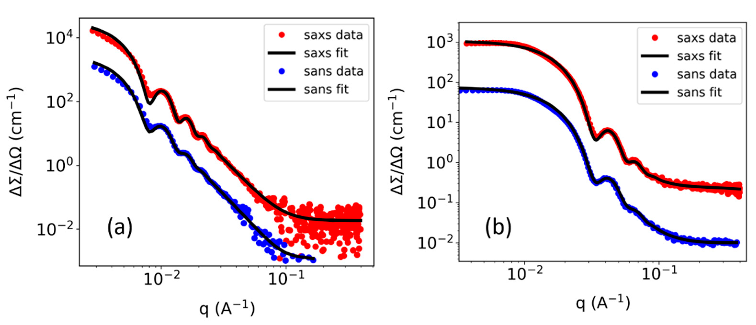
3.3. Overall SAXS Performance

For simultaneous SAXS/SANS experiments, it is important to achieve sufficient data quality in a given measuring time for both methods. For that purpose, it was important to reduce the background signal originating from gamma rays on the 2D X-ray detector of the SAXS instrument for all experimental conditions represented by different SANS collimation lengths and primary neutron beam fluxes and sizes, as well as different SDDX-ray. As demonstrated above, this could be achieved by a targeted enhancement of the neutron and magnetic field shielding, as well as using a double-energy threshold X-ray detector. To demonstrate the success of these upgrades for the performance of the SAXS instrument, two NP samples of nominal sizes of 100 nm and 26 nm were studied by simultaneous SAXS/SANS experiments. The scattering data was collected while the nuclear reactor was in operation (neutron shutter opened, reactor power = 43 MW). SAXS data of the same samples were also collected when the neutron reactor was in the shutdown time (neutron shutter closed, reactor power < 1.5 MW). The latter experiments allow a comparison of the SAXS data of the investigated samples under simultaneous SAXS/SANS data recording conditions with standard laboratory standalone SAXS experiments, where only natural cosmic background contributes to the gamma background of the SAXS data.
SAXS data of silica NP samples were collected for 15 min for both short (0.55 m) and long (1.63 m) SDDX-ray (Figure 4). For long SDDX-ray, SANS measurements of both silica NPs were simultaneously acquired at a long collimation length of 17.6 m. For short SDDX-ray, SANS data were simultaneously measured at two different collimation lengths of 8 m and 5.6 m for 100 nm and 26 nm silica NPs, respectively. The SAXS 2D detector patterns were corrected and normalized, respectively, for measuring time, sample thickness, transmission, flat field, solid angle covered by each detector pixel, and incident intensity. The data were calibrated to absolute intensities, and the q-scale was azimuthally averaged. Background (dark current) correction was performed by subtracting the background intensity measured on the detector at relevant operating conditions. For each neutron collimation length used, the background intensity on the X-ray detector was collected for several minutes while the neutron shutter was opened but the X-ray shutter was closed. Besides this, each SAXS data set was corrected for a constant background determined from a measurement of the corresponding sample with a closed neutron shutter (and reactor power < 1.5 MW).
At a long collimation length (17.6 m), the SAXS 1D profiles are well matching those collected in absence of the high-energy gamma radiation environment (Figure 4a,c). This reveals that the SAXS data quality of a simultaneous SAXS/SANS measurement at those conditions (collimation length = 17.6 m, SDDX-ray = 1.63 m) is not compromised with respect to the background level of a state-of-the-art lab-based SAXS measurement. At a short distance SDDX-ray (0.55 m), SANS images were collected in a synchronous mode at collimation lengths of 8 m (for 100 nm NPs) and 5.6 m (for 26 nm NPs). In the latter configurations, the SAXS profiles exhibited a background intensity approximately 4 and 8 times higher than the standard lab-based SAXS measurements for both 8 m and 5.6 m collimation lengths, respectively (Figure 4b,d). This gamma background originated predominantly from the gamma rays produced by neutron capture within the sample. Nevertheless, the overall background level even for the short neutron collimation lengths is still low and allows high quality simultaneous SAXS/SANS experiments. Based on the q-range of characteristic interference features, scattering power of investigated samples, and experimental conditions (such as collimation length, SDDX-ray, X-ray slit size, and measuring time), one needs to carefully optimize the experimental conditions to achieve the best quality simultaneous SAXS/SANS experiments.
Luckily, the D22 instrument is currently equipped with a second fixed detector at 1.4 m from the sample position in addition to a movable detector, enabling extended SANS q ranges (from 0.003 up to 0.7 Å−1 in a single configuration). Hence, typical SANS experiments are performed with collimation lengths > 5 m. Thus, the high signal-to-background level of the SAXS data detected at different collimation lengths (17.6, 8 and 5.6 m) allows state-of-the-art time-resolved in situ simultaneous SAXS/SANS measurements. This has been demonstrated by studies on the growth of gold nanorods using surfactant-assisted seed-mediated synthesis using simultaneous SAXS/SANS at a neutron collimation length of 8 m [23].
For SAXS/SANS data evaluation of the studied NP dispersions (Figure 5), a simple sphere model with a log-normally distributed radius coupled with a distribution width parameter implemented in SASfit software [33] was used. In SASfit, simultaneous fits of the model to the SAXS and SANS experimental data sets were performed with a convergence χ << 1. The scattering length density contrast ΔSLD = SLDNPs − SLDsolvent was fixed to the known values for silica to be 13.3 and 2.2 Å−2 for SAXS and SANS, respectively. The data can be nicely fitted with the simple model and reveals a particle diameter (with a distribution width) of 110 ± 7 nm (nominal 100 nm) and 26 ± 3 nm (nominal 26 nm), respectively. This simple example demonstrates that a simultaneous fit of a structural model to the SAXS and SANS data set can be easily performed on an absolute intensity scale and reveal the structural parameters, circumventing ambiguities which might be left if only one contrast (X-ray or neutron) is used. This is a particular advantage for in situ or operando studies of complex nanoscale systems which slowly change their structure, e.g., upon a reaction, crystallization, aggregation, gelation, particle formation, growth or aging process.

4. Conclusions

A customized SAXS system was successfully installed and operated in combination with the D22 SANS instrument of ILL, resulting in simultaneously collected SAXS and SANS data of the same part of a sample. Due to high-energy gamma radiation inside the D22 instrumental zone, the SAXS instrument was upgraded with a double-energy threshold detector and optimized lead shielding. Background suppression factors were evaluated for different collimation lengths and SDDX-ray. Extreme background reduction by factors up to 505 was achieved for short collimation lengths and SDDX-ray. Acceptable signal-to-background ratios of the SAXS patterns comparable to SAXS data collected in the absence of gamma background radiation could be achieved. For long neutron collimation lengths of up to 17.6 m and long SDDX-ray, the SAXS profiles did not exhibit an increase in background at all when compared to a typical lab experiment. An increase of the corresponding low background by factors of 4 and 8 were observed for neutron collimation lengths of 8 m and 5.6 m, respectively. In the latter experimental configurations, SAXS data of selected test samples prove the suitability of the SAXS instrument for combined SAXS/SANS experiments. The upgraded SAXS system is now compatible with all collimation settings of the D22 SANS instrument, enabling in situ and operando simultaneous SAXS/SANS measurements for a variety of complex nanoscale sample systems.

Author Contributions

Conceptualization, T.U., C.B., E.M., K.G., T.Z., L.P.; methodology, E.M., K.G., T.Z., L.P., I.S., C.B., A.M.; data curation, E.M., I.S.; formal analysis, E.M., I.S.; writing—original draft preparation, E.M.; writing—review and editing T.U., L.P., A.M., I.S.; technical details, C.B., E.M., T.U., L.P.; funding acquisition, T.U. All authors have read and agreed to the published version of the manuscript.

Funding

This research is primarily funded by the Federal Ministry of Education and Research of Germany (BMBF) in the framework of project No. 05K16WE1. As well, this research received funding from the Deutsche Forschungsgemeinschaft (DFG) through the ‘Cluster of Excellence En-gineering of Advanced Materials (EAM)’, the research training group GRK 1896 ‘In-situ Microscopy with Electrons, X-rays and Scanning Probes’ and the research unit FOR 1878 ‘Functional Molecular Structures on Complex Oxide Surfaces’.

Institutional Review Board Statement

Not applicable.

Informed Consent Statement

Not applicable.

Data Availability Statement

Data presented in this study can be obtained from the authors upon request.

Acknowledgments

We gratefully thank Herbert Lang (ICSP), Jürgen Grasser (ISCP), Mark Jacques (ILL) for their technical support. We acknowledge ILL for the beamtime DOI:10.5291/ILL-DATA.LTP-9-9.

Conflicts of Interest

The authors declare no conflict of interest.

Abbreviations

The following abbreviations are used in this manuscript:
SDDX-ray Sample-to-detector distance of SAXS
SDDNeutronSample-to-detector distance of SANS
SLDScattering length density
col Neutron collimation
diffDifference mode of double-energy threshold X-ray detector, 4–10 KeV
th1A single-energy threshold mode of X-ray detector, 4 KeV
SD1Sample-to-detector distance of SAXS at 0.55 m
SD2Sample-to-detector distance of SAXS at 1.632 m
shieldLead shielding on the SAXS system
NoShieldSAXS system without any lead shielding walls

References

  1. Schmutzler, T.; Schindler, T.; Zech, T.; Lages, S.; Thoma, M.; Appavou, M.S.; Peukert, W.; Spiecker, E.; Unruh, T. n-Hexanol Enhances the Cetyltrimethylammonium Bromide Stabilization of Small Gold Nanoparticles and Promotes the Growth of Gold Nanorods. ACS Appl. Nano Mater. 2019, 2, 3206–3219. [Google Scholar] [CrossRef]
  2. Schindler, T.; Lin, W.; Schmutzler, T.; Lindner, P.; Peukert, W.; Segets, D.; Unruh, T. Evolution of the Ligand Shell Around Small ZnO Nanoparticles During the Exchange of Acetate by Catechol: A Small Angle Scattering Study. ChemNanoMat 2019, 5, 116–123. [Google Scholar] [CrossRef]
  3. Schmiele, M.; Busch, S.; Morhenn, H.; Schindler, T.; Schmutzler, T.; Schweins, R.; Lindner, P.; Boesecke, P.; Westermann, M.; Steiniger, F.; et al. Structural Characterization of Lecithin-Stabilized Tetracosane Lipid Nanoparticles. Part I: Emulsions. J. Phys. Chem. B 2016, 120, 5505–5512. [Google Scholar] [CrossRef]
  4. Schmutzler, T.; Schindler, T.; Goetz, K.; Appavou, M.S.; Lindner, P.; Prevost, S.; Unruh, T. Concentration dependent morphology and composition of n-alcohol modified cetyltrimethylammonium bromide micelles. J. Phys. Condens. Mat. 2018, 30. [Google Scholar] [CrossRef] [PubMed]
  5. Schmutzler, T.; Schindler, T.; Schmiele, M.; Appavou, M.S.; Lages, S.; Kriele, A.; Gilles, R.; Unruh, T. The influence of n-hexanol on the morphology and composition of CTAB micelles. Colloid Surface A 2018, 543, 56–63. [Google Scholar] [CrossRef]
  6. Mohl, G.E.; Metwalli, E.; Bouchet, R.; Phan, T.N.T.; Cubitt, R.; Muller-Buschbaum, P. In Operando Small-Angle Neutron Scattering Study of Single-Ion Copolymer Electrolyte for Li-Metal Batteries. ACS Energy Lett. 2018, 3, 1–6. [Google Scholar] [CrossRef]
  7. Mohl, G.E.; Metwalli, E.; Muller-Buschbaum, P. In Operando Small-Angle X-ray Scattering Investigation of Nanostructured Polymer Electrolyte for Lithium-Ion Batteries. ACS Energy Lett. 2018, 3, 1525–1530. [Google Scholar] [CrossRef]
  8. Gehrer, S.; Schmiele, M.; Westermann, M.; Steiniger, F.; Unruh, T. Liquid Crystalline Phase Formation in Suspensions of Solid Trimyristin Nanoparticles. J. Phys. Chem. B 2014, 118, 11387–11396. [Google Scholar] [CrossRef] [PubMed]
  9. Schmiele, M.; Gehrer, S.; Unruh, T. Small-angle scattering simulations for suspensions of nanocrystals. Acta Crystallogr. A 2014, 70, C597. [Google Scholar] [CrossRef] [Green Version]
  10. Schuldes, I.; Noll, D.M.; Schindler, T.; Zech, T.; Gotz, K.; Appavou, M.S.; Boesecke, P.; Steiniger, F.; Schulz, P.S.; Unruh, T. Internal Structure of Nanometer-Sized Droplets Prepared by Antisolvent Precipitation. Langmuir 2019, 35, 13578–13587. [Google Scholar] [CrossRef]
  11. Unruh, T. Interpretation of small-angle X-ray scattering patterns of crystalline triglyceride nanoparticles in dispersion. J. Appl. Crystallogr. 2007, 40, 1008–1018. [Google Scholar] [CrossRef]
  12. Schmiele, M.; Schindler, T.; Westermann, M.; Steiniger, F.; Radulescu, A.; Kriele, A.; Gilles, R.; Unruh, T. Mesoscopic Structures of Triglyceride Nanosuspensions Studied by Small-Angle X-ray and Neutron Scattering and Computer Simulations. J. Phys. Chem. B 2014, 118, 8808–8818. [Google Scholar] [CrossRef] [PubMed]
  13. Wibmer, L.; Lages, S.; Unruh, T.; Guldi, D.M. Excitons and Trions in One-Photon- and Two-Photon-Excited MoS2: A Study in Dispersions. Adv. Mater. 2018, 30. [Google Scholar] [CrossRef] [PubMed]
  14. Putnam, C.D.; Hammel, M.; Hura, G.L.; Tainer, J.A. X-ray solution scattering (SAXS) combined with crystallography and computation: Defining accurate macromolecular structures, conformations and assemblies in solution. Q. Rev. Biophys. 2007, 40, 191–285. [Google Scholar] [CrossRef] [PubMed]
  15. Glatter, O.; Kratky, O. Small Angle X-ray Scattering; Academic Press: London, UK; New York, NY, USA, 1982; p. 515. [Google Scholar]
  16. Serdyuk, I.N.; Tsalkova, T.N.; Svergun, D.I.; Izotova, T.D. Determination of Radii of Gyration of Particles by Small-Angle Neutron-Scattering—Calculation of the Effect of Aggregates—Appendix. J. Mol. Biol. 1987, 194, 126–128. [Google Scholar] [CrossRef]
  17. Koch, M.H.J.; Vachette, P.; Svergun, D.I. Small-angle scattering: A view on the properties, structures and structural changes of biological macromolecules in solution. Q. Rev. Biophys. 2003, 36, 147–227. [Google Scholar] [CrossRef] [Green Version]
  18. Schindler, T.; Walter, J.; Peukert, W.; Segets, D.; Unruh, T. In Situ Study on the Evolution of Multimodal Particle Size Distributions of ZnO Quantum Dots: Some General Rules for the Occurrence of Multimodalities. J. Phys. Chem. B 2015, 119, 15370–15380. [Google Scholar] [CrossRef]
  19. Futscher, M.H.; Schultz, T.; Frisch, J.; Ralaiarisoa, M.; Metwalli, E.; Nardi, M.V.; Muller-Buschbaum, P.; Koch, N. Electronic properties of hybrid organic/inorganic semiconductor pn-junctions. J. Phys. Condens. Mat. 2019, 31. [Google Scholar] [CrossRef]
  20. Wang, X.Y.; Meng, J.Q.; Wang, M.M.; Xiao, Y.; Liu, R.; Xia, Y.G.; Yao, Y.; Metwalli, E.; Zhang, Q.; Qiu, B.; et al. Facile Scalable Synthesis of TiO2/Carbon Nanohybrids with Ultrasmall TiO2 Nanoparticles Homogeneously Embedded in Carbon Matrix. ACS Appl. Mater. Inter. 2015, 7, 24247–24255. [Google Scholar] [CrossRef]
  21. Wang, X.Y.; Zhao, D.; Wang, C.; Xia, Y.G.; Jiang, W.S.; Xia, S.L.; Yin, S.S.; Zuo, X.X.; Metwalli, E.; Xiao, Y.; et al. Role of Nickel Nanoparticles in High-Performance TiO2/Ni/Carbon Nanohybrid Lithium/Sodium-Ion Battery Anodes. Chem. Asian J. 2019, 14, 2169. [Google Scholar] [CrossRef] [Green Version]
  22. McAulay, K.; Wang, H.; Fuentes-Caparros, A.M.; Thomson, L.; Khunti, N.; Cowieson, N.; Cui, H.G.; Seddon, A.; Adams, D.J. Isotopic Control over Self-Assembly in Supramolecular Gels. Langmuir 2020, 36, 8626–8631. [Google Scholar] [CrossRef]
  23. Metwalli, E.; Gotz, K.; Lages, S.; Bar, C.; Zech, T.; Noll, D.M.; Schuldes, I.; Schindler, T.; Prihoda, A.; Lang, H.; et al. A novel experimental approach for nanostructure analysis: Simultaneous small-angle X-ray and neutron scattering. J. Appl. Crystallogr. 2020, 53, 722–733. [Google Scholar] [CrossRef]
  24. Deschler-Erb, E.; Lehmann, E.H.; Pernet, L.; Vontobel, P.; Hartmann, S. The complementary use of neutrons and X-rays for the non-destructive investigation of archaeological objects from Swiss collections. Archaeometry 2004, 46, 647–661. [Google Scholar] [CrossRef]
  25. Lehmann, E.H.; Mannes, D.; Kaestner, A.P.; Hovind, J.; Trtik, P.; Strobl, M. The XTRA Option at the NEUTRA Facility—More Than 10 Years of Bi-Modal Neutron and X-ray Imaging at PSI. Appl. Sci. 2021, 11, 3825. [Google Scholar] [CrossRef]
  26. Robuschi, S.; Tengattini, A.; Dijkstra, J.; Fernández, I.; Lundgren, K. A closer look at corrosion of steel reinforcement bars in concrete using 3D neutron and X-ray computed tomography. Cem. Concr. Res. 2021, 144, 106439. [Google Scholar] [CrossRef]
  27. Ziesche, R.F.; Arlt, T.; Finegan, D.P.; Heenan, T.M.M.; Tengattini, A.; Baum, D.; Kardjilov, N.; Markotter, H.; Manke, I.; Kockelmann, W.; et al. 4D imaging of lithium-batteries using correlative neutron and X-ray tomography with a virtual unrolling technique. Nat. Commun. 2020, 11. [Google Scholar] [CrossRef] [PubMed] [Green Version]
  28. Khaydukov, Y.; Soltwedel, O.; Keller, T. NREX: Neutron reflectometer with X-ray option. J. Large-Scale Res. Facil. 2015, 1, A38. [Google Scholar] [CrossRef] [Green Version]
  29. Mannes, D.; Schmid, F.; Frey, J.; Schmidt-Ott, K.; Lehmann, E. Combined Neutron and X-ray imaging for non-invasive investigations of cultural heritage objects. Phys. Procedia 2015, 69, 653–660. [Google Scholar] [CrossRef] [Green Version]
  30. Makarova, M.V.; Kravtsov, E.A.; Proglyado, V.V.; Khaydukov, Y.; Ustinov, V.V. Structure and Magnetism of Co/Dy Superlattices. Phys. Solid State 2020, 62, 1664–1666. [Google Scholar] [CrossRef]
  31. Murray, E.; Smith, A.G.; Pollitt, A.J.; Matarranz, J.; Tsekhanovich, I.; Soldner, T.; Koster, U.; Biswas, D.C. Measurement of Gamma Energy Distributions and Multiplicities Using STEFF. Nucl. Data Sheets 2014, 119, 217–220. [Google Scholar] [CrossRef]
  32. Dewhurst, C. GRASP. Available online: https://www.ill.eu/users/support-labs-infrastructure/software-scientific-tools/grasp (accessed on 15 March 2021).
  33. Bressler, I.; Kohlbrecher, J.; Thunemann, A.F. SASfit: A tool for small-angle scattering data analysis using a library of analytical expressions. J. Appl. Crystallogr. 2015, 48, 1587–1598. [Google Scholar] [CrossRef] [PubMed] [Green Version]
Figure 2. Background intensity collected using EIGER2 X 1M detector while neutron shutter is opened for two different neutron collimation lengths (2.8 m and 17.6 m). Two measurements for the single-energy threshold mode (dashed area, th1 mode = 4 keV) and two for the double-energy mode (solid area, diff = 4–10 keV) were performed for both the shielded and unshielded SAXS instrument. All background measurements were repeated for short (red, SD1 = 0.55 m) and long (blue, SD2 = 1.63 m) SDDX-ray. Lab environment data are based on background intensity measurements while the reactor power was off. The intensity (cts/s) data are shown on a logarithmic scale.
Applsci 11 05925 g002
Figure 3. X-ray beam intensity before (no magnetic shielding, green line) and after (magnetic field shielding, black symbols) installing a 1 mm thick plate of a soft ferromagnetic alloy at the X-ray source. A magnetic field strength of approximately 2–5 Gauss at the X-ray source causes an X-ray beam drift.
Applsci 11 05925 g003
Figure 4. 1D SAXS profiles at various neutron collimation lengths and SDDX-ray of (a,b) 100 nm and (c,d) 26 nm silica NPs collected with the neutron beam switched on (orange curves) or off (blue) (simultaneous and asynchronous modes). The asynchronous mode represents a standard laboratory SAXS experimental condition where neutron shutter was closed and the nuclear reactor was almost switched off (below < 1.5 MW). SAXS/SANS simultaneous mode represents an experimental condition where the neutron shutter of D22 was open (reactor power = 43 MW). At long SDDX-ray, the SAXS profiles in simultaneous mode are well matching those of the asynchronous mode, which leads to a good signal-to-noise ratio for the SAXS data. For short collimations, noticeable background which was approximately 4 and 8 times larger than that for standard lab SAXS measurements was found for both collimations 8 m (b) and 5.6 m (d), respectively.
Applsci 11 05925 g004
Figure 5. Solvent subtracted SAXS/SANS intensity profiles of 100 nm (a) and 26 nm (b) silica NPs acquired in a simultaneous mode. A common structural model is used for each sample to simultaneously fit both SAXS and SANS data collected from the same sample volume.
Applsci 11 05925 g005
Table 1. Background suppression factors (SFs) for all investigated experimental configurations in an optimized lead shielding setup with a double-energy threshold energy detector (total SF ≈ lead shielding SF × detector SF).
SDDX-ray
(m)
Neutron Collimation Length
(m)
X-ray Energy Mode
(keV)
Shielding Suppression FactorDetector Suppression FactorTotal Suppression Factor
0.552.8487-87
4–10895.5505
17.6458-58
4–10545.0275
1.642.8450-50
4–10495.1248
17.6417-17
4–10196.1116
Publisher’s Note: MDPI stays neutral with regard to jurisdictional claims in published maps and institutional affiliations.

