
Study of Immediate Implants Placed in Mandibular Alveolar Bone Reconstructed with Screw-Guided Bone Regeneration Technique: A 24-Months Follow-Up


Abstract
:1. Introduction
2. Materials and Method
2.1. Patient Selection and Study Design
- Test group (n = 10; 30 implants): simultaneous implant placement with ridge augmentation by S-GBR technique.
- Control group (n = 10; 32 implants): implant placement in naturally healed sites (non-grafted).
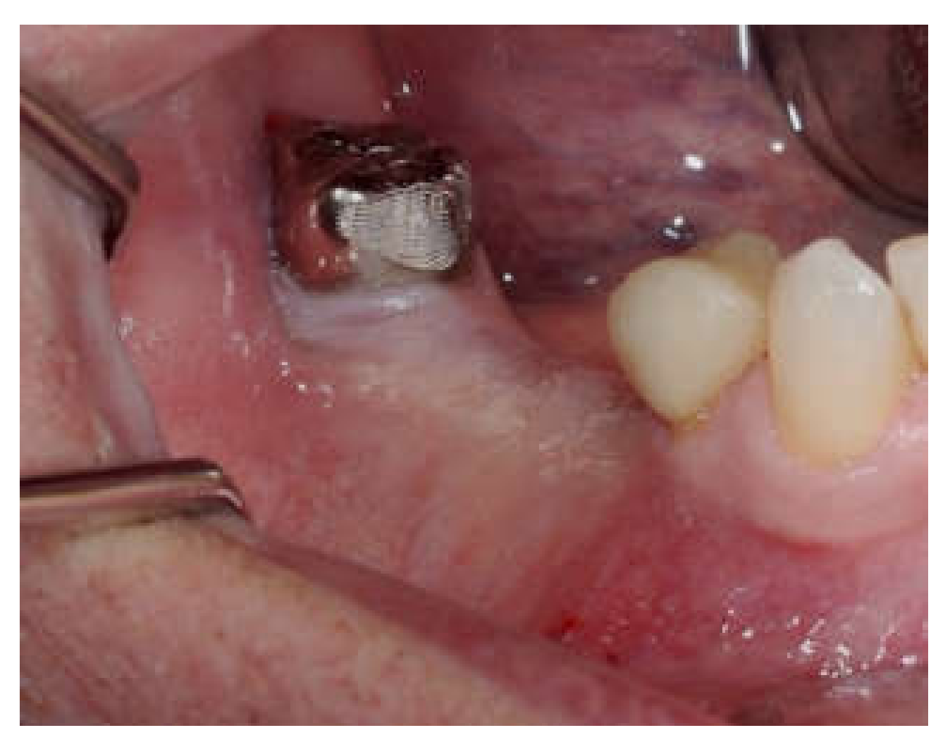
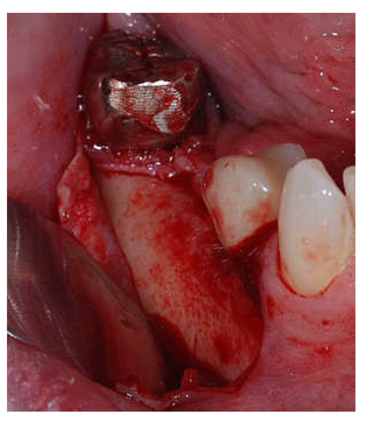
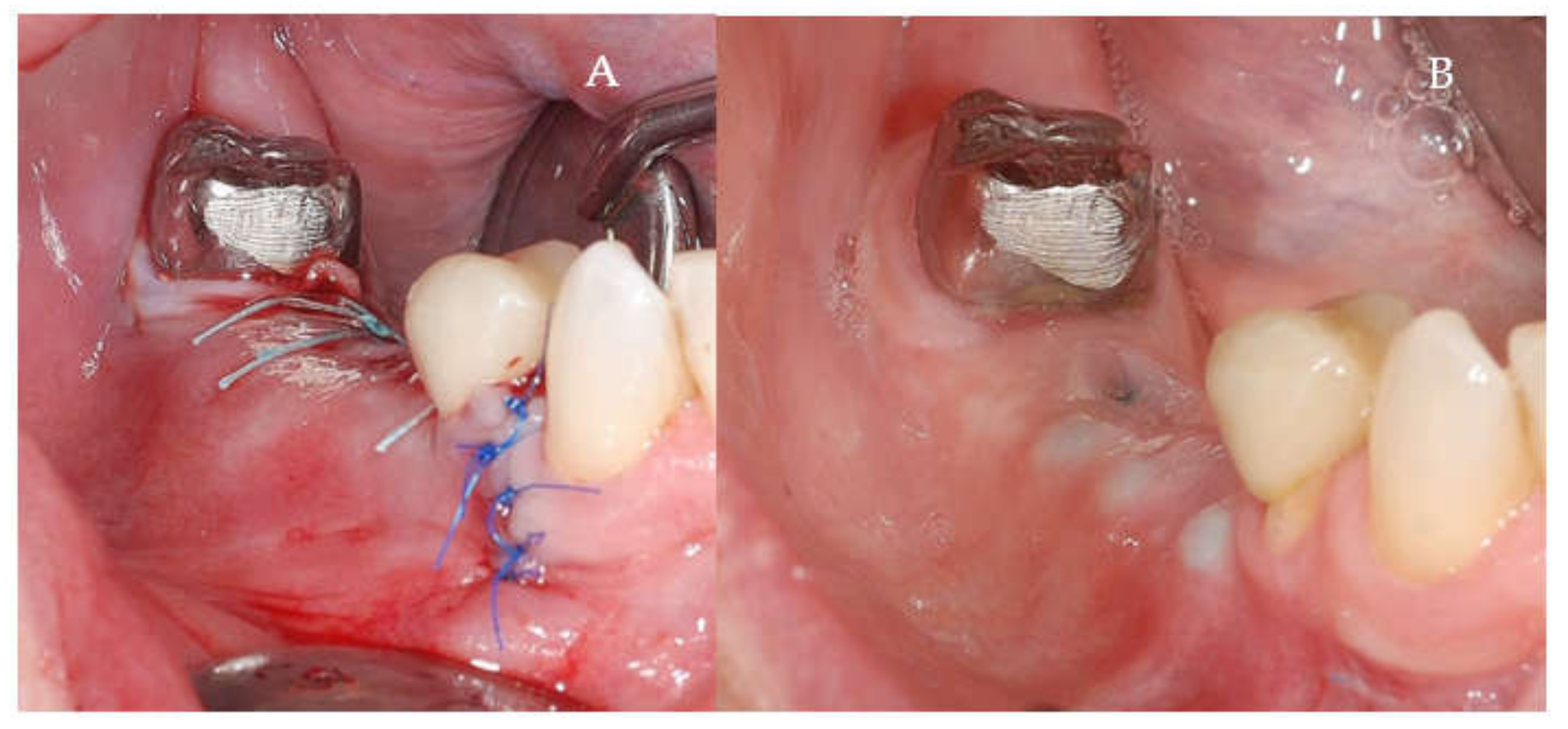
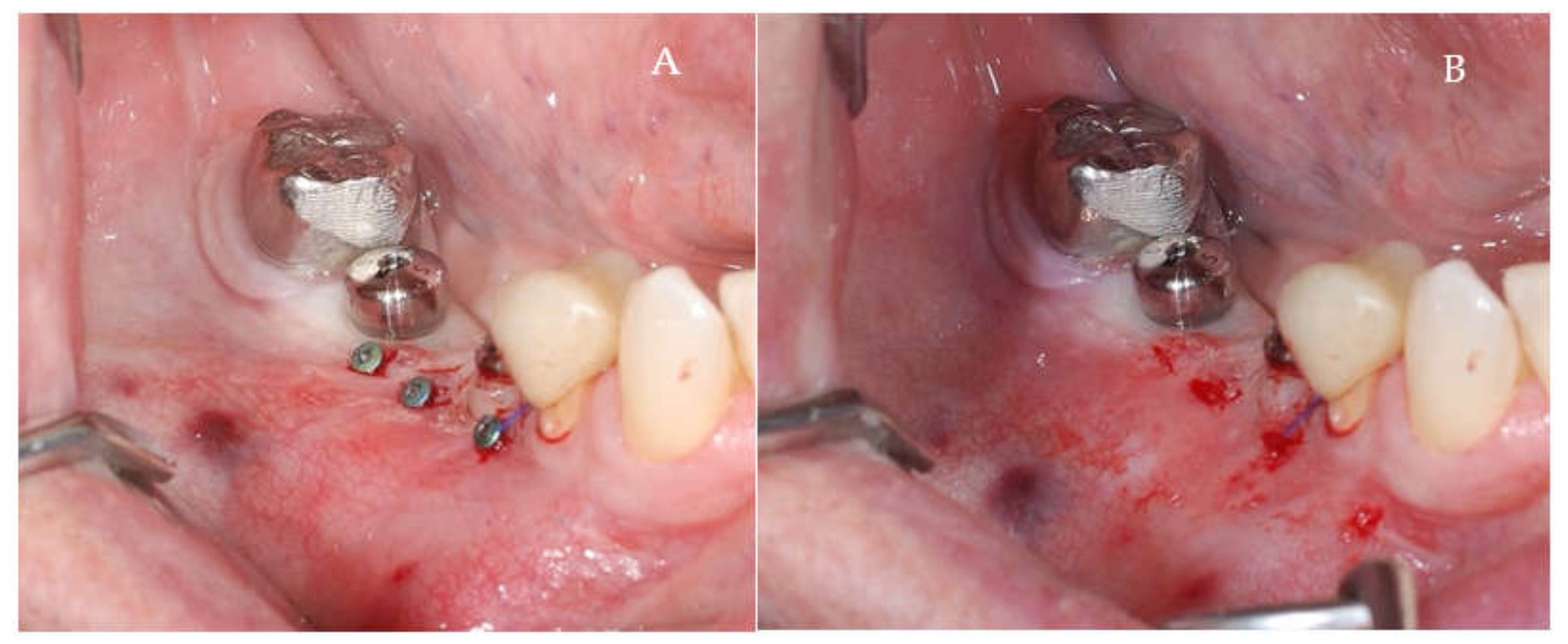
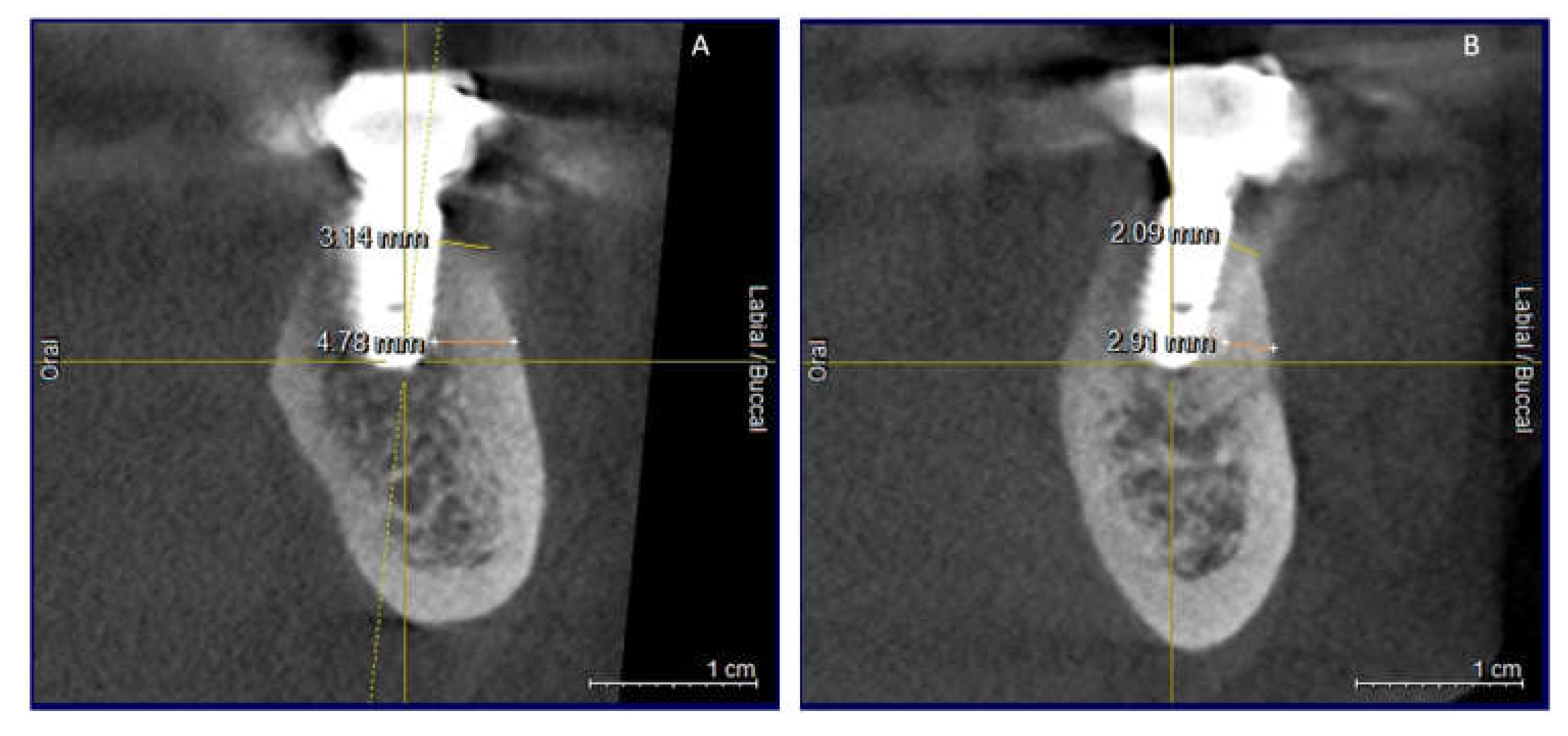
2.2. Surgical Procedures
- (a)
- Dental implants placement simultaneously with alveolar augmentation by SGBR technique (xenograft, or a mixture with autogenous bone; porcine collagen membrane) (test group);
- (b)
- Standard dental implants placement (control group).
2.3. Results Analysis
- (1)
- Clinical inflammation signs (erythema, swelling, bleeding on probing at force not overpassing 0.25 N, and/or suppuration) [12]
- (2)
- Mild increase of probing depth.
- (3)
- Absence of peri-implant bone loss (after post-loading stage of bone remodeling).
- (1)
- Clinical inflammation signs (erythema, swelling, bleeding on probing and/or suppuration);
- (2)
- Probing depth >3 mm, and/or recession of the mucosal margin.
- (3)
- Loss of peri-implant bone >3 mm (from implant shoulder).
2.4. Statistical Analysis
3. Results
4. Discussion
5. Conclusions
Author Contributions
Funding
Institutional Review Board Statement
Informed Consent Statement
Data Availability Statement
Conflicts of Interest
References
- Sammartino, G.; Bernard, J.P. A clinical round table about the treatment of the severely resorbed posterior mandible. Part 1: Challenges, endeavor and perspectives. POSEIDO 2013, 1, 65–67. [Google Scholar]
- Tolstunov, L.; Hamrick, J.F.E.; Broumand, V.; Shilo, D.; Rachmiel, A. Bone Augmentation Techniques for Horizontal and Vertical Alveolar Ridge Deficiency in Oral Implantology. Oral Maxillofac. Surg. Clin. N. Am. 2019, 31, 163–191. [Google Scholar] [CrossRef] [PubMed]
- Mittal, Y.; Jindal, G.; Garg, S. Bone manipulation procedures in dental implants. Indian J. Dent. 2016, 7, 86–94. [Google Scholar] [CrossRef] [Green Version]
- Chiapasco, M.; Casentini, P.; Zaniboni, M. Bone augmentation procedures in implant dentistry. Int. J. Oral Maxillofac. Implant. 2009, 24, 237–259. [Google Scholar]
- Le, B.; Burstein, J. Esthetic grafting for small volume hard and soft tissue contour defects for implant site development. Implant. Dent. 2008, 17, 136–141. [Google Scholar] [CrossRef] [Green Version]
- Toeroek, R.; Mazor, Z.; Del Corso, M.; Dohan Ehrenfest, D.M. The concept of Screw-Guided Bone Regeneration (S-GBR). Part 1: From sinus-lift to general applications in the resorbed maxilla and mandible. POSEIDO 2013, 1, 69–84. [Google Scholar]
- Toeroek, R.; Dohan Ehrenfest, D.M. The concept of Screw-Guided Bone Regeneration (S-GBR). Part 2: S-GBR in the severely resorbed preimplant posterior mandible using bone xenograft and Leukocyte and Platelet-Rich Fibrin (L-PRF): A 5-year follow-up. POSEIDO 2013, 1, 85–92. [Google Scholar]
- Liu, J.; Kerns, D.G. Mechanisms of Guided Bone Regeneration: A Review. Open Dent. J. 2014, 8, 56–65. [Google Scholar] [CrossRef] [Green Version]
- Buser, D.; Mericske-Stern, R.; Dula, K.; Lang, N.P. Clinical Experience with One-Stage, Non-Submerged Dental Implants. Adv. Dent. Res. 1999, 13, 153–161. [Google Scholar] [CrossRef]
- Berglundh, T.; Armitage, G.; Araujo, M.G.; Avila-Ortiz, G.; Blanco, J.; Camargo, P.M.; Chen, S.; Cochran, D.; Derks, J.; Figuero, E.; et al. Peri-implant diseases and conditions: Consensus report of workgroup 4 of the 2017 World Workshop on the Classification of Periodontal and Peri-Implant Diseases and Conditions. J. Periodontol. 2018, 89, S313–S318. [Google Scholar] [CrossRef] [PubMed]
- Heitz-Mayfield, L.J.A.; Heitz, F.; Lang, N.P. Implant Disease Risk Assessment IDRA–a tool for preventing peri-implant disease. Clin. Oral Implant. Res. 2020, 31, 397–403. [Google Scholar] [CrossRef] [PubMed] [Green Version]
- Lang, N.P.; Berglundh, T.; Working Group 4 of Seventh European Workshop on Periodontology. Periimplant diseases: Where are we now?—Consensus of the Seventh European Workshop on Periodontology. J. Clin. Periodontol. 2011, 38 (Suppl. 11), 178–181. [Google Scholar] [CrossRef] [PubMed] [Green Version]
- Mombelli, A.; van Oosten, M.A.; Schurch, E., Jr.; Land, N.P. The microbiota associated with successful or failing osseointegrated titanium implants. Oral Microbiol. Immunol. 1987, 2, 145–151. [Google Scholar] [CrossRef]
- Mombelli, A.; Lang, N.P. The diagnosis and treatment of peri-implantitis. Periodontol. 2000 1998, 17, 63–76. [Google Scholar] [CrossRef]
- Salvi, G.E.; Lang, N.P. Diagnostic parameters for monitoring peri-implant conditions. Int. J. Oral Maxillofac. Implant. 2004, 19, 116–127. [Google Scholar]
- Moy, P.K.; Aghaloo, T. Risk factors in bone augmentation procedures. Periodontol. 2000 2019, 81, 76–90. [Google Scholar] [CrossRef]
- Dastaran, M.; Bailey, D.; Austin, S.; Chandu, A.; Judge, R. Complications of augmentation procedures for dental implants in private practice, Victoria, Australia. Aust. Dent. J. 2019, 64, 223–228. [Google Scholar] [CrossRef]
- Elakkiya, S.; Ramesh, A.S.; Prabhu, K. Systematic analysis on the efficacy of bone enhancement methods used for success in dental implants. J. Indian Prosthodont. Soc. 2017, 17, 219–225. [Google Scholar] [CrossRef] [PubMed]
- Li, J.; Xuan, F.; Choi, B.H.; Jeong, S.M. Minimally invasive ridge augmentation using xenogenous bone blocks in an atrophied posterior mandible: A clinical and histological study. Implant Dent. 2013, 22, 112–116. [Google Scholar] [CrossRef]
- Pellicer-Chover, H.; Peñarrocha-Oltra, D.; Bagán, L.; Fichy-Fernandez, A.J.; Canullo, L.; Peñarrocha-Diago, M. Single-blind randomized clinical trial to evaluate clinical and radiological outcomes after one year of immediate versus delayed implant placement supporting full-arch prostheses. Medicina Oral Patología Oral y Cirugia Bucal 2014, 19, e295–e301. [Google Scholar] [CrossRef] [PubMed]
- Wessing, B.; Lettner, S.; Zechner, W. Guided Bone Regeneration with Collagen Membranes and Particulate Graft Materials: A Systematic Review and Meta-Analysis. Int. J. Oral Maxillofac. Implant. 2018, 33, 87–100. [Google Scholar] [CrossRef] [PubMed]
- Aghaloo, T.L.; Moy, P.K. Which hard tissue augmentation techniques are the most successful in furnishing bony support for implant placement? Int. J. Oral Maxillofac. Implant. 2007, 22, 49–70. [Google Scholar]
- Donos, N.; Mardas, N.; Chadha, V. Clinical outcomes of implants following lateral bone augmentation: Systematic assessment of available options (barrier membranes, bone grafts, split osteotomy). J. Clin. Periodontol. 2008, 35, 173–202. [Google Scholar] [CrossRef] [PubMed]
- Schwarz, F.; Giannobile, W.V.; Jung, R.E.; Groups of the 2nd Osteology Foundation Consensus Meeting. Evidence-based knowledge on the aesthetics and maintenance of peri-implant soft tissues: Osteology Foundation Consensus Report Part 2-Effects of hard tissue augmentation procedures on the maintenance of peri-implant tissues. Clin. Oral Implant. Res. 2018, 29 (Suppl. 15), 11–13. [Google Scholar] [CrossRef] [Green Version]
- Elnayef, B.; Porta, C.; Suárez-López Del Amo, F.; Mordini, L.; Gargallo-Albiol, J.; Hernández-Alfaro, F. The Fate of Lateral Ridge Augmentation: A Systematic Review and Meta-Analysis. Int. J. Oral Maxillofac. Implant. 2018, 33, 622–635. [Google Scholar] [CrossRef] [Green Version]
- Sakka, S.; Baroudi, K.; Nassani, M.Z. Factors associated with early and late failure of dental implants. J. Investig. Clin. Dent. 2012, 3, 258–261. [Google Scholar] [CrossRef]
- Smeets, R.; Stadlinger, B.; Schwarz, F.; Beck-Broichsitter, B.; Jung, O.; Precht, C.; Kloss, F.; Gröbe, A.; Heiland, M.; Ebker, T. Impact of Dental Implant Surface Modifications on Osseointegration. BioMed Res. Int. 2016, 2016, 6285620. [Google Scholar] [CrossRef] [Green Version]
- Topalo, E. Dental implants exposure prevention in their osseointegration period. Rom. J. Oral Rehabil. 2019, 11, 43–48. [Google Scholar]
- Ionescu, A.; Dodi, A.; Panagopoulos, V.; Nicolescu, M.I.; Mihai, A.T.; Tanase, G.; Vlasceanu, D. Biomechanical consequences of dental implants inserted in augmented alveolar ridges—A comparative study between tissue-level and bone-level implants: Finite elements analysis. Rom. J. Oral Rehabil. 2019, 11, 82–88. [Google Scholar]


| Test Group (S-GBR Sites) | Control Group (Non-Grafted Sites) | Total | |
|---|---|---|---|
| N of subjects (Ns) | 10 | 10 | 40 |
| N of sites (Ni) | 30 | 32 | 124 |
| Mean age (range) | (62.30 ± 16.059) | (56.60 ± 14.159) | (59.45 ± 15.220) |
| Gender | |||
| Men, Ns (Ni) | 4 (12) | 6 (18) | 10 (30) |
| Women, Ns (Ni) | 6 (18) | 4 (14) | 10 (32) |
| Component | Description |
|---|---|
| Population (P) | Mandibular edentulous patients requiring bone grafting and dental implants |
| Intervention (I) | Augmentation of alveolar bone by S-GBR technique. Immediate implant placement in grafted sites |
| Comparison (C) | Implant placement in non-grafted sites |
| Outcomes (O) | Primary outcomes: implants success; implants survival Secondary outcomes: clinical parameters of soft tissues: mPI, mGI, probing depth (mm), keratinized mucosa (mm), occurrence of peri-mucositis/peri-implantitis (%); marginal-bone-level (MBL) (mm); |
| No. | Procedure | Instruments and Materials | Role |
|---|---|---|---|
| 1. | Local anesthesia | Ultracain DS-Forte (Sanofi, Paris, France) | Comfort of patient and operator |
| 2. | Full-thickness flaps in the resorption bone area | DeveMed GmbH, (Tuttlingen, Germany) | Opening of the surgical field |
| 3. | Immediate implants insertion (3.5–4.5 mm diameter, 10–13 mm length) | BioSTI implants (Tafers, Switzerland) Dentium implants (Seoul, South Korea) | |
| 4. | Periosteal incisions on the flaps | AesculapAG (Tuttlingen, Germany) | Promotion of tension-free closure of the flaps |
| 5. | Insertion of the osteosynthesis screws (1.5 mm diameter, 8 mm length) on the buccal face of the alveolar bone (45° angle to the alveolar crest) | Implantology kits Synthes GmbH, (Zuchwil, Switzerland) DeveMed GmbH, (Tuttlingen,Germany) | Maintenance of the space for the grafted area |
| 6. | Covering of the exposed implant bone surface area with a small layer of autologous bone, followed by the placement of xenograft adjacent to the head of osteosynthesis screws. Covering of the grafted area with a resorbable collagen membrane | Xenograft CompactBone, (Dentegris GmbH, Rheinberg, Germany) Porcine pericardial tissue membrane BoneProtect Membrane (Dentegris GmbH) | Isolation and protection of the graft from the gingival tissue. Stimulation of the gingival healing. Protection of the surgical site from gingival dehiscence during the next 3–4 months and Reconstruction of the lateral resorbed bone area |
| 7. | Suture of the surgical site | Non-resorbable sutures (polypropylene 5.0, Hu-Friedy, Chicago, IL, USA) | Protection of grafted bone and peri-implant tissues |
| 8. | Post-operative care (7 days): antibiotherapy, analgesics, rinse of oral cavity with chlorhexidine 0.5% | Augmentin 625 mg, GlaxoSmithKline Pharma, Vienna, Austria) and analgesics (Ibuflam 600, Zentiva, Lichtenstein) | Control of post-operative pain and inflammatory processes |
| 9. | Removal of sutures at 9–11 days after the surgical intervention | ||
| 10. | Second stage surgery: removal of the osteosynthesis screws at 4 months after the surgical intervention |
| Score | Description |
|---|---|
| 0 | Undetected bacterial plaque |
| 1 | Bacterial plaque detected by peri-implant probing |
| 2 | Bacterial plaque detected by visual inspection |
| 3 | Peri-implant abundant white materia |
| Score | Description |
|---|---|
| 0 | No bleeding when the periodontal probe is passed along the peri-implant mucosal margin |
| 1 | Isolated bleeding spots |
| 2 | Bleeding line on mucosal margin |
| 3 | Heavy bleeding; profuse bleeding |
| S-GBR Group | Control | p-Value | |
|---|---|---|---|
| Mean Plaque Index (mPI) (mm) | 0.97 ± 0.882 (SD) | 0.66 ± 0.695 (SD) | 0.046 * |
| Keratinized mucosa (KM) | 4.13 ± 1.033 (SD) | 3.34 ± 0.821 (SD) | 0.000 ** |
| Probing Depth (PD) (mm) | 3.50 ± 1.372 (SD) | 2.56 ± 1.332 (SD) | 0.000 ** |
| Mean mGI | 0.90 ± 1.020 (SD) | 0.56 ± 0.794 (SD) | 0.061 |
| mGI (scores) | 0.075 | ||
| Score 0 | 43.3% | 56.3% | |
| Score 1 | 36.7% | 37.5% | |
| Score 2 | 6.7% | 0.0% | |
| Score 3 | 13.3% | 6.3% | |
| MBL (mm) | 2.20 ± 1.867 (SD) | 1.09 ± 1.678 (SD) | 0.000 ** |
| S-GBR Group | Control | p-Value | |
|---|---|---|---|
| Success rate | 76.7% | 90.6% | 0.001 ** |
| Survival rate | 86.7% | 93.8% | 0.182 |
Publisher’s Note: MDPI stays neutral with regard to jurisdictional claims in published maps and institutional affiliations. |
© 2021 by the authors. Licensee MDPI, Basel, Switzerland. This article is an open access article distributed under the terms and conditions of the Creative Commons Attribution (CC BY) license (https://creativecommons.org/licenses/by/4.0/).
Share and Cite
Török, B.; Török, R.; Dohan Ehrenfest, D.M.; Agop-Forna, D.; Dascălu, C.; Forna, N.C. Study of Immediate Implants Placed in Mandibular Alveolar Bone Reconstructed with Screw-Guided Bone Regeneration Technique: A 24-Months Follow-Up. Appl. Sci. 2021, 11, 6054. https://doi.org/10.3390/app11136054
Török B, Török R, Dohan Ehrenfest DM, Agop-Forna D, Dascălu C, Forna NC. Study of Immediate Implants Placed in Mandibular Alveolar Bone Reconstructed with Screw-Guided Bone Regeneration Technique: A 24-Months Follow-Up. Applied Sciences. 2021; 11(13):6054. https://doi.org/10.3390/app11136054
Chicago/Turabian StyleTörök, Bianca, Roland Török, David M. Dohan Ehrenfest, Doriana Agop-Forna, Cristina Dascălu, and Norina Consuela Forna. 2021. "Study of Immediate Implants Placed in Mandibular Alveolar Bone Reconstructed with Screw-Guided Bone Regeneration Technique: A 24-Months Follow-Up" Applied Sciences 11, no. 13: 6054. https://doi.org/10.3390/app11136054
APA StyleTörök, B., Török, R., Dohan Ehrenfest, D. M., Agop-Forna, D., Dascălu, C., & Forna, N. C. (2021). Study of Immediate Implants Placed in Mandibular Alveolar Bone Reconstructed with Screw-Guided Bone Regeneration Technique: A 24-Months Follow-Up. Applied Sciences, 11(13), 6054. https://doi.org/10.3390/app11136054