Article

Arabidopsis Disulfide Reductase, Trx-h2, Functions as an RNA Chaperone under Cold Stress

Division of Applied Life Science (BK21+) and Plant Molecular Biotechnology Research Center, Gyeongsang National University, Jinju 52828, Korea
*
Author to whom correspondence should be addressed.
These authors contributed equally to this work.
Appl. Sci. 2021, 11(15), 6865; https://doi.org/10.3390/app11156865
Submission received: 24 June 2021 / Revised: 23 July 2021 / Accepted: 23 July 2021 / Published: 26 July 2021

Abstract

:
The thioredoxin-h (Trx-h) family of Arabidopsis thaliana comprises cytosolic disulfide reductases. However, the physiological function of Trx-h2, which contains an additional 19 amino acids at its N-terminus, remains unclear. In this study, we investigated the molecular function of Trx-h2 both in vitro and in vivo and found that Arabidopsis Trx-h2 overexpression (Trx-h2OE) lines showed significantly longer roots than wild-type plants under cold stress. Therefore, we further investigated the role of Trx-h2 under cold stress. Our results revealed that Trx-h2 functions as an RNA chaperone by melting misfolded and non-functional RNAs, and by facilitating their correct folding into active forms with native conformation. We showed that Trx-h2 binds to and efficiently melts nucleic acids (ssDNA, dsDNA, and RNA), and facilitates the export of mRNAs from the nucleus to the cytoplasm under cold stress. Moreover, overexpression of Trx-h2 increased the survival rate of the cold-sensitive E. coli BX04 cells under low temperature. Thus, our data show that Trx-h2 performs function as an RNA chaperone under cold stress, thus increasing plant cold tolerance.

1. Introduction

Since plants are sessile organisms, they have evolved sophisticated defense mechanisms to cope with unfavorable environmental conditions. Environmental stresses trigger a variety of defense signaling pathways involved in the production of reactive oxygen species (ROS) [1,2]. Depending on the concentration, ROS function as a double-edged sword, playing beneficial or detrimental roles in living organisms [3]. At a normal level, ROS participate in a number of signaling cascades that regulate diverse physiological and metabolic processes in plants to ensure optimal growth and development. However, excessive ROS induce damage to intracellular macromolecules and result in ER stress and ultimately cell death [4,5]. Therefore, to prevent apoptosis, ER stress must be regulated through UPR, such as refolding and ERAD, and a homeostatic level of ROS need to be maintained to function as secondary messengers, such as hormones, cyclic nucleotides, and calcium ions. [6,7]. To balance the level of ROS derived from both exogenous sources (temperature, drought, UV light, and various environmental stresses) and endogenous sources (chloroplast, mitochondria, and peroxisome), plants produce diverse non-enzymatic and enzymatic antioxidant molecules, including ascorbic acid, glutathione, and tocopherol [8], and a number of antioxidant proteins, including many isoforms of peroxidases, catalases, glutaredoxins, peroxiredoxins, and thioredoxins (Trxs) [9,10,11,12,13].
Trxs are a group of low molecular weight redox proteins ubiquitously distributed in eukaryotic and prokaryotic cells, which perform highly important functions in cells [14]. In plants, Trxs control transcription factors, activate ribonucleotide reductase, enhance photosynthetic efficiency, and regulate enzyme activities [15]. The following six major groups of Trx proteins have been identified in higher plants: m, f, x, y, o, and h [16,17]. In Arabidopsis thaliana, the Trx-h family contains eight cytosolic and nuclear isotypes, and each of these proteins exhibits a unique expression pattern and performs a specific function in response to various external stresses [18]. Trx-h1 and Trx-h4 are involved in cell cycle control and proliferation [19]. Trx-h3 is involved in plant defense under oxidative stress [20,21]. Trx-h5 plays a critical role in the immune response, and participates in the structural and functional regulation of NPR1 [22,23]. However, the specific function of many Trx-h isoforms remains unclear.
Here, we investigated the biochemical and physiological properties of Trx-h2, which contains an additional 19 amino acids at its N-terminus [24]. Our results demonstrate that Trx-h2 functions as an RNA chaperone under cold stress and plays a critical role in enhancing the cold tolerance of plants

2. Materials and Methods

2.1. Plant Materials and Growth Conditions

Plants of Arabidopsis thaliana ecotype Columbia (Col-0; wild-type (WT)) were used for characterizing the physiological properties of Trx-hs. The T-DNA insertion knockout mutant, trx-h2 (SALK_079509), was obtained from the Arabidopsis Biological Resource Center (ABRC; Ohio State University, OH, USA) and used for generating Trx-h2 overexpression (Trx-h2OE) lines. Arabidopsis Trx-h3 overexpression lines and trx-h3 knockout mutant lines used as controls have been described previously [21]. All plants were grown under controlled conditions (22 °C temperature, 70% relative humidity, 16 h light/8 h dark photoperiod, and 100–120 μmol m−2s−1 photosynthetic flux).

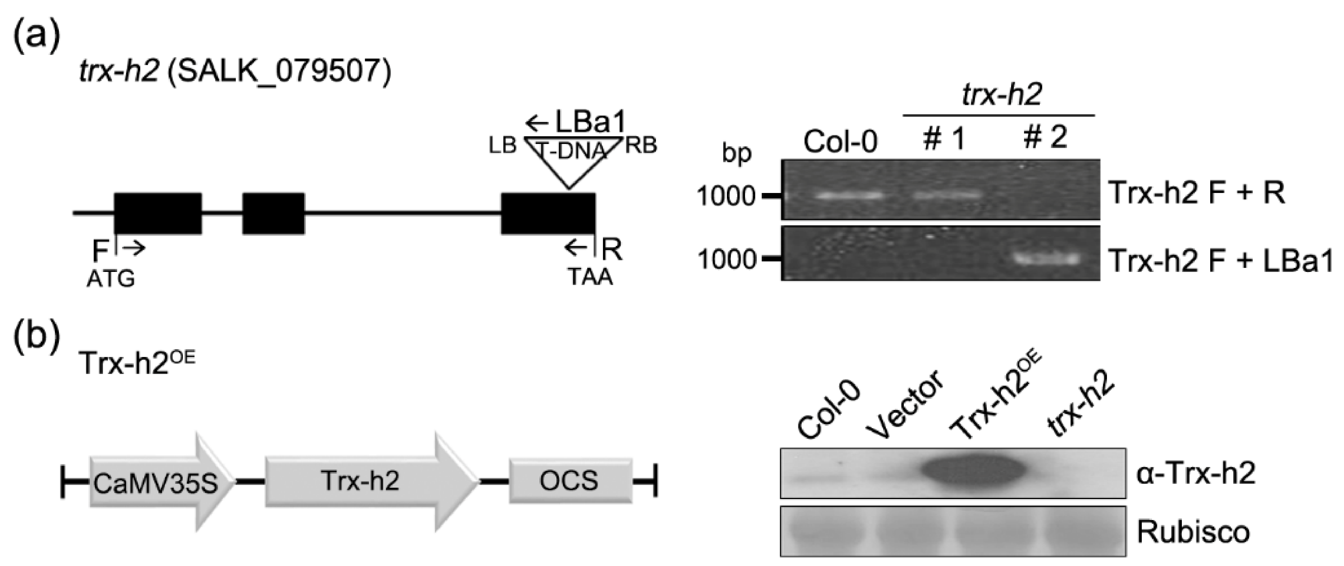
2.2. Identification of Trx-h2 Mutants and Generation of Trx-h2OE Lines

Homozygous trx-h2 mutant lines were identified by PCR-based genotyping using Trx-h2-specific primers (F: 5′-ATGGGAGGAGCTTTATCAAC-3′; R: 5′-TTATGCTCTGAGTTTGCTAA-3′) and T-DNA specific primer (LBa1) primer, (5′-TGGTTCACGTAGTGGGCCATCG-3′). To generate Trx-h2OE lines, Trx-h2 cDNA was amplified using sequence-specific primers (5′-ATCGATATGGGAGGAGCTTTATCAACT-3′ and 5′-TCTAGATGCTCTGAGTTTGCTAACTTTCTT-3′) and cloned into the pCAMBIA1300 binary vector. The plasmid was transformed into Agrobacterium tumefaciens strain GV3101, and the recombinant bacteria were used to transform trx-h2 mutants using the floral dip method. Homozygous T3 lines were selected and used for subsequent experiments.

2.3. Western Blot Analysis

Crude protein extract of Arabidopsis was prepared using extraction buffer (100 mM Tris-HCl (pH 7.5), 1 mM EDTA, 20% glycerol, and 1 mM PMSF). Arabidopsis proteins were separated by sodium dodecyl sulfate-polyacrylamide gel electrophoresis (SDS-PAGE) and then transferred to nitrocellulose membranes. Immunoblot analysis was carried out using anti-Trx-h2 antibody prepared in our laboratory, and the Trx-h2 protein was detected by chemiluminescence. The antibody specificity of anti-Trx-h2 was demonstrated by using the Col-0, Vector, Trx-h2OE, and trx-h2 plants.

2.4. Cold-Shock Treatment

Full-length cDNAs of Trx-h2 and Trx-h3 were amplified using the gene-specific primers (Trx-h2: 5′-CATATGATGGGAGGAGCTTTATCAACT-3′ and 5′-GGATCCTTATGCTCTGAGTTTGCTAACT-3′; Trx-h3: 5′-CATATGCATATGATGGGAGGAGCTTTATCAACT-3′ and 5′-TCAAGCAGCAGCAACAACTGTCTTCTAACT-3′) and cloned them into the pINIII vector. Each plasmid was transformed into the cold-sensitive E. coli BX04 cells that are deficient of the four cold-shock protein (csp) genes (cspA, cspB, cspE, and cspG) encoding the RNA chaperone proteins. The bacterial cells spreaded in Luria-Bertani (LB)-agar containing ampicillin were incubated at 37 °C, overnight. A single bacterial colony was picked and cultured for 12 h in LB-liquid media. Liquid culture cells were subjected to serial dilution and 5 μL of the cells were spotted onto LB-agar medium. Then the plates were incubated either at 37 °C for 1 day (control treatment) or at 19 °C for 5 days (cold-shock treatment).

2.5. Expression and Purification of Trx-h2 and Trx-h3 Recombinant Proteins

Full-length cDNAs of Trx-h2 and Trx-h3 were amplified by PCR using gene sequence-specific primers (Trx-h2: 5′-GGATCCATGGGAGGAGCTTTATCAACT-3′ and 5′-AAGCTTTTATGCTCTGAGTTTGCTAACTTT-3′; Trx-h3: 5′-GGATCCATGGCCGCAGAAG-3′ and 5′-TCAAGCAGCAGCAACAACTGTCTTAACTTT-3′) and cloned into the pGEX expression vector. The resulting plasmids were transformed into E. coli BL21 (DE3) pLysS cells. A single bacterial colony transforming the plasmid construct was grown in LB liquid medium at 37 °C. After the culture reached an optical density of 0.5–0.6 at 600 nm, protein expression was induced by addition of 0.5 mM isopropyl-β-D-thiogalactopyranoside (IPTG), and the culture was grown at 30 °C for 4 h. The cells were harvested by centrifugation and resuspended in a buffer containing 1.8 mM KH2PO4 (pH 8.0), 140 mM NaCl, 2.7 mM KCl, and 10 mM Na2HPO4. Cells were then disrupted by sonication, and recombinant proteins were purified using GSH agarose beads. The Trx-h2 and Trx-h3 proteins were eluted by thrombin cleavage, dialyzed against 20 mM HEPES-NaOH (pH 8.0) buffer, and used for biochemical analyses.

2.6. Electrophoretic Mobility Shift Assay (EMSA)

The purified recombinant Trx-h2 and Trx-h3 proteins were mixed with 100 ng of M13 mp8 phage single-stranded DNA (ssDNA) and double-stranded DNA (dsDNA), luciferase (Luc) mRNA, and Arabidopsis total RNAs in 10X EMSA DNA binding buffer (200 mM HEPES [pH 7.5], 750 mM NaCl, 10 mM EDTA, 25% glycerol, 2% Triton X-100, and 20 mM DTT), and incubated on ice for 15 min. Samples were then subjected to EMSA at a constant voltage of 100 V for ssDNA and dsDNA, and 50 V for Luc mRNA and Arabidopsis total RNAs. Nucleic acids (DNA and RNA) were then detected by ethidium bromide staining.

2.7. Melting Assay of Nucleic Acids

Molecular beacon used as a substrate in this melting assay contains two complementary DNA oligonucleotides. Each end of the two oligo-strands was labeled with fluorophore (FITC) or black hole quencher (BHQ1), respectively. To anneal the molecular beacon, two DNA oligonucleotides labeled with FITC and BHQ1 were mixed at 1:20 ratio as optimized in the previous paper [25] and denatured them at 95 °C for 2 min. The denatured DNAs dissolved in 10 mM Tris-HCl (pH 7.5) buffer containing 1 mM MgCl2 were annealed by incubating them on ice for 20 min. To detect the melting activity of Trx-h2, base-paired molecular beacon was reacted with Trx-h2, Trx-h3, CspA, or glutathione S-transferase (GST) protein (10 µM each) at room temperature. Additionally, the fluorescence was measured on a Spectra Max Gemini XPS spectrofluorometer (Molecular Devices) using a 96-well plate at excitation and emission wavelengths of 555 and 575 nm, respectively.

2.8. Transcription Anti-Termination Assay

The pINIII constructs containing the Trx-h2 and Trx-h3 cDNAs were used to perform the transcription anti-termination assay. Each plasmid was transformed into E. coli RL211 cells, and the transformed cells were grown overnight at 37 °C in LB liquid medium containing ampicillin and kanamycin. Each colony was spotted onto LB-agar medium supplemented with or without chloramphenicol. After growing the cells in the media at 37 °C for 2 days, we analyzed the transcription anti-termination activity by examining the cell growth in the presence of chloramphenicol.

2.9. In Situ Hybridization Assay

Poly(A) mRNA in situ hybridization was performed as described previously [26]. Briefly, leaves of 2-week-old WT (Col-0), trx-h2, and Trx-h2OE plants were harvested and fixed in a fixation buffer containing 120 mM NaCl, 7 mM Na2HPO4, 3 mM NaH2PO4, 2.7 mM KCl, 0.1% Tween-20, 80 mM EGTA, 5% formaldehyde, 10% DMSO, and 50% heptane. The fixed leaf samples were dehydrated and postfixed in the fixation buffer at room temperature for 30 min. Then, leaf samples were rinsed twice with fixation buffer containing no formaldehyde and once with 1 mL Hyb Plus hybridization buffer. Subsequently, the samples were prehybridized with 1 mL hybridization buffer at 50 °C for 1 h, and then hybridized with 5 pmol of 5′-fluorescein-labeled 45-mer oligo(dT) probe in hybridization buffer at 50 °C for more than 8 h. After hybridization, the fluorescence of leaf samples was observed under a confocal microscope at excitation and emission wavelengths of 488 and 522 nm, respectively. Each experiment was repeated at least three times, with similar results.

2.10. Seedling Growth under Cold Stress

WT (Col-0), trx-h2, and trx-h3 knock-out mutant plants, and overexpression lines of Trx-h2 (Trx-h2OE) and Trx-h3 (Trx-h3OE) Arabidopsis were sown on Murashige and Skoog (MS) medium supplemented with 3% sucrose. What is more, the seeds transforming pCAMBIA1300 vector only were used as a control. The plates were incubated at 4 °C for 3 days in the dark prior to germination. To test the effect of cold stress on seedling growth, plates were placed vertically in an incubator maintained at 12 °C.

2.11. Statistical Analysis

Root length data are presented as mean ± standard error (SE). Statistical analysis of the difference in root length between cold and normal conditions was examined using Student’s t-test (p < 0.05).

3. Results

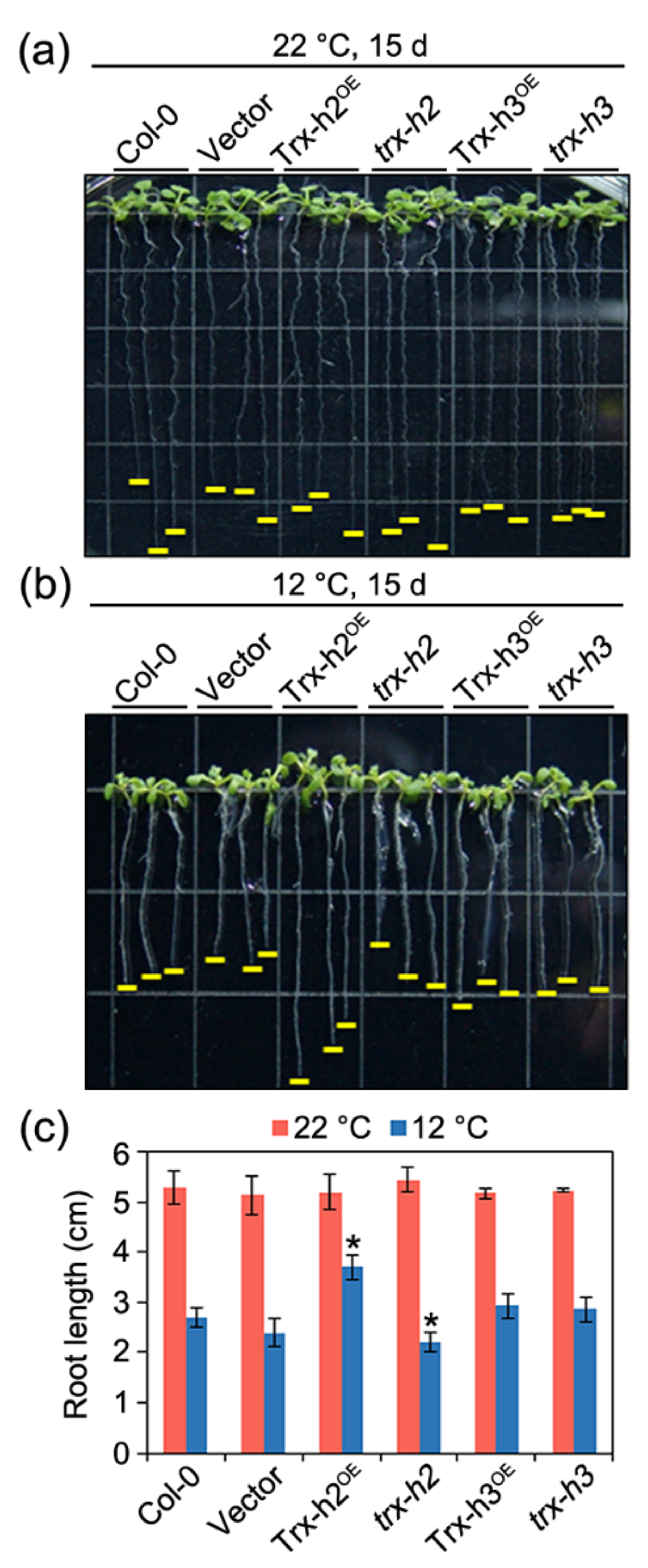
3.1. Role of Trx-h2 in Seedling Growth under Cold Stress

There are five representative Trx-h members in Arabidopsis distributed both in the cytoplasm and nucleus that have two conserved active-site cysteine (Cys) residues (Figure 1). Comparison of the amino acid sequences of Trx-hs revealed that Thx-h2 contains an additional 19 amino acids at its N-terminus and a putative nuclear localization signal (NLS) at their C-terminus estimated by the NLS-mapper database [27]. In contrast to Trx-h2, the other members of Trx-hs do not contain NLS, except Trx-h8, analyzed by the same program. The Trx-h2 protein has been shown to perceive intracellular ROS and to transmit the signal to downstream target molecules [24]. To investigate the physiological function of Trx-h2 in plants, we generated homozygous trx-h2 mutant lines from the trx-h2 knockout line (SALK_079507; Col-0 background) obtained from the ABRC (Ohio State University) and overexpressed the Trx-h2 gene in the trx-h2 mutant background. Knockout mutation of the Trx-h2 gene was confirmed by genotyping, and overexpression of Trx-h2 was verified by western blotting using anti-Trx-h2 antibody (Figure 2).
Next, we examined the stress resistance of trx-h2 and Trx-h2OE plants by measuring the root length of these plants under cold-shock treatments. The trx-h2, Trx-h2OE, and WT (Col-0) plants showed no difference in root length at normal temperature (22 °C) (Figure 3a). However, under cold conditions (12 °C), the root length of trx-h2 knockout mutant lines was shorter than that of WT (Col-0) plants, whereas roots of Trx-h2OE lines were significantly longer than those of the WT (Col-0) (Figure 3b,c). These results strongly suggest that Trx-2 plays a critical role in plant cold tolerance at low temperature.

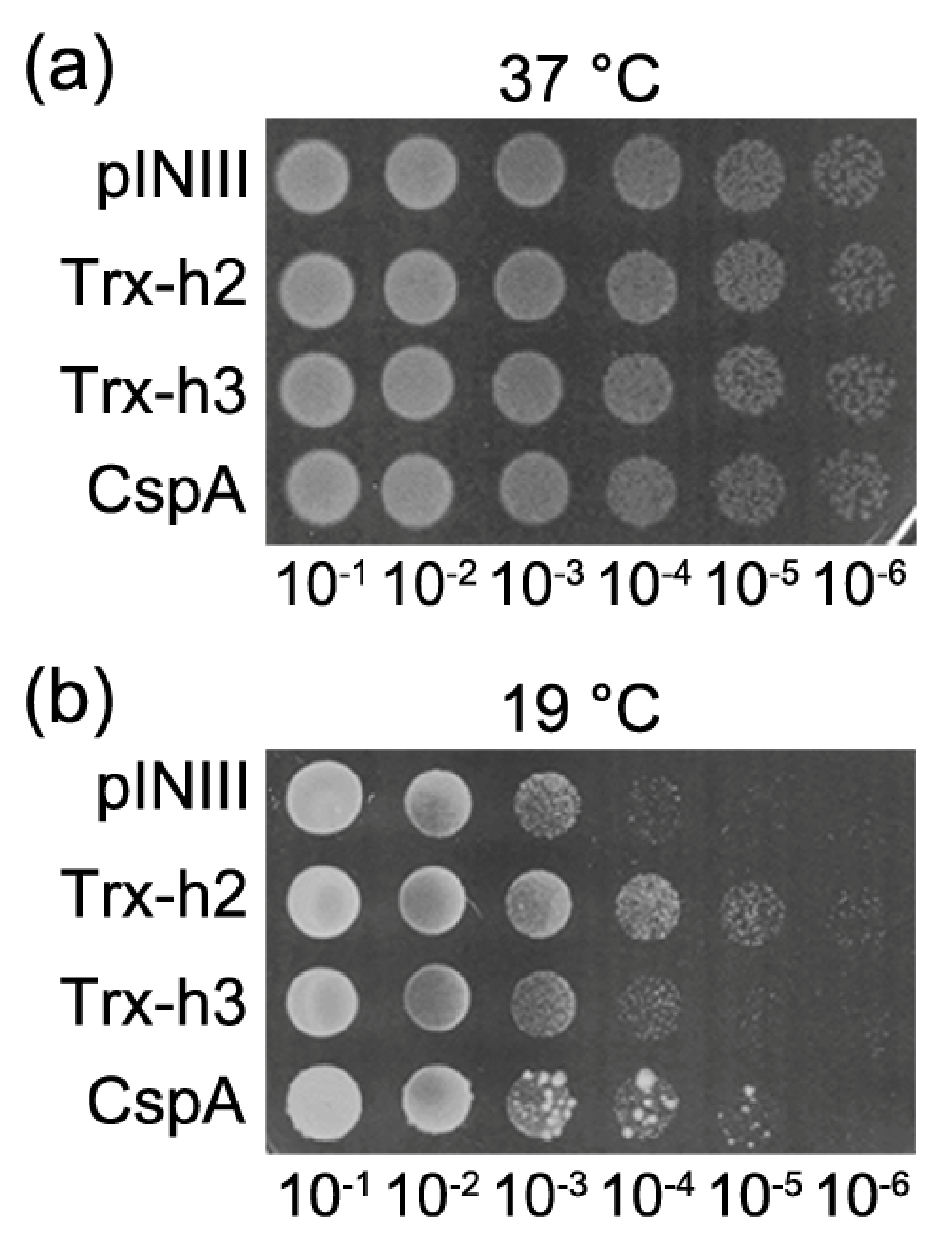
3.2. Trx-h2 Overexpression Complements the Cold-Sensitive Phenotype of the E. coli BX04 Mutant

Based on the results of the cold-shock treatment (Figure 3), we verified the ability of Trx-h2 to impart cold tolerance in the cold-sensitive E. coli BX04 mutant cells that lack four Csp genes (CspA, CspB, CspE, and CspG); proteins encoded by these genes are essential for bacterial growth at low temperature [28]. In this experiment, E. coli BX04 cells were transformed with the pINIII empty vector (control) or with pINIII vector carrying Trx-h2 or Trx-h3, and incubated at 37 °C or 19 °C; Trx-h3 served as a control because it contains no extra N-terminal amino acids, unlike Trx-h2. No difference was observed in the growth of BX04 cells expressing Trx-h2, Trx-h3, or pINIII empty vector at optimal growing conditions (37 °C) (Figure 4a). However, at low temperature (19 °C), the survival rate of E. coli expressing Trx-h2 was approximately 100-fold higher than that of E. coli mutants expressing the pINIII empty vector or Trx-h3 (Figure 4b). In addition, the survival of cells expressing the CspA gene used as a positive control was almost similar with Trx-h2 expressing cells at 19 °C. These results explicitly demonstrate that Trx-h2 complements the cold-sensitive phenotype of E. coli BX04 mutant cells at low temperatures.

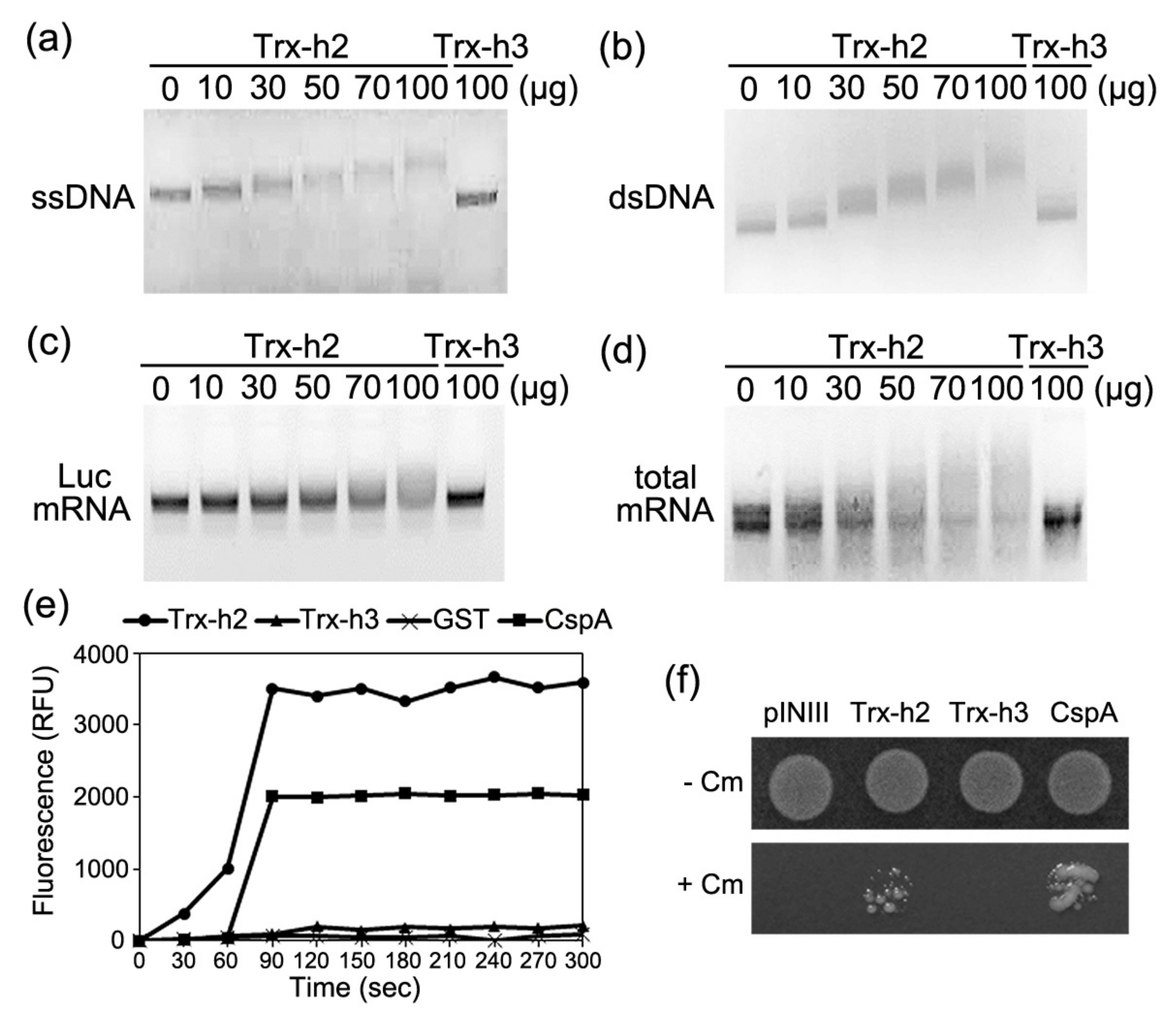
3.3. Trx-h2 Binds to Nucleic Acids

Next, we investigated how Trx-h2 confers enhanced cold tolerance in plants and microorganisms at the molecular level. Many studies have reported that the nucleic- and cytoplasmic-RNAs can be denatured at low temperature by their non-specific pairing to form misfolded hairpin structures [26]. Furthermore, resistance to cold shock is typically contributed by RNA chaperones, which exhibit several common characteristics. First, most of the RNA chaperones bind to nucleic acids including ssDNA and dsDNA. Second, RNA chaperones exhibit an anti-terminating mRNA transcription activity via their nucleic acid melting ability [29,30]. Based on these data, we examined whether the Trx-h2 protein expressed in E. coli interacts with M13 mp8 phage ssDNA and dsDNA, Luc mRNA, and Arabidopsis total RNAs (as substrates) in vitro [26].
Varying amounts of purified recombinant Trx-h2 were reacted with individual substrates in the reaction mixture containing binding buffer, and EMSAs were carried out using 0.8% agarose gels. Increasing amounts of Trx-h2 induced successive retardation of nucleic acid movement in agarose gels, indicating the binding of dsDNA, ssDNA, and total RNAs by Trx-h2 (Figure 5a–d) Additionally, excess of Trx-h3 (100 µg/µL; control) did not induce a shift in the position of nucleic acids, suggesting that Trx-h2 specifically interacts with ssDNA, dsDNA, and total RNAs. The results indicate that Trx-h2 binds to DNA and RNA, which is one of the peculiar properties of RNA chaperones.

3.4. Trx-h2 Melts to Nucleic Acids

To test whether Trx-h2 can dissociate double-stranded nucleic acids, we used two partially complementary double-stranded oligonucleotides, which together comprised a molecular beacon. In the synthetic DNA substrate of the molecular beacon, the end of one strand was labeled with a fluorophore, while that of the other strand was conjugated with the fluorescent quencher (Figure S1). Since the fluorophore and quencher labeled at the end of the molecular beacon form a stem-loop structure, fluorescence is significantly extinguished by the quencher when the ends of the two strands form a stem-loop structure. However, melting of the annealed hairpin-like beacon by RNA chaperone allows the emission of the fluorophore because it is spatially separated from the quencher. To determine the nucleic acid melting activity of Trx-h2, we incubated the Trx-h2 protein with the molecular beacon (substrate) and measured the fluorescence emitted by the fluorophore. We also reacted the molecular beacon with GST and CspA as negative and positive controls, respectively. Both Trx-h2 and CspA remarkably increased the fluorescence intensity of the molecular beacon (Figure 5e). By contrast, Trx-h3 and GST did not increase the fluorescence intensity of the beacon, suggesting that Trx-h2 exhibits a strong dsDNA melting activity in vitro.
Next, we examined the nucleic acid melting activity of Trx-h2 in vivo by using E. coli RL211 cells containing the chloramphenicol acetyltransferase (CAT) gene preceded by a ρ-independent trpL terminator. At the trpL termination sequences, two structural features of the transcribed RNA chain were detected. The transcribed RNA chain contained the GC-rich self-complementary area with several intruding nucleotides, which permitted to form a hairpin-like stem-loop structure. Immediately after the stem-loop secondary structure, several U bases were followed in the transcribed RNA. Then the hairpin-like structure interacted with the RNA polymerase and led to its stalling at the transcribing site. In particular, since the base pairs between the A bases in the template DNA and the 3′-U residues of the nascent RNA molecule are exceedingly precarious, they prompted the liberation of the RNA chain from the transcription complex at trpL termination sites, without proceeding the CAT gene transcription. The result makes the RL211 cells sensitive to chloramphenicol. However, when the RNA chaperone melted the hairpin-like stem-loop structure of the nascent RNA chain, it allows the continuous transcription of RNA polymerase for the CAT gene. Then the bacteria can express the CAT protein and make the cells tolerant to chloramphenicol (Figure S2). For investigating the RNA-melting activity of Trx-h2, we cloned Trx-h2 into the pINIII vector and transformed the DNA construct into RL211 cells. Using the bacterial cells expressing the protein, we examined their survival in LB-agar media containing chloramphenicol, comparing with the cells expressing the pINIII vector, CspA, and Trx-h3 as controls. Whereas the RL211 cells transforming the pINIII vector or Trx-h3 construct were not able to grow on LB-agar medium containing chloramphenicol, the cells expressing Trx-h2 and CspA grew well under the same conditions (Figure 5f). The results strongly suggested that Trx-h2 melted the hairpin-like secondary structure of trpL terminator located at immediately upstream of the CAT gene, which permitted the expression of CAT protein. Then, the bacterial cells expressing the CAT protein were able to grow at the presence of chloramphenicol. Taken together with the DNA-binding and melting activities of Trx-h2, our in vitro and in vivo data clearly show that the redox protein, Trx-h2 in Arabidopsis, functions as an RNA chaperone under cold conditions.

3.5. Trx-h2 Exports Nuclear mRNA to the Cytoplasm under Cold Stress

Because Trx-h2 harbors a putative NLS at its C-terminal end (Figure 1) and functions as an RNA chaperone (Figure 4 and Figure 5), we investigated its role in the export of mRNAs from the nucleus to the cytoplasm at different temperatures. Leaves of 14-day-old WT (Col-0), trx-h2, and Trx-h2OE Arabidopsis plants were incubated with a 45-mer oligo(dT) probe labeled with fluorescein at its end, and traced the localization of poly(A) mRNAs by in situ hybridization, as described previously [26].
Under normal conditions (22 °C), fluorescence signals were uniformly distributed throughout the nucleus and cytoplasm in WT (Col-0), trx-h2, and Trx-h2OE cells, indicating that mRNAs were effectively transported from the nucleus to the cytoplasm (Figure 6, upper panel). When incubated at 0 °C for 2 days, although WT (Col-0) and Trx-h2OE leaves showed no difference in the distribution of fluorescence signals between the cytoplasm and nucleus, parts of strong fluorescence signals were remained at the nucleus in trx-h2 leaves, suggesting that the export system of mRNAs transport was critically disrupted in the trx-h2 mutant plant, resulting in the accumulation of mRNAs in the nucleus (Figure 6, lower panel). Together, our data suggest that Trx-h2 has dual functions acting as a disulfide reductase and as an RNA chaperone, whose function should be controlled upon the environmental temperature changes.

4. Discussion

The Trx-h isoforms comprise the largest group in the plant Trx family. Although the physiological functions of most of these proteins have been predicted based on their intracellular localization and interaction partners [20,31,32], evidence proving these predictions is lacking [33]. Thus, determination of the localization patterns and specific interaction partners of Trx-h proteins under different environmental conditions is critical for understanding their roles in plant cells [16,21,22]. In this study, we investigated the physiological function of Trx-h2, which contains a 19-amino-acid extension at its N-terminus. Based on its subcellular localization in cytosol, ER/Golgi, and mitochondria [34,35], we found that Trx-h2 acts as an RNA chaperone under cold stress. Our previous result showed that one of the cold-tolerant nature of Arabidopsis is facilitated by Trx-h2-mediated expression of cold-responsive genes through its reductase activity [36]. However, in this study, we also propose that another function of Trx-h2 acting as an RNA chaperone can also contribute to the enhancement of cold tolerance in plants. In particular, the RNA chaperone function was also detected from the Trx-h2(C/S) mutant protein, suggesting that redox state of Trx-h2 did not give any effect on its RNA chaperone function.
To translate the genetic information of RNAs to proteins, they must maintain their structure as single-stranded poly-anionic forms. However, under cold stress conditions, RNA structures are misfolded to form non-native and non-functional conformations. Our results suggest that Trx-h2 functions as an RNA chaperone under cold stress by binding to misfolded and over-stabilized inactive RNAs. Then, Trx-h2 melts the hairpin-shaped RNAs and facilitates their refolding to form an active conformation. The resulting RNAs in their native form can be translated to produce cold resistant proteins, which confer enhanced cold tolerance in plants, similar to the rice (Oryza sativa L.) proteins, OsGRP4 and OsGRP6 [29].
Most of the RNA chaperones contain one of the following common motifs: RNA recognition motif, cold-shock domain, glycine-rich region, or Cys-Cys-His-Cys (CCHC) zinc-finger motif [37]. Although Trx-h2, originally defined as a disulfide reductase also functions as an RNA chaperone (according to our data), it does not contain any conserved motif or domain characteristic of RNA chaperones, such as the human peroxiredoxin 1 (hPrx1) protein [38]. While RNA chaperones enhance plant cold tolerance, they are also reported to play significant roles in plant growth and development; for example, Arabidopsis proteins AtRZ-1a and AtGRP4 regulate seed germination and seedling growth under cold stress [39,40], and the chloroplast-localized DEAD-box RNA helicase, AtRH3, participates in multiple steps of chloroplast development [41]. RNA chaperones, including AtGRP4 and AtGRP7 in Arabidopsis, OsZR2 in rice, and WCSP1 in wheat, also enhance plant tolerance to multiple stresses [30]. Therefore, the physiological functions of Trx-h2 in relation to diverse abiotic stresses and phytohormone signaling pathways should be investigated further. This study reveals a novel function of Trx-h2 as an RNA chaperone under cold stress. Thus, our results highlight the potential for generating cold tolerant crops by overexpressing the Trx-h2 gene using biotechnological approaches.

Supplementary Materials

https://www.mdpi.com/article/10.3390/app11156865/s1, Figure S1: Schematic representation of the molecular beacon used to test the DNA-melting activity of RNA chaperones. Figure S2: Schematic representation of the E. coli RL211 cells expressing the Trx-h2 for the chloramphenicol resistance.

Author Contributions

Conceptualization, E.S.L., J.H.P., and S.Y.L.; methodology, E.S.L., S.D.W., S.K.P., and K.A.T.P.; validation, E.S.L. and J.H.P.; former analysis, S.Y.L.; investigation, J.H.P.; resources, H.B.C., S.D.W., S.B.B., and K.A.T.P.; draft writing, review, and editing, E.S.L., J.H.P. and S.Y.L.; funding acquisition, E.S.L., J.H.P. and S.Y.L. All authors have read and agreed to the published version of the manuscript.

Funding

This work was supported by the grants (Grant # PJ015824 & PJ015674), RDA, (S.Y.L. and S.K.P), and by the Basic Science Research Program of the National Research Foundation (NRF), Korea (E.S.L.; Grant #NRF-2018R1A6A3A11048274 and J.H.P.; Grant no. NRF-2019R1I1A1A01040920).

Institutional Review Board Statement

Not applicable.

Informed Consent Statement

Not applicable.

Data Availability Statement

Not applicable.

Conflicts of Interest

The authors declare no conflict of interest.

References

  1. Choudhury, F.K.; Rivero, R.M.; Blumwald, E.; Mittler, R. Reactive oxygen species, abiotic stress and stress combination. Plant J. 2017, 90, 856–867. [Google Scholar] [CrossRef] [PubMed]
  2. Gilroy, S.; Bialasek, M.; Suzuki, N.; Gorecka, M.; Devireddy, A.R.; Karpinski, S.; Mittler, R. ROS, Calcium, and Electric Signals: Key Mediators of Rapid Systemic Signaling in Plants. Plant Physiol. 2016, 171, 1606–1615. [Google Scholar] [CrossRef] [PubMed]
  3. Gechev, T.S.; Van Breusegem, F.; Stone, J.M.; Denev, I.; Laloi, C. Reactive oxygen species as signals that modulate plant stress responses and programmed cell death. Bioessays 2006, 28, 1091–1101. [Google Scholar] [CrossRef] [PubMed]
  4. Asada, K. Production and scavenging of reactive oxygen species in chloroplasts and their functions. Plant Physiol. 2006, 141, 391–396. [Google Scholar] [CrossRef] [PubMed] [Green Version]
  5. Angelos, E.; Ruberti, C.; Kim, S.J.; Brandizzi, F. Maintaining the factory: The roles of the unfolded protein response in cellular homeostasis in plants. Plant J. 2017, 90, 671–682. [Google Scholar] [CrossRef] [PubMed] [Green Version]
  6. Imlay, J.A. Pathways of oxidative damage. Annu. Rev. Microbiol. 2003, 57, 395–418. [Google Scholar] [CrossRef] [PubMed]
  7. Park, J.H.; Kang, C.H.; Nawkar, G.M.; Lee, E.S.; Paeng, S.K.; Chae, H.B.; Chi, Y.H.; Kim, W.Y.; Yun, D.J.; Lee, S.Y. EMR, a cytosolic-abundant ring finger E3 ligase, mediates ER-associated protein degradation in Arabidopsis. New Phytol. 2018, 220, 163–177. [Google Scholar] [CrossRef] [PubMed] [Green Version]
  8. Jang, H.H.; Lee, K.O.; Chi, Y.H.; Jung, B.G.; Park, S.K.; Park, J.H.; Lee, J.R.; Lee, S.S.; Moon, J.C.; Yun, J.W.; et al. Two enzymes in one; two yeast peroxiredoxins display oxidative stress-dependent switching from a peroxidase to a molecular chaperone function. Cell 2004, 117, 625–635. [Google Scholar] [CrossRef]
  9. Hanschmann, E.M.; Godoy, J.R.; Berndt, C.; Hudemann, C.; Lillig, C.H. Thioredoxins, glutaredoxins, and peroxiredoxins--molecular mechanisms and health significance: From cofactors to antioxidants to redox signaling. Antioxid. Redox Sign. 2013, 19, 1539–1605. [Google Scholar] [CrossRef]
  10. Lee, E.S.; Kang, C.H.; Park, J.H.; Lee, S.Y. Physiological Significance of Plant Peroxiredoxins and the Structure-Related and Multifunctional Biochemistry of Peroxiredoxin 1. Antioxid. Redox Sign. 2018, 28, 625–639. [Google Scholar] [CrossRef]
  11. Meyer, Y.; Buchanan, B.B.; Vignols, F.; Reichheld, J.P. Thioredoxins and glutaredoxins: Unifying elements in redox biology. Annu. Rev. Genet. 2009, 43, 335–367. [Google Scholar] [CrossRef]
  12. Nordberg, J.; Arner, E.S. Reactive oxygen species, antioxidants, and the mammalian thioredoxin system. Free Radic. Bio. Med. 2001, 31, 1287–1312. [Google Scholar] [CrossRef]
  13. Chae, H.B.; Kim, M.G.; Kang, C.H.; Park, J.H.; Lee, E.S.; Lee, S.U.; Chi, Y.H.; Paeng, S.K.; Bae, S.B.; Wi, S.D.; et al. Redox sensor QSOX1 regulates plant immunity by targeting GSNOR to modulate ROS generation. Mol. Plant 2021. [Google Scholar] [CrossRef]
  14. Lee, J.R.; Lee, S.S.; Jang, H.H.; Lee, Y.M.; Park, J.H.; Park, S.C.; Moon, J.C.; Park, S.K.; Kim, S.Y.; Lee, S.Y.; et al. Heat-shock dependent oligomeric status alters the function of a plant-specific thioredoxin-like protein, AtTDX. Proc. Nati. Acad. Sci. USA 2009, 106, 5978–5983. [Google Scholar] [CrossRef] [Green Version]
  15. Aslund, F.; Beckwith, J. Bridge over troubled waters: Sensing stress by disulfide bond formation. Cell 1999, 96, 751–753. [Google Scholar] [CrossRef] [Green Version]
  16. Cabrillac, D.; Cock, J.M.; Dumas, C.; Gaude, T. The S-locus receptor kinase is inhibited by thioredoxins and activated by pollen coat proteins. Nature 2001, 410, 220–223. [Google Scholar] [CrossRef]
  17. Huber, H.E.; Tabor, S.; Richardson, C.C. Escherichia coli thioredoxin stabilizes complexes of bacteriophage T7 DNA polymerase and primed templates. J. Biol. Chem. 1987, 262, 16224–16232. [Google Scholar] [CrossRef]
  18. Gelhaye, E.; Rouhier, N.; Jacquot, J.P. The thioredoxin h system of higher plants. Plant Physiol. Bioch. 2004, 42, 265–271. [Google Scholar] [CrossRef] [PubMed]
  19. Menges, M.; Hennig, L.; Gruissem, W.; Murray, J.A. Cell cycle-regulated gene expression in Arabidopsis. J. Biol. Chem. 2002, 277, 41987–42002. [Google Scholar] [CrossRef] [PubMed] [Green Version]
  20. Brehelin, C.; Mouaheb, N.; Verdoucq, L.; Lancelin, J.M.; Meyer, Y. Characterization of determinants for the specificity of Arabidopsis thioredoxins h in yeast complementation. J. Biol. Chem. 2000, 275, 31641–31647. [Google Scholar] [CrossRef] [Green Version]
  21. Park, S.K.; Jung, Y.J.; Lee, J.R.; Lee, Y.M.; Jang, H.H.; Lee, S.S.; Park, J.H.; Kim, S.Y.; Moon, J.C.; Lee, S.Y.; et al. Heat-shock and redox-dependent functional switching of an h-type Arabidopsis thioredoxin from a disulfide reductase to a molecular chaperone. Plant Physiol. 2009, 150, 552–561. [Google Scholar] [CrossRef] [PubMed] [Green Version]
  22. Sweat, T.A.; Wolpert, T.J. Thioredoxin h5 is required for victorin sensitivity mediated by a CC-NBS-LRR gene in Arabidopsis. Plant Cell 2007, 19, 673–687. [Google Scholar] [CrossRef] [PubMed] [Green Version]
  23. Tada, Y.; Spoel, S.H.; Pajerowska-Mukhtar, K.; Mou, Z.; Song, J.; Wang, C.; Zuo, J.; Dong, X. Plant immunity requires conformational changes [corrected] of NPR1 via S-nitrosylation and thioredoxins. Science 2008, 321, 952–956. [Google Scholar] [CrossRef] [Green Version]
  24. Jung, Y.J.; Chi, Y.H.; Chae, H.B.; Shin, M.R.; Lee, E.S.; Cha, J.Y.; Paeng, S.K.; Lee, Y.; Park, J.H.; Kim, W.Y.; et al. Analysis of Arabidopsis thioredoxin-h isotypes identifies discrete domains that confer specific structural and functional properties. Biochem. J. 2013, 456, 13–24. [Google Scholar] [CrossRef] [Green Version]
  25. Melencion, S.M.B.; Chi, Y.H.; Pham, T.T.; Paeng, S.K.; Wi, S.D.; Lee, C.; Ryu, S.W.; Koo, S.S.; Lee, S.Y. RNA Chaperone Function of a Universal Stress Protein in Arabidopsis Confers Enhanced Cold Stress Tolerance in Plants. Int. J. Mol. Sci. 2017, 18, 2546. [Google Scholar] [CrossRef] [Green Version]
  26. Gong, Z.; Dong, C.H.; Lee, H.; Zhu, J.; Xiong, L.; Gong, D.; Stevenson, B.; Zhu, J.K. A DEAD box RNA helicase is essential for mRNA export and important for development and stress responses in Arabidopsis. Plant Cell 2005, 17, 256–267. [Google Scholar] [CrossRef] [PubMed] [Green Version]
  27. Kosugi, S.; Hasebe, M.; Tomita, M.; Yanagawa, H. Systematic identification of cell cycle-dependent yeast nucleocytoplasmic shuttling proteins by prediction of composite motifs. Proc. Nati. Acad. Sci. USA 2009, 106, 10171–10176. [Google Scholar] [CrossRef] [Green Version]
  28. Xia, B.; Ke, H.; Inouye, M. Acquirement of cold sensitivity by quadruple deletion of the cspA family and its suppression by PNPase S1 domain in Escherichia coli. Mol. Microbiol. 2001, 40, 179–188. [Google Scholar] [CrossRef]
  29. Kim, J.Y.; Kim, W.Y.; Kwak, K.J.; Oh, S.H.; Han, Y.S.; Kang, H. Glycine-rich RNA-binding proteins are functionally conserved in Arabidopsis thaliana and Oryza sativa during cold adaptation process. J. Exp. Bot. 2010, 61, 2317–2325. [Google Scholar] [CrossRef] [Green Version]
  30. Kim, J.Y.; Kim, W.Y.; Kwak, K.J.; Oh, S.H.; Han, Y.S.; Kang, H. Zinc finger-containing glycine-rich RNA-binding protein in Oryza sativa has an RNA chaperone activity under cold stress conditions. Plant Cell Environ. 2010, 33, 759–768. [Google Scholar] [CrossRef]
  31. Chi, Y.H.; Moon, J.C.; Park, J.H.; Kim, H.S.; Zulfugarov, I.S.; Fanata, W.I.; Jang, H.H.; Lee, J.R.; Lee, Y.M.; Kim, S.T.; et al. Abnormal chloroplast development and growth inhibition in rice thioredoxin m knock-down plants. Plant Physiol. 2008, 148, 808–817. [Google Scholar] [CrossRef] [Green Version]
  32. Lee, E.S.; Park, J.H.; Wi, S.D.; Kang, C.H.; Chi, Y.H.; Chae, H.B.; Paeng, S.K.; Ji, M.G.; Kim, W.-Y.; Kim, M.G.; et al. Redox-dependent structural switch and CBF activation confer freezing tolerance in plants. Nat. Plants 2021, 7, 914–922. [Google Scholar] [CrossRef] [PubMed]
  33. Laloi, C.; Mestres-Ortega, D.; Marco, Y.; Meyer, Y.; Reichheld, J.P. The Arabidopsis cytosolic thioredoxin h5 gene induction by oxidative stress and its W-box-mediated response to pathogen elicitor. Plant Physiol. 2004, 134, 1006–1016. [Google Scholar] [CrossRef] [Green Version]
  34. Meng, L.; Wong, J.H.; Feldman, L.J.; Lemaux, P.G.; Buchanan, B.B. A membrane-associated thioredoxin required for plant growth moves from cell to cell, suggestive of a role in intercellular communication. Proc. Nati. Acad. Sci. USA 2010, 107, 3900–3905. [Google Scholar] [CrossRef] [Green Version]
  35. Traverso, J.A.; Micalella, C.; Martinez, A.; Brown, S.C.; Satiat-Jeunemaitre, B.; Meinnel, T.; Giglione, C. Roles of N-terminal fatty acid acylations in membrane compartment partitioning: Arabidopsis h-type thioredoxins as a case study. Plant Cell 2013, 25, 1056–1077. [Google Scholar] [CrossRef] [Green Version]
  36. Park, J.H.; Lee, E.S.; Chae, H.B.; Paeng, S.K.; Wi, S.D.; Bae, S.B.; Thi Phan, K.A.; Lee, S.Y. Disulfide reductase activity of thioredoxin-h2 imparts cold tolerance in Arabidopsis. Biochem. Bioph. Res. Commun. 2021, 568, 124–130. [Google Scholar] [CrossRef] [PubMed]
  37. Cremers, C.M.; Reichmann, D.; Hausmann, J.; Ilbert, M.; Jakob, U. Unfolding of metastable linker region is at the core of Hsp33 activation as a redox-regulated chaperone. J. Biol. Chem. 2010, 285, 11243–11251. [Google Scholar] [CrossRef] [PubMed] [Green Version]
  38. Kim, J.H.; Lee, J.M.; Lee, H.N.; Kim, E.K.; Ha, B.; Ahn, S.M.; Jang, H.H.; Lee, S.Y. RNA-binding properties and RNA chaperone activity of human peroxiredoxin 1. Biochem. Bioph. Res. Commun. 2012, 425, 730–734. [Google Scholar] [CrossRef]
  39. Kim, Y.O.; Kang, H. The role of a zinc finger-containing glycine-rich RNA-binding protein during the cold adaptation process in Arabidopsis thaliana. Plant Cell Physiol. 2006, 47, 793–798. [Google Scholar] [CrossRef] [Green Version]
  40. Kim, Y.O.; Kim, J.S.; Kang, H. Cold-inducible zinc finger-containing glycine-rich RNA-binding protein contributes to the enhancement of freezing tolerance in Arabidopsis thaliana. Plant J. 2005, 42, 890–900. [Google Scholar] [CrossRef]
  41. Jiang, W.; Hou, Y.; Inouye, M. CspA, the major cold-shock protein of Escherichia coli, is an RNA chaperone. J. Biol. Chem. 1997, 272, 196–202. [Google Scholar] [CrossRef] [PubMed] [Green Version]
Figure 1. Comparison of the N-terminal amino acid sequences of the five representative Arabidopsis Trx-h proteins. (a) alignment of the N-terminal amino acid sequences of Arabidopsis Trx-h proteins. Two conserved cysteine (Cys) residues in the CXXC motif positioned between amino acids 59 and 62 are indicated in a blue box. The extra 19 amino acids at the N-terminus of Trx-h2 are outlined in a red box. Numbers above the alignment indicate the amino acid positions. (b) amino acid sequence of Trx-h2. Active Cys residues in the Trx motif are indicated in red, and the bipartite nuclear localization signal (NLS), identified using the NLS-mapper database, is outlined in red box. The asterisk (*) indicates the stop codon.
Applsci 11 06865 g001
Figure 2. Identification of trx-h2 knockout mutant lines and transgenic Trx-h2 overexpression (Trx-h2OE) lines by PCR. (a) schematic showing the location of the T-DNA insertion in the trx-h2 mutant (SALK_079507), and gel image showing the results of PCR-based genotyping. T-DNA was inserted in the third exon of the Trx-h2 gene. Primers (F, R, and LBa1) used for genotyping the plants are indicated in the schematic. The trx-h2 knockout mutant lines #1 and #2 were confirmed by PCR. (b) schematic showing the construct used to generate Trx-h2OE lines, and results of western blot analysis. The Trx-h2 gene was expressed under the control of the cauliflower mosaic virus 35S promoter (CaMV35S) and octopine synthase terminator (OCS). The Trx-h2 protein was detected by western blotting using anti-Trx-h2 antibody. Rubisco was used as a loading control.
Applsci 11 06865 g002
Figure 3. Trx-h2 increases the root growth of Arabidopsis seedlings under cold condition. (a,b) Comparison of the root growth of WT (Col-0), trx-h2 knockout mutant, and Trx-h2 overexpression (Trx-h2OE) plants at 22 °C (a) and 12 °C (b). Plants expressing Trx-h3 or pCAMBIA1300 vector were used as controls. Arabidopsis seedlings were grown on Murashige and Skoog (MS) medium at different temperatures for 15 days. Plates were oriented vertically and photographed. (c) root length of various genotypes at different temperatures. Data represent mean ± standard error (SE) of three biological replicates. Significant differences between the WT (Col-0) and other genotypes (n = 4) are indicated by an asterisk (* p < 0.05; Student’s t-test).
Applsci 11 06865 g003
Figure 4. Trx-h2 restores the cold-sensitive phenotype of E. coli BX04 mutant cells lacking four cold-shock protein (Csp) genes. (a,b) Images showing the growth of E. coli BX04 cells expressing Trx-h2 on LB-agar media at 37 °C for 1 day (a) and at 19 °C for 5 days (b).
Applsci 11 06865 g004
Figure 5. Nucleic acid binding and melting activity of Trx-h2. (ad) electrophoretic mobility shift assay (EMSA) using varying amounts of Trx-h2 incubated with M13mp8 ssDNA (a), M13mp8 dsDNA (b), luciferase (Luc) mRNA (c), or Arabidopsis total RNAs (d). A shift in the mobility of the Trx-h2/nucleic acid complexes was analyzed on 0.8% agarose gels. Trx-h3 (100 µg) was used as a control. (e) nucleic acid melting assay using a molecular beacon as a substrate. The molecular beacon was incubated with Trx-h2 (-●-), Trx-h3 (-▲-), GST (-x-), or CspA (-■-), and fluorescence intensity was measured at various time points at excitation and emission wavelengths of 555 and 575 nm, respectively. (f) DNA melting activity of Trx-h2 in RL211 E. coli cells in vivo. Anti-termination activity of Trx-h2 in E. coli RL211 cells. Trx-h2 was transformed into RL211 cells, which harbor the chloramphenicol (Cm) resistance gene (chloramphenicol acetyltransferase [CAT]) downstream of the trpL terminator, and was grown for 2 days on LB-agar medium supplemented with (+) or without (−) Cm at 37 °C. Cells transformed with the pINIII vector, CspA, and Trx-h3 were used as controls.
Applsci 11 06865 g005
Figure 6. Mutation of Trx-h2 in Arabidopsis disrupts mRNA export from the nucleus to the cytoplasm. The distribution of mRNAs was examined in the leaves of 2-week-old WT (Col-0), trx-h2, and Trx-h2OE Arabidopsis plants at 22 °C or 0 °C by in situ poly(A) mRNA hybridization. After fixation the leaves, 5′-fluorescein-labeled oligo(dT) probe was hybridized and the fluorescence of the leaf cells was observed using confocal microscopy (Olympus FV500) at excitation and emission wavelengths of 488 and 522 nm, respectively. Scale bars = 20 μm.
Applsci 11 06865 g006
Publisher’s Note: MDPI stays neutral with regard to jurisdictional claims in published maps and institutional affiliations.

