Article

Influence of Angiopoietin Treatment with Hypoxia and Normoxia on Human Intervertebral Disc Progenitor Cell’s Proliferation, Metabolic Activity, and Phenotype

by
Muriel C. Bischof
1,
Sonja Häckel
2,
Andrea Oberli
1,
Andreas S. Croft
1,
Katharina A. C. Oswald
2,
Christoph E. Albers
2,
Benjamin Gantenbein
1,2,* and
Julien Guerrero
1
1
Tissue Engineering for Orthopaedics & Mechanobiology (TOM), Bone & Joint Program, Department for BioMedical Research (DBMR), Faculty of Medicine, University of Bern, CH-3008 Bern, Switzerland
2
Department of Orthopaedic Surgery and Traumatology, Inselspital, Bern University Hospital, University of Bern, CH-3010 Bern, Switzerland
*
Author to whom correspondence should be addressed.
Appl. Sci. 2021, 11(15), 7144; https://doi.org/10.3390/app11157144
Submission received: 12 July 2021 / Revised: 23 July 2021 / Accepted: 30 July 2021 / Published: 2 August 2021
(This article belongs to the Special Issue Intervertebral Disc Regeneration II)

Abstract

:
Increasing evidence implicates intervertebral disc (IVD) degeneration as a major contributor to low back pain. In addition to a series of pathogenic processes, degenerated IVDs become vascularized in contrast to healthy IVDs. In this context, angiopoietin (Ang) plays a crucial role and is involved in cytokine recruitment, and anabolic and catabolic reactions within the extracellular matrix (ECM). Over the last decade, a progenitor cell population has been described in the nucleus pulposus (NP) of the IVD to be positive for the Tie2 marker (also known as Ang-1 receptor). In this study, we investigated the influence of Ang-1 and Ang-2 on human NP cell (Tie2+, Tie2 or mixed) populations isolated from trauma patients during 7 days in normoxia (21% O2) or hypoxia (≤5% O2). At the end of the process, the proliferation and metabolic activity of the NP cells were analyzed. Additionally, the relative gene expression of NP-related markers was evaluated. NP cells showed a higher proliferation depending on the Ang treatment. Moreover, the study revealed higher NP cell metabolism when cultured in hypoxia. Additionally, the relative gene expression followed, with an increase linked to the oxygen level and Ang concentration. Our study comparing different NP cell populations may be the start of new approaches for the treatment of IVD degeneration.

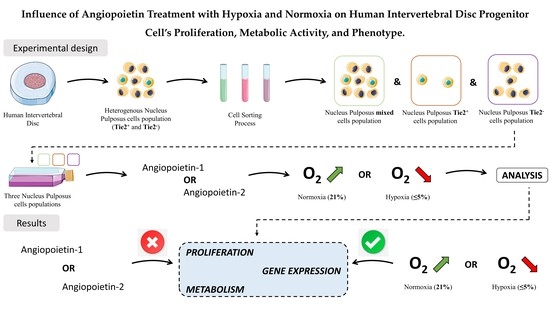
Graphical Abstract

1. Introduction

1.1. Low Back Pain and Intervertebral Disc Degeneration (IVDD)

Intervertebral disc degeneration is a significant and multifaceted cause of low back pain [1]. The so-called “discogenic pain” is described as a major cause of low back pain in 26–42% of cases [2]. Yet, it is known that the IVD undergoes degenerative changes earlier in life than do other tissues of major organ systems [1]. Whilst for the process of aging and degeneration, the IVD cells enter a state of cell senescence, showing an altered and often more catabolic metabolism [3], oftentimes the nucleus pulposus (NP) is primarily affected [3]. Moreover, the IVD possesses a very limited self-healing potential [4,5]. Conventional treatment strategies, such as analgetics, physical therapy, or surgical interventions, mainly address the symptoms of LBP, while no regeneration mechanisms in the IVD are activated [6]. The combination of tissue engineering strategies with stem cell-based therapies and approaches with native cells have gained significant momentum, being a desirable and promising novel treatment opportunity for degenerated IVDs since they aim to restore the properties of the IVD and, thus, their biological function [7].

1.2. Hypoxia vs. Normoxia in the Intervertebral Disc (IVD)

Another critical aspect in the IVD cell biology is that the NP is avascular, so its cell population does not receive a blood supply (nutrients diffuse through the vertebral endplates); therefore, oxygen levels in this central area are very low [8,9], causing a hypoxic environment [10]. As consequence, these NP cells may have adapted to these conditions. An increase in oxygen concentration during IVDD could be considered a pathological condition, unlike that observed in other tissues or wound healing [11].

1.3. Nucleus Pulposus Progenitor Cells (also Known as Tie2+ Cells)

A cell source of current major interest is the tissue-specific Tie2 (also known as TEK receptor tyrosine kinase or angiopoietin-1 receptor or CD202b) positive NP progenitor cell (NPPC) population. These cells are perfectly adapted to the hypoxic environment and can therefore uphold their regenerative potency [12]. Notably, NPPCs are attributed with having potential in regenerative medicine because of their remarkable in vitro multipotency capability through their osteogenic, chondrogenic, and adipogenic lineages in addition to their self-renewal potential [13]. The harsh IVD microenvironment forms an obstacle to exogenous cells (such as bone marrow-derived mesenchymal stem cells) to survive and exhibit a tissue-regenerative function. Thus, NPPCs might pose a promising target to stimulate and induce tissue repair [14]. However, a major challenge in the NPPC therapy approach is caused by the decrease in the percentage of the Tie2+ NPPC population with aging and the extent of disc degeneration [1]. The methodology for optimized NP cell isolation (by cell sorting) and cell expansion are already described [6,15,16].

1.4. Tie2 Receptor and Its Ligands; Angiopoietin-1 and Angiopoietin-2

The receptor tyrosine kinase Tie2 is mainly expressed in endothelial cells and is essential for embryonal and adult angiogenesis and vascular maturation and maintenance [17,18] and its also expressed by early hematopoietic cells and subsets of monocytes express Tie2 [19]. Ang-1–4 are the growth factor ligands of the Tie2 receptor. In this project, we focused on Ang-1 and -2, which are mainly produced by vascular smooth muscle cells, throughout the entire vascular system, and endothelial cells respectively [20,21,22]. The binding of Ang-1 to its extracellular receptor Tie2 results in autophosphorylation and subsequent activation of downstream signaling pathways including the PI3-kinase/Akt [18,19,23]. The Tie2 receptor activation mediates stabilization, remodeling, and repair of the vascular system [18,24]. The inactivation of this pathway protects endothelial cells from apoptosis and inhibits inflammatory responses [18]. Cell-based Ang-1 gene therapy may represent a new strategy for treating various endothelium disorders, including several severe human pulmonary diseases [25,26]. Ang-2 fulfills a dual function by acting as an agonist or antagonist in a context-dependent manner [17]. while showing similar biological outcomes to Ang-1. However, it has to be emphasized that they are weaker in potency and kinetics. As a consequence, Ang-2, thus, may act as a partial Ang-1 agonist [17].

1.5. The Role of Tie2/Ang-1 and Ang-2 Signaling Pathway in Human NPCs and NPPCs

Sakai et al. [1] suggested that Tie2 is a sensitive marker of aging and the degeneration of IVDs, and therefore is strongly relevant in IVDD. In this study, the elementary role of the Tie2/Ang-1 signaling was already demonstrated. Endogenous Ang-1 provides an innate anti-apoptotic effect on human NP cells and is crucial for their survival. Additionally, enhancement of the proliferation of Tie2+ populations was achieved when NP cells were cultured with Ang-1. More recently, it was discovered that with aging and increasing degeneration, the imbalance between the Ang-2/Ang-1 ratio significantly rises [23]. Downregulation by Ang can be associated with suppressing cell adhesion and viability and promoting the apoptosis of NPCs; therefore, the blockage of Ang-2 may represent another novel therapeutic approach to prevent NPC loss in IVD.

1.6. Hypothesis and Aims

This study aimed to investigate the effect of angiopoietin-1 and angiopoietin-2 in combination with culture under hypoxia and normoxia on three NP cell populations’ (mixed, Tie2 and Tie2+) proliferation, metabolism, and phenotype in the context of IVD regenerative medicine. The present work was undertaken to explore the hypothesis that, as NP tissue represents a heterogenous cell population, cells will react differently, in a dose-dependent effect to Ang-1 and Ang-2 stimulation. Secondly, we hypothesized that the variation in the measured parameters within those three cell populations is modified under normoxic and hypoxic conditions with or without stimulation with Ang-1 and Ang-2.

2. Materials and Methods

2.1. NPC Isolation

NP cells were isolated from IVD. The IVDs were obtained from human donors undergoing spinal surgery after trauma (Table 1). Written and informed consent was obtained from all participants. The tissue was dissected into nucleus pulposus tissue (NP), annulus fibrosus (AF) tissue, cartilaginous endplate (CEP) tissue, and cut into smaller fragments (0.3 cm3). To avoid drying out the sample fragments, a sufficient amount of phosphate-buffered salt solution (PBS) was given over the tissue fragments.
Subsequently, NP tissue samples were centrifuged for 5 min at 500× g (Eppendorf Centrifuge 5810, Vaudaux-Eppendorf, Schönenbuch, Switzerland). After centrifugation, PBS was removed and the samples were incubated with sterile 0.19% pronase solution (#10165921001; Roche Diagnostics, Basel, Switzerland) and digested on a shaker for 60 min at 12 rpm.
After one hour of digestion, tissue digested samples were centrifuged again for 5 min at 500× g. After removing the supernatant, the tissues were washed with PBS and digested with sterile collagenase type II (255 U/mg; #LS004174; Worthington Biochemical, Inc., London, UK). A solution of 0.025% collagenase type II was used, diluted in LG-DMEM (low glucose Dulbecco’s Modified Eagle Medium (1 g/L; LG-DMEM; #31600-083; Thermo Fisher Scientific, Basel, Switzerland) +10% FBS (fetal bovine serum; #10500-064; Gibco Life Technologies, Basel, Switzerland). Then, the tissue samples were transferred into T75 flasks and left on a shaker at 12 rpm for overnight digestion.
The following day, cells and the digested tissues were filtered through a 100 μm strainer (#431752; Falcon; Becton, Dickinson and Company, Allschwil, Switzerland) to remove debris and washed once again with PBS. The cells were then resuspended in a LG-DMEM complete medium (CM) containing 0.22% sodium hydrogen carbonate (#31437-500G-R; Sigma-Aldrich, Buchs, Switzerland), 10% FBS, 1 mM sodium pyruvate (#11360-039; Thermo Fisher Scientific), 1% penicillin/streptomycin/glutamine (#10378-016; P/S/G, 100 units/mL, 100 μg/mL and 292 μg/mL, respectively; Thermo Fisher Scientific), and 10 mM HEPES buffer solution (#15630-056; Thermo Fisher Scientific). The NP cell suspension was then transferred into T150 culture flasks (#90151; TPP, Trasadingen, Switzerland). Furthermore, cells from the AF and CEP tissues were frozen.

2.2. NPC Expansion

The cells were expanded with LG-DMEM (CM) supplemented with 2.5 ng/mL of FGF-2 (#100-18B, PeproTech, London, UK) [13]. The medium was changed twice per week. To obtain a sufficient number of cells for future experiments, NP cells were passaged until 100% confluency was reached, which corresponded to about 5–8 days of culture.

2.3. NPC Sorting

As soon as the NP cells reached 90% confluency, most of them were used to be sorted into a Tie2+ cell population and a Tie2 cell population with fluorescence-activated cell sorting (FACS) as previously described [6,15,16]. In short, freshly isolated NPCs were incubated with an anti-human Tie2 PE-conjugated monoclonal mouse antibody (#FAB3131P, clone 83715, R&D systems) with 2 µL/106 cells for 30 min on ice and protected from light in 100 μL of FACS buffer ([PBS] with 0.1% bovine serum albumin (BSA; #A7030-100G; Sigma-Aldrich), P/S (100 units/mL and 100 μg/mL; Thermo Fisher Scientific), and 0.5 M ethylenediaminetetraacetic acid (EDTA; #03610; Fluka, Buchs, Switzerland)). Propidium iodide (Sigma-Aldrich) was used to exclude dead cells. Cell sorting was performed by FACS Diva III (BD Biosciences) based on the procedure previously reported [6]. The mouse IgG1 PE-conjugated antibody (#IC002P, clone 11711, R&D systems) was used as an isotype control to set the gate for sorting. The NP cell population prior to the sorting process was considered a mixed NP cell population, and the NP cell population after the sorting process was considered Tie2+ or Tie2 NP cell populations.

2.4. NPC Seeding and Incubation with Ang-1 or Ang-2

In our study, 104 cells/cm2 NPCs were seeded on 12-well plates (#92012, TPP) and incubated for 7 days, either within a control medium (CM; LG-MEM) or supplemented medium (LG-MEM with either angiopoietin-1 or angiopoietin-2 (#130-06 and #130-07, respectively, Peprotech) at a concentration of 10 ng/mL, 50 ng/mL, and 100 ng/mL). For each donor, the plates were prepared as duplicates, such that one plate per condition could be tested under a normoxic condition (21% of O2) and the second plate was tested under 2% O2 hypoxia (Invivo2 400 #0612NCF05, Ruskinn Technology Ltd., London, UK).

2.5. Resazurin Sodium Salt Cell Activity Assay

The resazurin sodium salt (#R7017-1G, Sigma-Aldrich) assay was performed at a concentration of 10 µg/mL added to the respective medium for 4 h at 37 °C under normoxic or hypoxic conditions, respectively. All test conditions were run in triplicate (#655101_100, Greiner Bio-One, St. Gallen, Switzerland). The fluorescence signal was quantified with a SpectraMax M5 (Molecular Devices, distributed by Bucher Biotec, Switzerland) microplate reader with excitation and emission wavelengths of 560 nm and 600 nm, respectively. The resulting values were normalized to the respective day 7 control sample CM without any added growth factors and incubated in normoxia or hypoxia, respectively.

2.6. Papain Digestion and DNA Quantification

The quantity of DNA present in each condition was measured for each sample after overnight papain digestion. The absolute amount of DNA was determined, using the CyQUANTTM Cell Proliferation Assay Kit (#7026 Molecular Probes, Fisher Scientific) according to the manufacturer’s instruction. All test groups were analyzed in triplicate.

2.7. RNA Extraction and cDNA Synthesis

The total RNA was isolated from monolayer cultures using the GenEluteTM Mammalian Total RNA Miniprep Kit (#RTN70-1KT; Sigma-Aldrich), according to the manufacturer’s instruction. The following four steps were performed: releasing of the RNA from cells, binding of the RNA to the affinity column, washing to remove contaminants, and elution of the purified RNA. The purity of the RNA was assessed by measuring the A260/A280 ratio.
Reverse transcription of total RNA into double-stranded cDNA was performed with the High Capacity Reverse Transcription cDNA Kit (Thermo Fisher Scientific, Inc., cat #4368814), including 10X RT Buffer, 10X Random Primers, 25X dNTP Mix, and MultiScribe Reverse Transcriptase (50 U/mL), which were combined into a mastermix with additional RNase-free water and the addition of about 500 ng of total RNA.

2.8. Quantitative Polymerase Chain Reaction (qPCR)

To study the relative gene expression, cDNA (2 μL) was mixed with the PCR reaction solution (2X SYBR green master mix, #B21202; Bimake) containing 0.25 μM specific primers (Table 2). A real-time quantitative polymerase chain reaction (qPCR) was performed using the CFX96TM Real-Time System (#185-5096 C1000 Touch Thermal Cycler; Bio-Rad Laboratories) under standard thermal conditions. The qPCR program consisted of a two-step cycling protocol: an initial denaturation at 95 °C for 5 min, followed by 39 PCR cycles at 95 °C for 10 sec, and an annealing temperature of 61 °C for 30 sec. To control the specificity of the amplification products, a melting curve analysis was carried out for each reaction. The results were expressed relative to gene expression levels on day 0. Gene expression was calculated by the 2−ΔΔCt method [27], and the results were normalized to the reference genes glyceraldehyde-3-phosphate dehydrogenase (GAPDH) and 18S ribosomal RNA (18S) expression. All reactions were performed in triplicate.
We first assessed the effect of Ang-1/2 at different concentrations in hypoxia and normoxia on the relative expression of two genes that are associated with ECM production in the IVD, namely aggrecan (ACAN). We also examined the HIF1A gene, which is the crucial mediator of the adaptive response of cells to hypoxia. Furthermore, to estimate the expression of the Tie2 receptor, regarding the effect of the Ang-1/2 proteins and the microenvironments (normoxia vs. hypoxia) in the NP cells mixed population, and the NP Tie2 and NP Tie2+ cell populations, we analyzed the TEK gene.

2.9. Statistical Analysis

Statistics were performed using GraphPad Prism (version 8.0.1 for Windows, GraphPad Software; San Diego, CA, USA). All data were initially tested for normal distribution using the Shapiro–Wilk test. Due to lack of normality a nonparametric distribution was then assumed. Statistical significance was determined using the Kruskal–Wallis’s test followed by the Sidak’s multiple comparison test. Results were presented as mean ± standard deviation (SD). A p-value < 0.05 was considered significant. Asterisks within figures denote the degree of statistical relevance observed: * < 0.05; ** < 0.01; *** < 0.001; **** < 0.0001.

3. Results

3.1. Effect of Oxygen Tension and Angiopoietin-1/2 on NP Cell’s Proliferation

To explore the role of Ang-1 and Ang-2 in cell proliferation on the three NP cells populations (namely, Tie2+, Tie2and mixed populations) we assessed the amount of DNA under normoxic and hypoxic conditions for the NP cells mixed, NP Tie2 cells, and NP Tie2+ cells populations (Figure 1).
Concerning the NP cells mixed population (Figure 1a), under normoxic conditions, the control Day 7 and the stimulated samples showed a significant decrease in their amount of DNA in a range of 35 to 47%, compared to the control Day 0 sample. However, all the samples did not show any significant differences in their amount of DNA, compared to the Day 0 control sample in hypoxia. Moreover, over the whole NP cells mixed population, no concentration-dependent or protein-dependent trends were apparent.
In the NPCs Tie2 population (Figure 1b), a slight upregulation in the amount of DNA was observed in every sample, in normoxia and hypoxia, compared to the Day 0 sample. In normoxia, the amount of DNA was increased up to 1.5 folds and in hypoxia, it was increased 1.25- to 1.6-fold. However, no concentration-dependent or O2-dependent effect or trend was detectable for any sample condition, treated or either untreated with Ang-1 or Ang-2.
The entire collection of samples treated in the NP cells Tie2+ population (Figure 1c) showed an increased amount of DNA, compared to the Day 0 sample. However, no concentration-dependent stimulation or protein-dependent effect was observable either in normoxia. In the hypoxic condition, only the sample treated with Ang-1 at (10 ng/mL) showed a significant increase in their amount of DNA compared to the control.

3.2. Effect of Oxygen Tension and Angiopoietin-1/2 on NP Single Cell’s Metabolism

Further, we examined the metabolic activity/DNA ratio at Day 7, which is the most relevant parameter, as it refers to single-cell metabolic activity (Figure 2).
In the NP mixed population under normoxic conditions, the stimulated samples showed a 2- to 3-fold increase in the metabolism/DNA ratio, compared with the Day 7 control sample. In summary, for normoxia, a stimulation effect was detectable, but not specifically in concentration- or protein-dependency (Figure 2a). Furthermore, in hypoxic conditions, for the NP mixed population, the stimulated samples all presented an increase in their ratio metabolism/DNA, as they were 4 to 8 times higher than in normoxic conditions. Hence, no clear trend in concentration- or protein-dependency was observable. Concerning the Ang-1 stimulated samples, the metabolism/DNA ratio increased from 10 to 50 ng/mL concentrations and then decreased for the 100 ng/mL sample, with a significant increase for Ang-1 at 50 and 100 ng/mL. Regarding the Ang-2 treated samples, the metabolism/DNA ratio decreased from 10 to 50 ng/mL and then increased again for the Ang-2 for the 100 ng/mL treated sample, with a significant increase for Ang-2 at 10 and 100 ng/mL.
For the NP Tie2 cells population under normoxic condition, the ratios of the Ang-2 at 10 ng/mL and the Ang-1 at 50 ng/mL samples were 3- to 3.5-fold increased, compared to the Day 7 control sample (Figure 3b). For the other Ang-1 or Ang-2 treated samples, the ratios stayed at an equal level to the Day 7 control sample, or they even decreased. In hypoxia, all the stimulated samples showed a decrease in their metabolism/DNA ratio, except for the Ang-2 at the 100 ng/mL sample, whose ratio stayed at a similar level to the Day 7 control sample. as Additionally, for the mixed population, no trend in the effect of the concentration, nor the protein, was assessable. The Ang-1 treated samples presented an increase in their metabolism from the 10 to 50 ng/mL samples and then a decrease to the 100 ng/mL sample. The Ang-2 treated samples showed a decrease from the 10 ng/mL sample to the 50 ng/mL sample and then an increase to the 100 ng/mL sample. However, no significant increase or decrease was detected.
In the NP Tie2+ cells population, we observed a similar ratio for all the samples in normoxia, either stimulated with Ang-1/2 or not (Figure 2c). The same picture was present in hypoxia. All samples showed similar or slightly elevated ratios, compared to the day 7 control sample, except for the Ang-2 at the 10 ng/mL sample, which significantly decreased in its ratio, compared to the control sample. Therefore, a significant increase in stimulation with Ang-1 at 50 ng/mL and Ang-2 at 10, 50 and 100 ng/mL was detected.

3.3. Effect of Oxygen Tension and Angiopoietin-1/2 on ECM Related-Genes

3.3.1. Analysis of Aggrecan Relative Gene Expression

Concerning the relative gene expression of ACAN for the NP cells mixed population, we did not observe any significant difference for normoxia, compared to the control condition (Figure 3a). No effects were detected in a dose-dependent manner for angiopoietin on the relative gene expression nor for specific variation in the use of Ang-1 or Ang-2. Nonetheless, all treated samples, either with Ang-1 or Ang-2, showed an equal amount of ACAN as the control samples, or at least a 2- to 3-fold increase in their ACAN relative expression. However, Ang-2 samples at 100 ng/mL in normoxia presented a downregulation in their ACAN gene expression.
Like the NP cells mixed population, the control Day 0 and control Day 7 samples showed non-significant differences in their relative expression of ACAN in normoxia and hypoxia for the NP cells Tie2 population (Figure 3b). However, a remarkable effect in a dose-dependent manner was detectable for the Ang-1/2 sample at 10 ng/mL in normoxia, the Ang-1 sample at 100 ng/mL in normoxia and hypoxia, and the Ang-2 sample at 100 ng/mL in hypoxia.
The NP cells mixed, NP Tie2 cells, and NP Tie2+ cells populations showed no differences in the gene expression of ACAN, compared to the control Day 0 (Figure 3c). We detected no specific variation in the use of Ang-1 or Ang-2 in normoxia and hypoxia but detected a dose-dependent increase in relative gene expression for hypoxia. In normoxia, we detected no effect in a dose-dependent manner of angiopoietin on the relative gene expression or specific variation in the use of Ang-1 or Ang-2. All the stimulated samples in normoxia were downregulated in their ACAN gene expression by 40 to 75%, compared to the control Day 0 sample.

3.3.2. Analysis of Collagen Type II Relative Gene Expression

The relative gene expression of COL2 for every sample in normoxia and hypoxia increased compared to the control Day 0 sample (Figure 4a). The peak values in COL2 relative gene expression compared to the control Day 0 sample were reached on a significant level in the Ang-1 and Ang-2 at 10 ng/mL treated samples in normoxic condition. All stimulated hypoxia samples and the control Day 7 sample showed a 2- to 2.5-fold increase in their COL2 relative gene expression. Thus, we detected no effect in a dose-dependent manner of angiopoietin on the relative gene expression or specific variation in the use of Ang-1 or Ang-2 in hypoxia.
The COL2 relative gene expression for the Ang-1/2 at 10 ng/mL and Ang-1 at 100 ng/mL sample in normoxia followed the same patterns concerning the peak values as described above (Figure 4b). The NP Tie2 population showed a similar relative gene expression, compared to the NP cells mixed population for the control day 7 sample and the Ang-2 at 100 ng/mL sample in normoxia and hypoxia. As showed previously in the NP cells mixed population, the effect of the stimulation of the 10 ng/mL concentration for Ang-1 and Ang-2 in normoxia is noteworthy. Additionally, the Ang-1 at 100 ng/mL sample reacted remarkably to its stimulation in normoxia and hypoxia. For all the other samples, no trend in the Ang-1 and Ang-2 administration was detectable.
Concerning the NP Tie2+ population in normoxia, all the stimulated samples, as well as the control Day 7 sample, showed a 1.5- to 2.5-fold increase in their COL2 relative gene expression, compared to the normoxia control Day 0 sample (Figure 4c). In hypoxia only, the control Day 7 sample with a 3.5-fold increase and the Ang-1/2 at 10 ng/mL sample with a 2- to 2.25-fold increase in their COL2 relative gene expression showed a difference, compared to their control Day 0 sample. Interestingly, the 10 ng/mL concentration for Ang-1 and Ang-2 did not provoke a remarkable difference in the gene expression for normoxia as it did in the NP cells mixed population and the NP cells Tie2 population, compared to the normoxia control Day 0 sample. Summing up, the unstimulated control Day 7 sample in hypoxia reached the highest COL2 gene expression in the NP cells Tie2+ population.

3.4. Effect of Oxygen Tension and Angiopoietin-1/2 on Oxygen Level Related-Genes

Analysis of Hypoxia-Inducible Factor 1-Alpha Relative Gene Expression

The HIF1A relative gene expression in NP cells mixed population presented a similar trend, compared to the COL2 relative gene expression in the NP cells mixed population. As in the COL2 relative gene expression of NP cells mixed and NP Tie2 cells populations, the 10 ng/mL stimulation in normoxia of the Ang-1 and Ang-2 samples reached, also in this population, the highest amount of relative gene expression (Figure 5a). All stimulated hypoxia samples and the control Day 7 sample range in a 10% decrease and increase around the baseline in their COL2 relative gene expression. Thus, we detected no effect in a dose-dependent manner of angiopoietin on the relative gene expression or specific variation in the use of Ang-1 or Ang-2 in hypoxia. However, the stimulation in normoxia clearly induced a higher potency than in hypoxia for 4 out of the 6 samples, whereas, again, no clear trend in concentration- or protein-dependency could be evaluated.
The normoxia associated samples of the Ang-1 and Ang-2 at 10 ng/mL concentrations reached a 12-fold and 15-fold increase in their HIF1A relative gene expression, compared to the control Day 0 sample (Figure 5b). Remarkably, similar to the NP cells mixed population, the NP Tie2 cells population showed a similar pattern in relative gene expression, compared to the COL2 relative gene expression within the NP Tie2 cells population. It is also worth mentioning that the Ang-2 at 50 ng/mL and the Ang-1 at 100 ng/mL samples achieved the highest effect in stimulation in hypoxia for both COL2 and HIF1A relative gene expression within the NP cells Tie2 population.
In the NP cells Tie2+ population (Figure 5c), the incubation with Ang-1 and Ang-2 in their different concentrations does not seem to have any effect on the relative HIF1A gene expression. Notably, all of the stimulated samples showed downregulation in their gene expression, compared to the control conditions at Day 0.

3.5. Effect of Oxygen Tension and Angiopoietin-1/2 on NP Progenitor Cells Related-Genes

Analysis of Angiopoietin-1 Receptor Relative Gene Expression

In the NP cells mixed population, all stimulated samples in normoxia, as well as in hypoxia, showed an increase in their gene expression (Figure 6a). With a stimulation dose of 10 ng/mL for Ang-1/2, the gene expression in normoxia reached two peak values: the Ang-1 at 10 ng/mL sample presents a statistically relevant 400-fold increase and the Ang-2 at 10 ng/mL sample, a 200-fold upregulation of their TEK gene expression, compared to their control Day 0 sample. Thus, in this population, again, the 10 ng/mL dose in normoxia reached the highest values in gene expression. However, in hypoxia, no trend in gene expression upon stimulation by Ang-1 or Ang-2 and their different concentrations were detectable.
Concerning the NP cells Tie2 population (Figure 6b), no clear trends in dose-dependency nor protein-dependency are detectable. In normoxia, for all the NP cells Tie2+ samples treated with Ang-1 or Ang-2 and including the Day 7 control sample, the TEK relative gene expression was increased up to 2-fold, compared to the control Day 0 equivalent (Figure 6c). Therefore, normoxia upregulated the gene expression slightly. Yet, the stimulation with Ang-1 or Ang-2 in its different doses did not have any effect in normoxia, as the gene expression stays in a similar range for all normoxia samples. In addition, in the hypoxic condition, the stimulation did not have any upregulating effect. This missing reaction to stimulation in hypoxia was further confirmed by the control day 7 sample, which reached the maximum value of TEK relative gene expression for this population.

4. Discussion

Our study showed that the three different cells population studied here and present in the NP tissue act/behave differently in hypoxic and normoxic conditions. The increase in the O2 tension significantly decreases the NP cells mixed population proliferation, while, conversely, we observed an increase in proliferation of the NP cells Tie2+ population. Concerning the cell’s activity, the decrease in the O2 tension induced an increase in the cell’s metabolism for the NP cells mixed and Tie2+ populations.
Additionally, the three NP cells population showed variation in their phenotype/genotype, according to the presence of Ang-1 or Ang-2 in combination with normoxic or hypoxic conditions. To our knowledge, no other studies are comparing the influence of the oxygen tension combined on three distinct cells population, well-characterized in the human NP tissue, including NP progenitor cells. Moreover, to the best of our knowledge, as of today, there is no literature published about the different components of the heterogeneous NP tissue cell’s population, including NPPCs isolated from relatively young and healthy IVDs in relation to the activation of the receptor-ligand system Tie2/Ang.

4.1. Ang-1/2 and Its Effect on DNA Measurement in Normoxic and Hypoxic Conditions

Evidence has arisen in recent years suggesting that a tissue renin–angiotensin system (tRAS) is involved in the progression of various human diseases [28], including IVD degeneration [29]. Numerous reports and studies showed the positive effects of pathologic tRAS pathway inhibition and protective tRAS pathway stimulation on the treatment of cardiovascular, inflammatory, and autoimmune disease and the progression of neuropathic pain. Cell proliferation, senescence, and aging are known to be related to RAS pathways. In this context, the DNA concentration measured in our study in all the NP cells mixed and Tie2+ population samples cultured within normoxic conditions were statistically different from the controls at Day 0. None of the samples in the NP cells Tie2 population showed statistically significant differences. The amount of DNA in the NP cells mixed population decreased, whereas the amount was increased in the NP cells Tie2+ population on Day 7 for the control and the stimulated samples in normoxia, compared to their control Day 0 equivalents. In hypoxia, no effect on the proliferation was detected, except for the NP cells Tie2+ population treated with Ang-1 at 10 ng/mL.
In the NP cells Tie2 population, a slight upregulation in the amount of DNA was seen in every sample, in normoxia and hypoxia, compared to the Day 0 sample. The reason for this discrepancy between the NP cells mixed and NP Tie2 cells populations is not clear. As the NP cells mixed population consists of 95 to 99% of the exact cell types as the Tie2 cells population, the 1 to 5% difference in cell type should not provoke the observed difference [1]. Furthermore, both samples underwent the same schedule of incubation and medium change, which should result in a more similar outcome. However, a possible cell-to-cell interaction or co-culture between the Tie2+ and Tie2 cells within the mixed population could influence the proliferation as shown in other tissue and cell types [30,31].
All samples treated in the NP cells Tie2+ population showed an increased amount of DNA, compared to the Day 0 sample and a trend of higher amounts of DNA in normoxia, compared to hypoxia. A possible explanation for the lower amount of DNA detected within the cells population incubated under hypoxic conditions, compared to the normoxic condition, could be the potential differentiation within the chondrogenic lineage of the cells in hypoxia, reducing, at the same time, cell proliferation. As described by Lyu et al. [12], an explanation could be that hypoxia can exert either positive or negative effects on IVD progenitor cells. In particular, hypoxia can inhibit proliferation and induce apoptosis but might also promote the chondrogenic differentiation of IVD progenitor cells. Huang et al. [32] identified tissue-specific intervertebral disc progenitor cells (DPCs) from healthy Rhesus monkeys; in particular, they found that they are sensitive to low oxygen tension and undergo apoptosis under hypoxic conditions due to their inability to induce/stabilize hypoxia-inducible factors (HIF). These results confirm our findings. Our findings and the findings of the previously mentioned studies contradict the publication of Tekari et al. [13], who found a better cell survival of Tie2+ cells cultured in hypoxia (2%), which were based on young-aged bovine cells.
We identified in our results a trend for an increased amount of DNA in normoxia for the NP Tie2+ population. These are contradictory findings to several studies, in which it was shown that an oxemic shift would be expected to lead to a failure in progenitor cell activation [13,33]. The variation in oxygen tension is possibly mediated by alterations in the vascular supply to the endplate cartilage or even the annulus fibrosus. In turn, the cells no longer reside in their native associated microenvironment and are associated with a reduced cell proliferation capacity [34,35,36].
Concerning the amount of DNA, we detected either no effect in a dose-dependent manner of angiopoietin or specific variation in the use of Ang-1 or Ang-2 in normoxia and hypoxia for the three tested populations. Sakai et al. [1] demonstrated selective enhancement of the proliferation of Tie2+ GD2 CD24 (i.e., Tie2 single-cell sorted positive cells (T/sp)) and of Tie2+ GD2+ CD24 (i.e., Tie2 single-positive cells (TG/dp)) by using a co-culture with a cells feeder layer (murine stromal cells overexpressing human Ang-1 (AHESS-5). A marked Ang-1 dependent growth resulted in increased proliferation of the T/sp population by 3.1 ± 0.4 times and for the TG/dp population by 3.2 ± 0.4 times. Unfortunately, no information about the administrated Ang-1 dose or incubation time was published. Nevertheless, these results could not be confirmed with the NP cells Tie2+ population in this study. A possible explanation could be that the Tie2 receptor was not expressed anymore over in vitro culture time, as cells needed expansion before starting the experiment. Summing up, at the time of incubation with Ang-1 and Ang-2, the cells were supposedly not Tie2+ progenitor cells anymore, and therefore could not react to any stimulation, as the receptor for our ligands was not present anymore.
Ligand-deficient Ang1−/− or receptor-deficient Tie2−/− is embryonically lethal for transgenic mice. Both deletions showed common features in their phenotype, and both failed to develop a fully functional cardiovascular system [4]. Vasculogenesis proceeds normally, but remodeling and maturation of the vessels are defective [24]. Ang-2-deficient mice seem phenotypically normal at birth but die within 14 days or develop normally throughout adulthood. On the other hand, systemic embryonic Ang-2 overexpression results in an embryonic lethal phenotype of Ang-2 transgenic mice [18].
Furthermore, Wang et al. [37] determined the role of Ang-2 in the pathology of IVDD. They verified the expression of Ang-2 in human degenerative NPC and showed that Ang-2 expression was considerably higher in severe IVDD than in mild IVDD. Additionally, they demonstrated that NP cells treated with exogeneous Ang-2 resulted in marked downregulation of type II collagen and aggrecan and a significant increase in the expression of MMP13, ADAMTS4, and the pro-inflammatory cytokine IL-1β, both on protein and mRNA levels. The resulting dysregulation in ECM degradation in NPCs upon Ang-2 exposure contributed to the pathogenesis of IVDD. Conclusively, the study reveals Ang-2 inhibition as a potential novel therapeutic target for IVDD treatment.

4.2. Ang-1/2 and Its Effect on Metabolic Activity per DNA in Normoxic & Hypoxic Conditions

Out of all analyses, in cell metabolism relative to DNA amount, the difference between oxygen tension (≤ 5% vs. 21%) was in favor of hypoxia for every single sample tested in our three populations of interest, i.e., NP cells mixed, NP cells Tie2, and NP cells Tie2+ populations. In our study, the samples in the hypoxic environment all achieved a higher metabolism, compared to the normoxic environment. This is in accordance with previous studies upon oxygen levels in NPC cultures, such as those of Feng et al. [38], Mwale et al. [39], Stoyanov et al. [40], and Gantenbein et al. [31]. The authors demonstrated that hypoxia enhances NPCs phenotype, which results in greater ECM components production and, indeed, a higher metabolism.
Additionally, as the NP cells mixed population and NP Tie2 cells population only differ in 1 to 5% of their cell population (only depletion of Tie2+ cells), this could explain that stimulated samples showed comparable values in their metabolic activity [6]. In contrast, the NP cells Tie2+ population responded in higher potency to the normoxic and hypoxic environments, compared to the two others population (i.e., mixed and Tie2). On one hand, the upregulation of the cell’s metabolism would therefore seem logical, as this population expresses the Tie2 receptor and thus can react to the stimulation (i.e., presence/absence of Ang) by its ligands Ang-1 and Ang-2. However, on the other hand, the stimulation effect remains in question, as the treated samples do not show any trend either in a dose- or protein-dependent manner.
According to the findings of Wang et al. [23], Ang-2 plays a role in suppressing cell adhesion, decreasing cell viability, and promoting NP cell apoptosis. Concerning the cell’s metabolism, in our study, the findings for Ang-2 could not be confirmed, which supports the aforementioned lack of effect upon Ang-1 and Ang-2 stimulation. Thus, the more reliable and convincing explanation for the upregulation of fluorescence intensity (i.e., cell’s metabolism) in the NP cells Tie2+ population in normoxia and hypoxia, compared to the NP cells mixed and NP cells Tie2+ populations, could potentially be that progenitor cells show a higher metabolism, compared to mature cells [41].
Another aspect that has to be taken into consideration by comparing the findings of Wang et al. [23] with our study is that the cells from our study were isolated from trauma discs and not from degenerated IVDs. Thus, the cells in Wang’s study were already stimulated by Ang-1, due to potential blood vessel infiltration and inflammation in the degenerated disc as well as by Ang-2, as the Ang-2/Ang-1 ratio increases in degenerated discs for the same reason [37]. Whether such contradictory findings could be related to differences in IVD degeneration pathways between injuries (trauma), natural aging, or the degree of IVD degeneration is still unclear.

4.3. Ang-1/2 and Its Effect on the ECM Gene Expression in Normoxia & Hypoxia

Looking at the gene expression levels, we did not observe significant results between normoxia and hypoxia for the majority of the samples. The effect of hypoxia on gene expression associated with chondrogenic lineage and ECM production in the IVD is already well documented in the literature [38,39]. At least, in the NP cell Tie2+ population for ACAN, we detected a trend of upregulation in hypoxia, whereas for COL2, it was not the case. If Ang-1 and/or Ang-2 had a stimulatory effect, this would be the case for the NP cells Tie2+ population, as this population has the Tie2 receptor for the binding of the Ang-ligands. It is, therefore, astonishing and unexpected to find the highest upregulations for ACAN in normoxia in the NP cells mixed and Tie2 populations for Ang-1/2 at 10 ng/mL and Ang-1 at 100 ng/mL. A possible explanation for these particular increases could be related to any cross-reactions of the ligands Ang-1 and Ang-2 with another receptor. This hypothesis is difficult to defend in the sense that there are only two or three out of the six treated samples in normoxia that show an effect upon stimulation.
However, the main explanation for the stimulation of Ang-1 and Ang-2 not having any effect is the outcome for the NP cells Tie2+ population. The stimulation did not show any differences upon Ang-1 or Ang-2 protein stimulation, including their different concentrations. In the case of the NP cells Tie2+ population, several explanations can be considered. First, there could have been a cross-reaction of the Tie2 receptor with other ligands, which subsequently blocked the binding of Ang-1/2 to the receptor and thus the downstream pathways. Second, it would be possible that the Tie2 receptor was not expressed after the expansion culture phase. Briefly, at the time of incubation with Ang-1 and Ang-2, the cells were not Tie2+ progenitor cells anymore. To confirm the progenitor state of the cell right before the start of the experiment, another FACS analysis could have been performed. Third, the first FACS analysis itself could already have stressed the cells and favored the loss of their progenitor attributes.
The hypothesis for the lack of effect of Ang stimulation is encouraged by the examination of the TEK gene. It is eminently important to see that, similar to ACAN and COL2, we did miss out on any specific reaction in the relative gene expression of TEK to either Ang-1, Ang-2, including their three different doses in the Tie2+ cells population. This is unexpected for the same reasons as already mentioned for ACAN and COL2. In the NP mixed and NP Tie2 cells populations, the relative gene expression in TEK and HIF1A do show the same patterns as COL2 and ACAN. The peak values in normoxia deserve special mention, such as Ang-1/2 at 10 ng/mL and Ang 1 at 100 ng/mL. However, again, it was unexpected to find such upregulation in normoxia of the genes within the NP mixed and NP cells Tie2 populations. As we find the same pattern several times, there must be attractive or favorable attributes for these doses in normoxia. Up to that point, explanations remain elusive. As already mentioned before, the data must be interpreted with caution, due to the high donor variation. The downregulation of HIF1A in hypoxia in the NP cells mixed, NP cells Tie2 and Tie2+ populations can be explained, as hypoxia progressively decreases HIF1A expression at the mRNA level, due to the mRNA-destabilizing protein tristetraprolin [42].
However, Huang et al. [32] detected that the level of relative gene expression of HIF1A for DPCs of the NP remained constant under hypoxia, compared to normoxia. They suggest that incompetency of the DPCs of the NP to adapt to oxygen tension may be in part due to the inability to upregulate HIF expression. Their findings are consistent with our results of the HIF1A relative gene expression of the NP cells Tie2+ population.

4.4. Study Weaknesses and Limitations

One of the main limitations of this study was the relatively small sample size for the NP cells Tie2+ population. Sample expansion of the NP progenitor cells after a sorting process was a challenge, due to the relatively low percentage who represent this population in the NP tissue, and future studies should recruit IVD material from bigger cohorts and multiple populations and etiologies. Moreover, previous studies demonstrated the difficulties to expand, in vitro, this population [16,43]. Additionally, the “controls” used in the study were not truly healthy IVDs, as they were traumatic discs. In this respect, a comparison of fresh cadaveric IVD material of undegenerated nature would be essential for any future investigations. Moreover, the 2D culture was shown to not be the most accurate system for culture and expansion of NP progenitor cells [16]. Previous studies, such as that of Sakai et al., dealt with only Tie2+ cells from degenerated discs; Wang et al. investigated the effect of Ang-2 in degenerated discs. It remains unclear whether the Tie2+ cells from the degenerated or the trauma discs react in a comparable way upon Ang stimulation. Thus, it remains unclear whether we can compare our results (cells isolated from trauma patients) to the results from publications based on degenerated discs. We suspect that the results from degenerated discs are potentially biased, as discs subjected to degeneration present a higher Ang-2/Ang-1 ratio than healthy discs [23], and the Ang-1 release should be increased as the degenerated discs present blood vessels ingrowth.
Another strong limitation of the study is that we only used the Tie2 population as a control population. Previous studies [1,37] investigated Tie2 blocking antibodies as a negative control. For our study, a second negative control with a Tie2 blocking antibody would have made sense, as we observed, especially in the qPCR analysis, the effects in normoxia for Ang-1/2 at 10 ng/mL and Ang-1 at 100 ng/mL. Alternatively, other small inhibitory molecules could have been tested, which are known to interfere and block the Ang-1 and/or Ang-2 receptors. These inhibitors could have been tested by adding these in equimolar concentrations or by titration approaches. These are important negative controls that were not considered here.

4.5. Outlook and Clinical Relevance

In this study, we used human NP cells isolated from trauma patients’ IVDs. We demonstrated the significant impact of culturing NP cells under hypoxic conditions to improve the proliferation, metabolism and gene expression of NP progenitor cells. However, no impact or effects were observed concerning the implementation of Ang-1 or Ang-2 in our culture medium.
The ensuing findings could directly impact clinical and therapeutic strategies in contrast to preclinical studies in other species that often need to be verified in humans first. Additionally, we provided novel evidence that culture within a hypoxic environment can drastically influence the proliferation, metabolism, and gene expression of the different cell populations present in human NP tissue. Moreover, we demonstrated that the hypoxic conditions affect more the progenitor cell populations from the human NP tissue (also known as Tie2+) than the two other populations (mixed and Tie2). All those findings could lead to a new way to culture NP progenitors cells prior to clinical applications. Several studies have demonstrated Ang-1 and Ang-2 act in a concentration-dependent manner [17,44,45,46,47]. Beyond the crucial role in blood vessel development, recent studies suggest a broader role for Tie2, and the importance of Ang-1 and Ang-2 seem to be evident, as they are ligands (agonists) of the Tie2+ receptor and, therefore, expressed in NPPC. Nevertheless, previous studies, such as those of Sakai et al. and Wang et al., demonstrated the effects of Ang-1 on cell proliferation and of Ang-2 on cell viability and cell adhesion. Their findings prompt us to clarify the favorable methods, adjust and improve our study design, and to further investigate the effects of Ang-1 and Ang-2 on NP cells. Despite our results, Ang-1 and Ang-2 may constitute a novel therapeutic target for the treatment of IVDD. A better understanding of the influence of Ang-1 and Ang-2 on the NPPCs would be of eminent importance in the context of IVD tissue engineering and regenerative medicine.
Other parameters that could have been taken into consideration as well are the GAG content, and the measurement of inflammatory cytokines. Moreover, HIF1A should be investigated at the protein level with Western blot analysis or immunohistology, as the HIF1A mRNA expression could have a fast turnover, especially in prolonged hypoxia [42].

5. Conclusions

  • To the best of our knowledge, this study cannot provide evidence for stimulation of Ang-1 or Ang-2 or their dose-dependent administration influences on NP cell proliferation.
  • Neither on the NP cell’s metabolism, or relative gene expression.
  • However, our study demonstrated that hypoxia enhances NP cells phenotype, which resulted in greater ECM components production, in accordance with Feng et al. [38] and Mwale et al. [39].
  • Moreover, we also demonstrated a higher NP cell metabolism if cultured in hypoxia.
  • Regarding the differences in Tie2+ cell proliferation upon normoxia and hypoxia, our results confirmed the studies by Lyu et al. [12] and Huang et al. [32].

Author Contributions

Conceptualization, M.C.B., B.G. and J.G.; methodology, M.C.B. and A.O.; formal analysis, M.C.B. and J.G.; investigation, M.C.B. and J.G.; resources, C.E.A. and B.G.; data curation, M.C.B. and J.G.; writing—original draft preparation, M.C.B. and J.G.; writing—review and editing, M.C.B., B.G., A.S.C., S.H., K.A.C.O. and J.G.; visualization, J.G. and B.G.; supervision, J.G. and B.G.; project administration, J.G. and B.G.; funding acquisition, B.G. and C.E.A. All authors have read and agreed to the published version of the manuscript.

Funding

Financial support was received from the iPSpine H2020 project #825925 entrusted to B.G, www.iPSpine.eu (accessed on 1 August 2021), https://cordis.europa.eu/project/id/825925 (accessed on 1 August 2021). Further funds were received from the Swiss Society of Orthopedics (SGOT), the clinical trials unit of Bern University Hospital, and by a Eurospine Task Force Research grant #2019_22 entrusted to C.E.A.

Institutional Review Board Statement

The study was conducted according to the guidelines of the Declaration of Helsinki and approved by the Ethics Committee of University of Bern (# 2019-00097).

Informed Consent Statement

Informed consent was obtained from all subjects involved in the study.

Data Availability Statement

The data presented in this study are available on request from the corresponding author.

Acknowledgments

The cytometer equipment was from FACSlab core facility of the University of Bern (Available online: www.facslab.unibe.ch (accessed on 1 August 2021).

Conflicts of Interest

The authors declare no conflict of interest.

References

  1. Sakai, D.; Nakamura, Y.; Nakai, T.; Mishima, T.; Kato, S.; Grad, S.; Alini, M.; Risbud, M.V.; Chan, D.; Cheah, K.S.; et al. Exhaustion of nucleus pulposus progenitor cells with ageing and degeneration of the intervertebral disc. Nat. Commun. 2012, 3, 1264. [Google Scholar] [CrossRef] [PubMed] [Green Version]
  2. Baumgartner, L.; Wuertz-Kozak, K.; Le Maitre, C.L.; Wignall, F.; Richardson, S.M.; Hoyland, J.; Ruiz Wills, C.; Gonzalez Ballester, M.A.; Neidlin, M.; Alexopoulos, L.G.; et al. Multiscale regulation of the intervertebral disc: Achievements in experimental, in silico, and regenerative research. Int. J. Mol. Sci. 2021, 22, 703. [Google Scholar] [CrossRef] [PubMed]
  3. Roberts, S.; Evans, E.H.; Kletsas, D.; Jaffray, D.C.; Eisenstein, S.M. Senescence in human intervertebral discs. Eur. Spine J. 2006, 15 (Suppl. 3), S312–S316. [Google Scholar] [CrossRef] [PubMed] [Green Version]
  4. Adams, M.A.; Roughley, P.J. What is intervertebral disc degeneration, and what causes it? Spine 2006, 31, 2151–2161. [Google Scholar] [CrossRef] [PubMed] [Green Version]
  5. Binch, A.L.A.; Fitzgerald, J.C.; Growney, E.A.; Barry, F. Cell-based strategies for ivd repair: Clinical progress and translational obstacles. Nat. Rev. Rheumatol. 2021, 17, 158–175. [Google Scholar] [CrossRef] [PubMed]
  6. Frauchiger, D.A.; Tekari, A.; May, R.D.; Dzafo, E.; Chan, S.C.W.; Stoyanov, J.; Bertolo, A.; Zhang, X.; Guerrero, J.; Sakai, D.; et al. Fluorescence-activated cell sorting is more potent to fish intervertebral disk progenitor cells than magnetic and beads-based methods. Tissue Eng. Part C Methods 2019, 25, 571–580. [Google Scholar] [CrossRef] [PubMed] [Green Version]
  7. Kumar, D.; Gerges, I.; Tamplenizza, M.; Lenardi, C.; Forsyth, N.R.; Liu, Y. Three-dimensional hypoxic culture of human mesenchymal stem cells encapsulated in a photocurable, biodegradable polymer hydrogel: A potential injectable cellular product for nucleus pulposus regeneration. Acta Biomater. 2014, 10, 3463–3474. [Google Scholar] [CrossRef] [Green Version]
  8. Urban, J.P.; Smith, S.; Fairbank, J.C. Nutrition of the intervertebral disc. Spine 2004, 29, 2700–2709. [Google Scholar] [CrossRef]
  9. Kwon, W.K.; Moon, H.J.; Kwon, T.H.; Park, Y.K.; Kim, J.H. The role of hypoxia in angiogenesis and extracellular matrix regulation of intervertebral disc cells during inflammatory reactions. Neurosurgery 2017, 81, 867–875. [Google Scholar] [CrossRef] [PubMed]
  10. Bartels, E.M.; Fairbank, J.C.; Winlove, C.P.; Urban, J.P. Oxygen and lactate concentrations measured in vivo in the intervertebral discs of patients with scoliosis and back pain. Spine 1998, 23, 1–7, discussion 8. [Google Scholar] [CrossRef] [Green Version]
  11. Tejada, S.; Batle, J.M.; Ferrer, M.D.; Busquets-Cortes, C.; Monserrat-Mesquida, M.; Nabavi, S.M.; Del Mar Bibiloni, M.; Pons, A.; Sureda, A. Therapeutic effects of hyperbaric oxygen in the process of wound healing. Curr. Pharm. Des. 2019, 25, 1682–1693. [Google Scholar] [CrossRef] [PubMed]
  12. Lyu, F.J.; Cheung, K.M.; Zheng, Z.; Wang, H.; Sakai, D.; Leung, V.Y. Ivd progenitor cells: A new horizon for understanding disc homeostasis and repair. Nat. Rev. Rheumatol. 2019, 15, 102–112. [Google Scholar] [CrossRef]
  13. Tekari, A.; Chan, S.C.W.; Sakai, D.; Grad, S.; Gantenbein, B. Angiopoietin-1 receptor tie2 distinguishes multipotent differentiation capability in bovine coccygeal nucleus pulposus cells. Stem Cell Res. Ther. 2016, 7, 75. [Google Scholar] [CrossRef] [PubMed] [Green Version]
  14. Sakai, D.; Schol, J. Cell therapy for intervertebral disc repair: Clinical perspective. J. Orthop. Translat. 2017, 9, 8–18. [Google Scholar] [CrossRef] [PubMed]
  15. Sakai, D.; Schol, J.; Bach, F.C.; Tekari, A.; Sagawa, N.; Nakamura, Y.; Chan, S.C.W.; Nakai, T.; Creemers, L.B.; Frauchiger, D.A.; et al. Successful fishing for nucleus pulposus progenitor cells of the intervertebral disc across species. JOR Spine 2018, 1, e1018. [Google Scholar] [CrossRef] [PubMed]
  16. Guerrero, J.; Hackel, S.; Croft, A.S.; Albers, C.E.; Gantenbein, B. The effects of 3d culture on the expansion and maintenance of nucleus pulposus progenitor cell multipotency. JOR Spine 2021, 4, e1131. [Google Scholar] [CrossRef] [PubMed]
  17. Bogdanovic, E.; Nguyen, V.P.; Dumont, D.J. Activation of tie2 by angiopoietin-1 and angiopoietin-2 results in their release and receptor internalization. J. Cell Sci. 2006, 119, 3551–3560. [Google Scholar] [CrossRef] [PubMed] [Green Version]
  18. Fiedler, U.; Augustin, H.G. Angiopoietins: A link between angiogenesis and inflammation. Trends Immunol. 2006, 27, 552–558. [Google Scholar] [CrossRef] [PubMed]
  19. Thurston, G.; Daly, C. The complex role of angiopoietin-2 in the angiopoietin-tie signaling pathway. Cold Spring Harb. Perspect. Med. 2012, 2, a006550. [Google Scholar] [CrossRef] [PubMed] [Green Version]
  20. Wakui, S.; Yokoo, K.; Muto, T.; Suzuki, Y.; Takahashi, H.; Furusato, M.; Hano, H.; Endou, H.; Kanai, Y. Localization of ang-1, -2, tie-2, and vegf expression at endothelial-pericyte interdigitation in rat angiogenesis. Lab. Investig. 2006, 86, 1172–1184. [Google Scholar] [CrossRef] [PubMed]
  21. Akwii, R.G.; Sajib, M.S.; Zahra, F.T.; Mikelis, C.M. Role of angiopoietin-2 in vascular physiology and pathophysiology. Cells 2019, 8, 471. [Google Scholar] [CrossRef] [PubMed] [Green Version]
  22. Imanishi, Y.; Hu, B.; Jarzynka, M.J.; Guo, P.; Elishaev, E.; Bar-Joseph, I.; Cheng, S.Y. Angiopoietin-2 stimulates breast cancer metastasis through the alpha(5)beta(1) integrin-mediated pathway. Cancer Res. 2007, 67, 4254–4263. [Google Scholar] [CrossRef] [PubMed] [Green Version]
  23. Wang, K.; Liu, W.; Song, Y.; Wu, X.; Zhang, Y.; Li, S.; Gao, Y.; Tu, J.; Liu, Y.; Yang, C. The role of angiopoietin-2 in nucleus pulposus cells during human intervertebral disc degeneration. Lab. Investig. 2017, 97, 971–982. [Google Scholar] [CrossRef] [PubMed] [Green Version]
  24. Eklund, L.; Olsen, B.R. Tie receptors and their angiopoietin ligands are context-dependent regulators of vascular remodeling. Exp. Cell Res. 2006, 312, 630–641. [Google Scholar] [CrossRef] [PubMed]
  25. Makinde, T.O.; Agrawal, D.K. Increased expression of angiopoietins and tie2 in the lungs of chronic asthmatic mice. Am. J. Respir Cell Mol. Biol 2011, 44, 384–393. [Google Scholar] [CrossRef] [PubMed] [Green Version]
  26. McCarter, S.D.; Mei, S.H.; Lai, P.F.; Zhang, Q.W.; Parker, C.H.; Suen, R.S.; Hood, R.D.; Zhao, Y.D.; Deng, Y.; Han, R.N.; et al. Cell-based angiopoietin-1 gene therapy for acute lung injury. Am. J. Respir. Crit. Care Med. 2007, 175, 1014–1026. [Google Scholar] [CrossRef] [PubMed] [Green Version]
  27. Livak, K.J.; Schmittgen, T.D. Analysis of relative gene expression data using real-time quantitative pcr and the 2(-delta delta c(t)) method. Methods 2001, 25, 402–408. [Google Scholar] [CrossRef] [PubMed]
  28. Saravi, B.; Li, Z.; Lang, C.N.; Schmid, B.; Lang, F.K.; Grad, S.; Alini, M.; Richards, R.G.; Schmal, H.; Sudkamp, N.; et al. The tissue renin-angiotensin system and its role in the pathogenesis of major human diseases: Quo vadis? Cells 2021, 10, 650. [Google Scholar] [CrossRef] [PubMed]
  29. Li, Z.; Wystrach, L.; Bernstein, A.; Grad, S.; Alini, M.; Richards, R.G.; Kubosch, D.; Sudkamp, N.; Izadpanah, K.; Kubosch, E.J.; et al. The tissue-renin-angiotensin-system of the human intervertebral disc. Eur. Cell Mater. 2020, 40, 115–132. [Google Scholar] [CrossRef] [PubMed]
  30. Bertolo, A.; Thiede, T.; Aebli, N.; Baur, M.; Ferguson, S.J.; Stoyanov, J.V. Human mesenchymal stem cell co-culture modulates the immunological properties of human intervertebral disc tissue fragments in vitro. Eur. Spine J. 2011, 20, 592–603. [Google Scholar] [CrossRef] [PubMed] [Green Version]
  31. Gantenbein, B.; Calandriello, E.; Wuertz-Kozak, K.; Benneker, L.M.; Keel, M.J.; Chan, S.C. Activation of intervertebral disc cells by co-culture with notochordal cells, conditioned medium and hypoxia. BMC Musculoskelet. Disord. 2014, 15, 422. [Google Scholar] [CrossRef] [PubMed] [Green Version]
  32. Huang, S.; Leung, V.Y.; Long, D.; Chan, D.; Lu, W.W.; Cheung, K.M.; Zhou, G. Coupling of small leucine-rich proteoglycans to hypoxic survival of a progenitor cell-like subpopulation in rhesus macaque intervertebral disc. Biomaterials 2013, 34, 6548–6558. [Google Scholar] [CrossRef] [PubMed] [Green Version]
  33. Risbud, M.V.; Shapiro, I.M. Notochordal cells in the adult intervertebral disc: New perspective on an old question. Crit. Rev. Eukaryot. Gene Expr. 2011, 21, 29–41. [Google Scholar] [CrossRef] [PubMed] [Green Version]
  34. Yasen, M.; Fei, Q.; Hutton, W.C.; Zhang, J.; Dong, J.; Jiang, X.; Zhang, F. Changes of number of cells expressing proliferation and progenitor cell markers with age in rabbit intervertebral discs. Acta Biochim. Biophys. Sin. 2013, 45, 368–376. [Google Scholar] [CrossRef] [PubMed] [Green Version]
  35. Jia, Z.; Yang, P.; Wu, Y.; Tang, Y.; Zhao, Y.; Wu, J.; Wang, D.; He, Q.; Ruan, D. Comparison of biological characteristics of nucleus pulposus mesenchymal stem cells derived from non-degenerative and degenerative human nucleus pulposus. Exp. Ther. Med. 2017, 13, 3574–3580. [Google Scholar] [CrossRef] [PubMed] [Green Version]
  36. Zhao, Y.; Jia, Z.; Huang, S.; Wu, Y.; Liu, L.; Lin, L.; Wang, D.; He, Q.; Ruan, D. Age-related changes in nucleus pulposus mesenchymal stem cells: An in vitro study in rats. Stem Cells Int. 2017, 2017, 6761572. [Google Scholar] [CrossRef] [Green Version]
  37. Wang, K.; Kang, L.; Liu, W.; Song, Y.; Wu, X.; Zhang, Y.; Hua, W.; Zhao, K.; Li, S.; Tu, J.; et al. Angiopoietin-2 promotes extracellular matrix degradation in human degenerative nucleus pulposus cells. Int. J. Mol. Med. 2018, 41, 3551–3558. [Google Scholar] [CrossRef] [PubMed] [Green Version]
  38. Feng, G.; Li, L.; Liu, H.; Song, Y.; Huang, F.; Tu, C.; Shen, B.; Gong, Q.; Li, T.; Liu, L.; et al. Hypoxia differentially regulates human nucleus pulposus and annulus fibrosus cell extracellular matrix production in 3d scaffolds. Osteoarthr. Cartil. 2013, 21, 582–588. [Google Scholar] [CrossRef] [PubMed] [Green Version]
  39. Mwale, F.; Ciobanu, I.; Giannitsios, D.; Roughley, P.; Steffen, T.; Antoniou, J. Effect of oxygen levels on proteoglycan synthesis by intervertebral disc cells. Spine 2011, 36, E131–E138. [Google Scholar] [CrossRef]
  40. Stoyanov, J.V.; Gantenbein-Ritter, B.; Bertolo, A.; Aebli, N.; Baur, M.; Alini, M.; Grad, S. Role of hypoxia and growth and differentiation factor-5 on differentiation of human mesenchymal stem cells towards intervertebral nucleus pulposus-like cells. Eur. Cell Mater. 2011, 21, 533–547. [Google Scholar] [CrossRef]
  41. Guehring, T.; Wilde, G.; Sumner, M.; Grunhagen, T.; Karney, G.B.; Tirlapur, U.K.; Urban, J.P. Notochordal intervertebral disc cells: Sensitivity to nutrient deprivation. Arthritis Rheum 2009, 60, 1026–1034. [Google Scholar] [CrossRef] [PubMed]
  42. Chamboredon, S.; Ciais, D.; Desroches-Castan, A.; Savi, P.; Bono, F.; Feige, J.J.; Cherradi, N. Hypoxia-inducible factor-1alpha mrna: A new target for destabilization by tristetraprolin in endothelial cells. Mol. Biol. Cell 2011, 22, 3366–3378. [Google Scholar] [CrossRef] [PubMed]
  43. Zhang, X.; Guerrero, J.; Croft, A.S.; Albers, C.E.; Hackel, S.; Gantenbein, B. Spheroid-like cultures for expanding angiopoietin receptor-1 (aka. Tie2) positive cells from the human intervertebral disc. Int. J. Mol. Sci. 2020, 21, 9423. [Google Scholar] [CrossRef] [PubMed]
  44. Kanda, S.; Miyata, Y.; Mochizuki, Y.; Matsuyama, T.; Kanetake, H. Angiopoietin 1 is mitogenic for cultured endothelial cells. Cancer Res. 2005, 65, 6820–6827. [Google Scholar] [CrossRef] [PubMed] [Green Version]
  45. Hashimoto, T.; Wu, Y.; Boudreau, N.; Li, J.; Matsumoto, M.; Young, W. Regulation of tie2 expression by angiopoietin--potential feedback system. Endothelium 2004, 11, 207–210. [Google Scholar] [CrossRef] [PubMed]
  46. Kim, I.; Kim, J.H.; Moon, S.O.; Kwak, H.J.; Kim, N.G.; Koh, G.Y. Angiopoietin-2 at high concentration can enhance endothelial cell survival through the phosphatidylinositol 3’-kinase/akt signal transduction pathway. Oncogene 2000, 19, 4549–4552. [Google Scholar] [CrossRef] [PubMed] [Green Version]
  47. Kim, I.; Kim, H.G.; So, J.N.; Kim, J.H.; Kwak, H.J.; Koh, G.Y. Angiopoietin-1 regulates endothelial cell survival through the phosphatidylinositol 3’-kinase/akt signal transduction pathway. Circ. Res. 2000, 86, 24–29. [Google Scholar] [CrossRef] [PubMed] [Green Version]
Figure 1. Amount of DNA measured of NP cells populations under normoxia and hypoxia assessed by Hoechst DNA assay on Day 7. Ang-1 and Ang-2 were administrated in three concentrations: 10, 50 and 100 ng/mL. The control Day 0 samples were used as reference. (a) Amount of DNA measured of NP cells mixed population (N = 7); (b) amount of DNA measured of NP cells Tie2 population (N = 3); (c) amount of DNA measured of NP cells Tie2+ population (N = 2). Bars represent mean ± SD. A p-value < 0.05 was considered significant. Asterisks within figures denote the degree of statistical relevance observed: * < 0.05; ** < 0.01; **** < 0.0001.
Applsci 11 07144 g001
Figure 2. Metabolic activity on single-cell level of NP cells populations in normoxia and hypoxia on Day 7. Ang-1 and Ang-2 were administrated in three concentrations: 10, 50 and 100 ng/mL. The control Day 7 samples were used as reference. (a) Metabolic activity on single-cell level of NP cells mixed population; (b) metabolic activity on single-cell level of NP cells Tie2 population; (c) metabolic activity on single-cell level of NP cells Tie2+ population. Bars represent mean ± SD. A p-value < 0.05 was considered significant. Asterisks within figures denote the degree of statistical relevance observed: * < 0.05; ** < 0.01; *** < 0.001; **** < 0.0001.
Applsci 11 07144 g002
Figure 3. Relative gene expression of the three NP cells populations, comparing the effect of Ang1/2 at different concentrations in normoxia and hypoxia on the ACAN gene expression. The control (Day 0) samples were used as reference (set at 1). Ang-1 and Ang-2 were administrated in three concentrations: 10, 50 and 100 ng/mL. (a) ACAN relative gene expression of NP cells mixed population; (b) ACAN relative gene expression of NP cells Tie2 population; (c) ACAN relative gene expression of NP cells Tie2+ population. Bars represent mean ± SD.
Applsci 11 07144 g003
Figure 4. Relative gene expression of the three NP cells populations, comparing the effect of Ang1/2 at different concentrations in normoxia and hypoxia on the COL2 gene expression. The control (Day 0) samples were used as reference (set at 1). Ang-1 and Ang-2 were administrated in three concentrations: 10, 50 and 100 ng/mL. (a) COL2 relative gene expression of NP cells mixed population; (b) COL2 relative gene expression of NP cells Tie2 population; (c) COL2 relative gene expression of NP cells Tie2+ population. Bars represent mean ± SD. A p-value < 0.05 was considered significant. Asterisks within figures denote the degree of statistical relevance observed: ** < 0.01; **** < 0.0001.
Applsci 11 07144 g004
Figure 5. Relative gene expression of the three NP cells populations, comparing the effect of Ang1/2 at different concentrations in normoxia and hypoxia on the HIF1α gene expression. The control (Day 0) samples were used as reference (set at 1). Ang-1 and Ang-2 were administrated in three concentrations: 10, 50 and 100 ng/mL. (a) HIF1α relative gene expression of NP cells mixed population; (b) HIF1α relative gene expression of NP cells Tie2 population; (c) HIF1α relative gene expression of NP cells Tie2+ population. Bars represent mean ± SD. A p-value < 0.05 was considered significant. Asterisks within figures denote the degree of statistical relevance observed: * < 0.05.
Applsci 11 07144 g005
Figure 6. Relative gene expression of the three NP cells populations, comparing the effect of Ang-1/2 at different concentrations in normoxia and hypoxia on the TEK gene expression. The control (Day 0) samples were used as reference (set at 1). Ang-1 and Ang-2 were administrated in three concentrations: 10, 50 and 100 ng/mL. (a) TEK relative gene expression of NP cells mixed population; (b) TEK relative gene expression of NP cells Tie2 population; (c) TEK relative gene expression of NP cells Tie2+ population. Bars represent mean ± SD. A p-value < 0.05 was considered significant. Asterisks within figures denote the degree of statistical relevance observed: * < 0.05.
Applsci 11 07144 g006
Table 1. Overview of the sex and age of the patient when the sample was collected, IVD tissue location, cell’s passage, and IVD status from all patients used in this project. Abbreviations: Th, thoracic; L, lumbar; S, sacrum; P, passage.
Donor NumberSexAgeIVD Level (Indication for Surgery)Cell’s Passage
1Female26Th12/L1 (Trauma)P3
2Female25L1/L2 (Trauma)P6
3Male20L1/L2 (Trauma)P3
4Male24Th12/L1 (Trauma)P4
5Male24L3/L4; L4/L5; L5/S1 pooled (Trauma)P6
6Female19Th12/L1; L1/L2
pooled (Trauma)
P6
7Female20Th12/L1; L1/L2 pooled (Trauma)P2
8Male18L1/L2 (Trauma)P2
Table 2. Overview of human genes used for qPCR using glyceraldehyde-3-phosphate dehydrogenase (GAPDH) and 18S ribosomal RNA (18S) as a reference gene. Genes analyzed for the NPCs: receptor tyrosine kinase (TEK), collagen type II (COL2), hypoxia-inducible factor-1α (HIF1A), aggrecan (ACAN). (F = forward, R = reverse).
GeneFull Gene NamePrimer Nucleotide Sequence from 5′ to 3′
GAPDHGlyceraldehyde-3-Phosphate DehydrogenaseF—ATC TTC CAG GAG CGA GAT
R—GGA GGC ATT GCT GAT GAT
18S18S ribosomal RNAF—CGA TGC GGC GGC GTT ATT
R—TCT GTC AAT CCT GTC GTC CGT GTC C
TEKTEK Receptor Tyrosine KinaseF—TTA GCC AGC TTA GTT CTC TGT GG
R—AGC ATC AGA TAC AAG AGG TAG GG
COL2Collagen Type IIF—AGC AGC AAG AGC AAG GAG AA
R—GTA GGA AGG TCA TCT GGA
HIF1AHypoxia Inducible Factor 1 Subunit AlphaF—GTC GCT TCG GCC AGT GTG
R—GGA AAG GCA AGT CCA GAG GTG
ACANAggrecanF—CAT CAC TGC AGC TGT CAC
R—AGC AGC ACT ACC TCC TTC
Publisher’s Note: MDPI stays neutral with regard to jurisdictional claims in published maps and institutional affiliations.