Systematic Review

Subperiosteal Anchorage in Orthodontics: A Narrative Review

by
Marco Serafin
1,2,*,
Cinzia Maspero
1,2,
Salvatore Bocchieri
3,
Rosamaria Fastuca
4 and
Alberto Caprioglio
1
1
Department of Biomedical, Surgical and Dental Sciences, University of Milan, 20122 Milan, Italy
2
Fondazione IRCCS Ca’ Granda, Ospedale Maggiore Policlinico, 20100 Milan, Italy
3
Department of Medicine and Surgery, University of Insubria, 21100 Varese, Italy
4
Private Practice in Varese, 21100 Varese, Italy
*
Author to whom correspondence should be addressed.
Appl. Sci. 2021, 11(18), 8376; https://doi.org/10.3390/app11188376
Submission received: 23 June 2021 / Revised: 24 August 2021 / Accepted: 3 September 2021 / Published: 9 September 2021

Abstract

:
Orthodontic anchorage is a necessity for every treatment and must be carefully evaluated by the orthodontist. It is defined as the resistance to unwanted dental movement of a tooth or a number of teeth by using different techniques. The purpose of the present paper is to highlight the subperiosteal anchorage applied to orthodontics; this technique has been debated in the literature and the purpose here is to summarize the fields of application. During the first check of previous literature 548 results were found, which have been reduced to 19 selected papers after application of the inclusion criteria and the elimination of duplicates. Multiple electronic databases were searched from 1 January 1995 to 31 December 2020 in order to identify papers eligible for current review. The data obtained by this review underlined the versatility of onplants used as absolute anchorage during orthodontic treatments, the advantages and disadvantages, the biomechanical properties and survival rates, and the clinical procedure. Further clinical studies and research are required to explore other kinds of application and to state specific guidelines; however, this study represents an update and a starting point for clinicians who want to use these devices and for further improvement of the technique.

1. Introduction

Anchorage in orthodontics is described as the resistance to undesirable dental movement and the desire to have complete control over the anchorage is no doubt universal among orthodontists [1]. Many conventional tools used to improve orthodontic anchorage are not ideal because they rely on structures that are either conceivably mobile or depend on patient compliance. Nowadays, to obtain an absolute anchorage (e.g., skeletal anchorage), different types of intraoral implants have been developed and used in the palatal plate [2]. Unfortunately, traditional endosseous implants that are osseointegrated in the alveolar bone are excessively long for some intraoral sites such as the palate [3]; implants fitted in the palate for absolute anchorage of orthodontic devices must be shorter due to the reduced thickness of the palatal bone (2–8 mm, decreasing from anterior to posterior sites and from midline to medial and lateral areas) and to prevent the invasion of nasal cavities and the subsequent loss of osseointegration [4,5]. Short implants and miniscrews are commonly used in the palatal plate by orthodontists but one potential disadvantage of placement is the surgical risk of perforating the nasal floor or causing root injury [6,7]. If there is not sufficient bone mass to permit the positioning of a midpalatal short implant, then subperiosteal implants would be an alternative option because there is no restriction in terms of dental eruption stages and therefore no bone drilling is required [8].
The successful use of a subperiosteal thin disk, the Onplant Orthodontic System™ (Nobel Biocare, Gothenburg, Sweden) (henceforth onplant), as an orthodontic anchorage in an experimental in vivo study in animals was first described by Block and Hoffman in 1995 [9]. The onplant is a relatively thin (75 µm) titanium disk manufactured in two different sizes (7.7 or 10 mm in diameter) and less than 3 mm in height (Figure 1). The device has an internal hole into which a cover screw can be placed during the healing process; then the screw can be replaced by anchorage of the orthodontic device. Onplants used to be textured on a bone-interfacing surface and coated with a thin layer of hydroxyapatite; due to this bioactive surface the onplant is joined to the bone by a mechanical bond and osseointegration after a healing period of 3–4 months [10].
Even though the onplants are no longer available on the market, clinical and biological advantages of subperiosteal implants should be considered in the development of new types of absolute anchorage to provide clinicians with a safer technique compared to palatal short implants or orthodontic miniscrews. Therefore, the purpose of this narrative review is to offer the reader an overview of this type of skeletal anchorage that, although no longer available, represents a starting point for its use in orthodontics, especially in the most critical patients in whom the use of miniscrews is not recommended.

2. Materials and Methods

2.1. Study Design

The current investigation was carried out following the SPIDER tool for qualitative evidence synthesis. The foreground questions were in accordance with the SPIDER model (S: sample size; PI; phenomenon of interest; D: study design; E: evaluation; R: research type):
  • S: orthodontic patients treated with subperiosteal anchorage (onplants);
  • PI: type of intervention, survival rates, biomechanical properties, advantages and disadvantages of the technique;
  • D: in vitro studies, in vivo animal studies, in vivo human studies, case series, cohort studies, randomized controlled trials, systematic reviews and meta-analysis;
  • E: qualitative comparison of method and data;
  • R: qualitative or mixed method

2.2. Search Strategy

An analysis of published articles on subperiostal anchorage devices was performed in various electronic databases: MEDLINE (searched via PubMed), EMBASE (searched via ScienceDirect), OvidSP, Cochrane Database, Google Scholar and Scopus.
In order to extend the field of the research, a manual search was carried out on dentistry books to find relevant published studies or clinical reports. All the research methods were focused on subperiosteal orthodontic anchorage; the search strategy, supported by analysis of the reference lists of the considered manuscripts, was designed to include all relevant studies and to improve the sensitivity of the revision.

2.3. Keywords Used for Data Collection

Medical subject headings (MeSH) were considered for detecting the keywords used in this revision. The keywords chosen for the databases search were: Onplant AND orthodontic; Onplant AND anchorage; orthodontic AND subperiosteal. The last search was performed on 31 December 2020.

2.4. Manuscript Selections

Two different authors (M.S.; S.B.) independently reviewed and analyzed the articles selected according to the inclusion and exclusion criteria below. They combined their findings and evaluated the papers, involving a third experienced independent author (R.F.) when their evaluations did not match. During the full-text article revision, a comprehensive autonomic analysis of each paper was carried out by the two reviewers.

2.5. Research Categories

The article types included all meta-analyses, systematic reviews, reviews, human prospective and retrospective clinical studies, case series, case reports, case–control studies, split-mouth cohort studies and animal investigations published between January 1995 and December 2020 on subperiosteal orthodontic anchorage.

2.6. Exclusion and Inclusion Criteria

All the papers considered underwent an initial analysis and were selected accordingly to the following inclusion criteria:
  • investigations on subperiosteal orthodontic anchorage
  • in vitro and in vivo studies on animals and on humans
  • no minimal number of subjects included in the study
The exclusion criteria used to screen the results were:
  • inadequate information about the research topic
  • papers published before 1 January 1995
  • unable to obtain title and abstract

2.7. Strategy for Collecting Data

After analysis of the existing literature by the authors, all manuscript titles were inserted on a list and considered separately in order to exclude non-English and irrelevant publications. The second phase of exclusions that screened the remained papers was carried out on data contained in the abstracts. The final selection step was accomplished by reading the full texts of the papers considered in order to assess each study’s eligibility when considering the inclusion and exclusion criteria.

3. Results

Manuscript Collection and Search Strategy

A literature search on the cited databases was carried out that gave 548 results. Subsequently, filters and inclusion criteria were applied to refine the research and results more specifically to the topic of the present review. Primarily, studies from the last 25 years (since Onplant invention) were included in the collection and then those that had accessible full texts (to be able to analyze the results) and those written in English. Only 19 of the 548 studies were included and collected in this literature review (Figure 2).
The following MeSH keywords were used to search the databases: Onplant AND orthodontic; Onplant AND anchorage; orthodontic AND subperiosteal. The choice of these keywords was for increasing the number of results as much as possible in order to support the review and data collection. Also, a manual research was conducted on textbooks to increase the scientific support and accuracy of the study; there were no sources in support of the review but useful information was found for the Introduction and Discussion. Finally, at the end of the process, only 19 articles were considered for the review. Table 1 lists all 19 of the analyzed studies and types.

4. Discussion

4.1. Clinical Procedure and Survival Rates

As its name suggests, the onplant does not sit in a bone cavity but on the surface of the palatal bone. Onplant devices require three surgical procedures: the first to place them, the second to expose the onplant’s screw through the mucosa and the third to remove them. The onplant is placed under the periosteum on the posterior part of the hard palate during a “tunneling” procedure. A full-thickness mucoperiosteal incision must be made on the anterior part of the hard palate in order to place the onplant away from the incision; this aspect of the procedure is aimed at reducing soft tissue inflammation and creating the best condition for osseointegration [26]. The conditions for osseointegration, primary bone-to-onplant contact and avoidance of micro-motions are given by the midpalatal area and the thickness of the oral mucosa [17]; 10–12 weeks are needed for osseointegration. Patients are asked to wear a vacuum-formed stent to place pressure on the mucosa in order to maintain the correct position of the onplant. After the healing time, the implant is exposed with a punch, a healing screw is placed and after 1–2 weeks of healing the abutment is ready to be loaded, connecting it to the orthodontic or prosthetic device. A simple transfer impression technique of the abutment allows the fabrication of the orthodontic device. Finally, to remove the onplant, a portion of soft tissue overgrowth needs to be removed, which could cause postoperative discomfort for the patient. Most often the underlying bone shows a textured pattern that mirrors the textured undersurface of the onplant.
Since invention, onplants have been little investigated. Few studies are available on the success or failure rate of subperiosteal implants, both in vivo on animals or in patients. A clinical study on humans, reported by Feldmann and Bondemark, evaluated the success rate of Onplants as 82.8% in total, including only one failed onplant due to missing osseointegration [18]. In a previous pilot study, Hoffmann et al. reported a success rate of 87.5% [6]. An experimental test conducted by Crismani et al. on the implantation of Onplant in animal mandible resulted in a poor success rate of 33%. Due to these considerations, a stable Onplant device position is an important predictive factor for successful osseointegration and the mandible does not represent a proper anatomical location for subperiosteal implants [17]. Also, Schimd et al. reported a loss rate of 20.8% with the implantation of subperiosteal implants in domestic pigs [23], while Chen et al. reported a very high success rate of 95.8% during an experimental study on rabbit calvaria [15]. Despite the indication for onplant use being different from orthodontic use, a 100% loss rate was only reported in the latest available study on literature conducted by Heuberer et al. regarding the possibility of rehabilitating the anterior maxilla with cross-arch prosthetics supported by onplants in adult patients with severe maxillary atrophia [27]. Failure rates in human studies are summarized in Table 2.

4.2. Biomechanical Properties

Onplant devices are placed on relatively inactive bone surfaces, whereas dental implants are placed in freshly prepared cavities in the alveolar bone: this difference leads to a different possibility for osseointegration. The first in vivo study was conducted by Block and Hoffmann in dogs and monkeys [9]. It was shown that loaded and unloaded onplants revealed no significant histological differences; in all instances, bone was found directly fixed to the hydroxyapatite coating. In some regions, dense bone tissue was also found connecting the palatal bone to the device, and trabecular-type bone was found to underlay the titanium surface. Histological examination also did not reveal any presence of inflammatory cells such as macrophages or osteoclasts after osseointegration. In the same study, ex vivo mechanical tests were conducted. The shear force needed to dislodge onplants from the underlying mandible was around 72.5 kg. In addition, after the application of a continuous force of 0.3 kg, none of the onplants moved or failed [9].
The rate of success of the onplant as an orthodontic anchor unit depends on different factors, but the amount of force it can withstand before failure is one of the most important. Research on healing after osseointegration can provide clinicians with an optimum time for loading onplants.
The research group of Niwa et al. reported on the experimental use of an alpha-tricalcium phosphate coating on perforated subperiosteal implants to reduce the time for starting the loading when using the device as an absolute anchorage; the results of the study conducted on rabbits showed that the new coating provides greater tensile strength compared to a hydroxyapatite coating after a period of 3 weeks. This method could ensure reduced osseointegration time and faster orthodontic therapies [19].
Many improvements of surface coatings have been proposed since onplants were first commercialized. In order to reduce the healing time, a hydroxyapatite collagen bone-like nanocomposite coating was developed by Kikuchi et al. [18] and applied on the bone surface by Uezono et al. [19]; this surface reached rapid osseointegration onto rat calvarium in 4 weeks, with a bone-bonding strength of 16.4 N for a 6 mm circular Onplant [11,21]. Recently, Iwanami-Kadowaki et al. have tried to develop a novel bone-like nanocomposite coating of hydroxyapatite/collagen by modified electrophoretic deposition to surround bone formation [25]; the authors concluded that this kind of titanium coating showed excellent biological properties that may accelerate the osseointegration of subperiosteal implants threefold over previous techniques. Semicircular onplants were also tested and they have proved to have greater strength than circular onplants [22]. Ogasawara et al. performed a finite element analysis to determine the shape optimization of the subperiosteal devices; the study group concluded that a rectangular cross-section is the most mechanically favorable to guarantee higher resistance to bonding strength in simulated rat calvarium bone [24].
The use of bone morphogenetic protein-2 and dentin matrix protein-1 were tested like coating surface by Hassan at al. Bone morphogenetic protein-2 has an osteoinductive effect and it promotes the differentiation of mesenchymal stem cell to osteoblast and that proteins are involved in bone remodeling. It was seen that the bony activity of rabbit calvaria was significantly higher in experimental groups than original hydroxyapatite coating; the tensile force increased to an average of 4.6 kg when the bone morphogenetic protein-2 was used in addition to hydroxyapatite. Furthermore, the onplant diameter can be reduce to 5 mm with an appropriated tensile anchorage. Unfortunately, these treatments seem to not produce so faster healing but osseointegration can be achieved in 6 weeks [13].
Schmid et al. tried to study a way to increase performance of the onplant surface using a nanocrystalline diamond coating and bone morphogenetic protein-2 biofuctionalization to improve osseointegration; this study used domestic pigs, in fact these animals are considered to be similar to human body with regards of anatomy, morphology and even for bone healing and remodeling. The pilot study reported an accelerated osseointegration by coating with nanocrystalline diamond and functionalizating with bone morphogenetic protein-2 but the results need to be analyzed with caution because there was a high loss rate result [23].
Chen et al. used rabbits to understand the osseointegration mechanism and the biomechanical properties of the onplant devices. Onplants used were polished with sulphuric and hydrochloric acids [15]. Compared to Block and Hoffmann results, that obtained an interface to resist up to 711 N of shear debond force after a healing period of 5 months, the authors concluded that hydroxyapatite coating should be the reason for the improved shear force. It was discovered that healing time is an essential factor; not solely the area but also the density of newly formed bone determines the shear force after a minimum period of 12 weeks [9].
Since their development, onplants have been experimentally improved; it can be stated that surface has a primary role in the healing time and osseointegration outcome but, to date, does not exist any study that confirms these results clinically on human patients.

4.3. Advantages and Disadvantages

Animal tests and case series or case reports can only be beneficial as initial reports of a new technique but they have little validity in evidence-based dentistry. One of the advantages, reported by Hoffmann et al., is the minor pain or discomfort from the various procedures [6]. Despite this, it was reported by Feldmann et al. that the use of subperiosteal implants in the palatal area is quite well accepted by patients in the long term and can therefore be recommended because of the minimal discomfort they produce; onplants seem to be less invasive in the post-surgical period compared to a non-surgical treatment such as a transpalatal bar or headgear anchorage [20]. Feldmann et al. also reported a study on the amount of pain and discomfort following placement of onplants, with controversial results. It has been shown that onplants were tolerated less well than short implants used for orthodontic intent regarding pain intensity, discomfort and analgesic consumption. The pain was comparable to premolar extraction and so subperiosteal implants seem to be invasive regarding post-surgical installation. Furthermore, onplants can be used in patients with various stages of dental eruption. Nevertheless, the main advantage of the onplant system is that it does not require bone drilling or the possible consequences of teeth injuries.
The main problem with this kind of device is that primary stability often cannot be achieved by mechanical retention and clinical evaluation of integration is not easy; for this reason, a healing period is required. Placement might be significantly limited by anatomical structures such as toruses. To remove the implant, a large portion of soft tissue over the device needs to be removed and this could cause postoperative discomfort for the patient. Also, the system requires additional surgery to place the abutment [16].
Finally, infection is a common risk factor for failure of onplant osseointegration; micro-movements of the healing sites under the mucosa could be a factor risk of lost osseointegration. The high failure rate reported in some studies reflects the uncertainty in term of success [10,28].

4.4. Treatment Type

The effective clinical use of onplants has been demonstrated by the papers reported in Table 3. Hong et al. published a case report regarding the usage of onplants as absolute anchorage in the orthopedic treatment of class III malocclusion with a facemask; the results suggested that subperiosteal implants can be used as a stable anchorage for maxillary orthopedic traction, reducing the side effects of a dental anchor [14].
Janssens et al. reported a case of skeletal anchorage with onplants as direct and indirect anchorages; the device remained stable under indirect elastic traction of approximately 160 g applied for 17 weeks and two upper molars have been successfully extruded without any growth disturbances on maxillary development if it was used after an initial healing period of 5 months [7]. Feldmann and Bondemark compared osseointegrated and conventional anchorage systems; onplants were used to provide a posterior anchor to the upper first molars due to a transpalatal bar. Space closure has been performed with no anchorage loss and so onplants were effective in maintaining anchorage during fixed orthodontic therapy [18]. Clinically, these devices have been used successfully to provide anchorage in order to close space orthodontically [10] and move molars distally [10,12]. Although onplants were created with orthodontic indications, their use is also reported in the prosthetic field, as well as other technical strategies shown by several authors [29,30]; Hueberer et al. reported a retrospective evaluation of a large number of subjects with severe oligodontia, with the purpose of rehabilitating the maxilla; this study described Onplants as effective in upper arch prosthetical retention of growing patients as they are implanted in an osseous area of the palate that is not involved in the development of the craniofacial complex [31]. Hueberer et al. also reported a pilot study in 2016 demonstrating the possibility of preventing bone grafting and the usage of implants in conventional and zygomatic areas by using subperiosteal implants; the results showed that none of the onplants survived and, because of this, osseointegrated implants are not indicated for adult patients who need cross-arch prosthetic rehabilitations [27].

5. Conclusions

The present review aimed to show the main characteristics of onplant anchorage. Palatal implants are specially designed for orthodontic purposes and their application is useful and recommended when an absolute anchorage is required during orthodontic treatment. Further clinical studies and research is required to explore other kinds of application and to state specific guidelines; however, this study represents an update and a starting point for clinicians who want to use these devices and for further improvement of the technique, especially in critical situations when conventional orthodontic miniscrews should not be used.

Author Contributions

Conceptualization: M.S. and S.B.; methodology: R.F.; validation: C.M. and A.C.; formal analysis: R.F.; investigation: M.S. and S.B.; data curation: R.F.; original draft preparation: M.S. and S.B.; review and editing; M.S., S.B. and R.F.; visualization; A.C.; supervision: C.M. and A.C. All authors have read and agreed to the published version of the manuscript.

Funding

This research received no external funding.

Institutional Review Board Statement

Not applicable.

Informed Consent Statement

Not applicable.

Data Availability Statement

No new data were created or analyzed in this study. Data sharing is not applicable to this article.

Conflicts of Interest

The authors declare no conflict of interest.

References

  1. Liu, Y.; Yang, Z.J.; Zhou, J.; Xiong, P.; Wang, Q.; Yang, Y.; Hu, Y.; Hu, J.T. Comparison of Anchorage Efficiency of Orthodontic Mini-implant and Conventional Anchorage Reinforcement in Patients Requiring Maximum Orthodontic Anchorage: A Systematic Review and Meta-analysis. J. Evid. Based Dent. Pract. 2020, 20, 101401. [Google Scholar] [CrossRef] [PubMed]
  2. Kakali, L.; Alharbi, M.; Pandis, N.; Gkantidis, N.; Kloukos, D. Success of palatal implants or mini-screws placed median or paramedian for the reinforcement of anchorage during orthodontic treatment: A systematic review. Eur. J. Orthod. 2019, 41, 9–20. [Google Scholar] [CrossRef] [PubMed]
  3. Kim, H.J.; Yun, H.S.; Park, H.D.; Kim, D.H.; Park, Y.C. Soft-tissue and cortical-bone thickness at orthodontic implant sites. Am. J. Orthod. Dentofac. Orthop. 2006, 130, 177–182. [Google Scholar] [CrossRef]
  4. Wehrbein, H.; Feifel, H.; Diedrich, P. Palatal implant anchorage reinforcement of posterior teeth: A prospective study. Am. J. Orthod. Dentofac. Orthop. 1999, 116, 678–686. [Google Scholar] [CrossRef]
  5. Wehrbein, H.; Merz, B.R.; Diedrich, P. Palatal bone support for orthodontic implant anchorage—A clinical and radiological study. Eur. J. Orthod. 1999, 21, 65–70. [Google Scholar] [CrossRef] [PubMed] [Green Version]
  6. Hoffmann, O.; Suh, Y.I.; Caruso, J. Early healing events following placement of a palatal subperiosteal orthodontic anchor: A pilot study. Int. J. Oral Maxillofac. Implant. 2006, 21, 623–628. [Google Scholar]
  7. Janssens, F.; Swennen, G.; Dujardin, T.; Glineur, R.; Malevez, C. Use of an onplant as orthodontic anchorage. Am. J. Orthod. Dentofac. Orthop. 2002, 122, 566–570. [Google Scholar] [CrossRef]
  8. Chang, C.J.; Lin, W.C.; Chen, M.Y.; Chang, H.C. Evaluation of total bone and cortical bone thickness of the palate for temporary anchorage device insertion. J. Dent. Sci. 2021, 16, 636–642. [Google Scholar] [CrossRef]
  9. Block, M.S.; Hoffman, D.R. A new device for absolute anchorage for orthodontics. Am. J. Orthod. Dentofac. Orthop. 1995, 107, 251–258. [Google Scholar] [CrossRef]
  10. Armbruster, P.C.; Block, M.S. Onplant-supported orthodontic anchorage. Atlas Oral Maxillofac. Surg. Clin. N. Am. 2001, 9, 53–74. [Google Scholar] [CrossRef]
  11. Kikuchi, M.; Itoh, S.; Ichinose, S.; Shinomiya, K.; Tanaka, J. Self-organization mechanism in a bone-like hydroxyapatite/collagen nanocomposite synthesized in vitro and its biological reaction in vivo. Biomaterials 2001, 22, 1705–1711. [Google Scholar] [CrossRef]
  12. Bondemark, L.F.; Feldmann, I.; Feldmann, H. Distal molar movement with an intra-arch device provided with the Onplant System for absolute anchorage. World J. Orthod. 2002, 3, 117–124. [Google Scholar]
  13. Hassan, A.H.; Evans, C.A.; Zaki, A.M.; George, A. Use of bone morphogenetic protein-2 and dentin matrix protein-1 to enhance the osteointegration of the Onplant system. Connect. Tissue Res. 2003, 44, 30–41. [Google Scholar] [CrossRef] [PubMed]
  14. Hong, H.; Ngan, P.; Han, G.; Qi, L.G.; Wei, S.H. Use of onplants as stable anchorage for facemask treatment: A case report. Angle Orthod. 2005, 75, 453–460. [Google Scholar] [PubMed]
  15. Chen, X.; Chen, G.; He, H.; Peng, C.; Zhang, T.; Ngan, P. Osseointegration and biomechanical properties of the onplant system. Am. J. Orthod. Dentofac. Orthop. 2007, 132, 278-e1. [Google Scholar] [CrossRef] [PubMed]
  16. Feldmann, I.; List, T.; Feldmann, H.; Bondemark, L. Pain intensity and discomfort following surgical placement of orthodontic anchoring units and premolar extraction: A randomized controlled trial. Angle Orthod. 2007, 77, 578–585. [Google Scholar] [CrossRef]
  17. Crismani, A.G.; Bernhart, T.; Tangl, S.; Celar, A.G.; Fugger, G.; Gruber, R.; Bantleon, H.P.; Watzek, G. Osseointegration of a subperiosteal anchoring device in the minipig mandible. Am. J. Orthod. Dentofac. Orthop. 2008, 133, 743–747. [Google Scholar] [CrossRef]
  18. Feldmann, I.; Bondemark, L. Anchorage capacity of osseointegrated and conventional anchorage systems: A randomized controlled trial. Am. J. Orthod. Dentofac. Orthop. 2008, 133, 339-e19. [Google Scholar] [CrossRef]
  19. Niwa, K.; Ogawa, K.; Miyazawa, K.; Aoki, T.; Kawai, T.; Goto, S. Application of alpha-tricalcium phosphate coatings on titanium subperiosteal orthodontic implants reduces the time for absolute anchorage: A study using rabbit femora. Dent. Mater. J. 2009, 28, 477–486. [Google Scholar] [CrossRef] [Green Version]
  20. Feldmann, I.; List, T.; Bondemark, L. Orthodontic anchoring techniques and its influence on pain, discomfort, and jaw function—A randomized controlled trial. Eur. J. Orthod. 2012, 34, 102–108. [Google Scholar] [CrossRef] [Green Version]
  21. Uezono, M.; Takakuda, K.; Kikuchi, M.; Suzuki, S.; Moriyama, K. Hydroxyapatite/collagen nanocomposite-coated titanium rod for achieving rapid osseointegration onto bone surface. J. Biomed. Mater. Res. B Appl. Biomater. 2013, 101, 1031–1038. [Google Scholar] [CrossRef]
  22. Uezono, M.; Takakuda, K.; Kikuchi, M.; Suzuki, S.; Moriyama, K. Optimum cross-section for hydroxyapatite/collagen nanocomposite-coated subperiosteal devices. BioMed Res. Int. 2013, 26, 23. [Google Scholar]
  23. Schmid, J.; Brunold, S.; Bertl, M.; Ulmer, H.; Kuhn, V.; Crismani, A.G. Biofunctionalization of onplants to enhance their osseointegration. Int. J. Stomatol. Occlusion Med. 2014, 7, 105–110. [Google Scholar] [CrossRef]
  24. Ogasawara, T.; Uezono, M.; Takakuda, K.; Kikuchi, M.; Suzuki, S.; Moriyama, K. Shape Optimization of Bone-Bonding Subperiosteal Devices with Finite Element Analysis. BioMed Res. Int. 2017, 2017, 3609062. [Google Scholar] [CrossRef] [PubMed] [Green Version]
  25. Iwanami-Kadowaki, K.; Uchikoshi, T.; Uezono, M.; Kikuchi, M.; Moriyama, K. Development of novel bone-like nanocomposite coating of hydroxyapatite/collagen on titanium by modified electrophoretic deposition. J. Biomed. Mater. Res. A 2021, 109, 1905–1911. [Google Scholar] [CrossRef]
  26. Farhangfar, A.; Bogowicz, P.; Heo, G.; Lagravere, M.O. Palatal bone resorption in bone-anchored maxillary expander treatment. Int. Orthod. 2012, 10, 274–288. [Google Scholar] [CrossRef]
  27. Heuberer, S.; Ulm, C.; Zauza, K.; Zechner, W.; Watzek, G.; Dvorak, G. Effectiveness of subperiosteal bone anchor (Onplant) placement in the anterior highly atrophic maxilla for cross-arch prosthetic rehabilitation: Results from a pilot study. Eur. J. Oral Implantol. 2016, 9, 291–297. [Google Scholar]
  28. Dalessandri, D.; Salgarello, S.; Dalessandri, M.; Lazzaroni, E.; Piancino, M.; Paganelli, C.; Maiorana, C.; Santoro, F. Determinants for success rates of temporary anchorage devices in orthodontics: A meta-analysis (n > 50). Eur. J. Orthod. 2014, 36, 303–313. [Google Scholar] [CrossRef] [Green Version]
  29. Golovcencu, L.; Romanec, C.; Martu, A.; Anistoroaiei, D.; Pacurar, M. Particularities of Orthodontic Treatment in Patients with Dental Anomalies that Need Orthodontic—Restorative Therapeutic Approac. Rev. Chim. 2019, 70, 3046–3049. [Google Scholar] [CrossRef]
  30. Martu, A.; Agop-Forna, D.; Luchian, I.; Toma, V.; Picus, M.; Solomon, S. Influence of laser therapy as an adjunct to periodontaly optimizing orthodontic tooth movement. Rom. J. Oral Rehabil. 2019, 11, 161–169. [Google Scholar]
  31. Heuberer, S.; Dvorak, G.; Zauza, K.; Watzek, G. The use of onplants and implants in children with severe oligodontia: A retrospective evaluation. Clin. Oral Implant. Res. 2012, 23, 827–831. [Google Scholar] [CrossRef] [PubMed]
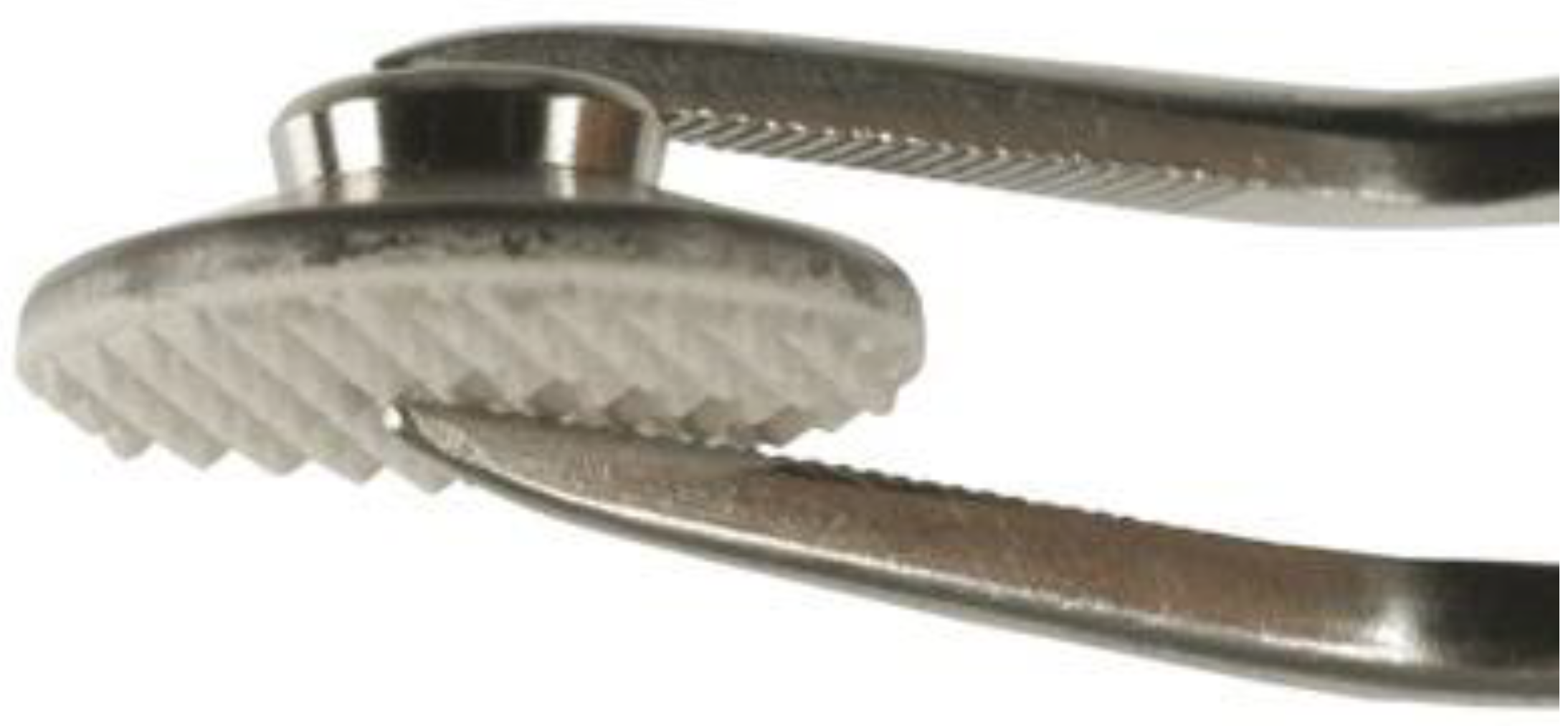
Figure 1. An Onplant device (Nobel Biocare, Gothenburg, Sweden).
Applsci 11 08376 g001
Figure 2. Flow diagram of SPIDER (tool for qualitative evidence synthesis). RCT: randomized controlled trial.
Applsci 11 08376 g002
Table 1. Results of the database search based on the SPIDER tool.
Authors (Year) [Reference]Type of Study
Block and Hoffmann (1995) [9]In vivo animal study
Armbruster and Block (2001) [10]Case series
Kikuchi et al. (2001) [11]In vitro study
Janssens et al. (2002) [7]Case report
Bondemark et al. (2002) [12]Case report
Hassan et al. (2003) [13]In vivo animal study
Hong et al. (2005) [14]Case report
Hoffmann et al. (2006) [6]Pilot study
Chen et al. (2007) [15]In vivo animal study
Feldmann et al. (2007) [16]Randomized controlled trial
Crismani et al. (2008) [17]In vivo animal study
Feldmann and Bondemark (2008) [18]Randomized controlled trial
Niwa et al. (2009) [19]In vivo animal study
Feldmann et al. (2012) [20]Randomized controlled trial
Uezono et al. (2013) [21]In vivo animal study
Uezono et al. (2013) [22]In vitro study
Schmid et al. (2014) [23]In vivo animal study
Ogasawara et al. (2017) [24]In vitro study
Iwanami-Kadowaki et al. (2021) [25]In vitro study
Table 2. Failure rates of Onplant studies in humans.
Authors (Year) [Reference]Type of StudyNo. of PatientsMean Patient Age (Years)No. of OnplantsPercentage FailureCause of Failure (No. of Failed Onplants)
Hoffmann et al. (2006) [6]Pilot study830.1 ± 15.6812.5%No osseointegration (1)
Feldmann and Bondemark (2008) [18]Randomized clinical trial2914.0 ± 1.532917.2%No osseointegration (1)
Technical problems (2)
Discontinued treatment (1)
Anchorage loss (1)
Table 3. Appliance used and treatment types performed with Onplants.
Authors (Year) [Reference]ApplianceNo. of OnplantsUse of Onplants
Armbruster and Block (2001) [10]Transpalatal bar2Anchorage for space closure
Janssens et al. (2002) [7]Transpalatal bar1Anchorage for molar extrusion
Bondemark and Feldmann (2002) [12]Transpalatal bar1Anchorage for molar distalization
Hong et al. (2005) [14]Facemask1Anchorage for maxillary orthopedic treatment
Feldmann and Bondemark (2008) [18]Transpalatal bar30Anchorage for space closure
Publisher’s Note: MDPI stays neutral with regard to jurisdictional claims in published maps and institutional affiliations.