Abstract
This paper aims to perform laser assisted size reduction to nanoparticles of gold (Au) sputtered layer on titanium (Ti) base material using an innovative method that could potentially be applied in novel blood contact and thromboresistive devices in the living body, such as ventricular assist devices (VADs). The enrichment of the surface layer of titanium with gold nanoparticles, due to its bioproperties, may contribute to the reduction of inflammatory reactions and infections occurring mainly in the first postoperative period causing implant failure. The possibility of obtaining superficial size reduction and/or bonding of nano gold particles with Ti micromachining by picosecond laser treatment was evaluated. The quantitative assessment of the particles has been made using SEM and are depicted on the histograms, whereby the appropriate number of particles determine the antibacterial properties and health safety. The initial analysis of micromachining process of the prepared material was focused on power-depth dependence by confocal microscopy. The evaluation of gold particles was conducted using scanning electron microscopy (SEM) using SE and QBSD detectors with energy dispersive spectroscopy (EDS) analysis. Attempts to reduce the deposited gold coating to the size of Au nanoparticles and to melt them into titanium matrix using a laser beam have been successfully completed. There seems to be no strict relationship between particle size distribution of gold onto Ti, probably due to too low energy to excite titanium enough, resulting from difference in Ti and Au melting point temperatures. However, the obtained results allow continuation of pilot studies for augmented research and material properties analysis in the future.
1. Introduction
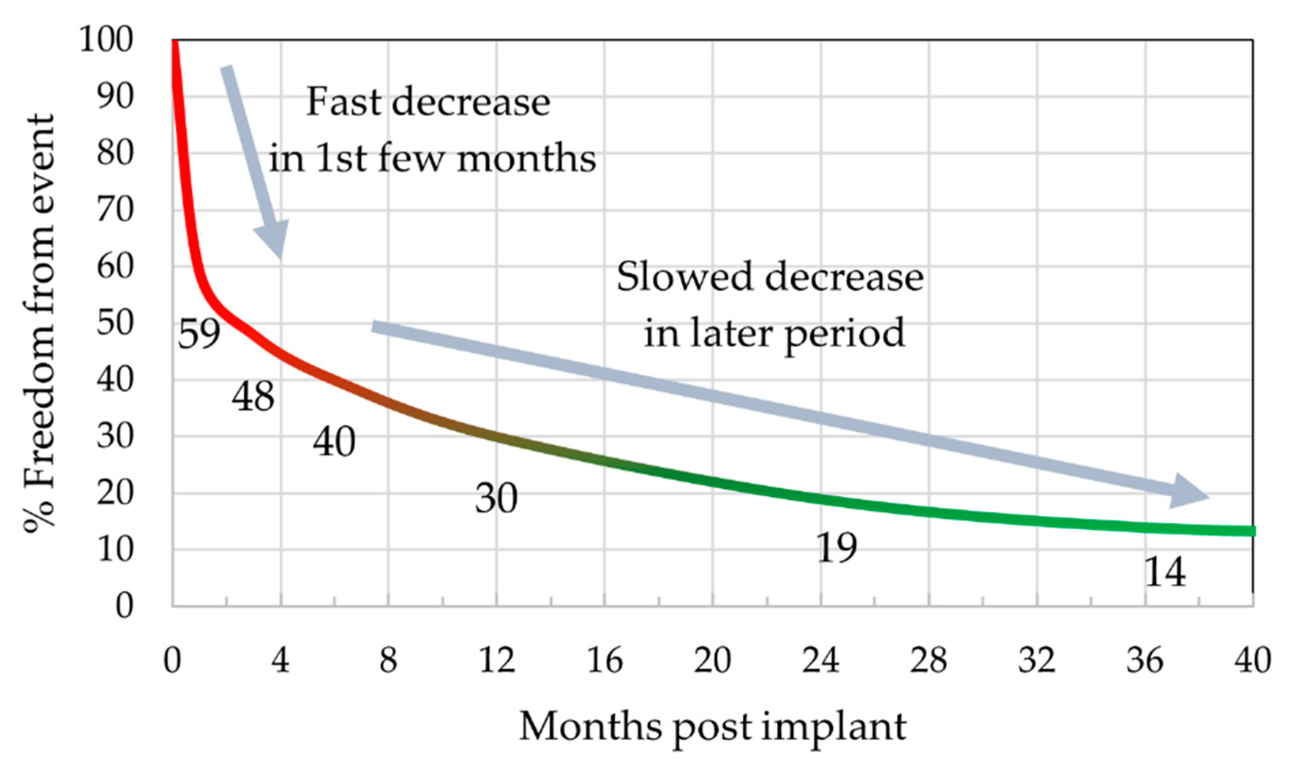
The intensive development of research to verify Feynman’s [1] idea of nanotechnology allowing observations of the world in the nanoscale has resulted in the rapid creation of numerous methods and applications for nanostructured materials. The basic division of methods for obtaining nanostructured materials includes bottom up (starting from the position of smaller materials, e.g., atoms, towards larger ones not exceeding 100 nm) and top down methods, in which dimensions of material change from larger to nano-sized [2,3]. One of the methods of effective particle size reduction is laser assisted particle reduction, the possibilities of which became the object of research of this paper. Laser superficial modification itself has a wide range of useful applications, due to its potential for changing surface chemistry through structuring morphology and being highly precise and repeatable [4,5,6,7,8,9]. The previous contribution of the authors described the possibility of obtaining nanosized Au particles infused in a polymer matrix with regular distribution and controlled amount of particles—both in size and quantity in laser irradiation area [10]. The promising results allowed continuation of the preliminary study on different types of base material and augmented laser power, suited to the needs of metallic bonding nature [11], however, with limitations due to the significant difference in the melting point of the system components reaching 604.18 °C (Ti ~1668.18 °C; Au ~1064 °C) [12,13]. Among other metallic materials, titanium and its alloys are considered as the most suitable material in medical application, due to fulfilling property requirements better than any other competing materials, e.g., stainless steel, niobium or tantalum [14]. In terms of these properties, biocompatibility, corrosion behavior, mechanical behavior, machinability and availability are of greatest interest [15]. In relation to the use of titanium in cardiovascular applications and the alarming number of early post-operative complications in the circulatory system, researchers are interested in the surface modification of titanium materials because of their direct contact with the body’s native tissues [16]. Literature data from 2017 that assesses the time of occurrence of complications in vascular assist devices (VADs) is shown in Figure 1. As can be seen, the highest number of events occurs up to 3 months after surgery.

Figure 1.
Freedom from postoperative VADs failure in the function of time based on literature data [16].
To improve the current state, there is a need to look for modern solutions that can meet both economic and processing demands, but above all that can effectively reduce the number of early postoperative complications. Gold (Au) can be expected as an element introduced for its antibacterial and biocompatible properties. It is worth to notice that gold particles have potential to control coagulation under nanoscale conditions. External laser excitation of nanoparticles, in particular, can cause payload release, e.g., thrombin inhibitors responsible for stopping blood clotting [17]. Au nanoparticles have gained considerable interest in therapeutic applications, as they can be selectively excited where tissue is transparent to release multiple species that can impact complex processes [18,19]. That implication suggests many advantages for controlling blood clotting after implantation of the device as a drug supplier or as a risk-reducing factor for infection/coagulation in itself [20,21]. The present work is an attempt to achieve the reduction of gold layer to the nanoscale particles onto a titanium base material, proceeding with their bonding on the surface [10]. This may contribute to the development of gold particle delivery methods for the subsequent control of thrombus formation through drug/protein release by selective laser irradiation of the patient at the site of the implanted device [17], or to the enhancement of the biocompatibility of titanium devices through gold’s characteristic antibacterial properties and lack of cytotoxicity [22,23]. A picosecond laser with a wavelength of 355 nm and a power reaching 22 mW was used for the study. The particle size distribution in the matrix were evaluated by statistical tests of images obtained by high-resolution scanning microscopy imaging. The topography of the structure and the effect of the laser power on the matter have been evaluated by confocal microscopy. The micro-areas of the beam interaction with gold layer and titanium base material were visualized by light microscopy to show the optical appearance of the laser irradiation areas and the tendency to particles agglomeration.
2. Materials and Methods
Samples from commercial pure Titanium grade II (CP Ti), were delivered as ø 30 mm rods (Wolften, Wrocław, Poland) cutted in slices by cutting on a lathe. The material is characterized by the ASTM B348/B348M standard, with the chemical composition presented in Table 1.

Table 1.
Chemical composition of pure titanium grade II used in experiment [24,25].
The rod was cut into 7 mm thick slices and prepared for sputtering. Each sliced sample was prepared according to metallographic procedure: grinding with paper grain size gradation sequentially 220,800, 1200 in time t = 4 min per each gradient and mechanical polished using 0.04 μm colloidal silica. The process was performed using the grinding-polishing machine TERGAMIN-30 (Struers, Willich, Germany).
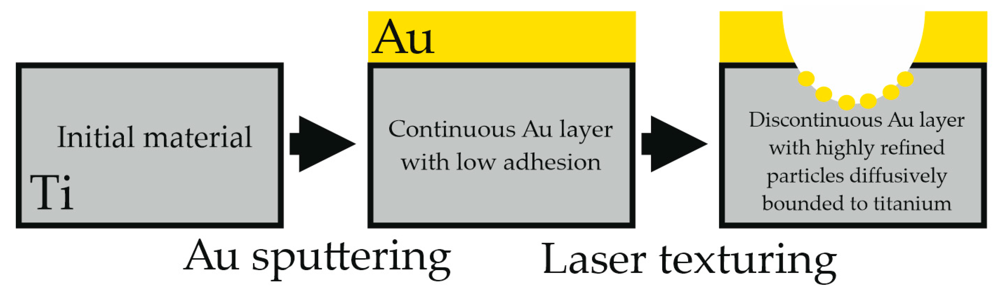
The investigation consisted of coating titanium with Au by sputtering. The next step was laser texturing which purpose was to refine sputtered layer to ultrafine particles and provide their adhesion to the substrate (Figure 2).

Figure 2.
Experiment scheme.
According to previous study of the authors [10], gold sputtering was performed on each sample. The device used for sputtering was Leica EM ACE600 device with 99.99% Au target. The current (I) parameter was set to 30 mA, sputter vacuum 8 × 10⁻³ mbar with the sputtering rate 0.18 nm/s [26].
2.1. Laser Micromachining
Different laser treatment was applied (Table 2) to investigate the influence of increasing power of photon beam. The experiment was carried out with the A-355 Laser Micromachining system (Oxford Lasers, Didcot, UK), which is based on a 355 nm diode-pumped solid-state picosecond laser.
Table 2.
Laser texturing parameters.
The laser micromachining system is based on G-code instructions language in order to realise users’ commands. Both, the device and the setup are shown in the Figure 3a,b. The system has a maximum energy density acquiring 3255 W/cm2, allowing it to ablate/melt the material. The A-355 picosecond laser micromachining system has a maximum pulse energy of 0.2 mJ, rated power of 23 mW, and a pulse length of approximately 6 ps. The laser texturing path was either a straight line (at frequency 400 Hz and speed 1mm/s), single laser pulse irradiation (at frequency 10 Hz and speed 1 mm/s). The laser texturing parameters are shown in Table 2.
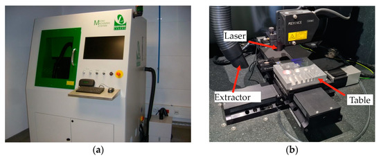
Figure 3.
Oxford laser micromachining system: (a) housing/radiation shield, (b) laser head, table, and fume extractor.
2.2. Microscopic Observations
Before microscopic analysis the surface of the sample has been cleaned with ≥99.6% pure acetone. The research was investigated in specific micro-areas to assess the infusion of gold into base material following laser treatment. Effects of laser micromachining was preliminary assessed by a metallographic light microscope Axio Observer (Zeiss, Oberkochen, Germany) and digital microscope Leica DVM6 (Leica, Wetzlar, Germany). The presence of particles melted/evaporated onto the Ti basement material ratio was determined using high-resolution scanning electron microscopy (HRSEM). The Supra 35 (Zeiss, Oberkochen, Germany) equipment incorporated EDS spectrum analysis, which allowed the surface texture and quality chemical composition to be determined. Samples were also examined using the Zeiss LSM Exciter 5 confocal microscopy equipment (Zeiss, Oberkochen, Germany). To obtain the highest optical resolution, the system was outfitted with a 405 nm diode laser with a 25 mW output. To study the selected structure of laser groove breakdown and acquired depth, each sample was analyzed using z-stack mode. After scanning, 3D pictures were extracted by combining photos of sequentially scanned planes.
In the course of creating the histograms, three selected microscope fields of view of 20, 50 and 100 k magnification each were used at the center of the crater for each of the 13.2, 17.6 and 22 mW powers analyzed.
3. Results
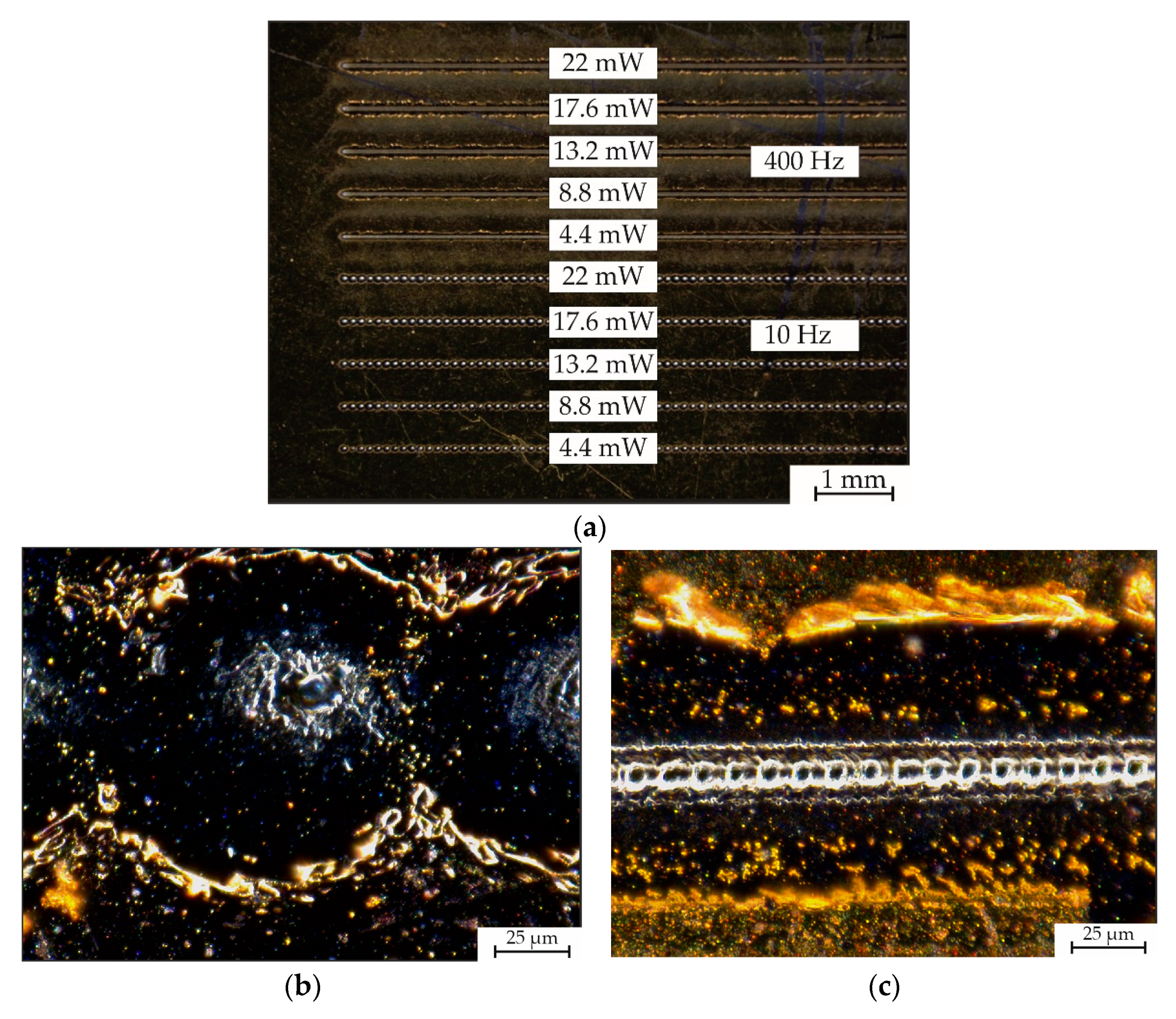
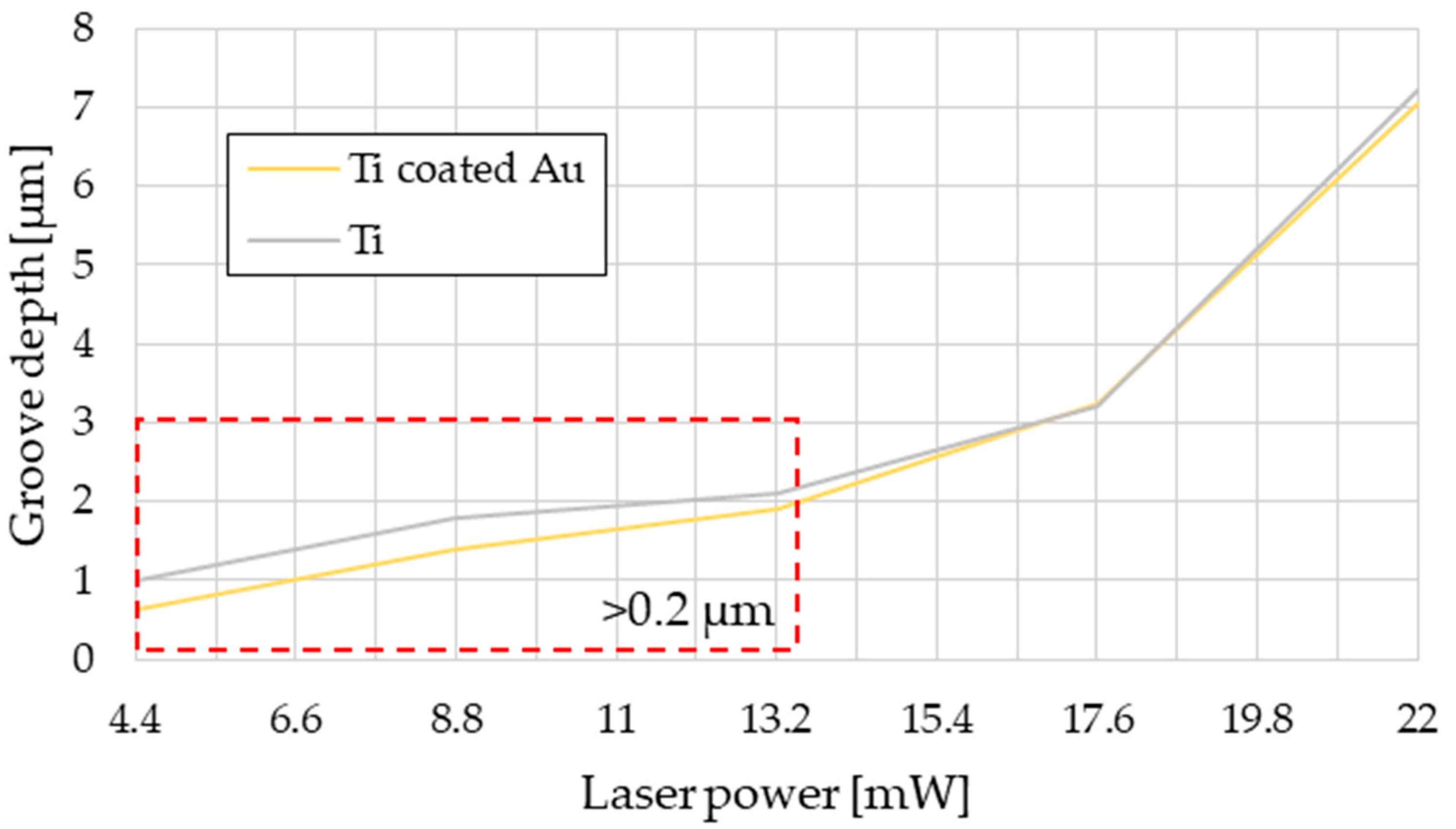
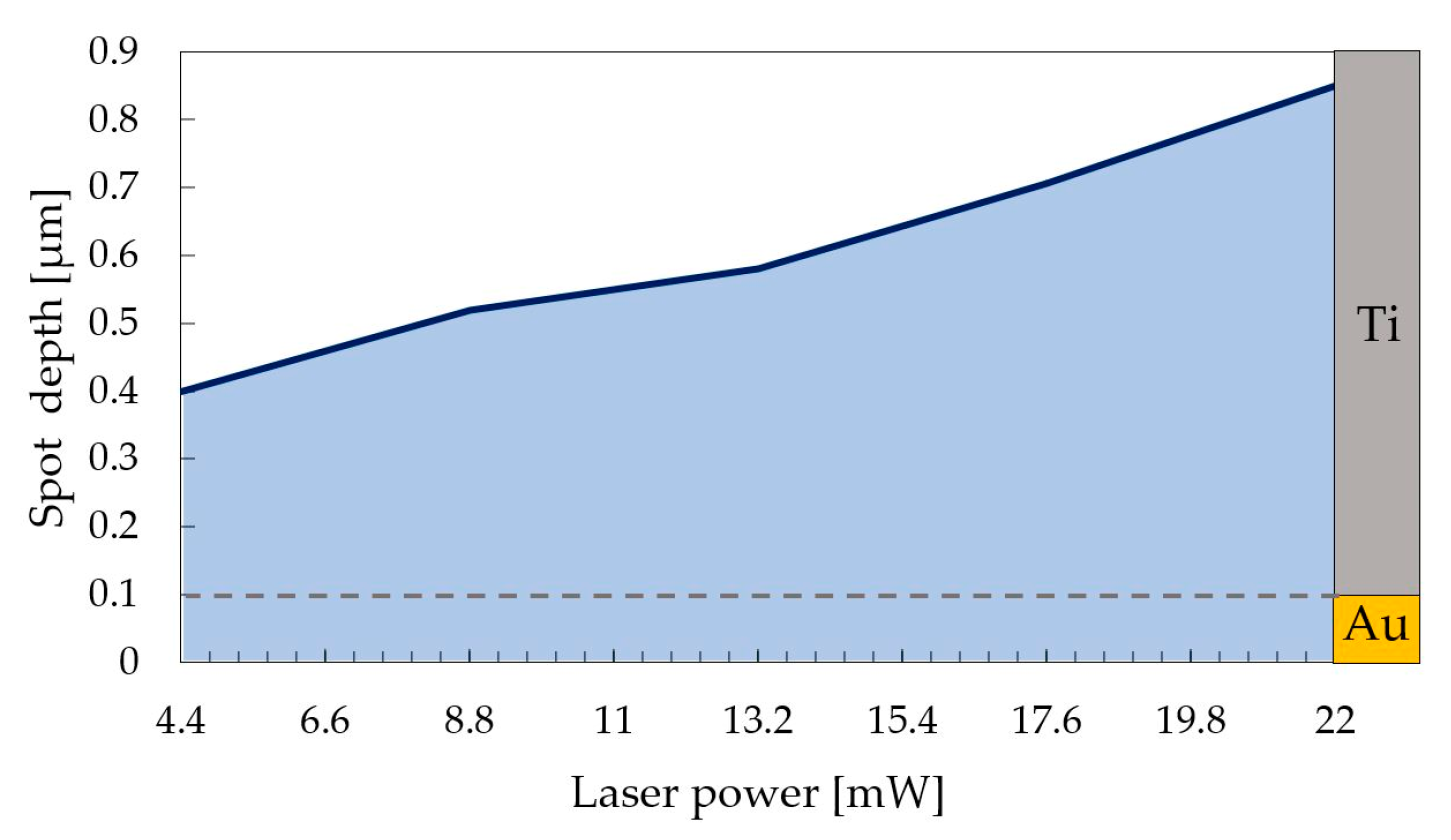
In the first stage of material testing, it was planned to determine the dependence of laser power on depth at frequencies of 400 and 10 Hz at the same beam feed rate of 1 mm/s. Based on the results obtained, the optimal parameters were selected to conduct the analyses, considering the initial objectives and the absorption of the laser beam by the material under study (Figure 4). The results obtained in the course of confocal analysis (Figure 5) for the variant with a frequency of 400 Hz provided the conclusion that the considered laser powers should be in the range from 13.2 to 22 mW due to the rapid growth of depth in this region (in the power range of 4.4 to 13.2 mW the trend should be considered as close to linear, while 13.2 to 22 mW grows exponentially) as is presented in Figure 6 and Figure 7. In the “single laser irradiation” approach, which corresponds to the 10 Hz variant, this trend was not visualized. The authors selected power of 13.2 mW as the minimum, and 17.6 and 22 mW as optimal values for the statistical evaluation of the number of surviving particles and their distribution in the matrix.
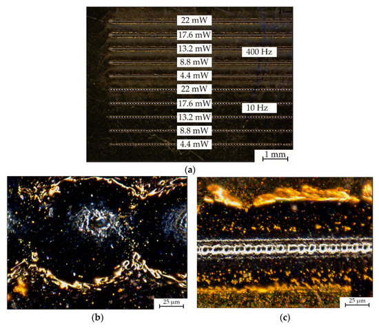
Figure 4.
Light microscopy images of the performed experiment with the presence of particles distribution: (a) full range of tested laser beam parameters; (b) single laser irradiation approach close up; (c) 400 Hz approach close up.
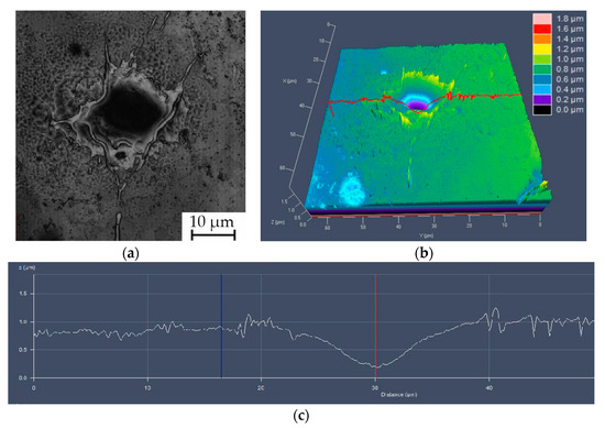
Figure 5.
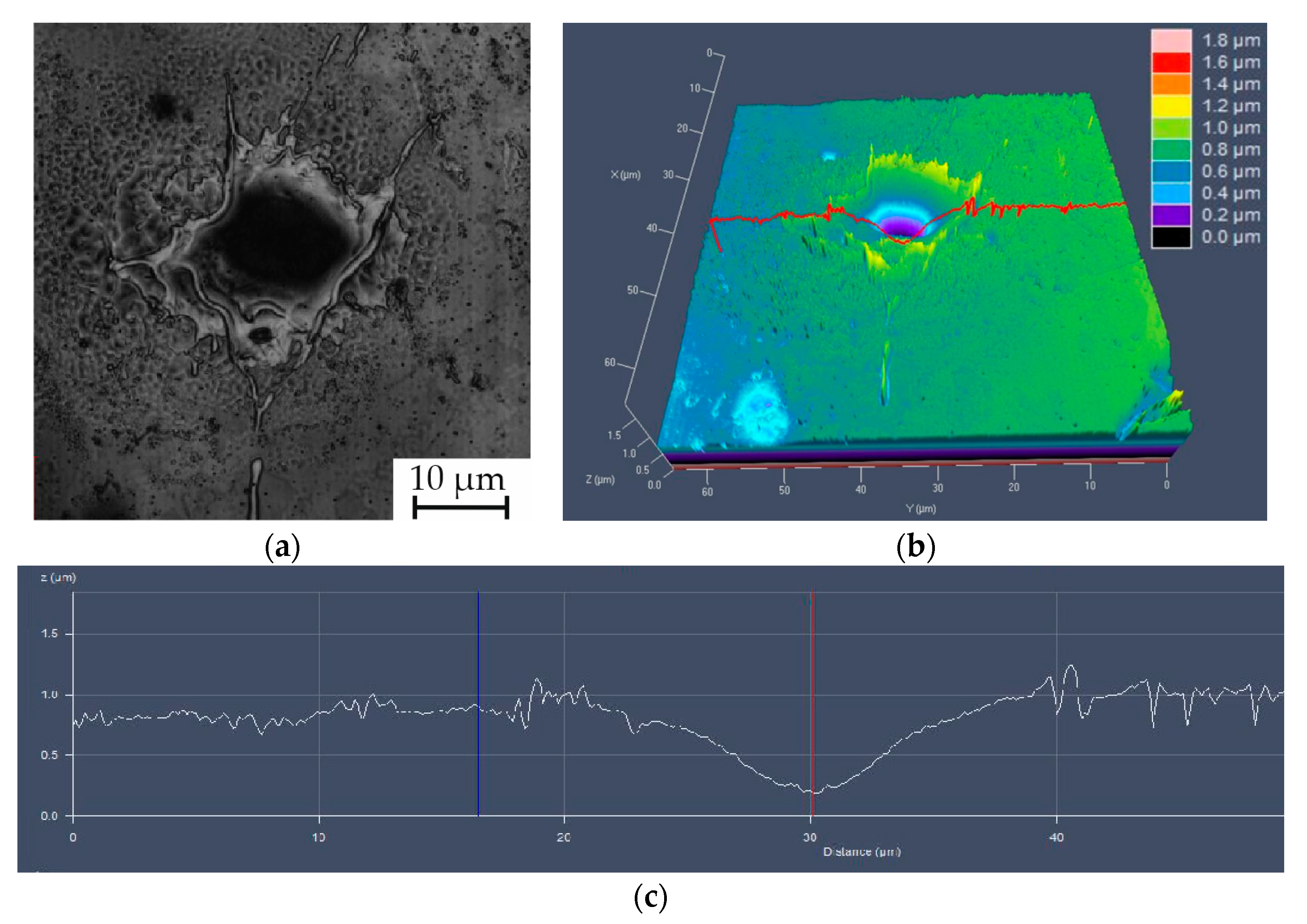
Confocal imaging and analysis of the laser irradiation depth: (a) image of single laser irradiation area; (b) Z-stack 3D reconstruction of the surface morphology of the single spot with range indicator; (c) topography profile traversing single irradiation area.

Figure 6.
Laser power to groove depth diagram based on confocal results, with the region excluded from statistical investigation (red frame).

Figure 7.
Laser power to spot depth diagram based on confocal results from single laser irradiation approach with visualization of coating thickness/depth ratio.
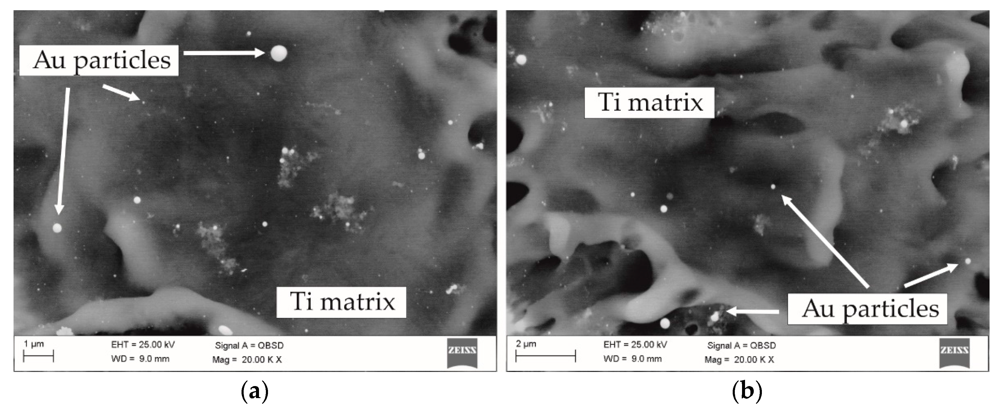
Statistical analysis of the particle distribution was conducted using SEM images (Figure 8) at magnifications of 20, 50 and 100 k.
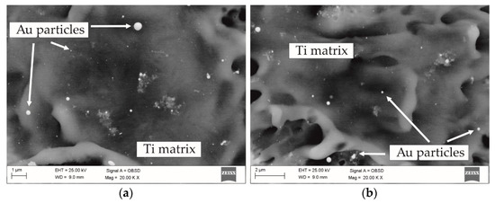
Figure 8.
Gold particles in center of the laser irradiation area: (a) laser power 22 mW; (b) laser power 17.6 mW.
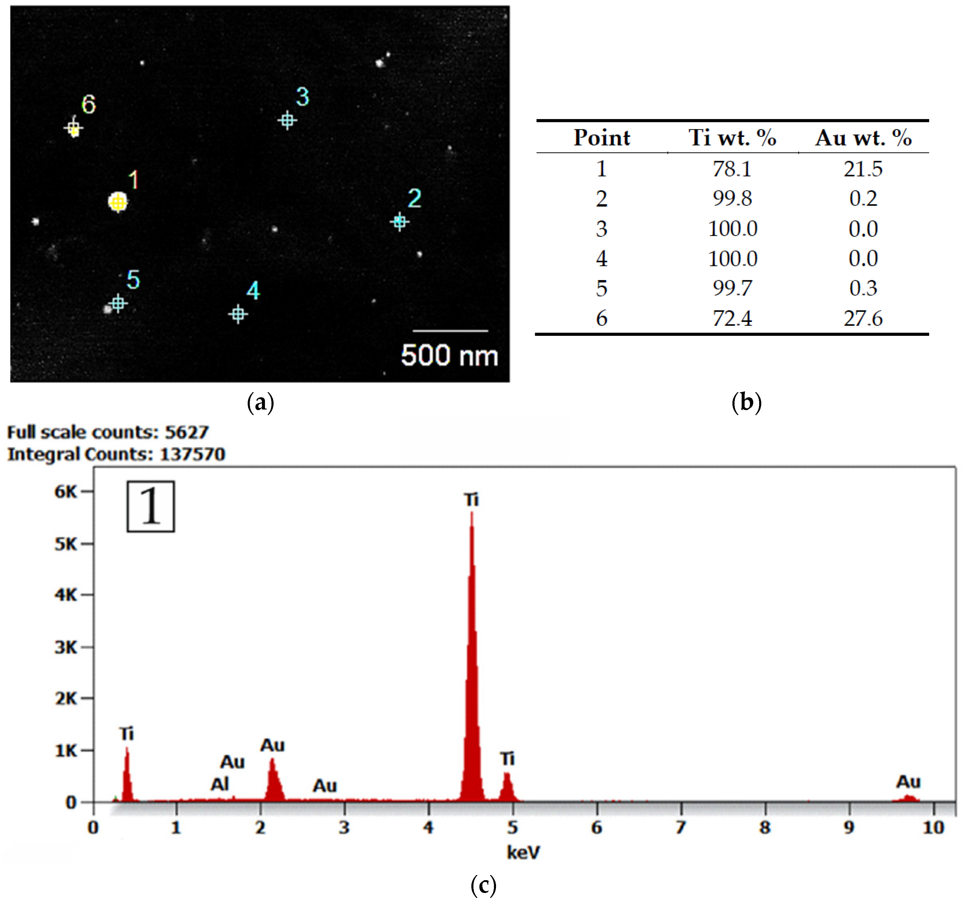
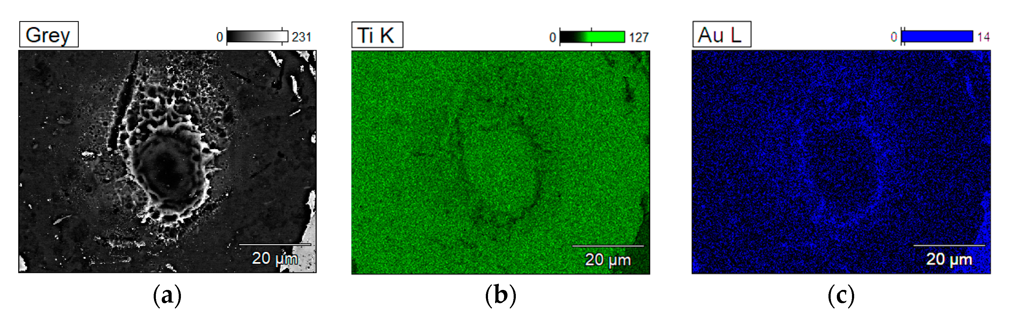
The qualitative composition of the observed individuum together with an indicative density map was validated by EDS analysis (Figure 9 and Figure 10). Bright spots according to the EDS analysis are gold, while the punctate background measurement indicates titanium. The EDS spectrum (Figure 9c) from point 1 shows peaks from gold and titanium, indicating a low gold content, characteristic of the presence of this element only in the form of small particles surrounded by titanium. The EDS qualitative chemical elements distribution map (Figure 10) highlight the change in the density of the gold particles as a function of distance from the laser irradiation site.

Figure 9.
EDS quantitative chemical elements composition and energy dispersion spectrum: (a) spectral collection points—assessment towards the gold particles; (b) results of collected spectrums in wt %; (c) EDS spectrum of the measurement point 1.
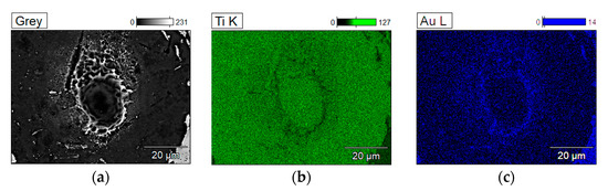
Figure 10.
EDS qualitative chemical elements distribution map: (a) SEM image; (b) EDS spectrum map of Ti; (c) EDS spectrum map of Au.
Statistical analysis of particles distribution (Figure 11) indicates the presence of nanometric particles in the whole range of tested laser powers. As the laser power increases, the proportion and size of particles differ rather slightly but, in every variant, there seem to be tendency to reduce either to very small particles hardly visible in SEM imaging or to reduce with size greater than 15 nm.
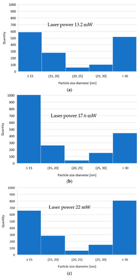
Figure 11.
Histograms of particles distribution: (a) 4 mW; (b) 15 mW; (c) 16 mW.
4. Discussion
The assessment of laser beam impact on the gold coated material was done using the whole range of the laser powers available in the A-355 laser micromachining system (Oxford Lasers, Didcot, UK). The treatment was performed using both the 400 Hz approach with a feed rate of 1 mm/s and the “single laser irradiation” method, which investigates the effects of a single laser pulse at a given power. When comparing the graphs of the two approaches, a certain analogy can be seen, but it is more evident when processing at 400 Hz (above 13.2 mW the groove depth increases exponentially). The recorded results for both power-depth relationship curves allowed us to select the optimal laser power range at 13.2, 17.6 and 22 mW. Additionally, in selecting the optimum parameters, it is important that even at the lowest power tested, the beam penetrated through a 100 nm thick layer of sputtered gold. Moreover, light microscopy images show an interesting tendency for particles to agglomerate outside craters, an agglomeration that is not visible in the case of a single irradiation.
Based on the microscopic images obtained and EDS maps, the presence of nano-size gold particles with a distribution dependent on the crater distance after the beam passage can be observed (Figure 9). The geometric character of the crater recorded by 3D analysis with a confocal microscope is typical for a single interaction of the Gaussian beam with matter as has been shown in numerous literature studies [27,28,29]. It can be observed that the debris which gathered around the ablation crater was a combination of re-solidified droplets of ejected molten material, globules, and clusters of small particles. This is because the laser used does not achieve pure ablation—the average energy flux density does not exceed 3.2 × 10⁷ W/m2. This relationship is described and confirmed by Pyatenko et al. [30]. They defined that if the energy flux density is less than 1012 W/m2, particles decrease their size in a mechanism following a strict order: heating–melting–evaporation. So, if the evaporation of the material is partial, the majority is melted. Following the statement of Inasawa et al. [31] that size reduction of the particles occurs in a layer-by-layer mechanisms based on bimodal particle distribution, the most important was to assess the size and amount of particles present in irradiation area already after one single pulse. Observation of the bimodal distribution of particle sizes that are slightly smaller and much smaller than the original ones allowed Inasawa to conclude that the irradiation by picosecond-laser pulses is the most efficient way to reduce the Au NPs size. Later, Pyatenko carried out the calculations to examine the melting and size reduction of AuNPs, stating that photothermal mechanisms prevails at low laser intensities, that occurs with best efficiency in 532 nm nanosecond lasers in comparison with wavelengths of 1064 and 355 nm, but he gave no experimental evidence for that finding. The 355 nm picosecond laser has been found efficient for splitting Ag NPs [32] and Au NPs in aqueous sodium dodecyl sulphates, as the picosecond excitation–relaxation electronic interband enables a faster absorption of a new photon that the conduction-band electrons can absorb. What is more, positively charged Au ions occurring during interband excitation allow the thermionic emission of electrons, which is responsible for the highly efficient splitting of the Au NPs [33,34].
Taking into account our previous study on Au particles infused into polyether ether ketone (PEEK) polymer [10], and the fact that picosecond laser tends to be the most suitable for an efficiently to inducing size reduction [31], single laser irradiation should be analyzed at the first step in gold reduction trials. As shown in Figure 9, the scale of reduction of the gold layer to particles occurred to a very large extent in the material we studied. In the image one can see particles of very small size standing out from the background at extremely high SEM magnification. There is also a larger approximately 25 nm particle with high sphericality in the field of view of the microscope. Figure 12 suggests that particles smaller than 15 nm are the most numerous in the material studied but are difficult to observe with conventional SEM observations. Investigations by Transmission Electron Microscopy (TEM) should be carried out in continuation of the study.
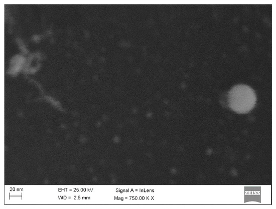
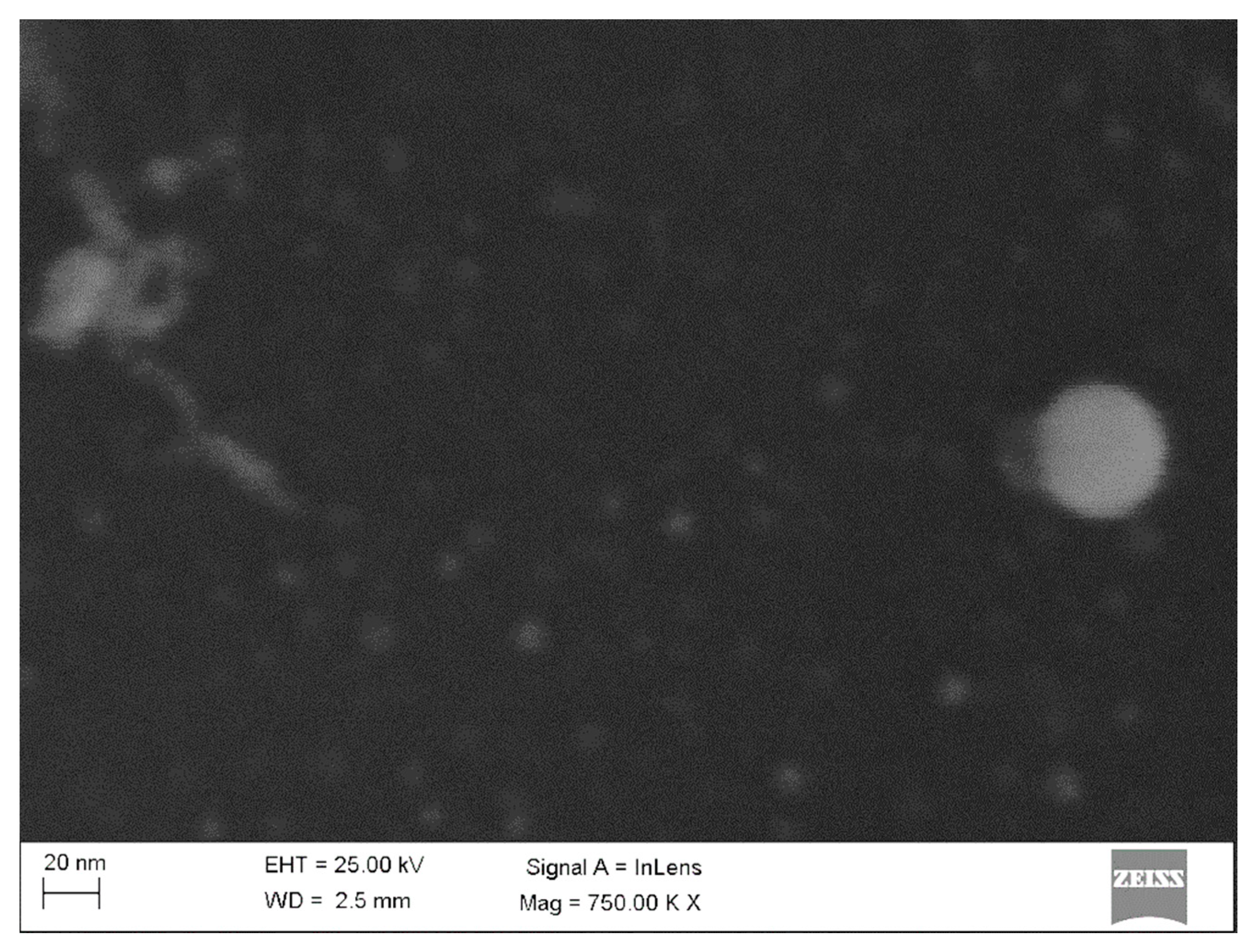
Figure 12.
Nanosized Au particles inside the irradiation area.
The EDS analysis confirmed the presence of Au particles as spherical particles appearing in the whole volume of the laser irradiation area. The EDS qualitative chemical elements distribution map shows Au particles density versus distance from the crater. The closer to the crater top the number of particles increases, but they are almost non-existent in the centre [35].
Despite the difference obtained in the depth of penetration of the beam into the material, no outstanding treatment variant can be distinguished. No unambiguous tendency to change the particle size was demonstrated, although in the variants with higher energies of 17.6 and 22 mW one can distinguish higher counts for the extreme ranges of the histogram, which may indicate a tendency to polarize the particle size into either large or very small ones, the intermediate sizes successively decreasing their proportion.
Previous studies [10] have established that the number and size of particles surviving in a polymer matrix depends on the laser power used, and so the next step was to see if this experiment could be repeated with similar efficacy on metallic materials. The obtained results allowed for a reduction in particle size, but although in the power vs. depth diagram (Figure 6) the influence of the beam on the surfaces is exponential, this was not reflected in the histograms (Figure 11). However, the observations on bimodal particle distribution made on other materials, by other researchers [30,31,32,33,34], were confirmed. This allows for comparative analysis and raises the suspicion that using higher beam energy could lead to greater superficial melting of titanium and thus fusion of gold particles into the titanium matrix (as the amount of surviving Au NPs in the range studied does not depend on power).
5. Conclusions
Preliminary research of laser assisted size reduction of Au particles onto the Ti surface allows for a positive assessment of the potential of the applied technique. The results of the research allow us to state:
- Results of power—depth measurements indicates that above 13.2 mW the depth of the obtained grove increases exponentially, while under this power the increase is linear
- 355 nm picosecond laser with power of 22 mW causes obtaining grove depth of 7 and 0.8 μm for 400 Hz and single laser irradiation, respectively
- The Au NPs formation (mainly between 15 to 30 nm) and partial remelting into matrix is possible for proposed in experiment laser powers and allowed obtainment of nano Au particles
- There seems to be no strict relationship between used power and particle size distribution of gold onto Ti, probably due to too low energy to excite titanium enough resulting from difference in Ti and Au melting point temperatures.
- The results show clear bimodal distribution of particle size
- EDS analysis confirmed presence and local distribution shape in laser irradiated area changing with the distance from laser source
Further analysis utilizing TEM to obtain the superficial properties resulting from the laser treatment of the material is required, as well as further optimization of the process to enhance the size reduction process control.
Author Contributions
Conceptualization, O.B. and M.A.; methodology, M.L. and O.B.; software, M.L. and O.B.; validation, J.G. and M.A.; formal analysis, M.A. and J.G.; investigation, M.L., T.P. and O.B.; resources, M.A. and J.G.; data curation, M.L.; writing—original draft preparation, O.B.; writing—review and editing, M.L. and A.S.; visualization, M.L. and A.S.; supervision, M.A. and J.G.; project administration, O.B. and M.A.; funding acquisition, J.G. All authors have read and agreed to the published version of the manuscript.
Funding
Co-financed by the European Union through the European Social Fund (grant POWR.03.05.00-00-Z305).
Conflicts of Interest
The authors are not aware of any conflicts of interest regarding this paper.
References
- Feynman, R.P. Plenty of Room at the Bottom; American Physical Society: Pasadena, CA, USA, 1959. [Google Scholar]
- Bayda, S.; Adeel, M.; Tuccinardi, T.; Cordani, M.; Rizzolio, F. The History of Nanoscience and Nanotechnology: From Chemical-Physical Applications to Nanomedicine. Molecules 2019, 25, 112. [Google Scholar] [CrossRef] [PubMed] [Green Version]
- Górka, J.; Czupryński, A.; Żuk, M.; Adamiak, M.; Kopyść, A. Properties and Structure of Deposited Nanocrystalline Coatings in Relation to Selected Construction Materials Resistant to Abrasive Wear. Materials 2018, 11, 1184. [Google Scholar] [CrossRef] [PubMed] [Green Version]
- Chang, Y.-Y.; Zhang, J.-H.; Huang, H.-L. Effects of Laser Texture Oxidation and High-Temperature Annealing of TiV Alloy Thin Films on Mechanical and Antibacterial Properties and Cytotoxicity. Materials 2018, 11, 2495. [Google Scholar] [CrossRef] [PubMed] [Green Version]
- Frostevarg, J.; Olsson, R.; Powell, J.; Palmquist, A.; Brånemark, R. Formation Mechanisms of Surfaces for Osseointegration on Titanium Using Pulsed Laser Spattering. Appl. Surf. Sci. 2019, 485, 158–169. [Google Scholar] [CrossRef]
- Hatzikiriakos, S.G.; Moradi, S.; Kamal, S. Superhydrophobic Laser-Ablated Stainless Steel Substrates Exhibiting Cassie–Baxter Stable State. Surf. Innov. 2015, 3, 151–163. [Google Scholar] [CrossRef]
- Woźniak, A.; Adamiak, M.; Chladek, G.; Bonek, M.; Walke, W.; Bialas, O. The Influence of Hybrid Surface Modification on the Selected Properties of CP Titanium Grade II Manufactured by Selective Laser Melting. Materials 2020, 13, 2829. [Google Scholar] [CrossRef]
- Kołodziejczak, P.; Golański, D.; Chmielewski, T.; Chmielewski, M. Microstructure of Rhenium Doped Ni-Cr Deposits Produced by Laser Cladding. Materials 2021, 14, 2745. [Google Scholar] [CrossRef]
- Wang, C.; Suder, W.; Ding, J.; Williams, S. Wire Based Plasma Arc and Laser Hybrid Additive Manufacture of Ti-6Al-4V. J. Mater. Process. Technol. 2021, 293, 117080. [Google Scholar] [CrossRef]
- Bialas, O.; Lis, M.; Woźniak, A.; Adamiak, M. Laser Superficial Fusion of Gold Nanoparticles with PEEK Polymer for Cardiovascular Application. Materials 2021, 14, 971. [Google Scholar] [CrossRef]
- Chmielewski, T.; Hudycz, M.; Krajewski, A.; Sałaciński, T.; Skowrońska, B.; Świercz, R. Structure Investigation of Titanium Metallization Coating Deposited onto AlN Ceramics Substrate by Means of Friction Surfacing Process. Coatings 2019, 9, 845. [Google Scholar] [CrossRef] [Green Version]
- Laing, M. Melting Point, Density, and Reactivity of Metals. J. Chem. Educ. 2001, 78, 1054. [Google Scholar] [CrossRef]
- Podor, R.; Pailhon, D.; Ravaux, J.; Brau, H.-P. Development of an Integrated Thermocouple for the Accurate Sample Temperature Measurement During High Temperature Environmental Scanning Electron Microscopy (HT-ESEM) Experiments. Microsc. Microanal. 2015, 21, 307–312. [Google Scholar] [CrossRef]
- Lisiecki, A. Study of Optical Properties of Surface Layers Produced by Laser Surface Melting and Laser Surface Nitriding of Titanium Alloy. Materials 2019, 12, 3112. [Google Scholar] [CrossRef] [Green Version]
- Balazic, M.; Kopac, J.; Jackson, M.J.; Ahmed, W. Review: Titanium and Titanium Alloy Applications in Medicine. IJNBM 2007, 1, 3. [Google Scholar] [CrossRef]
- Chaanine, A.H.; Pinney, S.P. Mechanical Circulatory Support as a Bridge to Heart Transplantation. In Heart Failure; Eisen, H., Ed.; Springer London: London, UK, 2017; pp. 639–663. ISBN 978-1-4471-4218-8. [Google Scholar]
- de Puig, H.; Cifuentes Rius, A.; Flemister, D.; Baxamusa, S.H.; Hamad-Schifferli, K. Selective Light-Triggered Release of DNA from Gold Nanorods Switches Blood Clotting On and Off. PLoS ONE 2013, 8, e68511. [Google Scholar] [CrossRef] [PubMed] [Green Version]
- Wijaya, A.; Schaffer, S.B.; Pallares, I.G.; Hamad-Schifferli, K. Selective Release of Multiple DNA Oligonucleotides from Gold Nanorods. ACS Nano 2009, 3, 80–86. [Google Scholar] [CrossRef] [PubMed]
- Huschka, R.; Zuloaga, J.; Knight, M.W.; Brown, L.V.; Nordlander, P.; Halas, N.J. Light-Induced Release of DNA from Gold Nanoparticles: Nanoshells and Nanorods. J. Am. Chem. Soc. 2011, 133, 12247–12255. [Google Scholar] [CrossRef] [Green Version]
- Ajdari, N.; Vyas, C.; Bogan, S.L.; Lwaleed, B.A.; Cousins, B.G. Gold Nanoparticle Interactions in Human Blood: A Model Evaluation. Nanomed. Nanotechnol. Biol. Med. 2017, 13, 1531–1542. [Google Scholar] [CrossRef] [PubMed]
- Fan, J.H.; Hung, W.I.; Li, W.T.; Yeh, J.M. Biocompatibility Study of Gold Nanoparticles to Human Cells. In 13th International Conference on Biomedical Engineering; Lim, C.T., Goh, J.C.H., Eds.; IFMBE Proceedings; Springer: Berlin, Germany, 2009; Volume 23, pp. 870–873. ISBN 978-3-540-92840-9. [Google Scholar]
- Liu, K.; Zheng, Y.; Lu, X.; Thai, T.; Lee, N.A.; Bach, U.; Gooding, J.J. Biocompatible Gold Nanorods: One-Step Surface Functionalization, Highly Colloidal Stability, and Low Cytotoxicity. Langmuir 2015, 31, 4973–4980. [Google Scholar] [CrossRef]
- Gu, X.; Xu, Z.; Gu, L.; Xu, H.; Han, F.; Chen, B.; Pan, X. Preparation and Antibacterial Properties of Gold Nanoparticles: A Review. Environ. Chem. Lett. 2020. [Google Scholar] [CrossRef]
- Camilo, C.C.; de Souza, E.C.; Di Lorenzo, P.L.; de Almeida Rollo, J.M.D. Measurement of the Grain Boundary Energy of Commercially-Pure Grade 2 Titanium at High Temperature. RBEB 2011, 27, 175–181. [Google Scholar] [CrossRef] [Green Version]
- Standard Specification for Titanium and Titanium Alloy Bars and Billets. ASTM B348/B348M. Available online: https://www.astm.org/Standards/B348.htm (accessed on 3 January 2021).
- Sample preparation for electron-microscopy. Available online: https://Www.Leica-Microsystems.Com/ (accessed on 3 January 2021).
- Ortiz, R.; Basnett, P.; Roy, I.; Quintana, I. Picosecond Laser Ablation of Polyhydroxyalkanoates (PHAs): Comparative Study of Neat and Blended Material Response. Polymers 2020, 12, 127. [Google Scholar] [CrossRef] [PubMed] [Green Version]
- Shaheen, M.E.; Gagnon, J.E.; Fryer, B.J. Laser Ablation of Iron: A Comparison between Femtosecond and Picosecond Laser Pulses. J. Appl. Phys. 2013, 114, 083110. [Google Scholar] [CrossRef]
- Milovanović, D.S.; Petrović, S.M.; Shulepov, M.A.; Tarasenko, V.F.; Radak, B.B.; Miljanić, Š.S.; Trtica, M.S. Titanium Alloy Surface Modification by Excimer Laser Irradiation. Opt. Laser Technol. 2013, 54, 419–427. [Google Scholar] [CrossRef]
- Pyatenko, A.; Yamaguchi, M.; Suzuki, M. Mechanisms of Size Reduction of Colloidal Silver and Gold Nanoparticles Irradiated by Nd:YAG Laser. J. Phys. Chem. C 2009, 113, 9078–9085. [Google Scholar] [CrossRef]
- Inasawa, S.; Sugiyama, M.; Yamaguchi, Y. Bimodal Size Distribution of Gold Nanoparticles under Picosecond Laser Pulses. J. Phys. Chem. B 2005, 109, 9404–9410. [Google Scholar] [CrossRef]
- Kamat, P.V.; Flumiani, M.; Hartland, G.V. Picosecond Dynamics of Silver Nanoclusters. Photoejection of Electrons and Fragmentation. J. Phys. Chem. B 1998, 102, 3123–3128. [Google Scholar] [CrossRef]
- Shoji, M.; Miyajima, K.; Mafuné, F. Ionization of Gold Nanoparticles in Solution by Pulse Laser Excitation as Studied by Mass Spectrometric Detection of Gold Cluster Ions. J. Phys. Chem. C 2008, 112, 1929–1932. [Google Scholar] [CrossRef]
- Muto, H.; Miyajima, K.; Mafuné, F. Mechanism of Laser-Induced Size Reduction of Gold Nanoparticles As Studied by Single and Double Laser Pulse Excitation. J. Phys. Chem. C 2008, 112, 5810–5815. [Google Scholar] [CrossRef]
- Kutrovskaya, S.; Arakelian, S.; Kucherik, A.; Osipov, A.; Evlyukhin, A.; Kavokin, A.V. The Synthesis of Hybrid Gold-Silicon Nano Particles in a Liquid. Sci. Rep. 2017, 7, 10284. [Google Scholar] [CrossRef]


Publisher’s Note: MDPI stays neutral with regard to jurisdictional claims in published maps and institutional affiliations. |
© 2021 by the authors. Licensee MDPI, Basel, Switzerland. This article is an open access article distributed under the terms and conditions of the Creative Commons Attribution (CC BY) license (https://creativecommons.org/licenses/by/4.0/).