Article

Ultrafast Ultrasound-Derived Muscle Strain Measure Correlates with Carotid Local Pulse Wave Velocity in Habitual Resistance-Trained Individuals

1
Department of Athletics, National Taiwan University, Taipei 10617, Taiwan
2
Department of Exercise and Health Science, National Taipei University of Nursing and Health Sciences, Taipei 11219, Taiwan
3
Department of Electrical Engineering & Graduate Institute of Biomedical Electronics and Bioinformatics, National Taiwan University, Taipei 10617, Taiwan
*
Author to whom correspondence should be addressed.
H.-F.L. and Y.-H.L. equally contributed to this work as co-first authors.
Appl. Sci. 2021, 11(18), 8783; https://doi.org/10.3390/app11188783
Submission received: 15 August 2021 / Revised: 15 September 2021 / Accepted: 17 September 2021 / Published: 21 September 2021

Abstract

:

Featured Application

This study demonstrated that muscle strain rate derived from ultrafast ultrasound imaging decreases as resistance intensity increases. Additionally, carotid artery stiffness measured by ultrafast local pulse wave velocity correlates with widely used carotid function measures and associates with the increased central pulse pressure following acute resistance exercise in the young resistance-trained population. All of this indicates the clinical implication of ultrafast ultrasound imaging in monitoring exercise training. Moreover, habitual resistance-trained individuals with higher upper-arm muscle stiffness tend to have higher carotid stiffness, suggesting the potential link between muscular and vascular remodeling along with regular resistance training.

Abstract

Purpose: this study investigated the effects of the intensity of machine-based bicep curl resistance exercise on ultrafast ultrasound-derived muscle strain rate and carotid ultrafast pulse wave velocity (ufPWV), and examined the association between muscle strain rate, ufPWV, and established carotid function measures in habitual resistance-trained individuals. Methods: twenty-three young habitual resistance-trained males (age: 24 ± 1 year, body mass index = 24 ± 1 kg/m2) were recruited to participate in two bouts of acute bicep curl exercise. After one-repetition maximum determination (1RM), the participants were randomly assigned to engage in bicep curls at 40 or 80%1RM intensity (10 reps × five sets) by a crossover study design. The muscle strain rate of bicep muscle, carotid ufPWV during systole(ufPWV-sys), and diastole (ufPWV-dia) were obtained pre- and post-exercise. In addition, carotid function measures were calculated by obtained carotid diameter and central blood pressure changes. Results: compared with pre-exercise, the reduction in post-exercise muscle strain rate and its area under the curve of 80%1RM was greater than those of 40%1RM. Both ufPWV-sys and ufPWV-dia increased regardless of exercise intensity. Baseline bicep muscle strain rate correlated not only with ufPWV-sys (r = −0.71, p = 0.001), ufPWV-dia (r = −0.74, p = 0.001), but also carotid compliance (r = 0.49, p = 0.02), distensibility (r = 0.54, p = 0.01) and ß stiffness (r= −0.84, p < 0.0001). The ufPWVs also correlated with ß stiffness (r = 0.64–0.76, p = 0.01). Conclusion: muscle stiffness measured by ultrafast ultrasound elastography increases positively with resistance exercise intensity, and it appears to correlate with carotid ufPWV and established carotid function measures in habitual resistance-trained individuals.

1. Introduction

Habitual resistance exercise has been shown to improve muscular fitness and health benefits through physiological adaptations such as muscle hypertrophy and enhanced muscle strength after training [1]; however, high-intensity resistance exercise has been shown to acutely [2] and chronically [3] result in impaired central arterial function in young adults. Greater sympathetic and fluctuation of arterial pressure resulting from intensive training are potential underlying mechanisms that affect central arterial stiffness [4]. Moreover, contraction-induced muscle damage occurring during intensive resistance exercise has also been reported to contribute to transient arterial stiffening due to exercise-induced inflammatory responses [5,6], as well as increased muscle stiffness [7,8]. However, the association between exercise-induced muscle stiffness and arterial stiffening in response to high-intensity resistance remains elucidated.
Ultrasound strain elastography is one of the methods in quantitating muscular stiffness based on the principle of tissue deformation in response to the amount of applied stress; softer tissue experiences larger strain than stiffer tissue [9]. Strain elastography uses the ultrasonic particle property of moving in a consistent pattern to calculate deformation through tracking ultrasonic particles. Chino et al. (2012) found that the elastic moduli obtained from ultrasound elastography were highly correlated with the mechanical displacement load compression method (r = 0.996) [10]. Gao et al. (2016) also demonstrated that stiffness could be measured when applying an external force on muscle tissue by quantitating the changed length from the original muscle length. This method can effectively present the anatomical properties of the muscles [11]. Measuring muscular stiffness using ultrasound strain elastography is a noninvasive analytical tool characterizing lower experimenters’ operational errors and participants’ subjective feedback to provide a practical and objective understanding of muscular properties [10]. With the growing accessibility of ultrasound imaging, it is imperative to investigate its application in different clinical settings. For example, Dankel and Razzano [12] summarized that acute but not regular resistance exercise increases muscle stiffness monitored by using ultrasound imaging; however, the extent to which how resistance exercise variables (i.e., intensity, duration, volume) would affect muscle stiffness is less characterized due to limited research evidence in this research topic.
Carotid–femoral pulse wave velocity (cfPWV) has been taken as a gold standard to measure central arterial stiffness; however, cfPWV as a research method has several limitations. First, the flow direction of blood pumped from the heart toward the carotid artery is opposite to the direction of blood flowing from the heart toward the femoral artery; blood flow travel may differ for each blood vessel segment [13]. In addition, blood vessel length differs from person to person [14], and the linear distance measured between two points does not represent the actual blood vessel length [15]. Therefore, accurate arterial stiffness cannot be calculated using cfPWV because this analysis method fails to consider individual blood vessel differences and blood flow speed [16]. In addition, this method assumes the whole vascular system to possess homogeneous and uniform physical properties, yet blood vessel diameter properties are not uniform. More importantly, the maximum deformation occurs during the systole when the arterial walls buffer against blood pumped from the heart; the velocity of pulse wave conduction during the systole can better reflect changes in the aortic structure [17]. Such limitations may introduce measured error when performing cfPWV in a clinical setting, even though this methodology is widely accepted. Therefore, characterizing local arterial stiffness might offer a potential alternative for clinical uses.
Ultrafast ultrasound imaging analysis is a recently developed technology known as plane wave imaging (24). Its main characteristic is that it uses unfocused transmission during the sampling process to reduce unnecessary focusing time and increase the frame rate as thousands of images per second [18]. As a result, ultrafast ultrasound imaging can detect the arterial pulse wave in an extremely short waveform time delay in cardiovascular imaging applications. Furthermore, combined with the phase-tracking method, the variation of the artery diameter can be calculated to measure the local artery pulse wave velocity (PWV), which can be used as an index for local arterial stiffness. For example, carotid ultrafast local PWV (ufPWV) has been recently introduced and demonstrated positive correlation with increased blood pressure [19,20]. However, currently, its relationships with established local carotid function measures (i.e., carotid compliance, distensibility, and ß stiffness) remain unknown. From resistance exercise perspective, as exercise intensity plays a critical role in determining the training effects and cardiovascular responses, whether ufPWV changes in a dose-dependent manner in response to different exercise intensities, also remains to be established first before its clinical use.
Taken together, we hypothesized that high-intensity resistance exercise produces higher muscular and arterial stiffening responses than the lower intensity. Therefore, the purpose of this study was to compare the acute effects of high-/low-intensity resistance exercise on muscle strain rate and ufPWV responses in the habitual resistance-trained population. In this study, we defined habitual resistance-trained as participants who have engaged at least one year (>3 times per week) at moderate to high intensity (60–80% one-repetition maximum) habitual whole-body resistance training to gain muscle mass and power without regular endurance or stretching exercise training (<1 time per week) after screening. Furthermore, we also examined the relationships among muscle strain rate, ufPWV indices and established local carotid function measures in this population.

2. Materials and Methods

2.1. Participants

A total of 23 apparently healthy young habitual resistance-trained male adults (24 ± 1 years, BMI = 24 ± 1 kg/m2) were recruited from the neighboring area. Exclusions from the study participants were due to: (1) obesity (BMI > 30 kg/m2); (2) cigarette smoking within past six months; (3) hypertension (high blood pressure > 140/90 mmHg); (4) personal history of diabetes (fasting blood glucose > 126 mg/dL), hyperlipidemia, heart disease or other cardiovascular problems; (5) orthopedic injury that may prevent him from completing the exercise, or (6) the use of over-the-counter supplements or vitamins. All participants gave their written informed consent, and all procedures were reviewed and approved by the Institutional Review Board of National Taiwan University Hospital.
Based on the previous study [21], we estimated that an approximately 13% change (exact value for the difference 0.43) would be sufficient for a significant effect. Therefore, a total of 15 participants were needed for determining a statistical difference at α = 0.05 based on the formula: n = [2 × σ2 × (Zα+ Zβ)2]/Δ2, where n is the estimated sample size, Zα is the value for the two-tailed α, Zβ is the value for the one-tailed β, σ is the standard deviation (S.D), and Δ is the smallest difference to be considered statistically significant. Therefore, the sample size of this study should be optimal.

2.2. Experimental Design

A crossover and counter-balanced experimental design was used in this study. Thus all participants underwent both trials with different resistance exercise intensity (40 or 80%1RM). The preparation period took place one week before the experiment commenced. The participants were invited to the laboratory to provide their demographic details and body circumference to be measured in the morning following at least 6 h of overnight fasting. After drawing blood for biochemical analysis, the participants’ height and weight were measured on the scale (HBF-701, Omron, Osaka, Japan). The average of brachial systolic blood pressure (SBP), brachial diastolic blood pressure (DBP) of both arms was obtained by using an automated vascular testing device (VP-1000 plus, Omron Healthcare). Waist and hip circumference data were acquired using a tape measure in duplicate on the narrowest point of the waist and the width of the hip; the average was reported in this study. The one-repetition maximum (1RM) of participants’ biceps brachii was also tested on the same day. The temperature of the experimental environment was controlled to between 24 and 26 °C. After the baseline measurements, the participants were randomly assigned to either 40 or 80%1RM exercise sessions according to their randomization crossover orders. Each session was conducted at least one week apart to wash out the residual effects of previous exercise.
Moreover, all participants were instructed to avoid any forms of exercise for at least 24 h before the resistance exercise trial to eliminate possible confounding effects. In each exercise session, all participants were required to fast and refrain from caffeine intake 8 h before the testing. They were instructed to rest quietly in the supine position at least 10 min before measurement, and post-exercise measurements were performed immediately (within ~3 min) after the exercise. In the present study, all data were analyzed by another independent researcher to avoid potential bias.

2.3. One-repetition Maximum Test

Before the experiment began, the participants were tested for 1RM of their biceps brachii. The testing equipment used in the present study was a bicep curl machine (Cybex International Inc., Medway, MA, USA). Before testing, the seat height was adjected accordingly so that the entire two arms of the participants were pressed against the inclined plate to prevent the body from swinging during the exercise and avoid compensation movements. The participants were required to operate the machine throughout the exercise without purposely decelerating or accelerating during the eccentric and concentric phase (1:1). The participants were instructed to perform full of range motion in each repetition with verbal encouragement of the investigators to encourage and monitor the participants reaching full range of motion during each repetition and to control the exercise stimulus. For the first set, the participants were asked to warm up by doing ten repetitions with a 10 kg weight, and the participants proceeded into the experimental procedures. According to participants’ exhaustion level, the weight increased gradually with 3 to 5 min of rest intervals between trials. For example, if a participant performed more than five repetitions with a certain weight, the weight increased gradually until the participant was nearly exhausted by the fifth repetition. The 1RM of the bicep curls was estimated from 5RM according to the American College of Sports Medicine (2018).

2.4. Acute Bicep Curl Exercise

The participants were randomly assigned to perform five sets of 10 repetitions on the biceps curl machine used for the 1RM test (Cybex International Inc., Medway, MA, USA) at 40 or 80%1RM of exercise intensity and were asked to exercise with a full range of motion during each repetition. Rest interval was set at 2–3 min between sets, and each exercise session was separated at least 24 h apart. In addition, the participants were asked to maintain regular breathing to avoid Valsalva effects during exercise.

2.5. Muscle Stiffness

All participants were required to measure the muscle stiffness of both upper arms before an exercise session. The measure of muscle stiffness was performed by following the previous muscle elastography protocol [22]. Measurements started once the alignment and position of the biceps brachii were confirmed using the B model of the ultrafast ultrasonic machine (Prodigy, S-Sharp, Taiwan). During measurement, the participants were instructed to relax, lie in the supine position, straighten their arms into a natural and relaxed position close to the body, and supinate the forearms supported by a towel pad. After ascertaining that the participants did not exert extra force, the independent investigator tied a sandbag weighing 2 kg to the probe, placed it on the midpoint of the biceps brachii perpendicularly to the skin surface (Figure 1A), and allowed the probe to subside naturally from the gravitational force produced by the sandbag [11]. To assure the consistent portion of biceps brachii was imaging, the probe location was marked along with the circumference throughout the whole testing session. Except for the gravitational force produced by the sandbag, the investigator needed to prevent other external forces from affecting the extent to which the probe subsided throughout the measurement. Therefore, the probe was retrieved when it subsided to the lowest point. Concurrently, the process from the beginning of subsidization to the moment of springing back to the original position was recorded in images. The whole testing was performed within 3–5 s each time. After the images of muscle deformation from both arms were acquired (Figure 1B), the muscle strain rate was calculated by the Lagrangian method and reported on average through pixel tracking using customized software (Figure 1C) (Muscle Tracer, S-Sharp, Taiwan). In addition to the absolute change in strain rate (Δ strain rate), we also calculated the difference of area under the strain rate curve (ΔAUC) before and after exercise to indicate continuous changes in muscle strain rate. The coefficient of variation for muscle strain measures in our laboratory was 7%.

2.6. Arterial Stiffness

The subjects were instructed to rest quietly in the supine position at least 10 min before measurement. Brachial blood pressure, brachial–ankle pulse wave velocity (baPWV), and carotid–femoral pulse wave velocity (cfPWV) were obtained using an automated vascular testing device (VP-1000 plus, Omron Healthcare). All measurements were made in duplicate, and average values were used for subsequent analyses.
The carotid ufPWV measurements were performed on the right carotid artery by using a Prodigy ultrasound imaging system with a 6.4 MHz linear array probe (S-Sharp Corporation, New Taipei City, Taiwan). The depth was fixed at 70 mm. Pulse repetition frequency (PRF) was set at 1K Hz. The number of received beams was fixed at 128. After confirming the location of the arterial wall (common carotid artery 1–2 cm proximal to the right carotid bulb), 128 channels were used simultaneously to generate two cycles of plane waves at a speed of 1540 m/s. Before the operation, we first used B mode to confirm if the carotid arterial wall images were clear. Subsequently, the participants were requested to hold their breath for 2 s during exhalation to acquire data for offline analysis as previously described [19,23]. We specifically instructed participants to avoid force exertion of the chest wall while breath-holding to eliminate possible intrathoracic pressure elevation and arterial pressure changes. The coefficient of variation for ufPWV measurement in our laboratory was below 5%.
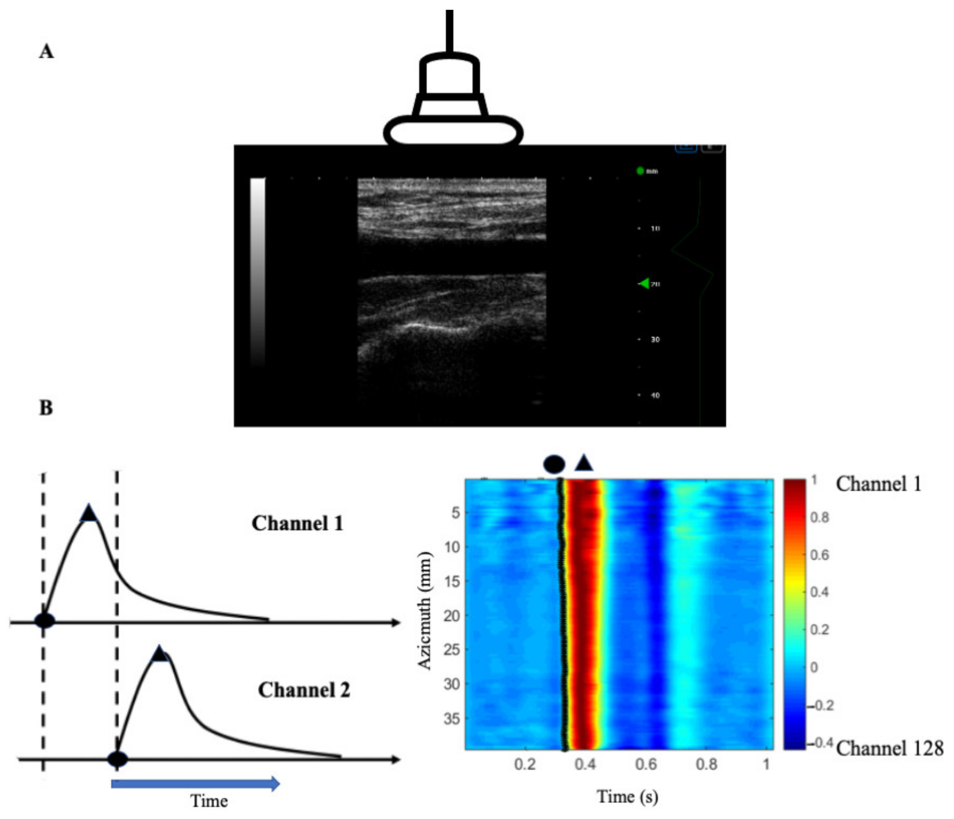
We measured the distance of the arterial wave by measuring the longitudinal velocity of the vessel wall to calculate the arterial velocity. A similar methodology was reported in the previous study [20]. Briefly, we used B-mode images for this measurement because the vessel wall signal is stronger than the blood signal, and therefore a more significant reflected signal can be seen on the ultrasound image (Figure 2A). In this study, the phase tracking method was used to demodulate the RF signal of each channel (128 channels in total) in the image after receiving delay and sum beamforming, and to obtain the complex signal (I-Q signal) in the fundamental frequency, which can be used to estimate the longitudinal velocity of the tube wall in the channel [24]. As shown in the left panel of Figure 2B, we connected the foot positions as the black line from 128 channels, followed by a robust bi-square linear regression analysis. The advantage of using a complex signal is that the phase change can be easily calculated and estimated. The systolic foot was defined as time points where the acceleration waveform reaches a maximum before and the systolic peak of the waveform. Carotid ufPWV velocity at the end of diastole (ufPWV-dia) and systole (ufPWV-sys) were calculated as the reciprocals of linear regression slope, respectively (right panel of Figure 2B).

2.7. Carotid Function and Waveform Analysis

Carotid pressure waveforms were measured noninvasively using a pulse wave tonometer (SPT301, Millar, Houston, TX, USA) and calibrated by brachial blood pressure for carotid systolic blood pressure (cSBP), diastolic (cDBP), and pulse pressure (cPP). Our previous work [6,25] has detailed that carotid compliance, ß stiffness, and distensibility were measured noninvasively and determined by simultaneously combining carotid diameter derived from ultrasound imaging and carotid blood pressure obtained by applanation tonometry after subjects rested well in the supine position pre- and post-exercise. The coefficients of variation for hemodynamic measures in our laboratory were all below 5%.

2.8. Statistical Analyses

Statistical analyses were performed using Graph Pad Prism 9.0 (La Jolla, CA, USA). All data are reported as mean ± SEM. Two-way repeated-measures ANOVA with a Bonferroni post hoc analysis was used to determine exercise intensity and time effects on measured variables. Spearman’s correlation was used to determine the associations between measured variables. Simple linear regression was used to determine the associations between hemodynamic changes in response to acute exercise challenges. Significance was set a priori at p < 0.05.

3. Results

3.1. Characteristics of Participants

As shown in Table 1, recruited resistance-trained subjects had elevated blood pressure, normal lipid profile, and fasting glucose.

3.2. Arterial Stiffness Measures and Muscle Strain Rate Changes before and after an Acute Bout of Exercise

In 40%1RM exercise session, cSBP, cDBP, cPP, cfPWV, strain rate, and AUC did not change, except that baPWV tended to increase after exercise (p = 0.07). In 80%1RM session, cPP (p = 0.04), baPWV (p < 0.0001) were significantly elevated after exercise; strain rate and AUC were also significantly reduced (p = 0.009, p < 0.0001). There were no changes to cSBP, cDBP, and cfPWV (Table 2).

3.3. Changes in Muscle and Local Carotid Stiffness in Response to Different Exercise Intensity

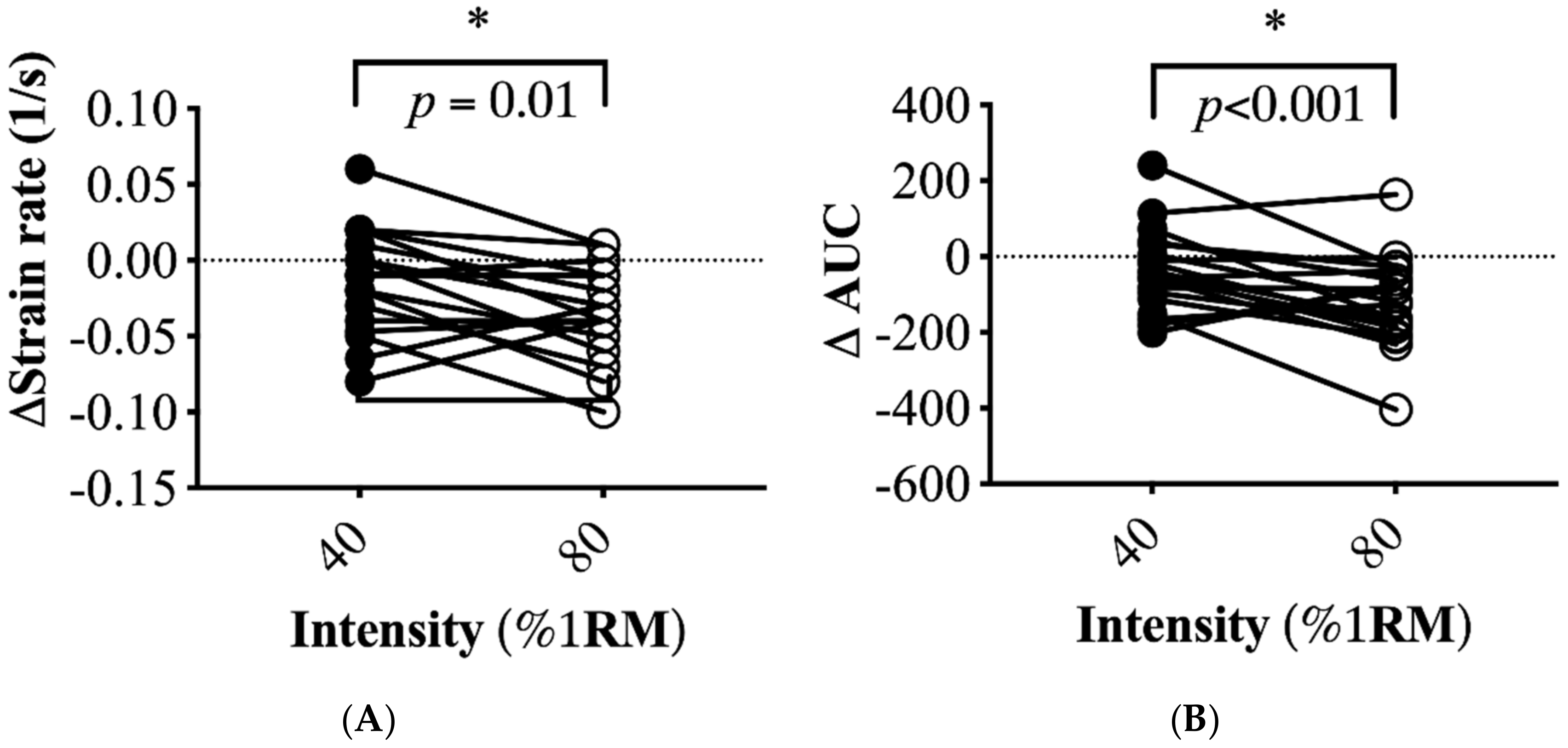
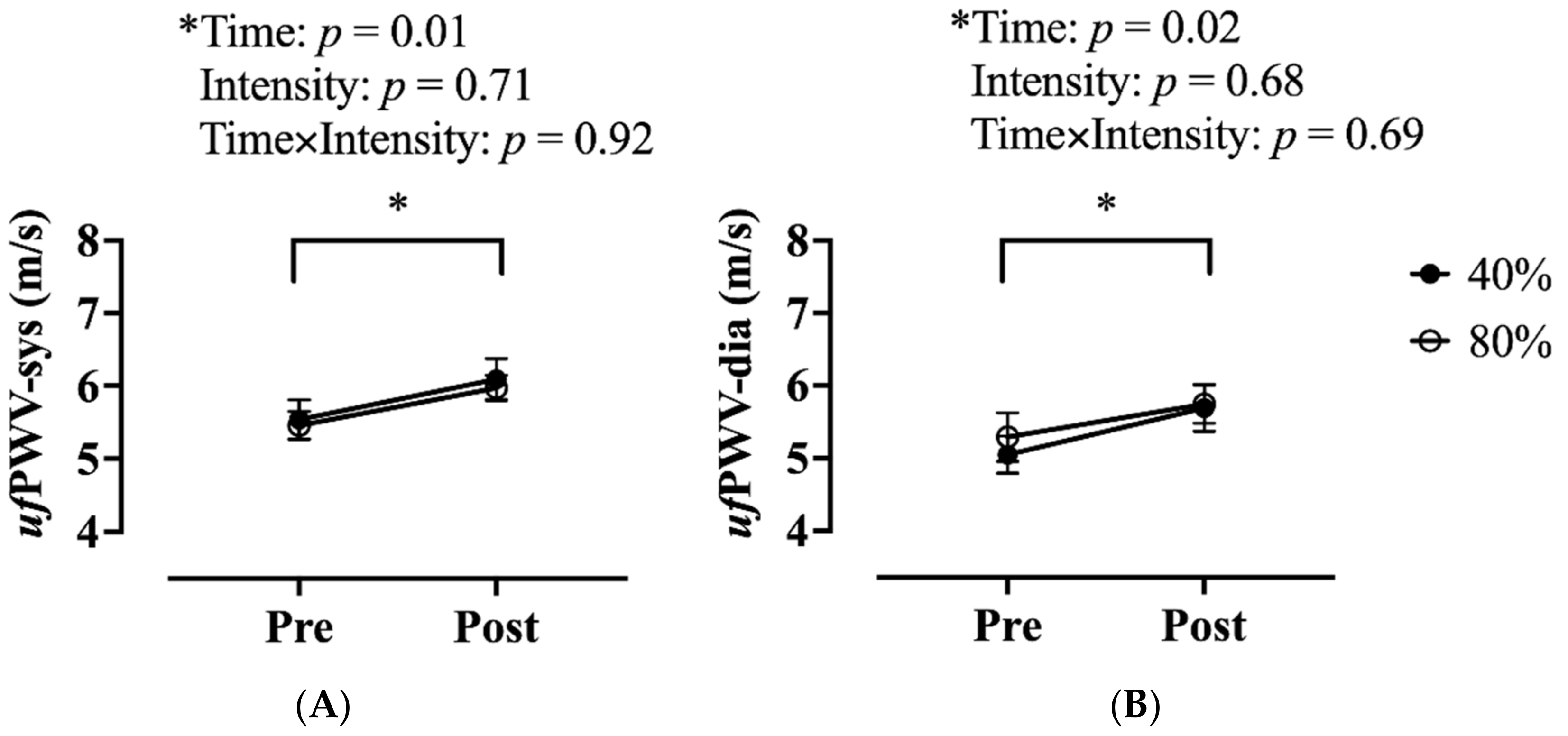
The intensity at 80%1RM elicited a greater magnitude strain rate (p = 0.01, d = 0.61) and AUC reductions (p < 0.001, d = 0.93) than those of 40%1RM, respectively (Figure 3). Both ufPWV-sys (p = 0.01, d = 0.78) and ufPWV-dia (p = 0.02, d = 0.63) significantly increased after an acute bout of exercise of intensity at 40 and 80% 1RM (Figure 4). There was no interaction between time and exercise intensity.

3.4. Associations of Muscle and Arterial Stiffness Measures

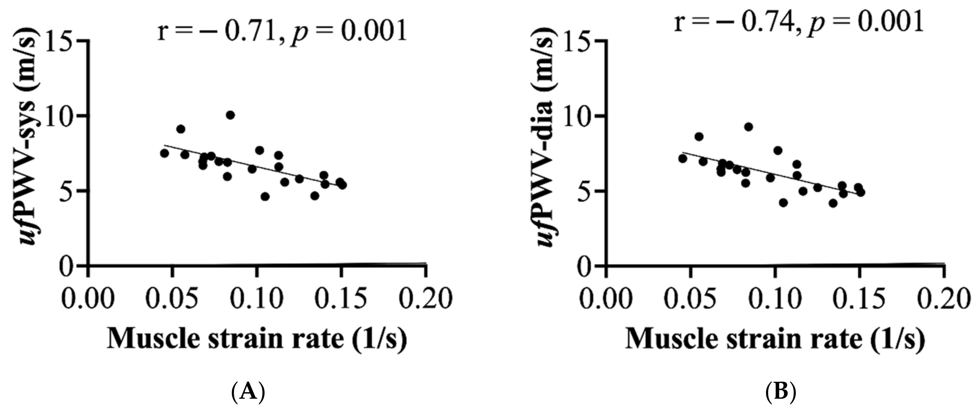
Spearman’s correlation analysis showed muscle strain rate significantly correlated with carotid compliance (r = 0.49, p = 0.02), ß stiffness (−0.84, p = < 0.0001), and carotid distensibility (r = 0.54, p = 0.01) (Table 3). The ufPWVs at systolic (r = 0.64) and disatolic phase (r = 0.67) were also moderately correlated with carotid ß stiffness (p = 0.01). Muscle strain rate and ufPWVs did not correlate with cfPWV and baPWV. As shown in Figure 5, muscle strain rate negatively correlated with ufPWV-sys (r = −0.71, p = 0.001), ufPWV-dia (r = −0.74, p = 0.001) respectively.

3.5. Contribution of ufPWV on the Increase in Central Pulse Pressure Following Exercise

After combing both exercise sessions, simple linear regression analysis showed that the increase in ufPWV-sys was the only variable contributing significantly to the increased cPP in response to resistance exercise (R2 = 0.30, p = 0.003) (Table 4).

4. Discussion

The main findings of the current study are as follows: (1) Bicep curl resistance exercise at a higher intensity (80%1RM) contributes to a more significant reduction in muscle strain rate (greater muscle stiffness) after exercise than that of lower intensity (40%1RM); (2) an acute bout of arm resistance exercise increases the carotid ufPWV parameters regardless of exercise intensity; (3) arm muscle stiffness was associated with ultrafast ultrasound-derived carotid local pulse wave velocity, but not central and systemic arterial stiffness indices.
Our findings on muscle strain elastography changes after exercise are consistent with the previous studies [26,27,28,29], showing that muscle stiffness increases after acute bouts of resistance exercise. Our data further suggest that a greater resistance exercise intensity results in lower mechanical deformation on muscle tissue. However, the average reduction in strain rate (40 vs. 80% = −0.1 vs. −0.3 in 1/s) and AUC (40 vs. 80% = −20 vs. −118) from our preliminary data did not clearly show a linear dose-response relationship between exercise intensity and induced muscle stiffness. This finding agrees with the finding of the previous in vivo study, demonstrating that the relationship between the applied strain to the developed strain in soft tissue elasticity imaging is nonlinear [30].
The underlying mechanism by which muscle stiffness increases following exercise could be primarily attributed to exercise-induced muscle alternations and their associated connective tissues. Indeed, resting intracellular calcium level might still be high immediately after exercise. Furthermore, the cross-bridge interactions [31] and engorged muscle bundles resulting from muscle recruitment and stretch reflex [32] are associated with augmented muscle stiffness. Therefore, higher neural activation and consequent structural alterations following higher exercise intensity might explain our finding on strain rate difference between 40 and 80%1RM. Moreover, we cannot exclude the possibility that muscle fiber swelling resulting from exercise also plays a role, although intramuscular swelling is not closely linked with the initial increased muscle stiffness following eccentric exercise [7,8]. We should be aware that the concentric exercise model was adopted in the present study and the extent to which exercise-induced muscle damage and inflammation are less pronounced than the eccentric. Interestingly, the nonlinear relationship between applied and developed strain in soft tissue is also inflammation dependent [30]. In this regard, the obtained muscle strain rate might also vary with exercise intensity and induced inflammatory status nonlinearly when applying strain elastography in an exercise setting. Future study needs to include more exercise intensities and different muscle contraction models (i.e., concentric vs. eccentric) to elucidate such relationship.
In the present study, we chose to report strain rate changes as the passive muscle stiffness, instead of strain ratio that takes subcutaneous tissue stiffness as the reference [22], since our participants were all resistance-trained and some of them had a skinny subcutaneous fat layer, which makes it challenging to quantitate reference strain in this population. However, the results from our pilot work showed that data expressed as strain and strain ratio were consistent in other participants of the same population. Furthermore, we quantitated muscle strain rate by using ultrafast imaging that allows us to measure transient displacement changes with the time with higher sensitivity than conventional techniques [18]; the identical external force (the probe tied with a sandbag) was carefully applied and lifted during the experiment to minimize possible variations by the same independent researcher (intraobserver coefficient of variation is about 7%). Therefore, the muscle strain rate obtained in this study was still valid as the indicator for muscle stiffness despite its strengths and limitations that have been addressed elsewhere [9].
To our knowledge, this is the first study characterizing ufPWV parameter changes in resistance exercise. The use of pulse wave imaging in assessing local carotid arterial stiffness has been studied in healthy and disease populations [19,20,24,33,34,35]. Although we did not include a healthy sedentary control group in our study design, compared to the previous study also using ultrafast ultrasound imaging [20], our average ufPWV-dia (6.3 m/s) is higher than those reported in healthy age-matched counterparts (5.3 m/s). Nevertheless, the changes of ufPWV during a cardiac cycle were relatively more minor but within a reasonable range (0.5 vs. 0.8 m/s). Such discrepancy might be attributed to exercise training and age. Indeed, habitual high-intensity resistance exercise training has been demonstrated to reduce central arterial compliance [2,3,36] compared with the sedentary control; the higher ufPWV-dia in the present study indicates greater diastolic arterial stiffness in this population, which supports these previous findings. Moreover, age is the powerful determinant of carotid local arterial stiffness [35], and the gapping between systolic and diastolic ufPWV has also been reported to increase with aging [20]. Therefore, it may not be surprising that the change of arterial stiffness over the cardiac cycle was small since our participants were relatively young. Nevertheless, our results showed that our measures on ufPWV were within the normal range.
To maximize the exercise-induced responses, we used a bicep curl exercise as the exercise model in the present study since upper limb exercise training was demonstrated to increase arterial stiffness compared with the lower limb [37,38]. Previous studies indicate the induced arterial stiffening by resistance exercise is intensity dependent; acute low-intensity resistance exercise favors arterial compliance [39], whereas high-intensity resistance exercise increases arterial stiffness [4]. Our results indicate that upper arm exercise may result in systemic arterial stiffening (increased baPWV) even at lower exercise intensity. Similar stiffening effects were observed on the carotid artery as we found both ufPWV-sys and ufPWV-dia increase regardless of intensity; the increase in stiffness during systolic phase (ufPWV-sys), but not during the diastolic phase, contributed significantly (~30%) to the increase in cPP following exercise, which was in line with the literature [17,35]. Our findings further support the notion that upper limb-biased resistance exercise may not favor vascular function.
Another novel finding of this study is that passive muscle stiffness correlates with conventional local carotid function measures and ufPWV parameters in the habitual high-intensity resistance-trained population. Moreover, passive muscle stiffness correlated with carotid ufPWV, but not baPWV or cfPWV. Currently, the exact underlying mechanisms remain to be determined, and it might be associated with greater chronic motor activation [40] that has been proposed as the result of high-intensity resistance exercise. From a molecular perspective, accumulating evidence showed that the extracellular matrix is vital in determining skeletal [41] and carotid stiffness [42]. Interestingly, recent human studies have demonstrated that the matrix metalloproteinase, the degrader of extracellular matrix, changes in response to acute [43,44,45] and short-term [46] resistance exercise training, suggesting that vascular structural and muscle stiffness might share common remodeling mechanisms. However, the association does not explain the causal effects, and future longitudinal studies adopting both neural and biochemical approaches would shed more insights.
Evaluation of mechanical properties of muscles is critical in clinical practice and the investigations of muscular responses to acute exercise. Thus, ultrasound strain elastography allows direct measurement of mechanical properties of muscle tissue. In addition, this study integrated ultrafast ultrasound strain elastography with ufPWV assessment, which can help understand the associations between acute resistance exercise challenges on skeletal muscle stiffness and vascular elasticity. We believe that linking these two techniques is essential for more comprehensive applications in sports physical medicine and rehabilitation practice, but the practical uses and limitations for appropriate subsequent clinical implementation and application still warrant further research.
There are some limitations of this study. First, the participants of this study were a self-selected group interested in improving their strength and muscular appearances through regular resistance training, limiting the ability to generalize the findings to females or the older population. In addition, participants who used to train with a bicep curl machine could have an advantage over those used to train with free weight only on the measured outcomes. However, to our understanding, the bicep curl machine was widely used among our studied participants. Most of the time, everyone used this machine for bicep curl exercises, which may not be the concern in our study. Second, we only reported strain rate changes as the passive muscle stiffness in the present study due to the uniqueness of participants. Third, we did not quantify the endurance and stretch component during their regular training regime, although all participants were reported to mainly engage in regular resistance training. Moreover, including an age-match control group and more exercise intensities in our study design would clarify the relationship between habitual exercise and acute local PWV, muscle stiffness response to exercise challenge.

5. Conclusions

In conclusion, acute bicep curl resistance exercise contributes to decreased biceps brachii muscle strain rate (increased muscle stiffness) and increased ufPWV. Thus, the use of ultrafast ultrasound-derived muscle strain rate can potentially help characterize or monitor the mechanical loading of working muscles during exercise training. Moreover, bicep muscle stiffness correlates with carotid vascular function measures, including ufPWV, in the young male habitual resistance-trained population.

Author Contributions

Conceptualization: H.-F.L., Y.-H.L., P.-C.L.; methodology: H.-F.L., Y.-H.L.; data collection and formal analysis: H.-F.L.; original draft preparation: H.-F.L., Y.-H.L.; writing—review and editing: H.-F.L., Y.-H.L., P.-C.L.; funding acquisition: H.-F.L., Y.-H.L. All authors have read and agreed to the published version of the manuscript.

Funding

This research was partially supported by the Ministry of Science and Technology (MOST, Taiwan; H.-F.L.: 107-2410-H-002-217, 108-2410-H-002-193-MY2; Y.-H.L.: 109-2628-H-227-002-MY3) and by National Taiwan University (NTU-CDP-106R7863). The APC was partially funded by the Ministry of Science and Technology (MOST) and the National Taipei University of Nursing and Health Sciences (NTUNHS).

Institutional Review Board Statement

The study was conducted according to the guidelines of the Declaration of Helsinki, and approved by the Institution Review Board of National Taiwan University Hospital (Protocol #201712133RINA).

Informed Consent Statement

Informed consent was obtained from all subjects involved in the study.

Data Availability Statement

The data presented in this study are available upon request from the corresponding author.

Acknowledgments

We would like to thank all the participants, the assistance with data collection of Shiou-Shou Huang, and the assistance of ufPWV image programming of Chih-Fan, Tang, Shang-Ju, Lee, and muscle strain elastography programming of Lin-Yi, Tseng. The authors wish to thank the National Taiwan University for providing us with space, materials, and resources throughout the experimental period.

Conflicts of Interest

The authors declare no conflict of interest.

References

  1. Agiovlasitis, S.; Riebe, D.; Ehrman, J.K.; Liguori, G.; Magal, M. American College of Sports Medicine Wolters Kluwer Health. In ACSM’s Guidelines for Exercise Testing and Prescription, 10th ed.; Wolters Kluwer: Philadelphia, PA, USA, 2018; ISBN 978-1-4963-3906-5. [Google Scholar]
  2. DeVan, A.E.; Anton, M.M.; Cook, J.N.; Neidre, D.B.; Cortez-Cooper, M.Y.; Tanaka, H. Acute effects of resistance exercise on arterial compliance. J. Appl. Physiol. 2005, 98, 2287–2291. [Google Scholar] [CrossRef] [Green Version]
  3. Miyachi, M.; Kawano, H.; Sugawara, J.; Takahashi, K.; Hayashi, K.; Yamazaki, K.; Tabata, I.; Tanaka, H. Unfavorable effects of resistance training on central arterial compliance: A randomized intervention study. Circulation 2004, 110, 2858–2863. [Google Scholar] [CrossRef] [Green Version]
  4. Miyachi, M. Effects of resistance training on arterial stiffness: A meta-analysis. Br. J. Sports Med. 2013, 47, 393–396. [Google Scholar] [CrossRef]
  5. Barnes, J.N.; Trombold, J.R.; Dhindsa, M.; Lin, H.F.; Tanaka, H. Arterial stiffening following eccentric exercise-induced muscle damage. J. Appl. Physiol. 2010, 109, 1102–1108. [Google Scholar] [CrossRef] [Green Version]
  6. Lin, H.F.; Chou, C.C.; Cheng, H.M.; Tanaka, H. Delayed onset vascular stiffening induced by eccentric resistance exercise and downhill running. Clin. J. Sport Med. 2017, 27, 369–374. [Google Scholar] [CrossRef]
  7. Chleboun, G.S.; Howell, J.N.; Conatser, R.R.; Giesey, J.J. Relationship between muscle swelling and stiffness after eccentric exercise. Med. Sci. Sports Exerc. 1998, 30, 529–535. [Google Scholar] [CrossRef]
  8. Howell, J.N.; Chleboun, G.; Conatser, R. Muscle stiffness, strength loss, swelling and soreness following exercise-induced injury in humans. J. Physiol. 1993, 464, 183–196. [Google Scholar] [CrossRef]
  9. Brandenburg, J.E.; Eby, S.F.; Song, P.; Zhao, H.; Brault, J.S.; Chen, S.; An, K.N. Ultrasound elastography: The new frontier in direct measurement of muscle stiffness. Arch. Phys. Med. Rehabil. 2014, 95, 2207–2219. [Google Scholar] [CrossRef] [PubMed] [Green Version]
  10. Chino, K.; Akagi, R.; Dohi, M.; Fukashiro, S.; Takahashi, H. Reliability and validity of quantifying absolute muscle hardness using ultrasound elastography. PLoS ONE 2012, 7, e45764. [Google Scholar] [CrossRef]
  11. Gao, J.; He, W.; Du, L.J.; Li, S.; Cheng, L.G.; Shih, G.; Rubin, J. Ultrasound strain elastography in assessment of resting biceps brachii muscle stiffness in patients with Parkinson’s disease: A primary observation. Clin. Imaging 2016, 40, 440–444. [Google Scholar] [CrossRef] [PubMed]
  12. Dankel, S.J.; Razzano, B.M. The impact of acute and chronic resistance exercise on muscle stiffness: A systematic review and meta-analysis. J. Ultrasound 2020, 23, 473–480. [Google Scholar] [CrossRef]
  13. Wang, J.J.; Parker, K.H. Wave propagation in a model of the arterial circulation. J. Biomech. 2004, 37, 457–470. [Google Scholar] [CrossRef]
  14. Sugawara, J.; Hayashi, K.; Yokoi, T.; Tanaka, H. Carotid-femoral pulse wave velocity: Impact of different arterial path length measurements. Artery Res. 2010, 4, 27–31. [Google Scholar] [CrossRef] [Green Version]
  15. Sugawara, J.; Hayashi, K.; Tanaka, H. Distal shift of arterial pressure wave reflection sites with aging. Hypertension 2010, 56, 920–925. [Google Scholar] [CrossRef] [Green Version]
  16. Sugawara, J.; Tomoto, T.; Tanaka, H. Arterial path length estimation for heart-to-brachium pulse wave velocity. Hypertens. Res. 2018, 41, 444–450. [Google Scholar] [CrossRef] [PubMed]
  17. Messas, E.; Pernot, M.; Couade, M. Arterial wall elasticity: State of the art and future prospects. Diagn. Interv. Imaging 2013, 94, 561–569. [Google Scholar] [CrossRef] [Green Version]
  18. Deffieux, T.; Gennisson, J.; Tanter, M.; Fink, M.; Nordez, A. Ultrafast imaging of in vivo muscle contraction using ultrasound. Appl. Phys. Lett. 2006, 89, 184107. [Google Scholar] [CrossRef]
  19. Li, X.; Jiang, J.; Zhang, H.; Wang, H.; Han, D.; Zhou, Q.; Gao, Y.; Yu, S.; Qi, Y. Measurement of carotid pulse wave velocity using ultrafast ultrasound imaging in hypertensive patients. J. Med. Ultrason. 2017, 44, 183–190. [Google Scholar] [CrossRef] [PubMed]
  20. Mirault, T.; Pernot, M.; Frank, M.; Couade, M.; Niarra, R.; Azizi, M.; Emmerich, J.; Jeunemaitre, X.; Fink, M.; Tanter, M.; et al. Carotid stiffness change over the cardiac cycle by ultrafast ultrasound imaging in healthy volunteers and vascular Ehlers-Danlos syndrome. J. Hypertens. 2015, 33, 1890–1896; discussion 1896. [Google Scholar] [CrossRef]
  21. Gao, J.; Li, P.C.; Chen, J.; He, W.; Du, L.J.; Min, R.; O’Dell, M. Ultrasound strain imaging in assessment of biceps muscle stiffness and dynamic motion in healthy adults. Ultrasound Med. Biol. 2017, 43, 1729–1736. [Google Scholar] [CrossRef] [PubMed]
  22. Gao, J.; Du, L.J.; He, W.; Li, S.; Cheng, L.G. Ultrasound strain elastography in assessment of muscle stiffness in acute Levodopa challenge test: A feasibility study. Ultrasound Med. Biol. 2016, 42, 1084–1089. [Google Scholar] [CrossRef] [PubMed]
  23. Huang, C.; Su, Y.; Zhang, H.; Qian, L.X.; Luo, J. Comparison of different pulse waveforms for local pulse wave velocity measurement in healthy and hypertensive common carotid arteries in vivo. Ultrasound Med. Biol. 2016, 42, 1111–1123. [Google Scholar] [CrossRef]
  24. Hasegawa, H.; Hongo, K.; Kanai, H. Measurement of regional pulse wave velocity using very high frame rate ultrasound. J. Med. Ultrason. 2013, 40, 91–98. [Google Scholar] [CrossRef] [PubMed]
  25. Lin, H.F.; Wang, S.C.; Cheng, H.M.; Sugawara, J. Homebased standing core exercise training improves femoral blood flow but not arterial stiffness in middle-aged to older adults. Artery Res. 2021, 27, 75–81. [Google Scholar] [CrossRef]
  26. Akagi, R.; Tanaka, J.; Shikiba, T.; Takahashi, H. Muscle hardness of the triceps brachii before and after a resistance exercise session: A shear wave ultrasound elastography study. Acta Radiol. 2015, 56, 1487–1493. [Google Scholar] [CrossRef] [PubMed]
  27. Agten, C.A.; Buck, F.M.; Dyer, L.; Fluck, M.; Pfirrmann, C.W.; Rosskopf, A.B. Delayed-onset muscle soreness: Temporal assessment with quantitative MRI and shear-wave ultrasound elastography. AJR Am. J. Roentgenol. 2017, 208, 402–412. [Google Scholar] [CrossRef]
  28. Lacourpaille, L.; Nordez, A.; Hug, F.; Couturier, A.; Dibie, C.; Guilhem, G. Time-course effect of exercise-induced muscle damage on localized muscle mechanical properties assessed using elastography. Acta Physiol. 2014, 211, 135–146. [Google Scholar] [CrossRef]
  29. Yanagisawa, O.; Niitsu, M.; Kurihara, T.; Fukubayashi, T. Evaluation of human muscle hardness after dynamic exercise with ultrasound real-time tissue elastography: A feasibility study. Clin. Radiol. 2011, 66, 815–819. [Google Scholar] [CrossRef]
  30. Xu, J.; Tripathy, S.; Rubin, J.M.; Stidham, R.W.; Johnson, L.A.; Higgins, P.D.; Kim, K. A new nonlinear parameter in the developed strain-to-applied strain of the soft tissues and its application in ultrasound elasticity imaging. Ultrasound Med. Biol. 2012, 38, 511–523. [Google Scholar] [CrossRef] [Green Version]
  31. Hill, D.K. Tension due to interaction between the sliding filaments in resting striated muscle. The effect of stimulation. J. Physiol. 1968, 199, 637–684. [Google Scholar] [CrossRef]
  32. Sinkjaer, T.; Toft, E.; Andreassen, S.; Hornemann, B.C. Muscle stiffness in human ankle dorsiflexors: Intrinsic and reflex components. J. Neurophysiol. 1988, 60, 1110–1121. [Google Scholar] [CrossRef] [PubMed]
  33. Luo, J.; Li, R.X.; Konofagou, E.E. Pulse wave imaging of the human carotid artery: An in vivo feasibility study. IEEE Trans. Ultrason. Ferroelectr. Freq. Control 2012, 59, 174–181. [Google Scholar] [CrossRef]
  34. Hermeling, E.; Reesink, K.D.; Kornmann, L.M.; Reneman, R.S.; Hoeks, A.P. The dicrotic notch as alternative time-reference point to measure local pulse wave velocity in the carotid artery by means of ultrasonography. J. Hypertens. 2009, 27, 2028–2035. [Google Scholar] [CrossRef] [PubMed]
  35. Hermeling, E.; Vermeersch, S.J.; Rietzschel, E.R.; de Buyzere, M.L.; Gillebert, T.C.; van de Laar, R.J.; Ferreira, I.; Hoeks, A.P.; van Bortel, L.M.; Reneman, R.S.; et al. The change in arterial stiffness over the cardiac cycle rather than diastolic stiffness is independently associated with left ventricular mass index in healthy middle-aged individuals. J. Hypertens. 2012, 30, 396–402. [Google Scholar] [CrossRef]
  36. Cortez-Cooper, M.Y.; DeVan, A.E.; Anton, M.M.; Farrar, R.P.; Beckwith, K.A.; Todd, J.S.; Tanaka, H. Effects of high intensity resistance training on arterial stiffness and wave reflection in women. Am. J. Hypertens. 2005, 18, 930–934. [Google Scholar] [CrossRef] [PubMed] [Green Version]
  37. Li, Y.; Bopp, M.; Botta, F.; Nussbaumer, M.; Schafer, J.; Roth, R.; Schmidt-Trucksass, A.; Hanssen, H. Lower body vs. upper body resistance training and arterial stiffness in young men. Int. J. Sports Med. 2015, 36, 960–967. [Google Scholar] [CrossRef] [PubMed]
  38. Okamoto, T.; Masuhara, M.; Ikuta, K. Upper but not lower limb resistance training increases arterial stiffness in humans. Eur. J. Appl. Physiol. 2009, 107, 127–134. [Google Scholar] [CrossRef]
  39. Okamoto, T.; Min, S.; Sakamaki-Sunaga, M. Arterial compliance and stiffness following low-intensity resistance exercise. Eur. J. Appl. Physiol. 2014, 114, 235–241. [Google Scholar] [CrossRef] [PubMed]
  40. Cashaback, J.G.; Cluff, T.; Potvin, J.R. Muscle fatigue and contraction intensity modulates the complexity of surface electromyography. J. Electromyogr. Kinesiol. 2013, 23, 78–83. [Google Scholar] [CrossRef]
  41. Wood, L.K.; Kayupov, E.; Gumucio, J.P.; Mendias, C.L.; Claflin, D.R.; Brooks, S.V. Intrinsic stiffness of extracellular matrix increases with age in skeletal muscles of mice. J. Appl. Physiol. 2014, 117, 363–369. [Google Scholar] [CrossRef] [Green Version]
  42. Lacolley, P.; Regnault, V.; Segers, P.; Laurent, S. Vascular smooth muscle cells and arterial stiffening: Relevance in development, Aging, and Disease. Physiol. Rev. 2017, 97, 1555–1617. [Google Scholar] [CrossRef] [PubMed]
  43. Wessner, B.; Liebensteiner, M.; Nachbauer, W.; Csapo, R. Age-specific response of skeletal muscle extracellular matrix to acute resistance exercise: A pilot study. Eur. J. Sport Sci. 2019, 19, 354–364. [Google Scholar] [CrossRef] [PubMed] [Green Version]
  44. Sullivan, B.E.; Carroll, C.C.; Jemiolo, B.; Trappe, S.W.; Magnusson, S.P.; Dossing, S.; Kjaer, M.; Trappe, T.A. Effect of acute resistance exercise and sex on human patellar tendon structural and regulatory mRNA expression. J. Appl. Physiol. 2009, 106, 468–475. [Google Scholar] [CrossRef] [Green Version]
  45. Gumpenberger, M.; Wessner, B.; Graf, A.; Narici, M.V.; Fink, C.; Braun, S.; Hoser, C.; Blazevich, A.J.; Csapo, R. Remodeling the skeletal muscle extracellular matrix in older age-effects of acute exercise stimuli on gene expression. Int. J. Mol. Sci. 2020, 21, 7089. [Google Scholar] [CrossRef]
  46. Urso, M.L.; Pierce, J.R.; Alemany, J.A.; Harman, E.A.; Nindl, B.C. Effects of exercise training on the matrix metalloprotease response to acute exercise. Eur. J. Appl. Physiol. 2009, 106, 655–663. [Google Scholar] [CrossRef] [PubMed]
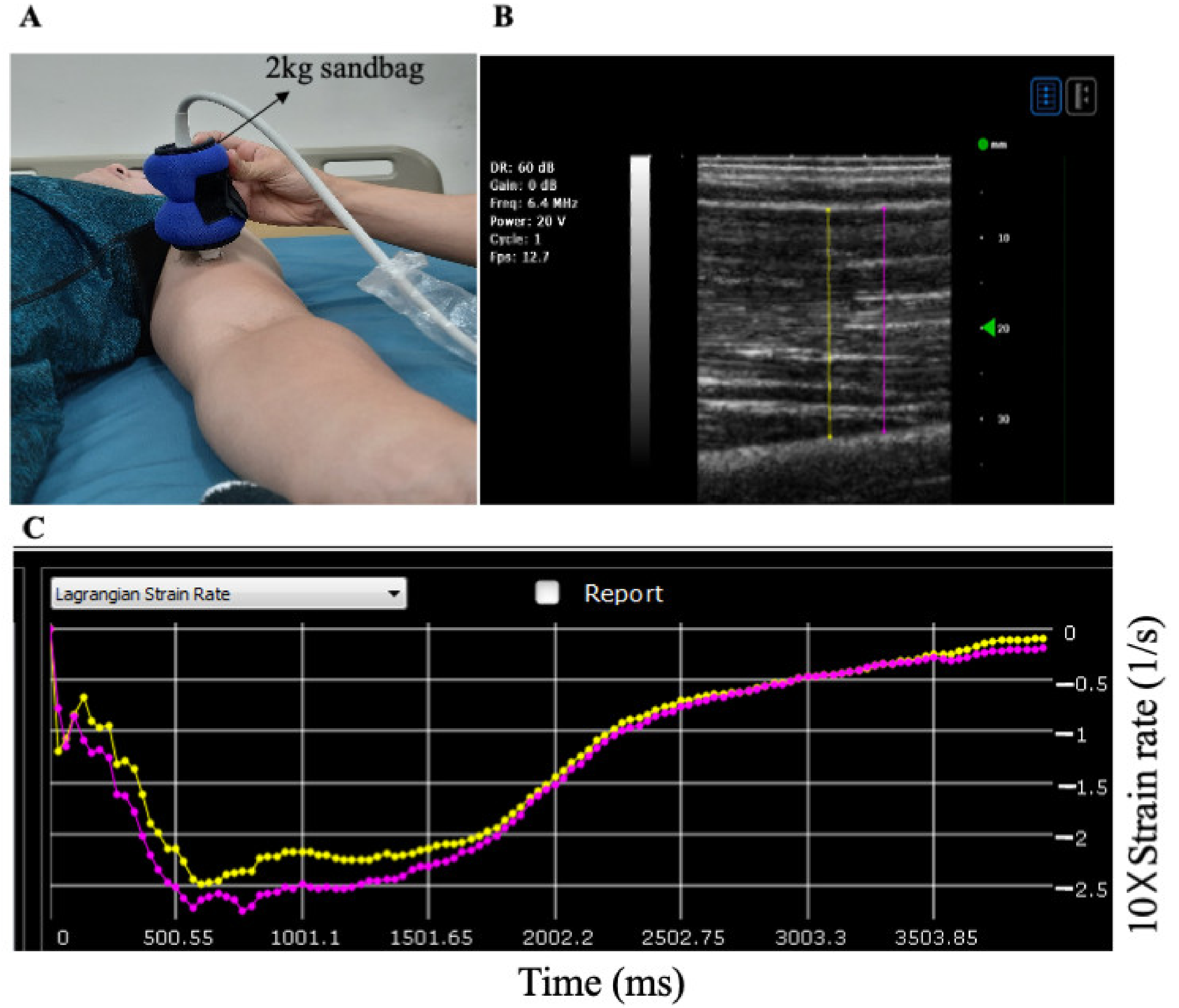
Figure 1. Muscle stiffness measurement. (A) The ultrasound probe was placed on the mid-point of the biceps brachii of the upper arms; (B) the pixel tracking of two different lines (~1 cm apart) was analyzed by the software (Muscle Tracer, S-Sharp Corp. Taiwan); (C) the average strain rate and area under the curve (AUC) calculated from two lines throughout the protocol were analyzed.
Applsci 11 08783 g001
Figure 2. (A) B-mode images of common carotid artery obtained by the ultrafast ultrasound machine; (B) the left panel illustrates artery diameter change obtained from each channel (filled circle = diastole; triangle = systole). The right panel represents the linear regression after receiving signals from 128 channels. Red represents movements toward the probe. Blue represents movements away from the probe. Color brightness represents speeds of movements, where the brightest red line indicates the fastest pulse wave (systole). The reciprocals of symbol line slopes represent ufPWV-dia (filled circle) and ufPWV-sys (triangle), respectively.
Applsci 11 08783 g002
Figure 3. The changes of strain rate (A) and AUC (B) after 40 and 80%1RM. * = vs. Pre.
Applsci 11 08783 g003
Figure 4. Pre- and post-exercise ufPWV-sys (A) and ufPWV-dia (B) in response to different exercise intensity. * = vs. Pre.
Applsci 11 08783 g004
Figure 5. Spearman’s correlation between muscle strain rate and ufPWV at systolic (A) and diastolic (B) phase.
Applsci 11 08783 g005
Table 1. Selected subject characteristics.
N = 23
Age, years24 ± 1
Height. cm175 ± 2
Weight, kg78 ± 3
Waist-hip ratio 0.81 ± 0.01
BMI, kg/m224 ± 1
1RM of bicep curl, kg63 ± 3
HDL cholesterol, mg/dL58 ± 3
LDL cholesterol, mg/dL97 ± 4
Total cholesterol, mg/dL171 ± 5
Fasting glucose, mg/dL83 ± 2
Brachial SBP, mmHg127 ± 5
Brachial DBP, mmHg68 ± 3
Brachial MAP, mmHg91 ± 4
ufPWV-sys, m/s6.8 ± 0.3
ufPWV-dia, m/s6.3 ± 0.3
Carotid compliance, mm2/mmHg × 10−20.14 ± 0.01
Carotid β-stiffness index, U6.1 ± 0.5
Carotid distensibility, mm2/KPa8.1 ± 0.7
Value = mean ± SEM; 1RM = 1 repetition maximum; SBP = systolic blood pressure; DBP = diastolic blood pressure; MAP = mean arterial pressure; ufPWV = ultrafast pulse wave velocity.
Table 2. Central blood pressure, pulse wave velocity, and muscle strain changes pre- and post-exercise.
40%1RM80%1RM
PrePostPrePost
cSBP, mmHg108 ± 5111 ± 4
NS
109 ± 5112 ± 4
NS
cDBP, mmHg68 ± 267 ± 1
NS
68 ± 366 ± 2
NS
cPP, mmHg41 ± 344 ± 3
NS
41 ± 246 ± 2 *
(p = 0.04)
cfPWV, cm/s775 ± 23809 ± 29
NS
762 ± 28807 ± 24
NS
baPWV, cm/s1195 ± 521247 ± 51
(p = 0.07)
1198 ± 581302 ± 72*
(p < 0.0001)
Muscle strain rate, 1/s 0.128 ± 0.0090.118 ± 0.010
NS
0.128 ± 0.0060.102 ± 0.009 *
(p = 0.009)
Muscle strain rate in AUC327 ± 33307 ± 25
NS
357 ± 23239 ± 19 *
(p < 0.0001)
Value = mean ± SEM; 1RM= 1 repetition maximum; cSBP = carotid systolic blood pressure; cDBP = carotid diastolic blood pressure; cPP = carotid pulse pressure; cfPWV = carotid–femoral pulse wave velocity; baPWV= brachial–ankle pulse wave velocity. AUC = area under curve. * = vs. Pre. NS = no significance vs. Pre.
Table 3. Spearman’s correlation between muscle strain rate, carotid vascular function, and pulse wave velocity measures.
Carotid ComplianceCarotid ß-StiffnessCarotid DistensibilitycfPWVbaPWV
Muscle strain rate0.49 *
(p = 0.02)
0.84 *
(p < 0.0001)
0.54 *
(p = 0.01)
−0.10
NS
−0.02
NS
ufPWV-sys−0.26
NS
0.64 *
(p = 0.01)
−0.26
NS
0.11
NS
0.14
NS
ufPWV-dia−0.27
NS
0.67 *
(p = 0.01)
−0.30
NS
0.15
NS
0.09
NS
Δ ufPWV0.24
NS
−0.26
NS
0.30
NS
−0.11
NS
−0.18
NS
* = significant, p < 0.05. NS = no significance.
Table 4. Simple linear regression analysis of increased cPP in response to bicep curl resistance exercise (two exercise sessions combined).
∆ cPP
∆ muscle strain rate. 1/sR2 = 0.04
ufPWV-sys, m/sR2 = 0.30 * (p = 0.003)
ufPWV-dia, m/sR2 = 0.04
∆ baPWV, cm/sR2 = 0.09
* = significant, p < 0.05
Publisher’s Note: MDPI stays neutral with regard to jurisdictional claims in published maps and institutional affiliations.