Article

Apoptotic Effect of Brassinin via Inhibition of CNOT2 and Activation of p53 and Its Combination Effect with Doxorubicin

College of Korean Medicine, Kyung Hee University, Seoul 02447, Korea
*
Author to whom correspondence should be addressed.
Appl. Sci. 2021, 11(21), 10036; https://doi.org/10.3390/app112110036
Submission received: 20 August 2021 / Revised: 20 October 2021 / Accepted: 22 October 2021 / Published: 26 October 2021

Abstract

:
Brassinin derived from Chinese cabbage has been reported to act as an anti-cancer agent on prostate, liver, and colon cancer cells. However, its mechanism and impact are largely unknown in colon cancer cells. Here, we first published a report that Brassinin induces apoptosis and inhibits the survival of colon cancer cells by activating p53. We found that Brassinin induces p53 and p21 dose- and time-dependent manner in wild type of p53 colon cancer cells. Interestingly, Brassinin induces apoptosis in wild-type of p53 cancer cells, but not in null-type of p53 cancer cells dose dependently. Additionally, Brassinin induces apoptosis through L5. Furthermore, Brassinin enhanced the apoptotic effect with doxorubicin by activating p53. Altogether, our findings suggest that Brassinin is a new p53 regulator via induce apoptosis and inhibit the proliferation in colon cancer cells.

1. Introduction

The tumor suppressor p53 prevents tumorigenesis, blocks metastasis, stops cancer cell proliferation, and induce apoptosis, resulting in mutations in cancer cells [1]. p53 is important to various biological functions include numerous target genes, such as p21, is involved in p53-dependent cell cycle arrest, while the Puma play important roles in p53 mediated apoptosis [2]. Furthermore, p53 is activated by regulator genes as well [1,3,4]. p53 is the primary regulator of cell cycle arrest or apoptosis in mammalian cell reactions to cellular stress, including nuclear or ribosomal stress [5,6,7]. Furthermore, p53 induce the anti-oxidant genes, such as GLS2. These genes remove the reactive oxygen species (ROS) and protect against oxidative insults [8]. Although many anti-cancer drugs involved in p53-dependent apoptosis have been studied, the p53-regulatory pathways are not completely understood.
Ribosomal biogenesis is essential for growth of cancer cell. Many ribosomal proteins, such as L5, L11, and L23, were able to inhibit growth of tumor cells by activating p53 in response to ribosomal stress. It appears that L5 and L11 work together to activate p53. Furthermore, L5, L11, and L23 are also bind to MDM2 [9,10,11,12].
The colon cancer is known as the third common cancer in the world [13]. Despite advances in modern medicine, about 50% of colon cancer patients have experienced cancer recurrence and mortality rate is about 40% [14]. Surgery is the main treatment for colon cancer and treatments, such as 5-FU and Oxaliplatin, are used. However, these treatments are accompanied by a variety of side effects such as anemia, loss of appetite, feeling sick, headache, etc. [15,16,17]. This is why the discovery and scientific proof of new colorectal cancer treatments is needed.
Brassinin is a phytoalexin found in crucible vegetables, which is anti-cancer effects [18,19]. Brassinin inhibits human liver cancer cell proliferation via mitochondrial dysfunction and inhibits invasive potential of lung cancer cells via PI3K/Akt/mTOR signaling cascade [18,20]. Recently, Brassinin enhances the anti-cancer actions of paclitaxel by targeting JAKs/STAT3 pathways in colon cancers [19]. However, the detail mechanism of Brassinin in colon cancer cells are not fully understand so far. Thus, in this study, we hypothesized that Brassinin may suppress the proliferation of colon cancer cells and induces apoptosis through activating p53.

2. Materials and Methods

2.1. Cell Culture and Reagents

HCT116p53+/+ (p53 wild-type human colon cancer), SW-480, HT-29 cells were purchased from the Korean Cell Line Bank (Seoul, Korea). Additionally, HCT116p53−/− (p53 knockout human colon cancer) cells were kindly obtained from Prof. Wonchae Choe (Kyung Hee University, Korea). Cells were cultured in RPMI1640 with 10% fetal bovine serum (FBS) and 2 μM L-glutamine and penicillin/streptomycin at 37 °C in incubator with 5% CO2. Brassinin (≥98% of purity) (product ID: B6801) was purchased from LKT laboratories (Minneapolis, MN, USA).

2.2. RNA Interference

p53 siRNA was purchased from Bioneer (ID: 7157-1) (Daejeon, Korea). RPL5 siRNA was purchased from Santa Cruz Biotechnologies (SC-78649) (Santa Cruz, CA, USA). HCT116p53+/+ cells were seeded on the 6-well plate for overnight and then cells were transfected with siRNA, as indicated in figure legends by INTERFERin® (Polyplus-transfection SA, Illkirch, France) follow the manufacturer’s protocol. After 48 h later, cells were treated with Brassinin (80 μM) or DMSO (control). Transfection assay was conducted as described earlier [21].

2.3. Cytotoxicity Assay

Cytotoxic effect of Brassinin in HCT116p53+/+, HCT116p53−/−, SW-480, and HT-29 cells were examined by CCK8 assay kit (Dojindo, Rockville, MD, USA) as described in Jung et al.’s paper [1]. Cells were seeded 1 × 10⁴ cells/well in 96-well culture plates for overnight. Additionally, treated with Brassinin (12.5, 25, 50, or 100 μM) or DMSO (control) for 24 h. The cell viability was calculated by the following equation: cell viability (%) = [OD (puromycin) − OD (blank)]/[OD (control) − OD (blank)] × 100.

2.4. Colony Formation Assay

HCT116p53+/+ cells were treated with Brassinin (40, 80 μM) or DMSO (control). After 24 h later, cells were distributed onto 6 well cell culture plate at 1000 cells/well. Additionally, then, we replaced the media (RPMI1640 with 10% fetal bovine serum (FBS) and 2 μM L-glutamine and penicillin/streptomycin at 37 °C in incubator with 5% CO2) every two days. After a week, the cells were washed with PBS several times and then fixed with Diff Quick Solution (Sysmex, Kobe, Japan). Then, we counted the colonies using the microscope.

2.5. Western Blotting

Cell lysates were separated in SDS-PAGE gel (8–15%). The detailed description of western blotting from Jung’s paper [6]. HCT116p53+/+ and HCT116p53−/− cells were treated with Brassinin (40, 80 μM) or DMSO (control) for 24 h. Furthermore, HCT116p53+/+ cells were treated with Brassinin (80 μM) for 24, 48, or 72 h. The membranes were incubated with various antibodies against CNOT2 (1:1000), RPL5 (1:1000) (Cell signaling Technology Inc., Danvers, MA, USA), PARP (1:1000), p53 (DO-1) (1:1000), (Santa Cruz Biotechnologies, Santa Cruz, CA, USA), p21 (1:500) (Ab frontier, Seoul, Korea) and β-actin (1:2000) (Sigma Aldrich Co., St. Louis, MO, USA).

2.6. Flow Cytometry Analysis

HCT116p53+/+ and HCT116p53−/− cells were treated with Brassinin (40, 80 μM) or DMSO (control) for 24 h. After then, the cells were collected and washed three times with PBS. The cells were fixed in 70% EtOH at −20 °C for overnight. Next day, the cells were incubated with RNase A (10 mg/mL) for 1 h at 37 °C. Additionally, then, cells were stained with propidium iodide (PI) for 30 min in dark. The stained cells were analysis with FACSCalibur by using Cellquest software (Becton Dickinson, Franklin Lakes, NJ, USA).

2.7. Statistical Analysis

The statistical analysis was performed as described previously [22]. All data were presented as means ± standard deviation (SD). Student t-test was used for comparison of two groups. Additionally, the one-way analysis of variance (ANOVA) followed by Tukey’s post hoc test was performed for multi-group comparison using GraphPad Prism software (Version 5.0, San Diego, CA, USA). The statistically differences between the DMSO (control) and Brassinin-treated groups were calculated using student’s t-test. All experiments were conducted three times. p-value of 0.05 or less was considered as significant.

3. Results

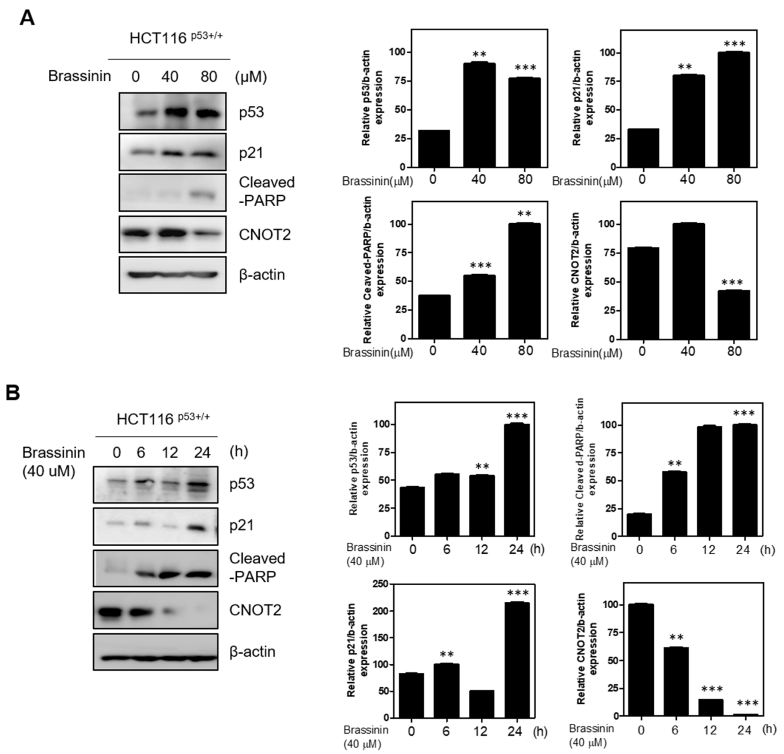
3.1. Brassinin Induces p53 and Its Target Gene p21

Brassinin has been shown to inhibit cancer cell proliferation, but needs to learn more about the role of Brassinin in cancer development. To solve this problem, we first treated Brassinin in HCT116p53+/+ cells. As shown in Figure 1A,B, Brassinin induced the expression of endogenous p53 in HCT116p53+/+ cells and expressed dose-dependent and time-dependent on the p53-target gene p21 and apoptotic marker cleaved-PARP. Furthermore, Brassinin inhibits CNOT2 expression, which is related to metastasis in cancer, and it is acting as a oncogene, dose-dependently.

3.2. Brassinin Induces Apoptosis by Activating p53

To confirm if Brassinin plays an important role in regulation of apoptosis by activating p53, we treated Brassinin both p53 wild type or null type cancer cells. First of all, we tested whether Brassinin induces apoptosis p53-dependent manner or not. As shown in Figure 2A, Brassinin induces apoptosis partly by activating p53 in cancer cells. Consistently, knockdown of p53 treated with Brassinin in HCT116p53+/+ cells has shown that Brassinin induces apoptosis by activating p53 (Figure 2B).

3.3. Effect of RPL5 Depletion with Brassinin on the Apoptosis in Colon Cancer Cells

Ribosomal proteins, such as L5, L11, and S14, related with MDM2 binding and activation of p53 in human cancer cells [23,24,25,26,27,28]. L5 was involved in p53 pathway and regulates apoptosis [22]. In attempt to address this question, we tested whether Brassinin induces apoptosis through L5. By performing western blotting assay, we found that Brassinin does not induces apoptosis marker cleaved-PARP via knockdown of L5 (Figure 3). This results suggest that Brassinin induces apoptosis via L5.

3.4. Brassinin Is Cytotoxic and Preservative to Colorectal Cancer Cells

To check the cytotoxicity and anti-proliferation effects of Brassinin, several colorectal cancer cells were employed MTT assay and colony formation assay. Here, Brassinin suppressed the cell viability of HCT116p53+/+ (p53 wild-type cell) cells in a concentration–dependent manner compared to HCT116p53−/− (p53 knockout cell), SW480 cells using an CCK8 assay (Figure 4A,B). Similarly, Brassinin suppressed the HCT116p53+/+ cells proliferation dose-dependent manner (Figure 4C).

3.5. Brassinin Regulates Apoptosis with a Growing Sub-G1 Population

To check cytotoxic effects of Brassinin are due to apoptosis, cell cycle analysis was performed in HCT116p53+/+ cells. As shown in Figure 5A,B, Brassinin increased the sub-G1 population in HCT116p53+/+ cells better than HCT116p53−/− cells. This results suggest that Brassinin induces more apoptosis in p53 wild type cells.

3.6. Combination Effect of Brassinin and Doxorubicin in HCT116p53+/+ Cells

A doxorubicin is potent drug to many kinds of solid tumors. However, the dosage of this drug which is essential for maximizing their anti-cancer effect [29,30]. Therefore, it is necessary to find new medications for combination therapy using compounds. To check the combination of Brassinin and doxorubicin in HCT116p53+/+ cells by western blotting. As shown in Figure 6A, Brassinin enhanced the apoptotic protein cleaved-PARP and tumor suppressor p53 expression with doxorubicin in a dose-dependent manner in HCT116p53+/+ cells.

4. Discussion

Brassinin have been study as anti-cancer drug in cancer cells [18,20]. The most recent paper found that Brassinin may be a novel anti-colon cancer target therapy through STAT3–JAK2 pathway. However, these studies did not discuss its fundamental mechanisms, especially p53 in cancer cells. As far as we know, this is the first study to discover the Brassinin induces apoptosis by activating p53 in colon cancer cells.
It is known that p53 acts as a tumor suppressor gene in various cancer cells.
Once p53 is activated, it causes apoptosis, ferroptosis, senescence, and cell cycle arrest; and inhibits migration, and metastasis in cancer cells. Many of drugs have been identified about p53 responsive regulation of cancer cell growth and apoptosis [1,4,31]. Our cell-based studies showed that Brassinin activating of p53 expression dose- and time-dependent manner. Additionally, Brassinin induced apoptosis dose- and time-dependent manner as well. Consistently, Brassinin increased the sub-G1 population in HCT116p53+/+ and HCT116p53−/− cells.
To check whether Brassinin induced apoptosis by activating p53, we treated Brssinin on p53 wild-type HCT116 and p53 null-type HCT116 cells. Furthermore, we treated Brassinin after treated with p53 siRNA for knockdown of p53 in p53 wild-type HCT116 cells. Interestingly, Brassinin induced apoptosis in p53 wild-type HCT116 cells more than p53 null-type HCT116 cells. Additionally, Brassinin needs L5 to cause apoptosis in cancer cells. According to previous studies, ribosomal RNA processing is critically involved in p53 activation. It is known that L5 or L11 plays a role in controlling the activity of p53 [24,25,32]. Here, our data suggested that Brassinin induced apoptosis through RPL5 in HCT116 cells.
CNOT2, a subunit of the CCR4-NOT complex, is known to be involved in apoptosis, metastasis, angiogenesis, and autophagy in various type of cancer cells as an oncogene [6,33,34]. We found that Brassinin reduced the expression of CNOT2 dose- and time-dependent manner in HCT116 cells.
Additionally, Brassinin enhanced the anti-cancer effect in HCT116 cells with a doxorubicin which is used to treat colon cancer drug. Brassinin induced p53 and cleaved-PARP in p53 wild-type HCT116 cells, demonstrating the combination effect of Brassinin and doxorubicin by western blotting. The results of this experiment show the possibility that Brassinin can be administered in combination with existing treatments as a new drug for colon cancer cells.

5. Conclusions

Thus, these new findings will provide more insight into the p53 activation function of Brassinin. In summary, our study demonstrates that Brassinin induced apoptosis by p53 activating in cancer cells. This information would be useful for anti-cancer drug discovery in the future.

Author Contributions

Conceptualization, J.H.J.; methodology, J.H.J. and W.Y.P.; software, W.Y.P.; formal analysis, W.Y.P. and J.E.P.; data curation, W.Y.P. and J.E.P.; investigation, J.H.J.; writing-original draft preparation, J.H.J. and W.Y.P.; writing-review and editing, J.H.J.; project administration, J.H.J.; funding acquisition, J.H.J. All authors have read and agreed to the published version of the manuscript.

Funding

J.H.J. was supported by the National Research Foundation of Korea (NRF) Grant funded by the Korea Government (MSIT) (no. 2020R1C1C1009348).

Institutional Review Board Statement

Not applicable.

Informed Consent Statement

Not applicable.

Acknowledgments

We appreciate Sung-Hoon Kim, Hyo-Jung Lee, Deok Yong Sim and Eunji Im.

Conflicts of Interest

The authors declare no conflict of interests.

References

  1. Jung, J.H.; Lee, H.; Cao, B.; Liao, P.; Zeng, S.X.; Lu, H. RNA-binding motif protein 10 induces apoptosis and suppresses proliferation by activating p53. Oncogene 2020, 39, 1031–1040. [Google Scholar] [CrossRef] [PubMed]
  2. Vousden, K.H.; Lu, X. Live or let die: The cell’s response to p53. Nat. Rev. Cancer 2002, 2, 594–604. [Google Scholar] [CrossRef] [PubMed] [Green Version]
  3. Jung, J.H.; Lee, H.; Zeng, S.X.; Lu, H. RBM10, a New Regulator of p53. Cells 2020, 9, 2107. [Google Scholar] [CrossRef] [PubMed]
  4. Chao, T.; Zhou, X.; Cao, B.; Liao, P.; Liu, H.; Chen, Y.; Park, H.W.; Zeng, S.X.; Lu, H. Pleckstrin homology domain-containing protein PHLDB3 supports cancer growth via a negative feedback loop involving p53. Nat. Commun. 2016, 7, 13755. [Google Scholar] [CrossRef] [PubMed] [Green Version]
  5. Horn, H.F.; Vousden, K.H. Cooperation between the ribosomal proteins L5 and L11 in the p53 pathway. Oncogene 2008, 27, 5774–5784. [Google Scholar] [CrossRef] [PubMed] [Green Version]
  6. Lee, J.; Jung, J.H.; Hwang, J.; Park, J.E.; Kim, J.H.; Park, W.Y.; Suh, J.Y.; Kim, S.H. CNOT2 Is Critically Involved in Atorvastatin Induced Apoptotic and Autophagic Cell Death in Non-Small Cell Lung Cancers. Cancers 2019, 11, 1470. [Google Scholar] [CrossRef] [Green Version]
  7. Sun, X.X.; Wang, Y.G.; Xirodimas, D.P.; Dai, M.S. Perturbation of 60 S ribosomal biogenesis results in ribosomal protein L5- and L11-dependent p53 activation. J. Biol. Chem. 2010, 285, 25812–25821. [Google Scholar] [CrossRef] [Green Version]
  8. Hao, Q.; Chen, Y.; Zhou, X. The Janus Face of p53-Targeting Ubiquitin Ligases. Cells 2020, 9, 1656. [Google Scholar] [CrossRef]
  9. Dai, M.S.; Shi, D.; Jin, Y.; Sun, X.X.; Zhang, Y.; Grossman, S.R.; Lu, H. Regulation of the MDM2-p53 pathway by ribosomal protein L11 involves a post-ubiquitination mechanism. J. Biol. Chem. 2006, 281, 24304–24313. [Google Scholar] [CrossRef] [Green Version]
  10. Zhou, X.; Hao, Q.; Zhang, Q.; Liao, J.M.; Ke, J.W.; Liao, P.; Cao, B.; Lu, H. Ribosomal proteins L11 and L5 activate TAp73 by overcoming MDM2 inhibition. Cell Death Differ. 2015, 22, 755–766. [Google Scholar] [CrossRef]
  11. Dai, M.S.; Lu, H. Inhibition of MDM2-mediated p53 ubiquitination and degradation by ribosomal protein L5. J. Biol. Chem. 2004, 279, 44475–44482. [Google Scholar] [CrossRef] [PubMed] [Green Version]
  12. Dai, M.S.; Zeng, S.X.; Jin, Y.; Sun, X.X.; David, L.; Lu, H. Ribosomal protein L23 activates p53 by inhibiting MDM2 function in response to ribosomal perturbation but not to translation inhibition. Mol. Cell Biol. 2004, 24, 7654–7668. [Google Scholar] [CrossRef] [Green Version]
  13. Rawla, P.; Sunkara, T.; Barsouk, A. Epidemiology of colorectal cancer: Incidence, mortality, survival, and risk factors. Prz. Gastroenterol. 2019, 14, 89–103. [Google Scholar] [CrossRef] [PubMed]
  14. Siegel, R.L.; Miller, K.D.; Goding Sauer, A.; Fedewa, S.A.; Butterly, L.F.; Anderson, J.C.; Cercek, A.; Smith, R.A.; Jemal, A. Colorectal cancer statistics, 2020. CA Cancer J. Clin. 2020, 70, 145–164. [Google Scholar] [CrossRef] [Green Version]
  15. Polk, A.; Vistisen, K.; Vaage-Nilsen, M.; Nielsen, D.L. A systematic review of the pathophysiology of 5-fluorouracil-induced cardiotoxicity. BMC Pharm. Toxicol 2014, 15, 47. [Google Scholar] [CrossRef] [Green Version]
  16. Kurokawa, Y.; Hasuike, Y.; Hattori, T.; Hayashi, S.; Fujitani, K.; Shin, E.; Mishima, H.; Sawamura, T.; Nishisho, I.; Kobayashi, K.; et al. Efficacy and side effect of continuous intra-arterial infusion of high-dose 5-FU for liver metastases of colorectal cancer. Gan Kagaku Ryoho 1999, 26, 1737–1740. [Google Scholar]
  17. Hijri, F.Z.; Arifi, S.; Ouattassi, N.; Mellas, N.; El Mesbahi, O. Oxaliplatin-induced ototoxicity in adjuvant setting for colorectal cancer: Unusual side effect. J. Gastrointest. Cancer 2014, 45, 106–108. [Google Scholar] [CrossRef] [PubMed]
  18. Hong, T.; Ham, J.; Song, J.; Song, G.; Lim, W. Brassinin Inhibits Proliferation in Human Liver Cancer Cells via Mitochondrial Dysfunction. Cells 2021, 10, 332. [Google Scholar] [CrossRef] [PubMed]
  19. Yang, M.H.; Baek, S.H.; Ha, I.J.; Um, J.Y.; Ahn, K.S. Brassinin enhances the anticancer actions of paclitaxel by targeting multiple signaling pathways in colorectal cancer cells. Phytother. Res. 2021, 35, 3875–3885. [Google Scholar] [CrossRef]
  20. Yang, M.H.; Lee, J.H.; Ko, J.H.; Jung, S.H.; Sethi, G.; Ahn, K.S. Brassinin Represses Invasive Potential of Lung Carcinoma Cells through Deactivation of PI3K/Akt/mTOR Signaling Cascade. Molecules 2019, 24, 1584. [Google Scholar] [CrossRef] [Green Version]
  21. Jung, J.H.; Kim, M.J.; Lee, H.; Lee, J.; Kim, J.; Lee, H.J.; Shin, E.A.; Kim, Y.H.; Kim, B.; Shim, B.S.; et al. Farnesiferol c induces apoptosis via regulation of L11 and c-Myc with combinational potential with anticancer drugs in non-small-cell lung cancers. Sci. Rep. 2016, 6, 26844. [Google Scholar] [CrossRef] [Green Version]
  22. Jung, J.H.; Lee, H.; Kim, J.H.; Sim, D.Y.; Ahn, H.; Kim, B.; Chang, S.; Kim, S.H. p53-Dependent Apoptotic Effect of Puromycin via Binding of Ribosomal Protein L5 and L11 to MDM2 and its Combination Effect with RITA or Doxorubicin. Cancers 2019, 11, 582. [Google Scholar] [CrossRef] [Green Version]
  23. Liu, Y.; Deisenroth, C.; Zhang, Y. RP-MDM2-p53 Pathway: Linking Ribosomal Biogenesis and Tumor Surveillance. Trends Cancer 2016, 2, 191–204. [Google Scholar] [CrossRef] [PubMed] [Green Version]
  24. Fregoso, O.I.; Das, S.; Akerman, M.; Krainer, A.R. Splicing-factor oncoprotein SRSF1 stabilizes p53 via RPL5 and induces cellular senescence. Mol. Cell 2013, 50, 56–66. [Google Scholar] [CrossRef] [Green Version]
  25. Zhang, Q.; Xiao, H.; Chai, S.C.; Hoang, Q.Q.; Lu, H. Hydrophilic residues are crucial for ribosomal protein L11 (RPL11) interaction with zinc finger domain of MDM2 and p53 protein activation. J. Biol. Chem. 2011, 286, 38264–38274. [Google Scholar] [CrossRef] [PubMed] [Green Version]
  26. Kayama, K.; Watanabe, S.; Takafuji, T.; Tsuji, T.; Hironaka, K.; Matsumoto, M.; Nakayama, K.I.; Enari, M.; Kohno, T.; Shiraishi, K.; et al. GRWD1 negatively regulates p53 via the RPL11-MDM2 pathway and promotes tumorigenesis. EMBO Rep. 2017, 18, 123–137. [Google Scholar] [CrossRef] [PubMed] [Green Version]
  27. Zheng, J.; Lang, Y.; Zhang, Q.; Cui, D.; Sun, H.; Jiang, L.; Chen, Z.; Zhang, R.; Gao, Y.; Tian, W.; et al. Structure of human MDM2 complexed with RPL11 reveals the molecular basis of p53 activation. Genes Dev. 2015, 29, 1524–1534. [Google Scholar] [CrossRef] [Green Version]
  28. Boultwood, J. The role of haploinsufficiency of RPS14 and p53 activation in the molecular pathogenesis of the 5q- syndrome. Pediatric Rep. 2011, 3, e10. [Google Scholar] [CrossRef] [PubMed] [Green Version]
  29. Zhang, Y.; Zhang, Q.; Zeng, S.X.; Hao, Q.; Lu, H. Inauhzin sensitizes p53-dependent cytotoxicity and tumor suppression of chemotherapeutic agents. Neoplasia 2013, 15, 523–534. [Google Scholar] [CrossRef] [Green Version]
  30. Zhang, Q.; Zeng, S.X.; Zhang, Y.; Zhang, Y.; Ding, D.; Ye, Q.; Meroueh, S.O.; Lu, H. A small molecule Inauhzin inhibits SIRT1 activity and suppresses tumour growth through activation of p53. EMBO Mol. Med. 2012, 4, 298–312. [Google Scholar] [CrossRef]
  31. Zhou, X.; Hao, Q.; Liao, P.; Luo, S.; Zhang, M.; Hu, G.; Liu, H.; Zhang, Y.; Cao, B.; Baddoo, M.; et al. Nerve growth factor receptor negates the tumor suppressor p53 as a feedback regulator. Elife 2016, 5, e15099. [Google Scholar] [CrossRef] [PubMed] [Green Version]
  32. Morgado-Palacin, L.; Llanos, S.; Urbano-Cuadrado, M.; Blanco-Aparicio, C.; Megias, D.; Pastor, J.; Serrano, M. Non-genotoxic activation of p53 through the RPL11-dependent ribosomal stress pathway. Carcinogenesis 2014, 35, 2822–2830. [Google Scholar] [CrossRef] [PubMed] [Green Version]
  33. Sohn, E.J.; Jung, D.B.; Lee, H.; Han, I.; Lee, J.; Lee, H.; Kim, S.H. CNOT2 promotes proliferation and angiogenesis via VEGF signaling in MDA-MB-231 breast cancer cells. Cancer Lett. 2018, 412, 88–98. [Google Scholar] [CrossRef] [PubMed]
  34. Jung, J.H.; Lee, H.J.; Kim, J.H.; Sim, D.Y.; Im, E.; Kim, S.; Chang, S.; Kim, S.H. Colocalization of MID1IP1 and c-Myc is Critically Involved in Liver Cancer Growth via Regulation of Ribosomal Protein L5 and L11 and CNOT2. Cells 2020, 9, 985. [Google Scholar] [CrossRef] [Green Version]
Figure 1. Brassinin induces p53 dose- and time-dependently. (A) HCT116p53+/+ cells were treated with Brassinin (0, 40, 80 μM). p53, p21, cleaved-PARP, and CNOT2 were determined by western blotting. (B) HCT116p53+/+ cells were treated with Brassinin (40 μM) for 0, 6, 12, and 24 h. Data represent means ± SD. * p < 0.05, ** p < 0.01, *** p < 0.001 vs. control. Values of Western blotting images represent relative level of protein expression/β-actin.
Applsci 11 10036 g001
Figure 2. Brassinin induces apoptosis partly by p53 activating. (A) HCT116p53+/+ and HCT116p53−/− cells were treated with Brassinin were incubated 24 h. Cleaved-PARP and p53 were determined by western blotting. *** p < 0.001 vs. untreated control. Values of Western blotting images represent relative level of protein expression/β-actin. (B) HCT116p53+/+ cells were transfected with p53 siRNA (40 nM) or scramble siRNA were incubated or 48 h then treated with Brassinin for 24 h. Additionally, then cleaved-PARP, p53 and p21 were confirmed by western blotting. ** p < 0.01, *** p < 0.001 vs. untreated control. Values of Western blotting images represent relative level of protein expression/β-actin.
Applsci 11 10036 g002
Figure 3. Brassinin induces apoptosis through the L5. HCT116p53+/+ cells were transfected with L5 siRNA (40 nM) or scramble siRNA for 48 h. After that, cells were treated with Brassinin (80 μM) for 24 h. Cleaved-PARP and L5 were determined by western blotting. ** p < 0.01, *** p < 0.001 vs. untreated control. Values of Western blotting images represent relative level of protein expression/β-actin.
Applsci 11 10036 g003
Figure 4. The cytotoxic effects of Brassinin in colon cancer cells. (A,B) Cytotoxicity of Brassinin in HCT116p53+/+, HCT116p53−/−, SW-480, and HT-29 cells in a dose-dependent manner by CCk8 assay. (C) Photo and bar graph for colony formation of Brassinin in HCT116p53+/+. The colonies were stained with crystal violet. * p < 0.05, ** p < 0.01, *** p < 0.001 vs. untreated control. Values of Western blotting images represent relative level of protein expression/β-actin.
Applsci 11 10036 g004
Figure 5. Brassinin increases the sub-G1 population partially p53-dependent manner. (AC) Effect of Brassinin (40 or 80 μM) on cell cycle distribution in HCT116p53+/+ and HCT116p53−/− cells. Cell cycle analysis was performed after prolidium iodide (PI) staining by flow cytometry system. Bar graphs showed the percentages of DNA contents undergoing sub-G1 and quantification of cell cycle related cancer cells population (%). Cell cycle division was analyzed by FACS.
Applsci 11 10036 g005
Figure 6. Combination effect of Brassinin and doxorubicin. (A) HCT116p53+/+ cells were treated with or without doxorubicin for 24 h. Western blotting was conducted with antibodies cleaved-PARP, p53 and β-actin. (B) A mechanistic scheme for Brassinin-induced p53 dependent apoptosis via L5.
Applsci 11 10036 g006
Publisher’s Note: MDPI stays neutral with regard to jurisdictional claims in published maps and institutional affiliations.

