Abstract
The aim of this work is the evaluation of the addition of Moringa leaf powder (MLP) in cookies in terms of antioxidant properties, dough processability and sensorial properties of the cookies. The total content of biophenols and flavonoids in MLP was detected and the identification of the bioactive molecules was performed by HPLC-ESI-TOF-MS measurements, before and after oven treatment at 180 °C for 20 min. After a preliminary evaluation of the MLP water soluble fraction (MLPsf) cytotoxicity, its protective effect against an oxidative injury induced in the SH-SY5Y cells was assessed. Data evidence that the bioactive molecules present in MLPsf are effective in preventing ROS production and in protecting neuronal cells against oxidative stress. Prototypes of cookies containing MLP in different concentrations were then produced and evaluated by a consumer panel. Selected doughs containing MLP were analysed to determine the total content of biophenols in the cookies after baking and their enrichment in terms of valuable chemical elements. The influence of MLP on the viscoelastic behaviour and morphology of the doughs was also assessed. Finally, the potential role in counteracting the insurgence of not treatable neurodegenerative pathologies of two main MLP components, glucomoringin and kaempferol derivatives, present also after the thermal treatment, was discussed.
1. Introduction
Recent evidence indicates that consumers are becoming increasingly demanding in their requests for healthy food [1]. This implies that food industries are forced to revolutionize their products to meet the consumers’ requests, introducing foods and beverages richer in bioactive molecules with proven benefits to human health. This trend originates by the discovery that many lethal pathologies, such as cancer, diabetes, neurodegenerative and cardiovascular diseases, share, at least in the first stage of the disease, the insurgence of a strong oxidative stress condition that is also intensely affected by diet [2,3]. Free radicals, be they reactive oxygen species (ROS) or reactive nitrogen species (RNS), are formed during metabolic reactions in living organisms [4]. In some physiological cases, they exert a functional action but when are either in excess or not detoxified by the endogenous antioxidant cell system, they can initiate noxious oxidative reactions [5,6]. Among the various organs, the brain is the most susceptible to redox reactions due to its high content of unsaturated fatty acids, high oxygen consumption and lack of an antioxidant defense system [7].
Antioxidant molecules present in vegetables and fruits are able to fight the poisoning effect of ROS or RNS due to their abilities in neutralizing and extinguishing the formation and propagation of free radicals [8,9,10]. Bioactive compounds are generally present in low amounts in the food items. The intake of functional foods enriched with bioactive molecules able to reduce inflammatory processes and ameliorate blood lipidic profiles, as part of a balanced diet, could be useful in the direction of improving physical condition and/or decreasing the risk of chronic disease appearance [8].
The plant of Moringa oleifera, belonging to the genus Moringaceae, unexplored until a few years ago, comes from the Asian and Africa tropical and subtropical regions and is gaining increasing popularity for its nutritional and pharmacological properties [11]. The tree’s height ranges from 5 to 12 m with an umbrella-like crown and a short trunk. The foliage can be evergreen or deciduous depending on the climate conditions. The leaves are 20–50 cm long with 4–6 pairs of pinnae distributed on the two sides of a long petiole [12]. Moringa Leaf Powder (MLP) is rich in β-carotene, proteins, vitamin C, calcium, potassium and a wide range of antioxidant molecules. It is used in the treatment of inflammatory conditions and cardiovascular, gastrointestinal and hepatorenal disorders [13]. Furthermore, some recent studies have indicated that moringa extracts are effective against neuronal damage [14].
The addition of 2.5% of MLP to white flour used for the traditional production of pasta caused an increase of the protein content from 10.7% to 15.6% [15]. Furthermore, the presence of MLP induced a reduction of the moisture content, guaranteeing an increased shelf-life of pasta [15]. Food supplementation with MLF was also proposed as a viable method to combat infant malnutrition in developing countries [16].
The aim of the present work is to evaluate if the health benefits of MLP can be retained when the powder is added in cookies. In particular, the protective effect of the bioactive compounds contained in MLP against oxidative stress as well as their ROS scavanging ability were evaluated, using SH-SY5Y cells as the neuronal system model. The impact of MLP addition in various amounts on dough processability was investigated by rheological and morphological analyses. Moreover, the sensorial properties of MLP-containing cookies were assessed by a blind test with a well-trained testing panel. The potential of two main components identified in MLP, glucomoringin and kaempferol in counteracting the insurgence of treatable neurodegenerative pathologies was also discussed.
2. Materials and Methods
2.1. Materials
The moringa leaf powder (MLP) was received from a local organic farming that does not make use of synthetic chemicals. Nitric acid was purchased from Merck. Methanol, acetonitrile used as eluent B, 2-propanol used in calibrant preparation and formic acid used in the preparation of the mobile phase and calibrant preparation were purchased from Fisher Scientific. Folin–Ciocalteu solution, sodium carbonate, gallic acid, methanol (HPLC grade), acetonitrile (LC-MS grade), phosphoric acid 85%, Luperox® TBH70X tert-butyl hydroperoxide solution (TBH) and 2′,7′-dichlorodihydrofluorescein diacetate (DCFH-DA) were purchased from Sigma-Aldrich (Milan, Italy). All the chemicals and solvents used were analytical grade reagents or LC-MS grade. CellTiter 96® Aqueous One Solution Assay (MTS) was purchased from Promega (Milan, Italy). The water used in all experiments was obtained from Millipore Milli-Q (Millipore, Bedford, MA, USA).
2.2. Biological Evaluation
2.2.1. Total Phenolic Content
In order to carry out the determination of the biophenols, a conventional solid-liquid extraction was performed on MLP before and after the thermal treatment of MLP at 180 °C for 20 min. Briefly, 3 g of the sample was added with 30 mL of 80:20 ethanol:water extraction sovent, kept for 30 min, and decanted. The supernatant phase was maintained in agitation at room temperature for 1 h. Samples were centrifuged at a relative centrifugal force (RCF) of 12,499 for 15 min at 4 °C in a Sorvall ST 16 R centrifuge (Thermo Scientific, Leicestershire, UK) and the supernatants were evaporated to dryness at 35 °C in the Savan SC250EXP Speed-Vac (Thermo Scientific, Leicestershire, UK). The total phenolic amount present was measured by the Folin–Ciocalteu assay in agreement with Hrncirik and Frische with a few minor modifications [17]. In brief, 0.2 mL of 1:100 water diluted extracts was added to 4.8 mL of water, followed by the addition of 0.5 mL Folin–Ciocalteu reagent. After 3 min, 1 mL 20 wt% sodium carbonate solution was added to the reaction mixture, and finally mixed and diluted with water to 10 mL total volume. The absorbance of the mixtures was measured after 2 h against a blank sample on a Shimadzu Spectrophotometer at a wavelength of 765 nm. A gallic acid calibration curve, linear between 62.5 ÷ 250 mg/mL, was used as a reference. The total phenolic content was expressed as mg of gallic acid equivalents per kilogram of dried sample. The flavonoids’ determination was spectrophotometrically evaluated. Then, 0.1 mL of the centrifuged MLP suspension, described above, was added to 5 mL of Millipore Super Q water. Then, 0.3 mL of a 5% NaNO2 and 3 mL of a 10% AlCl3 solutions were added in sequence for 5 min and mixed. Finally, 2 mL of 1 M NaOH were added. The solution was left to rest for 10 min. The absorbance was measured at 510 nm using a catechine calibration curve. All samples were stored at −20 °C until analysis. In order to assure reproducibility, the extraction was carried out in triplicate.
2.2.2. HPLC-ESI-TOF-MS Analysis
For the analysis by HPLC-MS, MLP before and after the thermal treatment was reconstituted in ethanol-water (80:20, v/v) at a concentration of 25 mg/mL. In order to avoid solid particles, the samples were passed through a 0.2 μm regenerate cellulose syringe filter (Millipore, Bedford, MA, USA). The analyses were performed in triplicate on an Agilent 1200 HPLC instrument (Agilent Technologies, Palo Alto, CA, USA) of the Rapid Resolution Series equipped with a binary pump, an online degasser, an auto-sampler and a thermostatically controlled column compartment, as well as a diode array detector. The samples were separated on an Agilent ZorBax Eclipse Plus C18 column (1.8 mm, 4.6 × 150 mm). The mobile phase consisted of water with 0.1% formic acid (eluent A) and acetonitrile (eluent B). The gradient elution for resolving the compounds from MLP was as follows: initial conditions 5% B; 10 min, 20% B; 21 min, 28% B; 30 min, 40% B; 35 min, 95% B; 37 min, 5% B; and 42 min, 5% B. The flow rate of mobile phase was 0.5 mL/min, the column temperature was maintained at 25 °C and the injection volume was 10 mL.
Detection was performed using a microTOF II mass spectrometer (Bruker Daltonik GmbH, Bremen, Germany) within a mass range of 50–1000 m/z operating in negative ion mode. The coupling was performed with an electrospray ionization (ESI) interface (Agilent Technologies, Palo Alto, CA, USA), using a “T” type splitter (split ratio 1:3) in order to ensure stable ionization conditions and, consequently, reproducible results. Nitrogen was used as the drying and nebulizing gas. The operating parameters were as follows: drying gas flow rate, 9 L/min; drying gas temperature, 210 °C; nebulizer, 2.3 bar; capillary, 3500 V; End Plate Offset, −500 V; Capillary exit voltage, −120 V; Skimmer 1, −40 V; Hexapole 1, −23 V; Hexapole RF, 80 Vpp; Skimmer 2, −22.5 V; Lens 1 transfer, 50 ms and Lens 1 Pre- Pulse Storage, 3 ms. In order to achieve accurate mass measurements with a precision of 5 ppm, external mass spectrometer calibration was performed with a 5 mM sodium formate solution, prepared in water/2-propanol (1:1, v/v) with 0.1% (v/v) formic acid. In order to recalibrate the mass spectra, the mixture was injected at the beginning of each run using a 74900-00-05 Cole Palmer syringe pump (Vernon Hills, IL, USA) directly connected to the interface. Each analysis was calibrated before identification in quadratic plus high-precision calibration (HPC) regression mode.
All data acquisition and processing operations for chemical characterization of the raw and heat-treated MLP were controlled with HyStar 3.2 and Data Analysis 4.0 software, respectively (Bruker Daltonics, Bremen, Germany). This software provided a list of possible elemental formula by using the Generate Molecular FormulaTM Editor, as well as a sophisticated comparison of the theoretical with the measured isotopic pattern (Sigma Aldrich St. Louis, MO, USA) for increased confidence in the suggested molecular formula. Thus, the tentative compound identification was performed by the molecular formula provided by the software in combination with the information reported in literature [18] and from data bases such as SciFinder, Scopus or SciDirect.
2.2.3. Extraction of Water-Soluble Fractions
MLPsf was obtained from raw or heat-treated MLP by dissolving 10 mg of MLP in 1 mL of PBS buffer (pH = 7.4; 137 mM NaCl, 2.7 mM KCl, 8 mM Na3PO4). The suspension was twice sonicated in ultrasonic bath at 59 kHz and 198 W for 30 s and magnetically stirred for two hours at room temperature. The insoluble fraction was removed by centrifugation at 14,000 rpm for 30 min at 4 °C and resulted 41.5% of the sample. The supernatant was collected, filtered by using a 0.45 μm Sartorius filter, aliquoted (1 mL/vial) and stored at −20 °C.
2.2.4. Cell Cultures and Treatment
Neuronal SH-SY5Y (ATCC® CRL-2266TM) cells were cultured in T25 tissue culture flasks. Complete Dulbecco’s Modified Eagle’s Medium and F12 (Corning, DMEM/F12; 1:1) were used, supplemented with 10% fetal bovine serum (FBS), 100 U/mL penicillin and 100 U/mL streptomycin (Sigma) and 2 mM l-glutamine in a humidified atmosphere of 95% air and 5% CO2 at 37 °C. All treatments were performed at least 24 h after plating in the 96-well plates. The cells were treated with 2.5, 5, 10 and 15 µL/mL of MLPsf (hereafter identified as MP2.5, MP5 and MP10, MP15) or heat-treated MLPsf (180 °C for 20 min) (HT-MP2.5, HT-MP5, HT-MP10 and HT-MP15). The control groups (Control) received an equal volume of PBS.
2.2.5. Cell Viability and Morphology
Cells were grown at a density of 2 × 10⁴ cell/well on the 96-well plates in a final volume of 100 mL/well. Cell viability was assessed by MTS assay, measuring the formazan absorbance at the wavelength 490 nm after 2 h incubation at 37 °C. Cell viability was expressed by normalization with the appropriate control. For the analysis of cell morphology, cells were grown at a density of 5 × 10³ cell/well on 96-well plates in a final volume of 100 mL/well. At the end of the experiments, the cells were washed twice with PBS and the cellular images were obtained using a Zeiss Axio Scope 2 microscope (Carl Zeiss, Oberkochen, Germany).
2.2.6. Antioxidant Effect against ROS
To assess the ROS generation, SH-SY5Y cells were plated at a density of 1 × 10⁴ cells/well on 96-well plates in a final volume of 100 µL/well. To induce the oxidative stress 500 µM tert-butyl hydroperoxide (TBH, Luperox® 70×) was used alone or in combination with 10 mg/mL of MLPsf or heat-treated MLPsf for 24 h. Previous data (here not shown) indicated that a 24 h treatment with 500 mM of TBH is able to induce a reduction of about 40% of the cell viability. At the end of the treatment, each sample had dichlorofluorescein diacetate (DCFH-DA, 1 mM) added and was placed in the dark for 1 h at room temperature. After washing with PBS, the cells were analyzed by measuring the fluorescence intensity with a Microplate Reader GloMax fluorimeter (Promega Corporation, Madison, WI, USA) at the excitation wavelength of 475 nm and emission wavelength 555 nm. Results were expressed after normalization with the appropriate control.
2.2.7. Statistical Analysis
All values reported were obtained as the mean of at least three independent experiments ± standard errors (SE). Results were compared using one-way analysis of variance with pairwise comparisons among treatments made using t-test. The analyses were performed using the SigmaPlot 11.0 statistical program (Systat Softwar, San Jose, CA, USA). Results were considered statistically significant at p < 0.05.
2.3. Cookie Dough Preparation and Characterization
2.3.1. Cookie Composition and Preparation
The cookies were produced by a local company (“Le Farine dei Nostri Sacchi”, Palermo, Italy) specialized in food manufacture for celiac patients. Dough ingredients per 1 kg, in descending amount, are rice flour, potato starch, sucrose, vegetable margarine, 3 whole eggs, water, apple pulp, glucose powder, modified potato starch, locust bean gum, baking powder and guar gum. The dough percentage nutritional value, as indicated by the producer company, are: carbohydrates 64 wt%, fats 12 wt%, proteins 4.5 wt%, fibers 2 wt% and minerals 0.15 wt%. The exact recipe is protected by a trade secret.
MLP was added at 1.5 wt%, 2.5 wt% and 5 wt% at the expense of rice flour. Hereafter, doughs containing the MLP at 1.5, 2.5 and 5% will be indicated as M1.5, M2.5 and M5. The reference dough is indicated as BD. For the cookie preparation, the doughs were kept in the oven at the temperature of 180 °C for 20 min.
2.3.2. Panel Test
Thirty volunteer subjects (both celiac and healthy) were enrolled for the sensory characterization of the four different cookie formulations, obtained by the untreated BD, M1.5, M2.5 and M5, and trained in the use of the sensory profiles method [19]. The panelists were trained with commercial biscuits and prototypes prepared by the local company. The blind panel was requested to express its judgment through the evaluation of nine different descriptors (color, smell, sweetness, sapidity, compactness, friability, crunchiness, adhesion to the palate and persistence of flavor), assessing the appearance, the flavor and the chewiness of the products. The evaluation was done by giving a score from 1 (very low intensity) to 5 (very high intensity) for each descriptor.
2.3.3. Chemical Element Determination with ICP-OES Technique
The measurements were carried out according to the European Standard ENI 16943:2017 using optical emission spectrometry with inductively coupled plasma (ICP-OES) after pressure digestion. The doughs treated at 120 °C for 20 min were homogenized and weighted in appropriate microwave vessels. Then, 3 mL HNO3, 1 mL H2O2 and MilliQ water up to 100 mL were added. The mineralization process occurred inside the Milestone Ethos Touch Control microwave with a power ramp of 100 (5 min), 600 (5 min) and 1000 W (10 min); the temperature was maintained at 200 °C and the pressure was 40 Bar. The obtained material was dissolved in 100 mL of distilled water. The solution was filtered using 0.45 mm Whatmann filter paper. Then, the sample was analyzed by ICP-MS model Perkin Elmer Ais 200 to determine the chemical elements concentration. An analytical-grade multi element standard solution containing known concentration of K (50 mg/L), Ca (100 mg/L), Cu (1 mg/L), Fe (1 mg/L), Mg (50 mg/L), Mn (1 mg/L), Na (50 mg/L) and Zn (1 mg/L) was used and the absorbances were read at the wavelegths of 766.49, 317.93, 327.40, 238.20, 285.21, 257.61, 589.59 and 206.20 nm, respectively. Lastly, the standard solution and a blank were analyzed. The value of the chemical element concentration in the sample was obtained by the following equation:
where A = volume of extraction (g); B = dilution factor; C = chemical element concentration (µg/mL); and W = weight of sample (g).
mg/kg = [(A × B)/W] × C
2.3.4. Dynamic Rheological Tests
Dynamic rheological tests were carried out on the fresh raw doughs using a AR-G2 (TA Instruments, New Castle, DE, USA) rheometer equipped with an acrylic plate geometry (diameter: 40 mm, gap height: 3500 mm) at the temperature of 25 ± 0.1 °C, controlled with a built-in Peltier system.
The strain sweep measurements were performed at constant frequency (1 Hz) in the strain range 5 × 10⁻⁶–3. The frequency sweeps were performed in the LVE regime at constant strain (8 × 10⁻⁵) in the frequency range 0.02 ÷ 100 Hz.
2.3.5. SEM Morphological Analysis
Fresh raw dough microstructures were investigated using a Field Emission Scanning Electron Microscope (SEM) Phenom ProX desktop at an accelerating voltage of 15kV. The samples were frozen at −20 °C, freeze-dried, mounted on aluminum stubs and gold coated by JFC-1300 gold coater (JEOL) for 120 s at 30 mA before scanning.
3. Results
3.1. Identification of Phytochemicals Present MLP
The Folin–Ciocalteu assay performed on MLP indicated 9031 ± 735 mg/kg and 2266 ± 165 mg/kg as total concentration of biophenols and flavonoids, respectively.
The single compounds present in the MLP were identified by HPLC-ESI-TOF-MS analysis. Data are reported in Figure 1 and Table 1. The qualitative HPLC-ESI-TOF-MS profile (Figure 1) showed a total of 35 compounds whose peaks were numbered according to the elution order. Among these, 23 compounds were identified and classified by their chemical structure, as displayed in Table 1. The same analysis was performed on the heat-treated MLP to ascertain whether significant chemical modifications would occur due to the thermal treatment. Interestingly, the heat-treated and not-treated samples did not evidence any qualitative difference in the phytochemical profile (data not shown).
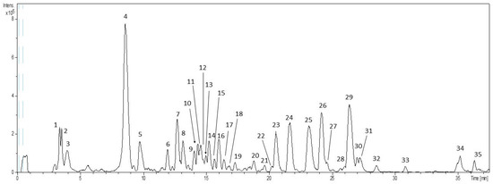
Figure 1.
HPLC-ESI-TOF-MS chromatogram of MLP.

Table 1.
Proposed compounds in MLP, identified by HPLC-ESI-TOF-MS. The numbers in the first column correspond to the peaks illustrated in Figure 1.
3.2. In-Vitro Biological Evaluation of MLP Soluble Fraction (MLPsf)
The in-vitro biological evaluation was carried out with the MLPsf using SH-SY5Y cells as the neuronal system model. The first investigation was devoted to the determination of the highest dose of MLPsf that did not induce cytotoxic effects. The analysis was carried out with both the extracts from MPL, raw and oven-treated. In Figure 2A, the results of the neurotoxicity tests obtained after addition of solutions containing MLPsf or heat-treated MLPsf at the concentrations of 2.5 mg/mL, 5 mg/mL, 10 mg/mL and 15 mg/mL are reported.
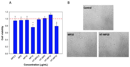
Figure 2.
Toxicity test in SH-SY5Y cell culture. (A) 24-h addition effect of 2.5 μg/mL, 5 μg/mL, 10 μg/mL and 15 μg/mL raw (MP2.5, MP5, MP10, MP15) and heat-reated (HT- MP2.5, HT-MP5, HT-MP10 and HT-MP15) MLPsf solutions. Values are obtained after normalization with the control values (red dashed line). The control error was within 0.05%. (B) Morphological inspection of the SH-SY5Y cells incubated for 24 h with 10 μg/mL of raw (MP10) or heat-treated (HT-MP10 HT) MLPsf. Magnification 20×. * p < 0.05.
The solutions were kept in direct contact with the cells for 24 h. Data indicated that the presence of the MLPsf, thermally treated or not, did alter neither the viability of the cells nor their morphology up to a concentration of 10 mg/mL (Figure 2B). This concentration was then chosen for testing the effects of MLPsf against ROS generation. To this aim, a DCFH-DA assay in SH-SY5Y cells was performed. DCFH-DA is a cell-permeable fluorogenic probe that, after crossing cell membranes, is hydrolyzed by intracellular enzymes to non-fluorescent DCFH. When ROS are present, DCFH is oxidized to fluorescent DCF originating an intracellular green fluorescence that is more or less intense, depending on the ROS concentration. Two different tests were performed; in the first one, aimed to verify the protective effect of the bioactive compounds present in MLPsf, SH-SY5Y cells were pre-treated with the MLPsf solution and, then TBH was added to induce an oxidative stress (Figure 3A,B). In the second one, for the investigation of MLPsf scavenging activity against ROS, TBH was added together with the MLPsf solution (Figure 3C,D). Control experiments (here not shown) indicated that the addition of TBH at the concentration used for the experiments was able to induce a reduction of the cell viability of about 40%. Our results (Figure 3) showed that both the pre-treatment and co-treatment of cells with MLP were effective in neutralizing neurotoxic effects induced by TBH.
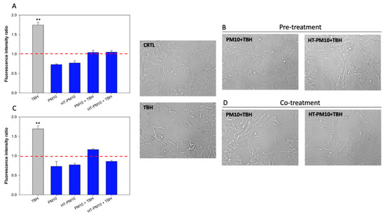
Figure 3.
Antioxidant effect of the compounds present in the MLPsf. (A) DCF assay revealing the recovery of the oxidative stress induced by the addition of 500 μM TBH in SH-SY5Y cell line. Cells were pre-treated for 24 h with 10 μg/mL MLPsf solutions obtained from raw and heat-treated MLP. Data values are obtained after normalization with the appropriate control (red dashed line; SD < 0.06%). Data concerning the PM10 and HT-PM10 samples are reported as control. (B) Morphological inspections of the SH-SY5Y cells after the treatment described in (A). Magnification 40×. (C) DCF assay illustrating the reduced oxidative stress after co-treatment with 500 μM TBH and 10 μg/mL MLPsf solutions containing raw and heat-treated MLP in SH-SY5Y cell line for 24 h. Data values are reported after normalization with the control (red dashed line; error < 0.06%). Data concerning the PM10 and HT-PM10 samples are reported as control. (D) Morphological inspections of the SH- SY5Y cells after the treatment described in (C). Magnification 40× ** p < 0.02.
3.3. Sensory, Chemical and Rheological Properties of MLP-Containing Cookies
MLP was mixed with the typical ingredients used to formulate gluten-free cookies at three concentrations, 1.5 wt% (M1.5), 2.5 wt% (M2.5) and 5 wt% (M5). A blind test was performed to check appearance, taste and chewiness of the MLP-containing cookie samples in comparison to the MLP-free blank dough (BD) that is the reference commercial product. Results (Figure 4) indicated a slight preference of the panel for the sample without addition of MLP for sweetness and smell. For the other properties, the products with the lower MLP concentrations (M1.5 and M2.5) were not statically different from those of the reference product. The lowest rating was obtained for the cookies with the highest powder concentration, M5. The panel expressed overall agreeableness towards M1.5 and M2.5 products and, for this reason, the M5 system was discarded. Further experiments were then performed on M1.5 and M2.5 systems only.
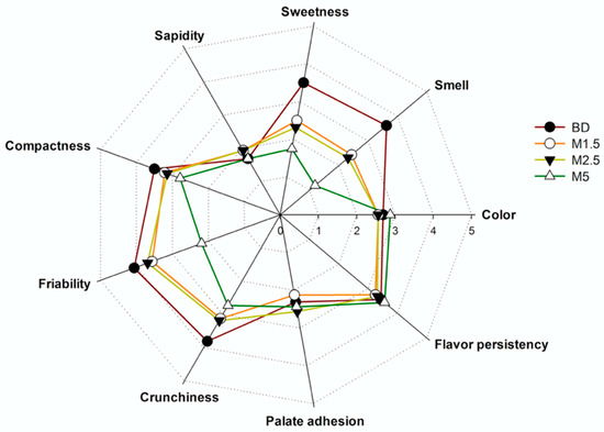
Figure 4.
Results of the blind panel evaluation, testing appearance, taste and chewiness of three different cookie samples containing MLP at the concentrations of 1.5 (M1.5), 2.5 (M2.5) and 5 (M5) wt%. The commercial blank dough (BD) was used as reference.
The IPC-OES analysis after thermal treatment was performed to assess dough enrichment in terms of valuable chemical elements. Results are reported in Figure 5. The presence of MLP, even at the lowest concentration, caused a significant increase of calcium (36%) and iron (28%) and a moderate rise of magnesium (16%) and potassium (12%) ions.
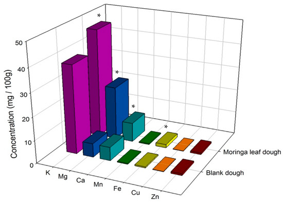
Figure 5.
Quantitative IPC-OES analysis of the chemical elements present in the blank dough (BD) and in the dough with 1.5 wt% of MLP (M1.5). * p < 0.05.
The enrichment of antioxidant compounds in the M1.5 dough compared to the blank dough after backing was verified by the Folin–Ciocalteu assay (see Table 2), resulting in about a doubling of the biophenols and flavonoids content.
Table 2.
Concentrations (mg/kg) of biophenols, flavonoids and anthocyanins in the blank dough (BD) and in the dough with 1.5 wt% MLP (M1.5). Both doughs were baked at 180 °C for 20 min.
Doughs for biscuit preparation are characterized by (i) low concentration of proteins and water; (ii) high content of fat and sugars; (iii) poor resistance to deformation; (vi) high cohesion degree; and v) low elasticity and extensibility, if compared with the doughs used for bread preparation [20]. If, on one hand, a dough with high elasticity is not desirable for cookies preparation because it tends to shrink during lamination, on the other hand, too low elasticity may cause excessive spreading during baking due to the decrease of the viscosity at the increase of temperature [20]. For this reason, knowing the rheological behavior of the dough is of primary importance. In Figure 6A, the profile of the elastic modulus as a function of the oscillatory stress applied for the BD, M1.5 and M2.5 doughs is reported. This measurement gives information about the stability of the system structure, investigating the resistance and resilience of the material when the applied stress or strain increase.
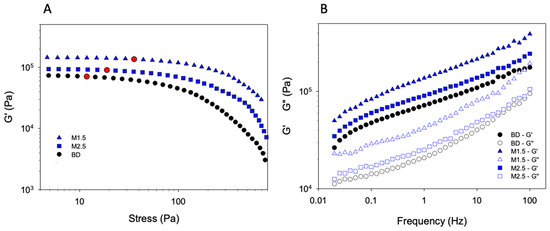
Figure 6.
Rheological behavior of the raw BD, M1.5, and M2.5 doughs. (A) Elastic modulus vs. stress plots. The full red dots show the stress limiting values indicating the departure from the linear regime. (B) Mechanical spectra.
For each sample, the limit of the linear viscoelastic region, that is, the region above which the viscoelastic response became nonlinear, was selected for a departure from the G’ plateau value lower than 5%. It can be observed that the blank dough had a lower critical threshold in comparison with both systems containing MLP (Figure 6A).
Mechanical spectra evidenced a solid-like behavior with frequency-dependence (Figure 6B). Moreover, although the addition of MLP acted as mechanical reinforcement for the dough with respect to the reference dough, the increment of the MLP content from 1.5 wt% to 2.5 wt% caused a reduction of the cohesive strength of the dough (Figure 6B). These results are in agreement with the observation of Dachana and collaborators who observed a reduction of the cohesiveness of the dough at increasing the dried moringa leaf powder up to 15 wt% [21].
This hypothesis was supported by the SEM analysis results (Figure 7). The M1.5 dough showed a greater degree of compactness than that of the M2.5 sample, in which a coarse structure with large fissures and poorly integrated starch granules can be recognized.
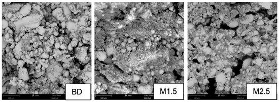
Figure 7.
SEM morphology of the BD, M1.5 and M2.5 raw doughs. Magnification 500×.
On the contrary, the M1.5 sample presented a continuum of very small particles where a higher number of small starch granules are visible. The BD sample appeared morphologically similar to M2.5 with large fissures and very large and poorly anchored starch granules. These differences could perfectly account for the observed rheological behavior, with the highest moduli for the system that has the highest ratio of aggregated to dispersed particles and particles with different size that can pack more densely than monodispersed particles. An explanation of the MLP content on the morphology and hence on the mechanical properties of the dough will require further investigations.
4. Discussion
It is well known that not-neutralized free radicals, generated by excessive reactive oxygen or nitrogen species’ (ROS and RNS) production, stimulate in cells and tissues a mechanism known as oxidative stress. This triggers the activation of a great variety of transcription factors that, in turn, stimulate the expression of genes involved in the inflammation process [22]. Antioxidant molecules, present in plants, can interact with free radicals, extinguishing the chains that increase their productions [22].
The phytochemicals that have been recognized in the leaves of Moringa oleifera can be grouped in three main chemical families: flavonoids, biophenols and their derivatives, and glucosinolates (GLs) and their derivatives [23,24]. In particular, glucomoringin, belonging to the glucosinolate group, possesses several biological properties, being effective against oxidation, viruses, fungi and bacteria [25]. Effects against inflammation, diabetes and cancer have also been described [26,27]. In aqueous environments, GLs are enzymatically hydrolyzed in glucose and unstable fragments; the latter undergo chemical modifications giving origin to nitriles, epithionitriles and isothiocianates (ITCs) [28]. There are indications that the latter strongly counteract the insurgence of not treatable neurodegenerative pathologies, such as Alzheimer’s (AD), Parkinson’s (PD) and Huntington diseases, Multiple Sclerosis and Amyotrophic Lateral Sclerosis [29,30].
Other mostly present compounds, in different chemical rearrangements, that we found in MLP are the kaempferol derivatives (Kmp) (see Table 1). Kmp belongs to the flavonoid group and it can act as scavenger of free radicals and in protecting antioxidant enzymes [30]. Furthermore, it is able to cross the blood-brain barrier (BBB), and for this reason it contributes to lowering the brain oxidation level [31]. A recent study demonstrated that the intraperitoneal injection of 10 mg/kg of Kmp was able to efficiently reverse the cognitive dysfunction induced in streptozotocin-injected mice and increased the superoxide dismutase (SOD) and glutathione (GSH) concentration in the hippocampal region [31]. The Kmp 3-O-glucoside was found to possess anti-amyloidogenic properties by inhibiting the fibrillation propensity of amyloid-beta peptide. Kmp 3-O-glucoside was capable to interact with amyloid-beta protein, originating off-pathway non-amyloidogenic conformational structures. As a consequence, an accumulation of smaller, soluble non-β-sheet and non-toxic oligomeric structural species was recorded. [32]. It is also worth mentioning that the inhibition of acetylcholinesterase, catalyzing the hydrolysis for acetylcholine (Ach) at the synaptic level, is one of the therapies used for treatment of AD patients. As a result, ACh remains active in the synaptic space reducing the symptoms of the disease. It has demonstrated in vivo, both in drosophila and in mice models, that the oral administration of Kmp exerted the same action, thus reducing the neurotoxic effect at the motor and cognitive levels [33]. The experimental evidence of the presence of GLs and Kmp among other phytochemicals, also after the heat-treatment, together with the above-presented experimental evidence from other studies of the important potential neurological effects of these molecules, prompted us to perform some further preliminary biological evaluations of MLP extracts and validate the possibility of adding MPL in the dough of cookies without altering its processing and organoleptic features.
5. Conclusions
The progressive lengthening of the life expectancy requires dietary strategies that may be able to counteract the aging phenomena that mainly occur at the level of the Central Nervous System. Many phytochemicals have been recognized in the leaves of Moringa oleifera and they can be grouped in three main chemical families: flavonoids, biophenols and their derivatives, and glucosinolates (GLs) and their derivatives. The results of this work indicate that doughs enriched with MLP possess nutraceutical valuable compounds that are still present after baking at a high temperature (180 °C) for 20 min. The MLP bioactive compounds added in the cookies are not toxic and are able to prevent oxidative stress in SH-SY5Y cells and protect them against ROS toxicity. Furthermore, the doughs enriched with MLP have suitable mechanical properties that allow their lamination and provide them with adequate resistance during baking. All gathered evidence encourages to proceed with further biological and clinical investigations.
Author Contributions
Methodology and investigation, D.N. and P.P.; Formal analysis and validation, J.L.S. and I.B.-L.; Data curation, A.G. and E.M.; Supervision, Writing, Funding acquisition P.L.S.B.; Supervision, Writing, Review and Editing, C.D.; Supervision, Writing—Original Draft preparation, D.B.; Supervision, Writing, Review and Editing, D.G. Supervision, Writing, Review and Editing, R.L. All authors have read and agreed to the published version of the manuscript.
Funding
This research received no external funding.
Institutional Review Board Statement
Not applicable.
Data Availability Statement
Data are presented in the article.
Acknowledgments
Authors wish to thank Fulvio Ferrante, Roberto Megna, and Alessia Provenzano for their valuable technical contribution. A special thank goes to Gloria Bosco, owner of the “Le Farine dei Nostri Sacchi” company for her fundamental and unquestionable contribution to the present research but above all for her kindness and willingness to comply with the requests of the authors.
Conflicts of Interest
The authors declare no conflict of interest.
References
- Singh, R.B.; Sharma, J.P.; Takahashi, T.; Juneja, L.R.; Watson, R.R.; Tomar, R.S.; Singh, M.; Jaglan, P.; Singh, M.; Chauhan, A.K.; et al. Modernization of policy for food manufacturing and farming may be necessary for global health. In The Role of Functional Food Security in Global Health; Singh, R.B., Watson, R.R., Takahashi, T., Eds.; Academic Press, Elsevier: Amsterdam, The Netherlands, 2019; Chapter 38; pp. 653–664. [Google Scholar] [CrossRef]
- Afshin, A.; Sur, P.J.; Fay, K.A.; Cornaby, L.; Ferrara, G.; Salama, J.S.; Mullany, E.C.; Abate, K.H.; Abbafati, C.; Abebe, Z.; et al. Health effects of dietary risks in 195 countries, 1990–2017: A systematic analysis for the global burden of disease study 2017. Lancet 2019, 393, 1958–1972. [Google Scholar] [CrossRef]
- Liguori, I.; Russo, G.; Curcio, F.; Bulli, G.; Aran, L.; Della-Morte, D.; Gargiulo, G.; Testa, G.; Cacciatore, F.; Bonaduce, D.; et al. Oxidative stress, aging, and diseases. Clin. Interv. Aging 2018, 13, 757–772. [Google Scholar] [CrossRef]
- Di Meo, S.; Reed, T.T.; Venditti, P.; Victor, V.M. Role of ROS and RNS sources in physiological and pathological conditions. Oxid. Med. Cell. Longev. 2016, 2016, 1245049. [Google Scholar] [CrossRef]
- Bokkon, I. Hot topic recognition of functional roles of free radicals. Curr. Neuropharmacol. 2012, 10, 287–288. [Google Scholar] [CrossRef][Green Version]
- Sharma, P.; Kapoor, S. Biopharmaceutical aspects of Brassica vegetables. J. Pharmacogn. Phytochem. 2015, 4, 140–147. [Google Scholar]
- Poon, H.F.; Calabrese, V.; Scapagnini, G.; Butterfield, D.A. Free radicals: Key to brain aging and heme oxygenase as a cellular response to oxidative stress. J. Gerontol. 2004, 59A, 478–493. [Google Scholar] [CrossRef] [PubMed]
- Bonnefoy, M.; Day, J.; Kostka, T. Antioxidants to slow aging, facts and perspectives. Press Med. 2002, 27, 1174–1184. [Google Scholar]
- Martinez-Gonzales, M.A.; Gea, A.; Ruiz-Canela, M. The Mediterranean diet and cardiovascular health. Circ. Res. 2019, 124, 779–798. [Google Scholar] [CrossRef]
- Nuzzo, D.; Galizzi, G.; Amato, A.; Terzo, S.; Picone, P.; Cristaldi, L.; Mulè, F.; Di Carlo, M. Regular intake of pistachio mitigates the deleterious effects of a high fat-diet in the brain of obese mice. Antioxidants 2020, 9, 317. [Google Scholar] [CrossRef]
- Anwar, F.; Latif, S.; Ashraf, M.; Gilani, A.H. Moringa oleifera: A food plant with multiple medicinal uses. Phytother. Res. 2007, 21, 17–25. [Google Scholar] [CrossRef]
- Ghazali, H.M.; Mohammed, A.S. Moringa (Moringa oleifera) seed oil: Composition, nutritional aspects and health attributes. In Nuts and Seeds in Health and Disease Prevention; Preedy, V.R., Watson, R.R., Patel, V.B., Eds.; Academic Press, Elsevier: Amsterdam, The Netherlands, 2010; Chapter 93; pp. 787–793. [Google Scholar] [CrossRef]
- Singh, K.K.; Kumar, K. Ethnotherapeutics of some medicinal plants used as antipyretic agent among the tribals of India. J. Econ. Taxon. Bot. 1999, 23, 135–141. [Google Scholar]
- Zeng, K.; Li, Y.; Yang, W.; Ge, Y.; Xu, L.; Ren, T.; Zhang, H.; Zhuo, R.; Peng, L.; Chen, C.; et al. Moringa oleifera seed extract protects against brain damage in both the acute and delayed stages of ischemic stroke. Exp. Geron. 2019, 122, 99–108. [Google Scholar] [CrossRef]
- Getachew, M.; Admassu, H. Production of pasta from Moringa leaves—Oat—Wheat composite flour. Cogent Food Agric. 2020, 6, 1724062. [Google Scholar] [CrossRef]
- Boateng, L.; Quarpong, W.; Ohemeng, A.; Asante, M.; Steiner-Asiedu, M. Effect of complementary foods fortified with Moringa oleifera leaf powder on hemoglobin concentration and growth of infants in the Eastern Region of Ghana. Food Sci. Nutr. 2019, 7, 302–311. [Google Scholar] [CrossRef] [PubMed]
- Hrncirik, K.; Frische, S. Comparability and reliability of different techniques for the determination of phenolic compounds in virgin olive oil. Eur. J. Lipid Sci. Technol. 2004, 106, 540–549. [Google Scholar] [CrossRef]
- Rodriguez-Perez, C.; Quintares-Piné, R.; Fernandez-Gutierrez, A.; Segura-Carretero, A. Optimization of the extraction method to obtain a phenolic compounds-rich extract from Moringa oleifera Lam leaves. Ind. Crop. Prod. 2015, 66, 246–254. [Google Scholar] [CrossRef]
- Lawless, H.T.; Heymann, H. Sensory Evaluation of Food. Principles and Practices, 2nd ed.; Food Science Text Series; Springer: New York, NY, USA; Dordrecht, The Netherlands; Heidelberg, Germany; London, UK, 2010; pp. 57–76. [Google Scholar]
- Maache-Rezzoug, Z.; Bouvier, J.M.; Allal, K.; Patras, C. Effect of principal ingredients on rheological behaviour of biscuit dough and on quality of biscuits. J. Food Eng. 1998, 35, 23–42. [Google Scholar] [CrossRef]
- Dachana, K.B.; Rajiv, J.; Indrani, D.; Prakash, J. Effects of dried Moringa (Moringa oleifera Lam) leaves on rheological, microstructural, nutritional, textural and organoleptic characteristics of cookies. J. Food Qual. 2010, 33, 660–677. [Google Scholar] [CrossRef]
- Hussain, T.; Tan, B.; Yin, Y.; Blachier, F.; Tossou, M.C.B.; Rahu, N. Oxidative stress and inflammation: What polyphenols can do for us? Oxford Med. Cell. Long. 2016, 2016, 7432797. [Google Scholar] [CrossRef]
- Jaafaru, M.S.; Abd Karim, N.A.; Enas, M.E.; Rollin, P.; Mazzon, E.; Abdull Razis, A.F. Protective effect of glucosinolates hydrolytic products in neurodegenerative diseases (NDDs). Nutrients 2018, 10, 580. [Google Scholar] [CrossRef]
- Borgonovo, G.; De Petrocellis, L.; Schiano Moriello, A.; Bertoli, S.; Leone, A.; Battezzati, S.; Mazzini, S.; Bassoli, A. Moringin, a stable isothiocyanate from Moringa oleifera, activates the somatosensory and pain receptor TRPA1 channel in vitro. Molecules 2020, 25, 976. [Google Scholar] [CrossRef]
- Galuppo, M.; De Nicola, G.R.; Iori, R.; Dell’Utri, P.; Bramanti, P.; Mazzon, E. Antibacterial activity of glucomoringin bioactivated with myrosinase against two important pathogens affecting the health of long-term patients in hospitals. Molecules 2013, 18, 14340–14348. [Google Scholar] [CrossRef] [PubMed]
- Jaja-Chimedza, A.; Graf, B.L.; Simmler, C.; Kim, Y.; Kuhn, P.; Pauli, G.F.; Raskin, I. Biochemical characterization and anti-inflammatory properties of an isothiocyanate-enriched moringa (Moringa oleifera) seed extract. PLoS ONE 2017, 12, e0182658. [Google Scholar] [CrossRef]
- Rajan, T.S.; De Nicola, G.R.; Iori, R.; Rollin, P.; Bramanti, P.; Mazzon, E. Anticancer activity of glucomoringin isothiocyanate in human malignant astrocytoma cells. Fitoterapia 2016, 110, 1–7. [Google Scholar] [CrossRef]
- Giacoppo, S.; Galluppo, M.; Montaut, S.; Iori, R.; Rollin, P.; Bramanti, P.; Mazzon, E. An overview on neuroprotective effects of isothiocyanates for the treatment of neurodegenerative diseases. Fitoterapia 2015, 106, 12–21. [Google Scholar] [CrossRef] [PubMed]
- Zandi, P.P.; Anthony, J.C.; Khachaturian, A.S.; Stone, S.V.; Gustafson, D.; Tschanz, J.T.; Norton, M.C.; Welsh-Bohmer, K.A.; Breitner, J.C. Reduced risk of Alzheimer Disease in users of antioxidant vitamin supplements. Arch. Neurol. 2004, 61, 82–88. [Google Scholar] [CrossRef]
- Kouhestani, S.; Jafari, A.; Babaei, P. Kaempferol attenuates cognitive deficit via regulating oxidative stress and neuroinflammation in an ovariectomized rat model of sporadic dementia. Neural Degener. Res. 2018, 3, 1827–1832. [Google Scholar] [CrossRef]
- Banjarnahor, S.D.; Artanti, N. Antioxidant properties of flavonoids. Med. J. Indones. 2014, 23, 239–244. [Google Scholar] [CrossRef]
- Sharoar, G.; Thapa, A.; Shahnawaz, M.; Ramasamy, V.S.; Woo, E.R.; Shin, S.Y. Keampferol-3-O-rhamnoside abrogates amyloid beta toxicity by modulating monomers and remodeling oligomers and fibrils to non-toxic aggregates. J. Biomed. Sci. 2012, 19, 104. [Google Scholar] [CrossRef]
- Beg, T.; Jyoti, S.; Naz, F.; Rahul; Ali, F.; Ali, S.K.; Reyad, A.M.; Siddique, Y.H. Protective effect of kaempferol on the transgenic drosophila model of Alzheimer’s Disease. CNS Neurol. Disord. Drug Targets 2018, 17, 421–429. [Google Scholar] [CrossRef] [PubMed]


Publisher’s Note: MDPI stays neutral with regard to jurisdictional claims in published maps and institutional affiliations. |
© 2021 by the authors. Licensee MDPI, Basel, Switzerland. This article is an open access article distributed under the terms and conditions of the Creative Commons Attribution (CC BY) license (https://creativecommons.org/licenses/by/4.0/).