1. Introduction
Surface plasmon polaritons (SPPs) is a phenomenon whereby a metal surface charges when interacting with a light wave electromagnetic field; they oscillate collectively, so that the electromagnetic field is limited to a small range and is enhanced [
1]. SPPs cannot only break through the diffraction limit of light, but is also highly sensitive to metal types, the dielectric environment, nano-shape, and size [
2]. Hence, photonic devices designed based on SPPs cannot only realize the integration of the sub-wavelength size [
3,
4,
5], but also provide the possibility of studying micro-nanophotonic devices with complex functions. It is worth mentioning that many optical phenomena have been observed in the plasmon waveguide coupling system, such as phase-coupled plasmon-induced transparency [
6] and Fano resonance [
7,
8,
9]. Fano resonance generally comes from the destructive interference between the wide-band mode (bright mode) and the narrow-band mode (dark mode) in plasmon resonance, which has a small radiation loss, a narrow full width at half maximum (FWHM), and an asymmetric spectral line shape [
10,
11]. Therefore, it has great application potential in refractive index sensors [
12,
13], slow light devices [
14], and optical switches [
15].
Nowadays, many waveguide coupling structures based on SPPs have been designed to fabricate various photonic devices, including metal−insulator−metal (MIM) waveguides [
16], insulator−metal−insulator waveguides [
17], channel waveguides [
18,
19], and nanoparticle chain waveguides [
20]. Among them, MIM waveguides are widely considered and reported by scholars and the media at home and abroad because of their sub-wavelength size, simple structure, easy integration, and high reliability [
16,
21]. Yang et al. [
22] designed a coupling structure with a double-gap ring cavity and MIM waveguides with two triangular baffles with a sensitivity of 1500 nm/RIU and figure of merit (FOM) of 65.2. Tang et al. [
23] proposed and studied a plasmonic structure that includes a ring nanocavity, two bus waveguides, and a rectangular nanocavity, with a sensitivity of 1125 nm/RIU. Su et al. [
24] devised a plasmonic sensor coupled with an elliptical ring cavity and a MIM waveguide with two rectangle baffles; its sensitivity is 1550 nm/RIU and FOM is 43.05. As shown in
Table 1, although the difference of FOM is not obvious, the sensitivity of the designed structure is obviously better than that of other structures. In addition, various photonic devices based on MIM waveguide structure design, such as optical splitters [
25,
26], filters [
27,
28], and Bragg reflectors [
29,
30], have achieved remarkable results.
Herein, a plasmonic structure consisting of a MIM waveguide with two symmetrical rectangle baffles coupled with a connected concentric double rings resonator (CCDRR) is presented and investigated. The transmission characteristics and the normalized magnetic field distribution were calculated, introducing the finite element method (FEM). In addition to the influence of the refractive-index of the dielectric on the transmission characteristics of Fano resonance, the influence of the geometric parameters of the structure was also studied. These parameters include the external radii of the outer ring and inner ring of the CCDRR, the separation between the two symmetrical rectangular baffles, the heights of the two rectangular baffles, and the coupling gap between the CCDRR and the bus waveguide. Additionally, applications of the designed structure in refractive-index sensing and temperature sensing were studied in detail. The designed structure provides new detection positions, which may be helpful for meeting special requirements for detection wavelengths.
2. Structural Model and Analysis Methods
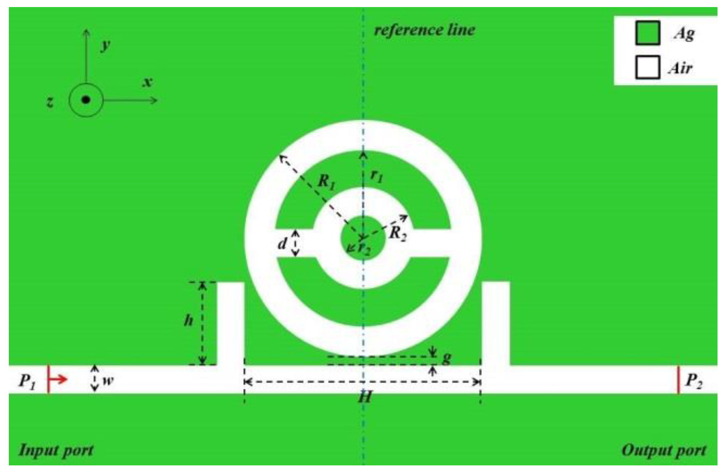
A schematic diagram of the presented MIM bus waveguide coupled with two rectangular baffles and a CCDRR is displayed in
Figure 1. The width
w of the MIM waveguide, two rectangular baffles, and two annulus cavities remained invariable at 50 nm to ensure that the waveguide only supports the transverse magnetic field (TM
0) mode.
g represents the coupling gap between the bus waveguide and the CCDRR. The heights of the two rectangle baffles and the separation between them are signified as
h and
H, respectively.
R1 and
r1,
R2 and
r2 express the external and internal radii of the outer ring and inner ring, respectively.
d is defined as the width of the baffles connecting two rings in the concentric ring, which is fixed at 40 nm.
The white part and green part in
Figure 1 represent air and sliver, respectively. The relative permittivity
εd of air is 1. Based on the Debye−Drude dispersion model [
31], the description of the relative dielectric constant of Ag is as follows:
where
is the boundless frequency dielectric constant and
is the static dielectric constant. The relaxation time and the conductivity of Ag are regarded as
and
, respectively.
The formula of the TM
0 mode of the MIM waveguide is as follows [
32]:
where
expresses the wave vector in the waveguide, and in free-space,
k is taken as
;
and
; among them,
εin and
εm are the permittivity of the insulator and metal, respectively.
By analyzing the shifts of the Fano resonance wavelength, the sensing performance of the proposed structure in the waveguide coupled system was investigated. The transmission wavelengths and the effective refractive index’s real part in the MIM waveguide can be expressed on the foundation of the standing wave theory as follows [
33,
34]:
where
L indicates the circumference of the ring cavity;
ψr signifies the phase shift caused by the reflection of SPPs at the metal−insulator boundary surface; and
m is a positive integral number, i.e., the number of antinodes of SPPs.
The characteristics of the sensor can be evaluated by two important parameters, namely, sensitivity (
S) and
FOM, which are expressed by the following equation [
35]:
where Δ
λ and Δ
n are the variation of resonant wavelength and refractive index, respectively.
In the next part of the paper, a simulation was run using COMSOL Multiphysics 5.4a. With the comparability of the operating principle of the two-dimensional (2D) mode and three-dimensional (3D) mode, a 2D geometric model with greatly reduced computational complexity was established, and the finite element method (FEM) was used to analyze the propagation characteristics. Then, hyperfine meshing was used to guarantee the accuracy of the emulation. In addition, the absorbing boundary condition was established by the perfect matched layer, which can absorb the outward reflected waves.
3. Simulations and Results
When comparing the performance of the double-ring cavity and the CCDRR, it was found that their sensitivity was almost the same in the range of the agreed refractive index change, but that the CCDRR had a higher FOM, so the CCDRR was chosen for further study.
To gain a distinct understanding of the propagation characteristics of the proposed structure, it was necessary to compare the whole system with the single CCDRR structure and the unitary two rectangular baffle structure. The unitary two rectangular baffle structure and the unitary CCDRR structure are shown in
Figure 2a,b, respectively. The transmission spectra of the three structures are shown in
Figure 2c. The geometric parameter settings are as follows:
R1 = 190 nm,
R2 = 130 nm,
H = 540 nm,
h = 150 nm, and
g = 10 nm. The transmission spectra of the unitary two rectangular baffle structure, the unitary CCDRR structure, and the whole system are represented by the black, red, and blue solid lines, respectively. In
Figure 2c, the black solid line representing the unitary two rectangular baffle structure has a positive slope, and it has relatively high transmittance in the range of 0.35 to 0.6. Hence, it can be regarded as a continuous wide-band mode. The transmission spectrum of the unitary CCDRR structure approximates the Lorentz line, which is considered as representing the discrete narrowband mode. It is obvious that the transmission spectrum of the whole structure (blue line) has an asymmetric shape, which indicates that Fano resonance is generated by the interaction of the successive wide-band mode and discrete narrow-band mode.
To better comprehend the inner theory of the black line’s role in the Fano resonance of the whole structure, the magnetic field distributions and the electric field distributions of the unitary two rectangular baffles and the whole system at the resonance dip point (
λ = 1459 nm) were demonstrated, which are shown in
Figure 3a–d, respectively. In
Figure 3a, there is a distinct resonance in the MIM waveguide, with only one bus waveguide and two rectangular baffles. As shown in
Figure 3b, for the whole structure, the firm resonance occurs only on the left side, while infirm resonance occurs on the right. Additionally, the upper and lower parts of the outer ring in the CCDRR are anti-phase.
Figure 3b,c provides insight into the actual energy distribution in the waveguides and the CCDRR cavity. In
Figure 3c, there is an obvious energy distribution in the MIM waveguide. However, as shown in
Figure 3d, the energy of SPPs is intensified at the CCDRR cavity and decreased at the right side of the waveguide. It was found that SPPs was directly coupled to the unitary two rectangular baffle structure and stimulated the resonance corresponding to the successive wide-band state, while the discrete narrow-band state in the CCDRR was indirectly stimulated by the SPPs in the two symmetrical rectangular baffles. Thus, the interaction of the two states produced the Fano resonance.
For further investigation of the influences of the different refractive indexes (
n) on the transmission spectrum of the Fano resonance, six refractive indexes were simulated: 1.00, 1.01, 1.02, 1.03, 1.04, and 1.05 RIU. The structural arguments were as follows:
R1 = 240 nm,
R2 = 130 nm,
H = 540 nm
, h = 150 nm, and
g = 10 nm.
Figure 4a,b shows the simulation results. In
Figure 4a, with the increase of
n, the transmission spectra have an approximately equidistant red-shift. As shown in
Figure 4b, when the refractive index changes, the change of dip wavelength-shift is linear. Therefore, the sensitivity of the sensor, which was 2260 nm/RIU with a FOM of 56.5, could be obtained from the slope after linear fitting, leading us to obtain the best result for the optimal parameter of the structure.
To investigate the influences of different external radii of the outer ring of the CCDRR on Fano resonance,
R1 was set to increase from 200 nm to 240 nm at an interval of 10 nm, while keeping other arguments fixed at
R2 = 110 nm,
H = 540 nm,
h = 150 nm, and
g = 10 nm. The transmission spectra are displayed in
Figure 5a. With the increase of
R1, there is an obvious red shift at the dip point of Fano resonance, and the transmittance of this position increases slightly. The simulation result shows that
R1 determines the dip wavelength of Fano resonance. This phenomenon can be explained in other words as a scenario where the dip wavelength depends on the CCDRR corresponding to the narrowband pattern, with
R1 as an important parameter of the CCDRR. As shown in
Figure 5b, by linear fitting, five solid lines representing the sensitivities of the different structures were obtained. As the external radius
R1 of the outer ring increases, the sensitivity becomes higher. The maximum sensitivity was obtained via calculation, which was 2200 nm/RIU when
R1 was 240 nm, and the maximum FOM was 47.8. Thus, in practical applications, the radius
R1 should be appropriately increased to obtain a better sensing performance.
The effects of different external radii
R2 of the inner ring of the CCDRR on the transmission capabilities were investigated as 90, 100, 110, 120, and 130 nm, while setting the parameter value
R1 as 240 nm and keeping the other parameters the same. As shown in
Figure 6a, the dip wavelength of Fano resonance was almost constant. When
R2 increases, the dip point of Fano resonance has a slight red-shift and the transmittance of the dip marginally decreases, and there is a slight increase in sensitivity, which is described in
Figure 6b. When
R2 = 130 nm, the sensitivity of the structure attained the highest value: 2260 nm/RIU with a FOM of 56.5.
Afterward, the effect of the separation
H of the two symmetric rectangular baffles on the propagation performance was studied. The transmission spectra that are displayed in
Figure 7a were calculated at different separations for
H = 540, 560, 580, 600, and 620 nm, while the other geometric parameters were kept the same. It was found that no matter how
H changed, the dip wavelength of Fano resonance remained almost unchanged, though the FOM obviously decreased, as represented in
Figure 7b. There was an optimal simulation result: the sensitivity was 2260 nm/RIU and FOM was 56.5. Then, the other geometric parameters were kept the same, except for increasing the height of the rectangular baffle
h from 130 to 170 nm in steps of 10 nm. The transmission spectra and the change of FOM of the diverse heights of the rectangle baffles are shown in
Figure 7c,d, respectively. As shown in
Figure 7c, with the increase of
h, the dip wavelength only demonstrates a slight blue-shift, while the Fano line shape changes significantly. According to the calculation, when
h = 150 nm, the maximum sensitivity is 2260 nm/RIU with a FOM of 56.5. As
h continues to increase, the sensitivity will decrease as well as the FOM.
The separation of the two symmetrical rectangular baffles H and the heights of the two baffles h are pivotal to the waveguide with two rectangle baffles. According to the simulation results, the successive wide-band mode has a significant effect on the line shape of Fano resonance, but not on the wavelength of the dip point.
To further investigate the effects of the coupling gap between the CCDRR and the waveguide on the propagation properties,
g was increased from 10 nm to 30 nm while the other geometric arguments were fixed at
R1 = 240 nm,
R2 = 130 nm,
H = 540 nm, and
h = 150 nm. The transmission performances of the structure with different coupling gaps between the CCDRR and the waveguide for
g = 10, 15, 20, 25, and 30 nm can be seen in
Figure 8a. With increasing
g, the dip wavelength of the Fano resonance shows a blue-shift, the FWHM tends to narrow and the transmittance of the dip position of Fano resonance tends to move higher. The fact that the coupling intensity weakens as the coupling gap increases can account for this phenomenon. Additionally, the sensitivity of the system decreased with the increase of
g, as shown in
Figure 8b. Thus, the optimal performance parameters can be obtained when the sensitivity is 2260 nm/RIU and the FOM is 56.5.
4. Application of the Proposed Structure in Temperature Sensing
The presented system can also be used as a nanoscale temperature sensor, which is realized by using the variation of the refractive indices of the temperature sensing medium caused by an ambient temperature, with the temperature sensing material viewed as a liquid. Ethanol was chosen as the temperature sensing medium to fill the CCDRR and the MIM waveguide with a bus waveguide and two symmetrical rectangular baffles because of its high refractive-index temperature parameter of 3.94 × 10
−4 (°C
−1). The refractive-index temperature coefficients of Ag and quartz are 9.30 × 10
−6 (°C
−1) and 8.60 × 10
−6 (°C
−1), respectively, which are two orders of magnitude smaller than that of ethanol. Thus, the variation of temperature largely affects ethanol, and the effects of thermal expansion of Ag and quartz can be ignored. The schematic diagram of three-dimensional (3D) structure is shown in
Figure 9. The blue part represents ethanol, the green part represents silver, and the black part represents the quartz substrate.
Commonly, the functional connection between the refractive index, temperature coefficient, and ambient temperature of a liquid temperature sensing material can be expressed as follows [
36]:
where
is the refractive index of the liquid corresponding to room-temperature,
;
is the refractive index temperature coefficient; and T represents the ambient temperature. Thus, the refractive index formula, with ethanol as the filling material of the temperature sensor, and the sensitivity formula can be expressed as follows:
The geometric arguments of the structure were fixed at
R1 = 240 nm,
R2 = 130 nm,
d = 40 nm,
H = 540 nm,
h = 150 nm,
g = 10 nm, and
w = 50 nm. The transmission spectrum for the disparate temperatures of the sensor is plotted in
Figure 10a. As the melting and boiling points of ethanol are −144.3 °C and 78.4 °C, respectively, the temperature sensor has good stability in the working range of −80–60 °C. As the temperature drops from 60 °C to −80 °C with an interval of 20 °C, the transmission spectrum displays a red-shift phenomenon and the sensitivity, which is shown as
Figure 10b, has a remarkable linear fit with the value of 1.48 nm/°C.
Although the temperature sensor has some advantages, such as a high sensitivity, a simple structure, and easy integration, it still has some limitations. Due to the bounds of the boiling and melting points of ethanol, the sensor is only suitable for low-temperature sensing, and it cannot solve sensing problems when the temperature is too high. As a liquid substance, ethanol cannot meet the needs of solid-state sensing equipment under some special conditions. In some practical applications, thermal materials such as lithium niobate can be used instead of ethanol to manufacture solid-state equipment. In future research, we will also consider adding a grapheme strip into the CCDRR cavity to allow for dynamic adjustment of the sensitivity.


