Article

Particulate Matter Contamination of Bee Pollen in an Industrial Area of the Po Valley (Italy)

1
Dipartimento di Scienze delle Produzioni Vegetali Sostenibili (DIPROVES), Università Cattolica del Sacro Cuore, 29122 Piacenza, Italy
2
Dipartimento di Scienze dell’Ambiente e della Terra (DISAT), Università Milano Bicocca, 20126 Milano, Italy
3
KOINE, Consulenze Ambientali, 43022 Montechiarugolo, Italy
*
Author to whom correspondence should be addressed.
Appl. Sci. 2021, 11(23), 11390; https://doi.org/10.3390/app112311390
Submission received: 7 October 2021 / Revised: 21 November 2021 / Accepted: 24 November 2021 / Published: 1 December 2021

Abstract

:
The global demand for bee pollen as a dietary supplement for human nutrition is increasing. Pollen, which comprises proteins and lipids from bees’ diets, is rich in essential amino acids, omega fatty acids, and bioactive compounds that can have beneficial effects on human health. However, bee pollen may also contain contaminants due to environmental contamination. To date, data on bee pollen contamination by environmental pollutants refer almost exclusively to pesticides and heavy metals, and very little information is available on the potential contamination of bee pollen by airborne particulate matter (PM), a ubiquitous pollutant that originates from a wide range of anthropogenic sources (e.g., motor vehicles, industrial processes, agricultural operations). In the present study, pollen grains collected by forager bees living in an industrial area of the Po Valley (Northern Italy) were analyzed for contamination by inorganic PM. The morpho-chemical characterization of inorganic particles using SEM/EDX allowed us to identify different emission sources and demonstrate the potential risk of PM entering the food chain and exposing bees to its ingestion.

1. Introduction

Honeybees are important providers of ecosystem services by providing pollination to plants and delivering many products to humans (e.g., honey, pollen, propolis, and royal jelly). In recent years, the global demand for bee pollen as a dietary supplement in human nutrition has increased. Pollen, which represents the protein and lipid sources of a bee’s diet, is rich in essential amino acids, omega fatty acids, and bioactive compounds [1,2,3]. Dietary antioxidant intake derived from bee pollen has been linked to the prevention and clinical treatment of several diseases, including liver and kidney fibrosis [4]. In addition, the incorporation of bee pollen into the diet may promote the inhibition of inflammation and oxidative stress [4]. However, bee pollen intake is also associated with health risks due to contamination by bacterial and fungal toxins, pesticides, and heavy metals. Previous data confirmed that bee pollen may also be contaminated by airborne particulate matter (PM) [5]. Airborne PM is a ubiquitous pollutant that may originate from either natural (e.g., soil erosion, volcanic eruptions, forest fires) or anthropogenic sources (e.g., motor vehicles, industries, agriculture). Particles are commonly classified according to their aerodynamic diameter, ranging from several micrometers (PM10, i.e., PM with an aerodynamic diameter less than 10 μm) to a few micrometers (PM2.5) or nanometers (PM0.1). Airborne PM inhalation is associated with inflammation, cardiopulmonary diseases, and lung cancer [6], but exposure to airborne dust can also occur by swallowing inhaled particles or ingesting contaminated food, causing harmful effects on the digestive tract [7,8,9,10]. Recent studies have demonstrated that, in heavily trafficked areas, bee pollen is contaminated by ultrafine PM originating from vehicle braking systems, thus endangering the safety of food produced at traffic-influenced sites and the health of bees [5]. Pollen contamination by PM may also threaten the overall health of bees. Previous studies on the oral exposure of bees to airborne particles indicate cytological and histological modifications of the gut epithelium or alterations in the gut microbial community [11,12].
The study area of the present work is in the peri-urban area of the city of Parma (Po Valley, Northern Italy). The Po Valley is one of the most important industrial and agricultural areas in Europe, characterized by high levels of PM10 and PM2.5 that mainly originate from traffic and agriculture [13,14,15]. A previous source apportionment study carried out in the area using honeybees has been used as an alternative sampling system for airborne PM. Agricultural operations and high-temperature combustion processes, probably linked to the Parma incineration plant, have been identified as a major sources of PM [16]. The honeybee (Apis mellifera L.), which is an important control in the biomonitoring of environmental pollutants [17,18,19,20], can act also as an efficient sampler of airborne dust [5,16,21,22,23]. The generation of static electricity on a bee’s body during flight attracts airborne particles that can be analyzed by scanning electron microscopy (SEM) coupled with X-ray spectroscopy (EDX) to evaluate size, morphology, and chemical composition [21,22]. Such information can facilitate the identification of PM emission source(s) and potential health hazards [5,16,21,22].
In the present work, airborne PM contamination of pollen grains collected by honeybees in the study area was investigated by SEM/EDX, with the aim of characterizing the nature, morphology, and mineralogical composition of particles and evaluating their possible origin. Such analysis also allowed us to demonstrate the potential risk of PM entering the food chain and exposing bees to its ingestion.

2. Materials and Methods

2.1. Sample Collection and Preparation

In February 2017, a hive was placed in the peri-urban area of the city of Parma (44.834357, 10.353865), close to wheat and alfalfa crops and about 100 m from the A1 Milan–Naples highway. The highway exit and the Parma incinerator plant were located approximately 400 m from the hive (Figure 1).
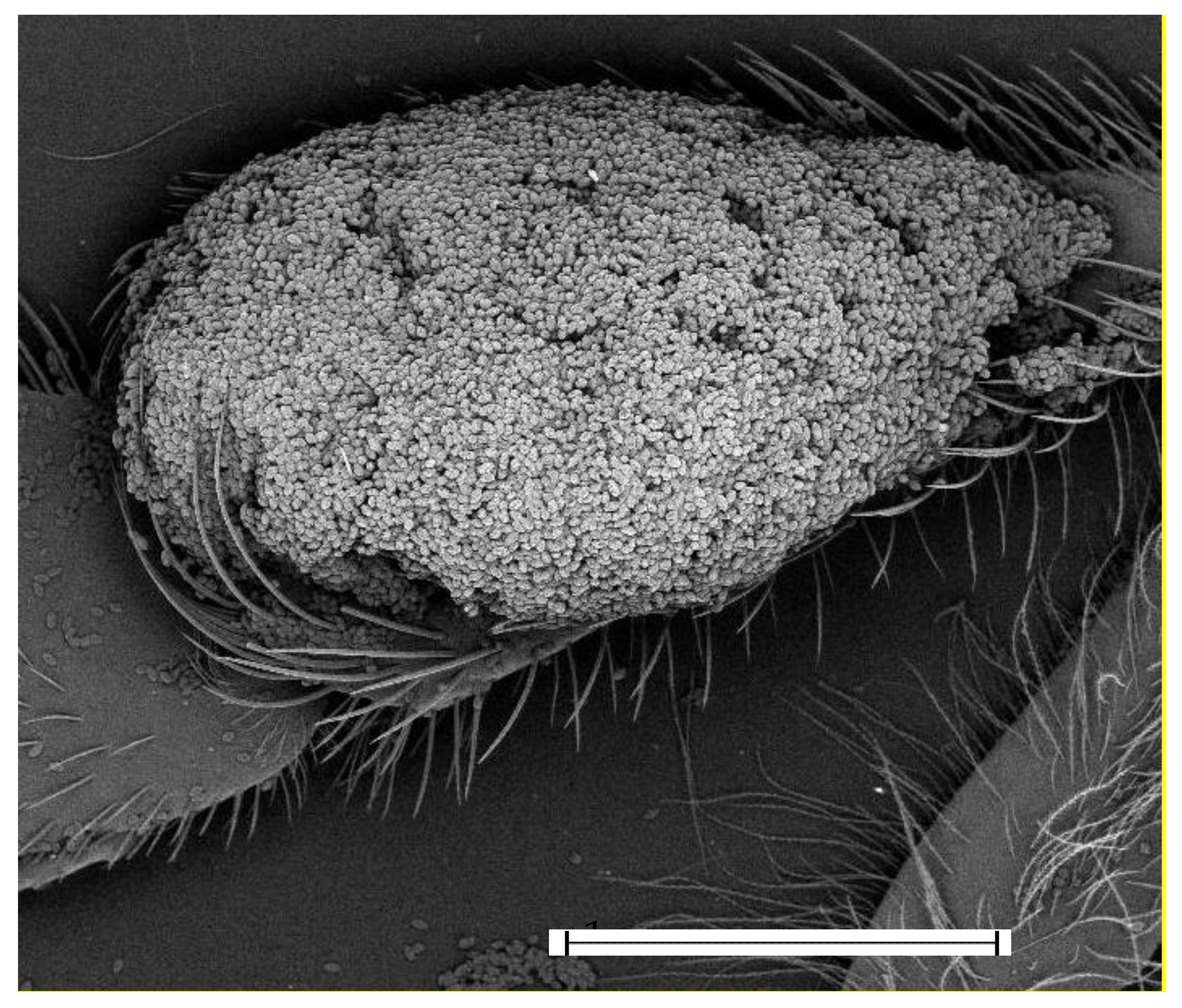
To avoid possible contamination of pollen caused by using pollen traps, analyses were carried out directly on pollen grains attached to the hindlegs of worker bees (Figure 2).
Four sampling sessions were carried out monthly from May to August. During each session, ten forager bees carrying pollen grains were sampled while returning to their hives. Bees were immediately placed in soda-glass-capped vials (Chromacol Limited, Welwyn Garden City, UK) stored on ice to inactivate them and quickly transferred to the laboratory for sample preparation. Hind legs with pollen grains (total number of pollen grains analyzed in this study = 40) were dissected under a laminar flow hood to avoid external contamination. Two pairs of legs were mounted onto SEM stubs using double-adhesive carbon tape for SEM/EDX analysis. Stubs were carbon coated, as previously described [16].
A few days after each sampling session, negative control bees carrying pollen grains were collected close to hives in a rural area located 10 km south of Parma (Northern Italy) near the bed of Parma creek and close to the foothills of the Apennine Mountains (44.690099, 10.336713). The control site was far from any known emitting source of PM (i.e., vehicular traffic, incinerators, agricultural lands).

2.2. Scanning Electron Microscopy with Energy Dispersive X-ray Spectrometry Investigation

The SEM/EDX investigation was carried out using both a Tescan VEGA TS5136XM (Brno-Kohoutovice, Czech Republic) and a Zeiss Gemini 500 field-emission instrument (Oberkochem, Germany), both equipped with an energy dispersive X-rays (EDX) analyzer [16]. Secondary electrons (SE) and backscattered electron (BSE) images, as well as EDX point analyses, were acquired in alternating sequence under the same conditions of 20 kV with a nominal beam current of approximately 1 nA. For each pollen grain, a panoramic BSE image was acquired. Airborne PM that could be distinguished from the pollen grain surface according to brightness and contrast was selected for the analysis [16]. Each analyzed PM was attributed to a phase according to the relative EDX spectrum, as well as the morphology of the particle itself, as previously described [21,22]. To account for contributions of pollen grains to the EDX spectra, point analyses were carried out directly on the surface of the pollen.

3. Results

During the monitoring period, forager bees were observed collecting pollen from wild and cultivated plants present in the surroundings (e.g., Taraxacum officinale or dandelion, Prunus spinosa or blackthorn, Brassicaceae, Zea mays or corn, Medicago sativa or alfalfa) in the industrial area. All pollen grains collected from both the control and the industrial site displayed contamination by inorganic particles, with controls appearing to be scarcely contaminated (no more than three particles for grain pollen were detected), while the pollen from the industrial site was heavily contaminated. Airborne PM detected on pollen from the control site was composed exclusively of natural mineral phases (i.e., clay minerals and calcite, Figures S1 and S2). All pollen samples from the industrial site were contaminated with clay minerals, calcite, iron oxides/hydroxides, barite, and quartz (Table 1, Figure 3).
Pollen grains contaminated by feldspar (Figure 4a), and Fe alloys (Fe-Cr and Fe-Cr-Mn) appeared less frequently (Table 1, Figure 4b–d).
PM containing Zn, Pb, and Sb (Figure 5a–c) and spherical PM composed of silicon dioxide (Figure 6d,e) were only occasionally present on bee pollen.
Interestingly, some grains collected in May appeared to be contaminated with gold particles (Figure 6).

4. Discussion

Airborne PM of natural and anthropogenic origin was observed on pollen grains collected by worker bees from the monitoring station. Naturally derived minerals included calcite, clay minerals, quartz, and feldspars, which are abundant in the neighboring soils [16]. Natural erosion and agricultural activities, such as sowing and harvesting that may have occurred in the adjacent crop fields, may have contributed to the environmental spread of soil minerals. Regarding calcite, this mineral is also widespread in incinerator bottom ash and represents one of the most common products of ash weathering [25]. Even if our data suggest that handling ash may represent a critical step for dust leakage, further work is needed to assess specific contributions of the waste-treatment process. The incinerator plant may also be the source of the spherical dusts of SiO2 contaminating bee pollen [16]. The spherical morphology is, indeed, a well-known marker of high-temperature combustion, such as that occurring at the incinerator plant [25,26].
Furthermore, the unexpected presence of gold particles on bee pollen could also be attributed to the incineration plant, as bottom ash may contain economically significant levels of this mineral [27,28]. According to the literature, the presence of precious metals is mainly derived from waste of electrical and electronic equipment and many efforts are currently being made to recover precious metals in bottom ash [27,28]. Airborne PM of quartz contaminating bee pollen may also have originated from the incineration plant, as SiO2 fragments are often abundant in bottom ash [25,29].
Other mineral phases frequently found in bee pollen included Fe oxides/hydroxides, metallic Fe, and Fe alloys (e.g., Fe, Cr, ±Mn, ±Sn). In natural soils, Fe oxides/hydroxides are common in mafic and ultramafic rocks, in lateritic horizons, and following alteration of sulfide ores, but they are rare in the alluvial deposits that characterize the area of investigation [16]. Thus, the natural origin of such dusts should be excluded. In addition, an anthropogenic origin should be considered for metallic Fe and Fe alloys. These compounds are present in several manufactured goods, especially in the automotive industry [30,31]. According to previous studies, Fe compounds and their alloys may therefore originate from the wearing of mechanical parts or the braking systems of vehicles [5,16,22,31].
Antimony oxides contaminating pollen grains may also be derived from the braking system. This compound is known to originate from the oxidation of antimony sulfide used to improve friction stability at elevated temperatures [32]. One possible origin of metallic Zn is vehicular traffic, as pure Zn is widely used in the automotive industry to protect steel from corrosion (International Zinc Association, www.zinc.org, 19 January 2021).
Another mineral phase frequently found in pollen grains was barite (BaSO4). Barite occurs naturally in hydrothermal deposits; therefore, this mineral should not be present in the area of investigation. However, barite (BaSO4) is widely used in vehicle tires and brakes as it improves the thermal and wear resistance and reduces noise [33,34,35]. An anthropogenic origin for TiO2 particles found on bee pollen was suggested: titanium dioxide occurs in nature as the mineral rutile, usually concentrated in the heavy fraction of sediments; however, synthetic titanium dioxide (TiO2) is widely used as a filler and a whitening agent in plastics, paints, and vehicle components.
It is widely acknowledged that waste incineration may be an important source of heavy metals, including Pb [36]. However, Pb is environmentally persistent and its presence in airborne dust may be the consequence of its common use in the past as a gasoline additive [36].

5. Conclusions

Our data provide evidence of the contamination of bee pollen by specific airborne PM of natural or anthropogenic origin. The morpho-chemical characterization of the dusts allowed us to clearly distinguish among different anthropogenic sources, namely vehicular traffic and incineration plants. Among the markers of traffic are particles originating from the wearing of mechanical parts and brakes (Fe-based compounds, metallic Zn, barite, and antimony oxide) of vehicles travelling on the A1 motorway. Emissions from the brake system may indeed be exceptionally high in areas close to motorway exits. However, it is also possible that the incineration plant may be responsible for the environmental presence of peculiar PM (gold and spherical SiO2) contaminating bee pollen, as this PM is characteristic of bottom and fly ash. PM containing lead is persistent in the environment and may derive from different past and present anthropic activities; further studies involving Pb isotopic analyses may help constrain their relative contributions [37]. In addition, the presence of TiO2 should be further investigated, given that this compound is used in a wide range of manufactured objects. The presence of calcite, clay minerals, quartz, and feldspars on the bee pollen may be due to both the erosion of soils and agricultural operations; however, further work is needed to assess the specific contributions of the waste-treatment process (e.g., handling of bottom ash) that may be responsible for the environmental spread of quartz and calcite.
In a previous work, similar contamination was found on the wings of worker bees living in the area [5,16], confirming that airborne PM pollutants are widespread and people living in the area may be chronically exposed to specific airborne PM. The present research also suggests a potential risk for PM entering the food chain and exposing honeybees and other pollinators to its ingestion. Therefore, ecotoxicological studies are urgently needed to specifically evaluate the exposure levels and health effects on humans and ecosystem services.

Supplementary Materials

The following are available online at https://www.mdpi.com/article/10.3390/app112311390/s1, Figure S1: Representative EDX spectra of particles of iron oxides/hydroxides, barite, quartz, and of a clay mineral; Figure S2: Representative EDX spectra of a feldspar, a Fe-Cr alloy, a Fe-Cr-Mn alloy, and metallic Zn; Figure S3: representative EDX spectra of a particle containing Pb, antimony oxide, and gold.

Author Contributions

Conceptualization, I.N. and M.P.; methodology, G.P., G.C., M.P., I.N.; validation, M.P. and G.C.; formal analysis, G.P. and G.C.; writing—original draft preparation, I.N.; writing—review and editing, G.P., G.C., M.P. and I.N.; funding acquisition, I.N. All authors have read and agreed to the published version of the manuscript.

Funding

This project was partly funded by the Project from Seed to Spoon − S2S (2019-1-IT02-KA201-06239) Erasmus Plus 2019 KA2–Cooperation for innovation and the exchange of good practices KA201–Strategic Partnerships for school education.

Institutional Review Board Statement

Not applicable.

Informed Consent Statement

Not applicable.

Data Availability Statement

Data are available upon request to the corresponding author.

Conflicts of Interest

The authors declare no conflict of interest.

References

  1. Thakur, M.; Nanda, V. Composition and functionality of bee pollen: A review. Trends Food Sci. Technol. 2020, 98, 82–106. [Google Scholar] [CrossRef]
  2. Denisow, B.; Denisow-Pietrzyk, M. Biological and therapeutic properties of bee pollen: A review. J. Sci. Food Agric. 2016, 96, 4303–4309. [Google Scholar] [CrossRef]
  3. Khalifa, S.A.M.; Elashal, M.H.; Yosri, N.; Du, M.; Musharraf, S.G.; Nahar, L.; Sarker, S.D.; Guo, Z.; Cao, W.; Zou, X.; et al. Bee Pollen: Current Status and Therapeutic Potential. Nutrients 2021, 13, 1876. [Google Scholar] [CrossRef]
  4. Mărgăoan, R.; Stranț, M.; Varadi, A.; Topal, E.; Yücel, B.; Cornea-Cipcigan, M.; Campos, M.G.; Vodnar, D.C. Bee Collected Pollen and Bee Bread: Bioactive Constituents and Health Benefits. Antioxidants 2019, 8, 568. [Google Scholar] [CrossRef] [Green Version]
  5. Papa, G.; Capitani, G.; Capri, E.; Pellecchia, M.; Negri, I. Vehicle-derived ultrafine particulate contaminating bees and bee products. Sci. Total Environ. 2021, 750, 141700. [Google Scholar] [CrossRef]
  6. Brook, R.D.; Rajagopalan, S.; Pope, C.A.; Brook, J.R.; Bhatnagar, A.; Diez-Roux, A.V.; Holguin, F.; Hong, Y.; Luepker, R.V.; Mittleman, M.A.; et al. Particulate matter air pollution and cardiovascular disease: An update to the scientific statement from the american heart association. Circulation 2010, 121, 2331–2378. [Google Scholar] [CrossRef] [PubMed] [Green Version]
  7. Lomer, M.C.E.; Thompson, R.P.H.; Powell, J.J. Fine and ultrafine particles of the diet: Influence on the mucosal immune response and association with Crohn’s disease. Proc. Nutr. Soc. 2002, 61, 123–130. [Google Scholar] [CrossRef] [PubMed] [Green Version]
  8. Salim, S.Y.; Jovel, J.; Wine, E.; Kaplan, G.G.; Vincent, R.; Thiesen, A.; Barkema, H.W.; Madsen, K.L. Exposure to Ingested Airborne Pollutant Particulate Matter Increases Mucosal Exposure to Bacteria and Induces Early Onset of Inflammation in Neonatal IL-10–Deficient Mice. Inflamm. Bowel Dis. 2014, 20, 1129–1138. [Google Scholar] [CrossRef] [PubMed]
  9. Kish, L.; Hotte, N.; Kaplan, G.G.; Vincent, R.; Tso, R.; Gänzle, M.; Rioux, K.P.; Thiesen, A.; Barkema, H.W.; Wine, E.; et al. Environmental Particulate Matter Induces Murine Intestinal Inflammatory Responses and Alters the Gut Microbiome. PLoS ONE 2013, 8, e62220. [Google Scholar] [CrossRef] [Green Version]
  10. Woodby, B.; Schiavone, M.L.; Pambianchi, E.; Mastaloudis, A.; Shelly, N.H.; Wood, S.M.; Pecorelli, A.; Valacchi, G. Particulate Matter Decreases Intestinal Barrier-Associated Proteins Levels in 3D Human Intestinal Model. Int. J. Environ. Res. Public Health 2020, 17, 3234. [Google Scholar] [CrossRef]
  11. Papa, G.; Di Prisco, G.; Spini, G.; Puglisi, E.; Negri, I. Acute and chronic effects of Titanium dioxide (TiO2) PM1 on honey bee gut microbiota under laboratory conditions. Sci. Rep. 2021, 11, 5946. [Google Scholar] [CrossRef]
  12. AL Naggar, Y.; Dabour, K.; Masry, S.; Sadek, A.; Naiem, E.; Giesy, J.P. Sublethal effects of chronic exposure to CdO or PbO nanoparticles or their binary mixture on the honey bee (Apis millefera L.). Environ. Sci. Pollut. Res. 2020, 27, 19004–19015. [Google Scholar] [CrossRef]
  13. Marcazzan, G.M.; Vaccaro, S.; Valli, G.; Vecchi, R. Characterisation of PM10 and PM2.5 particulate matter in the ambient air of Milan (Italy). Atmos. Environ. 2001, 35, 4639–4650. [Google Scholar] [CrossRef]
  14. Pietrogrande, M.C.; Bacco, D.; Ferrari, S.; Ricciardelli, I.; Scotto, F.; Trentini, A.; Visentin, M. Characteristics and major sources of carbonaceous aerosols in PM2.5 in Emilia Romagna Region (Northern Italy) from four-year observations. Sci. Total Environ. 2016, 553, 172–183. [Google Scholar] [CrossRef] [PubMed]
  15. Pozzer, A.; Bacer, S.; Sappadina, S.D.Z.; Predicatori, F.; Caleffi, A. Long-term concentrations of fine particulate matter and impact on human health in Verona, Italy. Atmos. Pollut. Res. 2019, 10, 731–738. [Google Scholar] [CrossRef]
  16. Capitani, G.; Papa, G.; Pellecchia, M.; Negri, I. Disentangling multiple PM emission sources in the Po Valley (Italy) using honey bees. Heliyon 2021, 7, e06194. [Google Scholar] [CrossRef]
  17. Satta, A.; Verdinelli, M.; Ruiu, L.; Buffa, F.; Salis, S.; Sassu, A.; Floris, I. Combination of beehive matrices analysis and ant biodiversity to study heavy metal pollution impact in a post-mining area (Sardinia, Italy). Environ. Sci. Pollut. Res. 2012, 19, 3977–3988. [Google Scholar] [CrossRef] [PubMed]
  18. Perugini, M.; Manera, M.; Grotta, L.; Abete, M.C.; Tarasco, R.; Amorena, M. Heavy Metal (Hg, Cr, Cd, and Pb) Contamination in Urban Areas and Wildlife Reserves: Honeybees as Bioindicators. Biol. Trace Elem. Res. 2011, 140, 170–176. [Google Scholar] [CrossRef]
  19. van der Steen, J.J.M.; de Kraker, J.; Grotenhuis, T. Spatial and temporal variation of metal concentrations in adult honeybees (Apis mellifera L.). Environ. Monit. Assess. 2012, 184, 4119–4126. [Google Scholar] [CrossRef] [Green Version]
  20. Zarić, N.M.; Ilijevic, K.; Stanisavljevic, L.; Grzetic, I. Use of honeybees (Apis mellifera L.) as bioindicators of spatial variations and origin determination of metal pollution in Serbia. J. Serb. Chem. Soc. 2018, 83, 773–784. [Google Scholar] [CrossRef]
  21. Negri, I.; Mavris, C.; Di Prisco, G.; Caprio, E.; Pellecchia, M. Honey bees (Apis mellifera, L.) as active samplers of airborne particulate matter. PLoS ONE 2015, 10, e0132491. [Google Scholar] [CrossRef] [PubMed] [Green Version]
  22. Pellecchia, M.; Negri, I. Particulate matter collection by honey bees (Apis mellifera, L.) near to a cement factory in Italy. PeerJ 2018, 2018, e5322. [Google Scholar] [CrossRef] [Green Version]
  23. QGIS. A QGIS Geographic Information System. 2021. [Google Scholar]
  24. Edo, C.; Fernández-Alba, A.R.; Vejsnæs, F.; van der Steen, J.J.M.; Fernández-Piñas, F.; Rosal, R. Honeybees as active samplers for microplastics. Sci. Total Environ. 2021, 767, 144481. [Google Scholar] [CrossRef] [PubMed]
  25. Kutchko, B.G.; Kim, A.G. Fly ash characterization by SEM-EDS. Fuel 2006, 85, 2537–2544. [Google Scholar] [CrossRef]
  26. Alam, Q.; Schollbach, K.; van Hoek, C.; van der Laan, S.; de Wolf, T.; Brouwers, H.J.H. In-depth mineralogical quantification of MSWI bottom ash phases and their association with potentially toxic elements. Waste Manag. 2019, 87, 1–12. [Google Scholar] [CrossRef]
  27. Morf, L.S.; Gloor, R.; Haag, O.; Haupt, M.; Skutan, S.; Di Lorenzo, F.; Böni, D. Precious metals and rare earth elements in municipal solid waste—Sources and fate in a Swiss incineration plant. Waste Manag. 2013, 33, 634–644. [Google Scholar] [CrossRef]
  28. Muchova, L.; Bakker, E.; Rem, P. Precious metals in municipal solid waste incineration bottom ash. Water Air Soil Pollut. Focus 2009, 9, 107–116. [Google Scholar] [CrossRef] [Green Version]
  29. Tang, Z.; Li, W.; Tam, V.W.Y.; Xue, C. Advanced progress in recycling municipal and construction solid wastes for manufacturing sustainable construction materials. Resour. Conserv. Recycl. X 2020, 6, 100036. [Google Scholar] [CrossRef]
  30. Carrero, J.A.; Arana, G.; Madariaga, J.M. Chapter 6. Use of Raman spectroscopy and scanning electron microscopy for the detection and analysis of road transport pollution. In Spectroscopic Properties of Inorganic and Organometallic Compounds; Royal Society of Chemistry: London, UK, 2014; Volume 45, pp. 178–210. ISBN 9781849739191. [Google Scholar]
  31. Gonet, T.; Maher, B.A. Airborne, Vehicle-Derived Fe-Bearing Nanoparticles in the Urban Environment: A Review. Environ. Sci. Technol. 2019, 53, 9970–9991. [Google Scholar] [CrossRef]
  32. Lee, W.K.; Rhee, T.H.; Kim, H.S.; Jang, H. Effects of antimony trisulfide (Sb2S3) on sliding friction of automotive brake friction materials. Met. Mater. Int. 2013, 19, 1101–1107. [Google Scholar] [CrossRef]
  33. Österle, W.; Griepentrog, M.; Gross, T.; Urban, I. Chemical and microstructural changes induced by friction and wear of brakes. Wear 2001, 251, 1469–1476. [Google Scholar] [CrossRef]
  34. Neis, P.D.; Ferreira, N.F.; Fekete, G.; Matozo, L.T.; Masotti, D. Towards a better understanding of the structures existing on the surface of brake pads. Tribol. Int. 2017, 105, 135–147. [Google Scholar] [CrossRef]
  35. Menapace, C.; Leonardi, M.; Matějka, V.; Gialanella, S.; Straffelini, G. Dry sliding behavior and friction layer formation in copper-free barite containing friction materials. Wear 2018, 398–399, 191–200. [Google Scholar] [CrossRef]
  36. Adamiec, E.; Jarosz-Krzemińska, E.; Wieszała, R. Heavy metals from non-exhaust vehicle emissions in urban and motorway road dusts. Environ. Monit. Assess. 2016, 188, 369. [Google Scholar] [CrossRef] [PubMed] [Green Version]
  37. Smith, K.E.; Weis, D.; Amini, M.; Shiel, A.E.; Lai, V.W.M.; Gordon, K. Honey as a biomonitor for a changing world. Nat. Sustain. 2019, 2, 223–232. [Google Scholar] [CrossRef]
Figure 1. Area of investigation. Red dots represent the location of the hive with respect to the A1 highway and the incinerator plant [24].
Applsci 11 11390 g001
Figure 2. Example of a hindleg of a worker bee with packed pollen ready for the scanning electron microscope/energy dispersive X-ray (SEM/EDX) analysis. Bar = 1 mm.
Applsci 11 11390 g002
Figure 3. Airborne PM collected on all pollen grains. (a) Two particles composed of iron oxides/hydroxides (arrowheads; 1.5 and 1.4 μm) on alfalfa pollen. (b) A particle composed of iron oxides/hydroxides (arrowhead; 1.5 μm) on pollen of a Brassicacea. (c) PM of barite (1.2 μm). (d) A particle of quartz (10 μm) on pollen grains of Taraxacum officinale. (e) PM of a clay mineral (6.9 μm) on a pollen grain of T. officinale. (f) Pollen grains of Medicago sativa and a Brassicacea contaminated by PM of calcite (4.7 μm). Corresponding EDX spectra are provided in the Supplementary Materials.
Applsci 11 11390 g003
Figure 4. (a) Particulate matter (PM) of feldspar (asterisk). (b) Particles of Fe-Cr-Mn (arrows, 1.3 μm and 2.7 μm) and Fe oxides/hydroxides (arrowhead, 1 μm). (c) a submicrometric PM of Fe-Cr (arrowhead, 200 nm). (d) a particle of Fe-Cr-Mn (6.3 μm). Corresponding EDX spectra are provided as Supplementary Materials.
Applsci 11 11390 g004
Figure 5. (a) Pollen grains of Medicago sativa contaminated with PM of quartz (arrow, 4.2 μm), clay mineral (asterisk, 2.9 μm), and metallic Zn (arrowhead, 3.8 μm). (b) PM containing lead (brighter areas). (c) PM of antimony oxide (2.7 μm). (d,e) Round-shaped SiO2 particles (4.2 μm and 4.3 μm).
Applsci 11 11390 g005
Figure 6. Fine and ultrafine gold PM found on the pollen surfaces. (a) Secondary electron micrograph of corn pollen contaminated by gold particles. (b) Corresponding backscattered electron (BSE) micrograph of corn pollen, highlighting bright spots that correspond to gold particles. (c) BSE micrograph showing PM of gold scattered on several pollen grains. (d) Higher magnification of gold PM on pollen grains showing the presence of fine and ultrafine PM.
Applsci 11 11390 g006
Table 1. Mineralogical phases and other PMs contaminating bee pollen collected in different months.
Analyzed PMNatural SourceAnthropogenic SourceMayJuneJulyAugust
Clay minerals +agriculture++++
Calcite +incinerator, agriculture++++
Quartz +incinerator, agriculture++++
Iron oxides/Hydroxides traffic++++
Barite traffic++++
Feldspars +agriculture+ +
Fe-alloy (Fe-Cr; Fe-Cr-Mn) traffic+ +
Spherical dioxide incinerator+
Heavy metals (Zn, Pb, Sb, Sn) traffic, incinerator+
Gold incinerator +
Publisher’s Note: MDPI stays neutral with regard to jurisdictional claims in published maps and institutional affiliations.