Article

Design and In Vivo Evaluation of a Novel Transdermal Hydrogen/Oxygen-Generating Patch

1
Master’s Program in Wellbeing Technology and Biomedical Engineering, Yuanpei University of Medical Technology, Hsinchu 300, Taiwan
2
TO2M Corporation, Hsinchu 350, Taiwan
3
Brain Research Center, National Yang Ming Chaio Tung University, Taipei 112, Taiwan
4
Department of Nursing, Hsin-Sheng College of Medical Care and Management, Taoyuan 325, Taiwan
5
Department of Nuclear Medicine, Camillian Saint Mary’s Hospital Luodong, Yilan 265, Taiwan
*
Author to whom correspondence should be addressed.
Ming-Hsien Lin contributed equally as the Corresponding author.
Appl. Sci. 2021, 11(24), 11680; https://doi.org/10.3390/app112411680
Submission received: 20 October 2021 / Revised: 27 November 2021 / Accepted: 3 December 2021 / Published: 9 December 2021
(This article belongs to the Special Issue Biomedical Optics and Engineering)

Abstract

:
Hydrogen/oxygen-generating biomaterials, a new trend in regenerative medicine, generate and supply hydrogen/oxygen to increase the local levels of hydrogen/oxygen to support tissue healing and regeneration. In this study, we carefully defined a strategic plan to develop a gas-permeable layer suitable for use in sanitary products that is capable of supplying hydrogen or oxygen in situ using calcium hydroxides as chemical oxygen sources. In vitro physicochemical evaluations of hydrogen- and oxygen-generation efficiency were performed to determine the amount of hydrogen and oxygen produced. An in vivo permeation study was conducted to assess biological parameters, including blood oxygen (O2) and hydrogen (H+) levels. The stress hormone corticosterone and inflammation marker interleukin 6 (IL-6) were also quantified. The hydrogen/oxygen-generating patch (HOGP) continuously generated H+ or O2 for up to 12 h after activation by water. An in vivo evaluation showed blood H+ peaked at 2 h after application of the HOGP and then progressively decreased until the end of study (24 h), whereas oxygen content (O2(ct)) and oxygen saturation (SO2(SAT)) continuously increased up to 6 h. Hematological and electrolyte parameters did not significantly change compared to baseline. Wearing the stretch fabric used to secure the patch did not significantly increase serum corticosterone or interleukin 6 (IL-6) in the animals. This novel design of a hydrogen/oxygen-generating biomaterial for supplying topical H+/O2 may hold potential for increasing in situ or circulating H+/O2 levels to improve healthcare outcomes.

1. Introduction

As a biological antioxidant, molecular H+ can selectively neutralize free radicals and exert anti-oxidative, anti-inflammatory, anti-apoptotic, and therapeutic effects on cells from various biological tissues and organs [1,2,3]. Another advantage of H+ is its strong diffusion ability [4]. Cell membranes and various biological barriers do not affect the diffusion and penetration of H+. H+ can reach any part of the body and is considered to exert therapeutic effects in several common acute and chronic diseases, such as apoplexy [5], diabetes [6], arteriosclerosis [7], and Parkinson’s disease [8]. H+ gas has recently been recognized as an important gaseous signaling molecule (GSM) in biology and holds appealing potential in healthcare due to its ability to prevent various types of cellular injury [9,10,11,12]. More recently, Wang et al. reported that inhalation of hydrogen gas (XEN) once for 45 min attenuated airway inflammation in patients with asthma and chronic obstructive pulmonary disease (COPD), mainly by inhibiting the pro-inflammatory cytokines monocyte chemoattractant protein-1 (MCP-1), interleukin-4 (IL-4), and IL-6 [13].
H+ can be administered by inhaling H+ [12], drinking H+ water [2], or injecting H+ brine [14]. Inhalation of H+ has been shown to improve the symptoms of neurological [15] and cardiovascular diseases, such as apoplexy and myocardial infarction [16,17]. Drinking H+ water has been reported to have therapeutic effects in diabetes and metabolic diseases [6,18]. In addition, drinking H+ water has also been proven to improve hypersensitivity in animal models of diseases, such as atopic dermatitis [19]. Direct injection of H+ brine into retinopathic eyeballs can treat fundus oculi diseases [20]. Runtuwene et al. (2015) administered high-concentration H+ water in combination with the anticancer drug 5-fluorouracil in both cytological and animal models and reported that H+ not only promoted tumor cell apoptosis but also significantly increased the anti-tumor effects of 5-fluorouracil and prolonged the lifespan of tumor-bearing animals [21]. As described above, molecular hydrogen can be ingested/delivered in various convenient ways, including inhalation [13], drinking hydrogen-dissolved water [2,22], injection of hydrogen-saturated saline [23] and taking a hydrogen bath [24]. Moreover, in contrast to other medical gases, such as carbon monoxide (CO), hydrogen sulfide (H2S), and nitric oxide (NO), H2 offers the advantages of being non-cytotoxic [25], and can readily diffuse into tissues and cells [26] and cross the blood-brain barrier [27], suggesting this gas could potentially be used to treat brain tumors.
However, first, a H+ production machine is needed, which is bulky and requires a power source, and it is not easy to store the H+ water produced. A high-pressure H+ steel cylinder is usually used to produce high-purity H+. However, in addition to the problem of non-portability due to the volume and weight of the cylinders, the potential safety risks of high-pressure gas steel cylinders are also of concern. H+-producing machines and high-pressure H+ steel cylinders are inconvenient to transport, which limits the use of H+ in practical applications [28].
Oxygen’s critical role in the healing of wounds is well understood [29]. Wound healing is an energy-demanding process and oxygen is needed to support the respiration essential to release the required energy [30,31]. There has been significant progress in the transition from refining vascularization techniques to engineering oxygen-releasing biomaterials over the past decade [32]. Oxygen-releasing biomaterials can be fabricated through the incorporation of solid peroxides, liquid peroxides, and fluorinated compounds into the scaffold polymer. Micro- and nanoparticles of solid peroxides (e.g., calcium, magnesium, and sodium peroxide) interact with water and undergo hydrolytic degradation to release oxygen [33]. The introduction of these particles has led to significant successes both in vitro and in vivo. The need for oxygen-releasing materials has led to the development of new substrates that release oxygen in a sustained and controlled manner. Controlling the oxygen-release kinetics can significantly influence the differentiation, viability, and proliferation of the surrounding cells. Solid inorganic peroxides, liquid peroxides, and fluorinated compounds can be used to achieve sustained oxygen release and maintain high cell viability in tissue constructs [32]. The strategy of doping scaffolding polymers with oxygen-releasing peroxides and fluorinated compounds has been increasingly used to enhance the viability of tissue constructs [32]. Overall, to date, no H+/O2-generating product has been designed for medical use. Therefore, to solve these issues, we aimed to develop an easy-to-use H+/O2-generating product that can be used anytime, anywhere.
Pay-load deliveries across the skin barrier to the systemic circulation have been one of the most challenging delivery options [34]. The skin as a route for systemic drug delivery has become a potentially non-invasive, continuous, and controllable alternative especially for the pediatric population and premature neonates [35]. Hence, the transdermal delivery approach would be an appropriate strategy to overcome the limitations, such as the inconvenience of the transport of H+ or controllable oxygen-release kinetics and tissue viability described above. In recent years, researchers and manufacturers in this field have striven for improvements and breakthroughs by aiming to achieve synergistic effects, such as the supply of H+ or O2, and other beneficial biomaterials/drugs for medical use (i.e., wound healing) through internal material reactions [36,37]. However, as the gas supply is produced by internal chemical reactions between materials, product designers often need to include additional materials or combinations of multiple types of materials in order to improve the healthcare efficacy of the products; however, these strategies may increase the risk of material degradation or lead to the generation of unsafe by-products [34].
Therefore, the creation of a gas-permeable layer capable of supplying H+ or O2 in sanitary products is an important task to be solved. In this study, we designed and created a novel hydrogen/oxygen-generating patch (HOGP) for transdermal application to the skin, and then examined the effects of the HOGP on the levels of H+ or O2 in vivo using physiological and psychological measurements.

2. Materials and Methods

2.1. Reagents

The chemicals and reagents were obtained from Sigma-Aldrich (St. Louis, MO, USA), unless otherwise stated. Porcine-derived gelatin was obtained from Fisher Scientific (Pittsburgh, PA, USA). To prepare the silicone film, Sylgard 184 silicone elastomer kit was obtained from DowCorning (Midland, MI, USA).

2.2. Preparation of Hydrogen/Oxygen-Generating Granules

The H+/O2 producing formula can be in the form of a powder or granules comprised of metal peroxides, metal hydroxides, metal hydrides and aluminum powder, or micro-silica.
In this study, calcium peroxides were selected as the metal peroxides and calcium hydroxides as the metal hydroxides. The H+/O2 producing formula can also include compounds that form metal hydroxides when exposed to moisture, such as magnesium hydrides, calcium hydrides, and silicon hydrides.
Optionally, a solid acid that neutralizes the H+/O2 producing formula can be included with the H+/O2 producing formula, so that the pH value remains between 4–9 in order to avoid irritating the skin and enhance the H+/O2 absorption effect. The solid acid can be solid citric acid, solid lactic acid, solid oxalic acid, solid hydrochloric acid, solid phytic acid, or solid silicic acid.
The following reactions describe the mechanism of the H+ and O2 producing formula:
2 CaO 2 + H 2 O 80 %   RH     Ca ( OH ) 2 + O 2   Ca ( OH ) 2 +   Al                                       Ca ( Al ( OH ) 4 ) 2 +   H 2   e + O 2                                     O 2
Metal hydrides react with moisture to form H+/O2 and metal hydroxides, and metal hydroxides then react with water to produce H+/O2 continuously. The peroxide reacts with water to form hydroxides and release oxygen.
In order to determine the preferred dosage ratio, the H+/O2 generation efficiency of each reactant was tested over the range of 0.01–100 g. The H+/O2 producing formula with a weight ratio of metal peroxides or metal hydroxides:aluminum powder of 100–100:1 could react to produce H+/O2, and weight ratios of 1:10–10:1 had the best H+/O2 generation efficiency.

2.3. Preparation of the Hydrogen/Oxygen-Generating Patch (HOGP)

As shown in Figure 1A,B, the gas permeable patch (I) was comprised of a thin layer (II) encapsulating an H+/O2 producing formula (III), and the outer side of the thin layer (II) is airtight and the inner side of the thin layer is air-permeable. The inner, skin-friendly surface has a plurality of small holes (IV), and the thin layer can be a single layer or a composite layer. The H+/O2 producing formula (III) encapsulated in the thin layer (II) does not dissipate, and absorbs moisture from the air or liquid water, thereby generating H+/O2.
The H+/O2 producing formula is encapsulated by the thin layer (II) between the airtight outer side and the inner air-permeable side. The diameter of the small holes (III) in the inner side of the thin layer is smaller than the diameter of the H+/O2 producing formula, to ensure that the H+/O2 producing formula does not leak.
For storage, the gas-permeable layer is packaged in an airtight material to ensure that moisture in the air does not react with the H+/O2 producing formula in advance; when being used, the airtight package is torn off and the patch is applied to the desired position on the skin. Sweat and moisture cannot leak through the airtight material of the outer side of the gas-permeable layer, which allows the H+/O2 producing formulation to react sufficiently with water and supply H+/O2 to the skin.
The material on the inner side of the thin layer that encapsulates the H+/O2 producing formula can be a silica gel, silicone rubber, non-woven fabric, or a plastic gas-permeable membrane. The material on the outer side of the thin layer is polypropylene (PP).
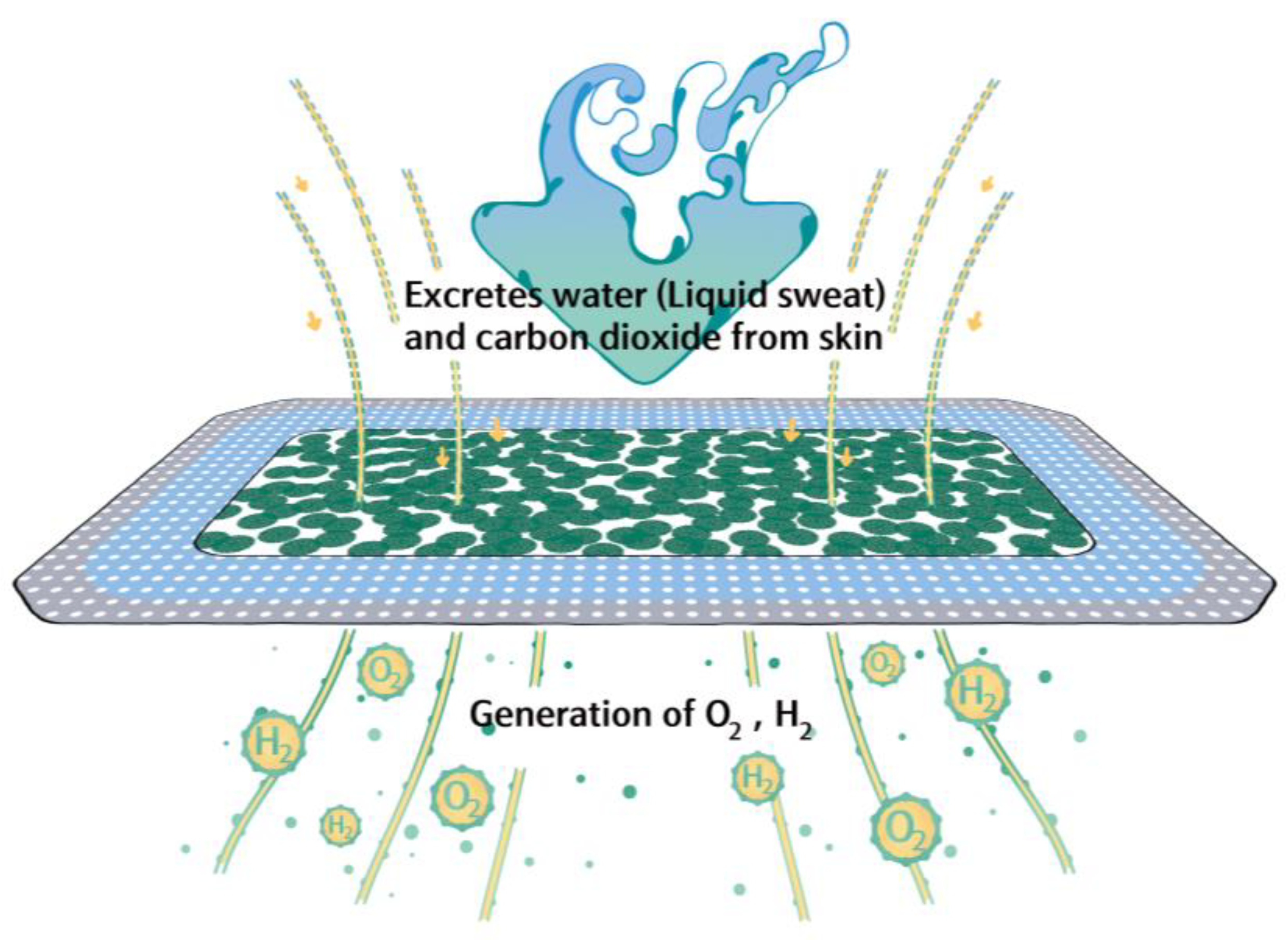
As shown in Figure 2, the H+ or O2 released through the small holes can be absorbed into the animal body through the skin in contact with the patch.

2.4. Physicochemical Evaluation of Hydrogen- and Oxygen-Generation Efficiency

The weight of the tested H+/O2 generating film (10 cm × 10 cm) was 1.23 g, which contained 0.7 g of H+/O2 generating granules. Double distilled water (5 mL) was added directly to the patch to activate the chemical reactions, and the patch was placed in a sealed 3 L container (25 ± 2 °C, 100% humidity). The sealed container was placed under a vacuum of up to −0.2 kg/cm2 using an automatic pump in a vacuum desiccator (FUJIWARA PC-3, Fujiwara, Zhejiang, China) and low-volume air sampler (XH-CYQ, Shanghai Xin Lu, Shanghai, China). To measure the release of H+ or O2 from the patch, the concentrations of H+ and O2 were measured every 10 min for 12 h using a H+/O2 m (PM80-H2, Yiyuntian, Guangdong, China) and oxygen meter (SKY2000-O2, Yiyuntian). The oxygen concertation (X) was calculated as X% × 10,000 = Y ppm, where X was the oxygen meter reading.

2.5. In Vivo Permeation Study

2.5.1. Animals

Six- to eight-week-old male ICR mice (n = 6 per group/time point) were randomly assigned to the following groups: (1) non-transdermal patch wearing (baseline); (2) patch-wearing 2 h (2 h); (3) patch-wearing 6 h (6 h); (4) patch-wearing 24 h (24 h); and (5) patch-wearing 48 h (48 h). The animals in each group were kept in separate cages.
The fur on the dorsal skin was shaved 24 h before the experiment. On the day of the experiment, the shaved skin was cleaned carefully with warm water and the patch was applied. Stretch fabric was sewn to the HOGP to secure the patches onto the animals’ skin. Simple Velcro jackets were designed and made to further secure the patches on the animals’ dorsum during the experiment (Figure 1C).
All animals were housed in a temperature (21 ± 2 °C) and humidity (55% ± 5%) controlled room with a 12/12 h light/dark cycle, with food and water provided ad libitum. The body weight, food and water consumption of each mouse were recorded weekly. All animal studies were performed under the approval of National Yang Ming Chaio Tung University, Taipei, Taiwan (IAUIC number: 1091203).

2.5.2. Blood Gas Analysis

Blood samples (0.2 mL) were collected from the adult animals at 0, 2, 6, 24, and 48 h post-application of the transdermal patch into heparinized 1-mL syringes. Pre-samples of 0.2 mL were collected, then re-injected after the blood gas analysis sample was taken. Finally, the catheter was flushed with heparinized saline (0.1 mL). The collected blood samples were analyzed within 15 min of collection to prevent equilibration of the blood in the syringe with room air. No more than two blood samples were taken from a mouse on a single day.
The pH, PCO2, and PO2 were measured directly, and arterial oxyhemoglobin saturation (SaO2) was calculated by assuming a standard oxyhemoglobin dissociation curve. The blood gas analysis unit (OPTI Critical Care analyzer, OPTIMedical, Roswell, GA, USA) was calibrated daily via two-point calibration, and hourly by single-point calibration. Blood samples and presamples for animals were 0.12 mL each and were analyzed and handled similarly to the adult samples.

2.5.3. Stress Hormone and Inflammation Assays

The patches were sewn to stretch fabric to create a jacket for the experimental mice, in order to secure the patches to the skin. Therefore, we tested whether this stretch fabric induced stress or stress-induced inflammation since mice are sensitive to restraint and odors. ELISA kits for corticosterone (Arbor Assays Company K014-H1; Michigan, Ann Arbor, MI, USA) and IL-6 (Abcam, ab100712; Cambridge, UK), were used to quantify the serum levels of the stress hormone and inflammation marker, according to the instructions provided by the manufacturers.
Briefly, 50 μL of standards or samples were added in duplicate to the wells of the micro-titer plate, 75 μL of assay buffer was added to the non-specific binding (NSB) wells, and 50 μL assay buffer was added to the wells to act as maximum binding wells. Then, 25 μL of DetectX Corticosterone Conjugate and 25 μL of DetectX Corticosterone Antibody (except the NSB wells) were added to each well and the titer plate was shaken for 1 h at room temperature. After the plate was washed using wash solution and dried by tapping the plate onto a paper towel, 100 μL of TMB Substrate was added to each well and incubated for 30 min at room temperature. The optical density (O.D.) values were read at 450 nm using a plate reader within 15 min of terminating the reaction by adding 50 μL of Stop Solution. The concentrations of corticosterone or IL-6 were calculated from standard curves.

2.6. Statistical Analysis

The differences between the two groups were compared using t-tests. p-values less than 0.05 (*), or less than 0.01 (**) or less than 0.005 (***) indicate significant differences. Data are expressed as mean ± standard deviation (mean ± SD).

3. Results

3.1. The Patches Continuously Release Hydrogen and Oxygen for Up to 12 h

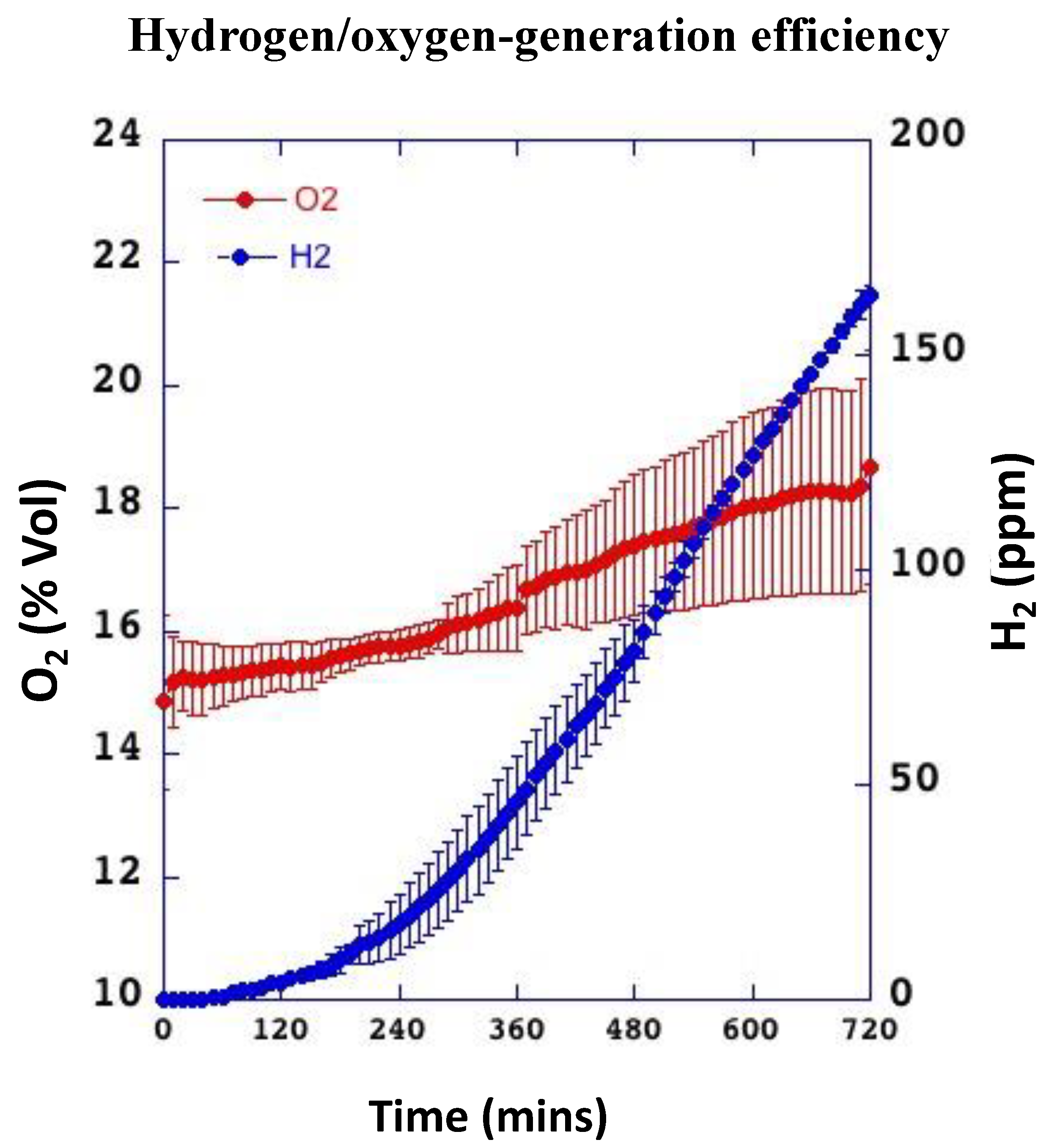
We first confirmed the H+ and O2 generating ability of the gel beads by activating the beads using double distilled water and monitoring the concentrations of dissolved H+ or O2 using H+ or O2 m. Over the 12-h study period, the dissolved oxygen concentration increased gradually from 0 to over 49,200 ppm (Figure 3; red dots). The oxygen release rate was 68.3 ppm/min and the amount of oxygen generated in the 12-h test period was 49,200 ppm.
H+ and O2 started to be released at 50 min and gradually increased from ~0 to 165.6 ppm at 12 h, suggesting that conversion of aluminum powder into stable compounds containing calcium led to the generation of H+ and O2 (Figure 3; blue dots). The H+ and O2 generation rate was 0.23 ppm/min and the amount of H+ and O2 generated in the 12-h test period was 165.6 ppm. The oxygen and H+ and O2 curves continued to increase upwards at 12 h, which indicates the H+/O2 generating granules were still generating gas.
The results of the H+ and O2 concentration monitoring experiment were used to define the operational lifetime of the patch model and establish the experimental parameters used to assess the biological effectiveness of the patches in vivo.

3.2. Wearing the Patch Significantly Increases Blood H+ and O2 Levels In Vivo

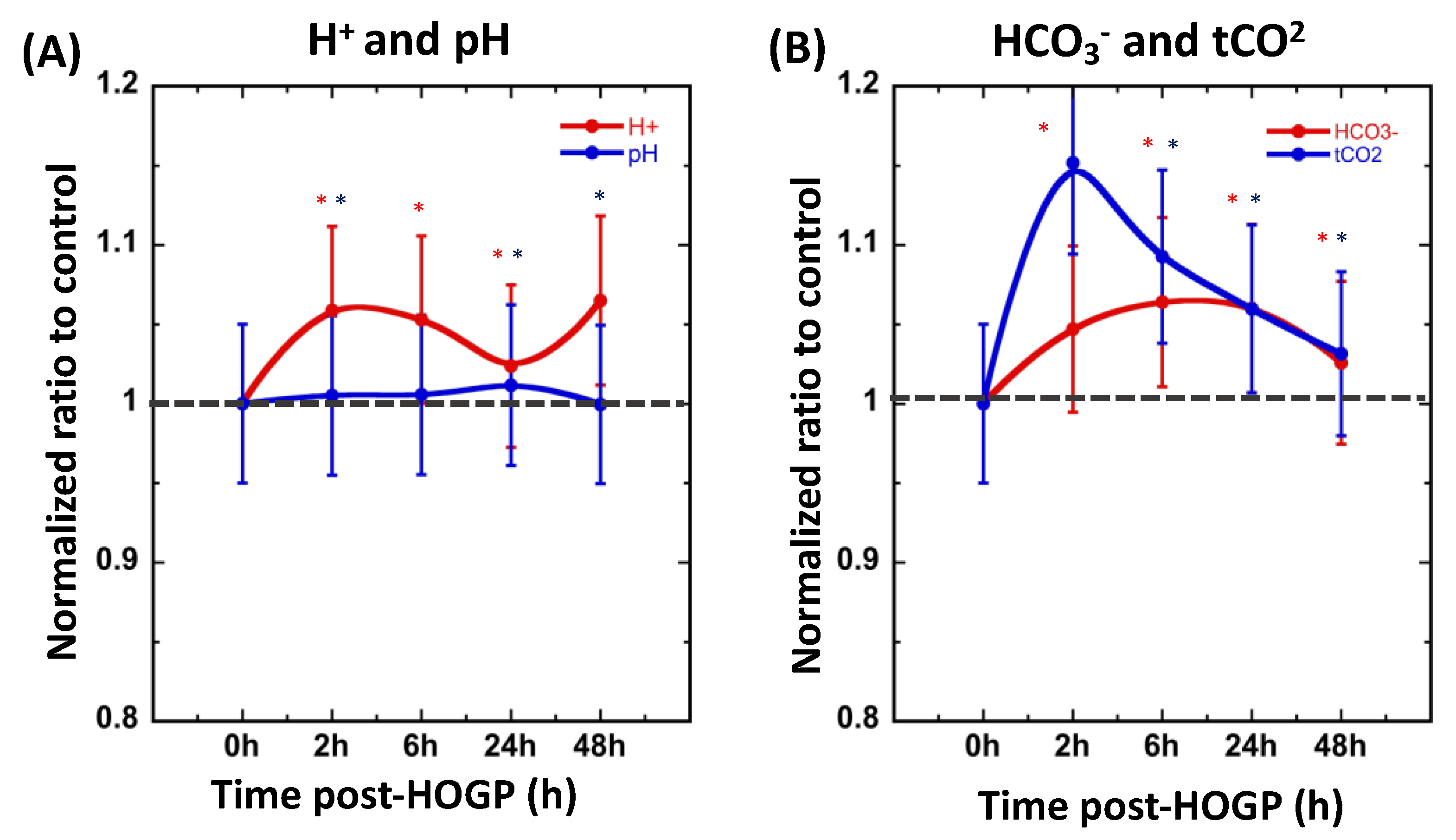
Blood H+ peaked at 2 h after application of the patch, and progressively decreased to ~1.05-fold (5% difference) until the end of study at 48 h (Figure 4A; red line), whereas the blood pH values slightly changed over time (Figure 4A; blue line). As shown in Figure 4B, the concretions of bicarbonate (HCO3) and total carbon dioxide (tCO2) exhibited similar patterns as the levels of blood H+.
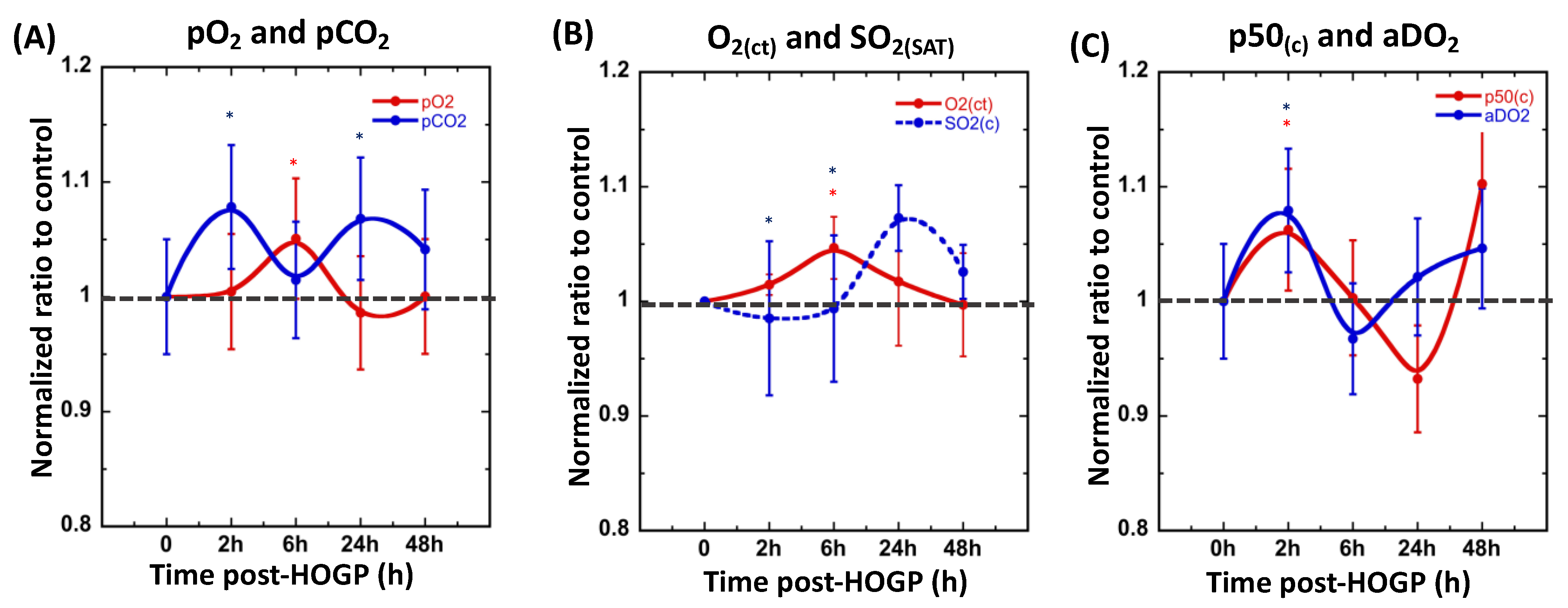
The partial pressure of oxygen (pO2) started to increase at 2 h after application of the patch, reached pO2-max at post 6 h (p < 0.05 compared to baseline), and then declined between 24 and 48 h (Figure 5A; red line). The partial pressure of oxygen (pCO2; relative to blood H+) significantly increased at 2 h after application of the patch (p < 0.05 compared to baseline) and then slightly reduced and eventually remained at ~1.05-fold (5% difference) up to the end of the 48-h study period (Figure 5A; blue line). The mean plasma concentration versus time profiles of the oxygen content (O2(ct)) and oxygen saturation (SO2(SAT)) showed similar patterns as the levels of pO2 after application of the transdermal patch (Figure 5B). Moreover, hemoglobin-oxygen affinity (p50(c)) and the alveolar-arterial oxygen gradient (aDO2) also increased at 2 h after application of the patch (p < 0.05 compared to baseline) and then gradually returned to baseline (Figure 5C).
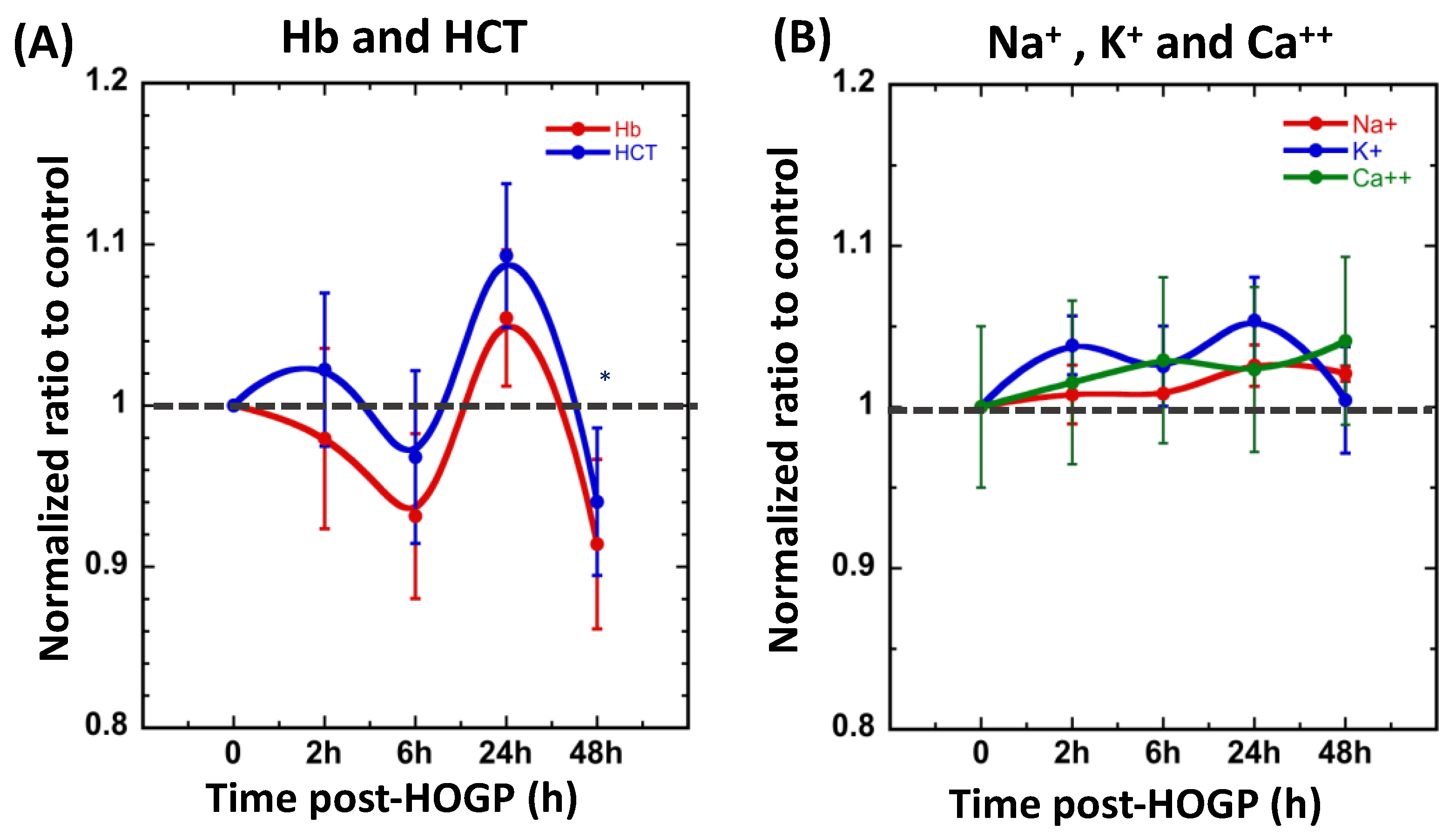
Other biological parameters, such as hemoglobin (Hb), hematocrit (HCT), sodium (Na+), potassium (K+), and calcium (Ca++) were also measured; however, there were no significant differences in these parameters after application of the patch compared to baseline (Figure 6A,B).

3.3. Effect of Acute Restraint Stress While Wearing Stretch Fabric Patch

The serum concentrations of corticosterone gradually increased at 2 h after application of the patch, peaked at 6 h, and then reduced at 24 h, whereas the levels of IL-6 remained constant over time (Figure 7).

4. Discussion

We report the creation of HOGP that continuously produces H+ and O2, which can effectively penetrate the skin. Due to the controllable chemical reactions within the H+ and O2-generating granules in a moist environment, the gases produced can be absorbed into and permeate the skin tissue, which may further improve H+ and O2 penetration to stimulate aerobic metabolism and angiogenesis within hypoxic tissues [38].
The H+ and O2 generating film contain H+ and O2 generating granules with a diameter of 18~38 μm; the granules consume calcium peroxide (CaO2) to produce H2) and O2 in response to the moisture in sweat. A polypropylene (PP) membrane with 2~3 μm apertures is used as the lining of the patch, to ensure bidirectional permeation of clean gases. When the HOGP is affixed to the skin, the impermeable polyurethane film (PE) creates a sealed system and microenvironment between the H+ and O2 generating film and the skin (Figure 1B,C). We assume that the levels of H+ and O2 in the circulation can be sufficiently raised by the HOGP. Both topical dissolved oxygen (TDO) and topical gaseous oxygen (TGO) can penetrate hydrophilic polytetrafluoroethylene (PTFE) or polypropylene (PP) membranes, which are widely used in medicine, while dissolved H+ and O2 from a microalgae-gel patch promoted chronic wound healing [37].
Interestingly, after application of the HOGP, the sweat and moisture-activated H+ and O2 generating granules released gas, which resulted in continued, significant increases in the circulating H+ and O2 levels for up to 6 h post-administration in vivo compared to control mice. These results indicate the H+ and O2 produced by the HOGP were transdermally delivered into blood vessels.
Moreover, the application of the HOGP did not alter the levels of hemoglobin (Hb), hematocrit (HCT), sodium (Na+), potassium (K+), or calcium (Ca++). Although the HOGP was sewn to an odorless stretch fabric and applied to the animals, increased levels of stress or stress-induced inflammation markers were not observed in the experimental animals, suggesting the materials or methods employed for the in vivo evaluation were safe and acceptable to the animals.
These results imply the potential clinical utility of the HOGP. The depth of transdermal delivery of the gases—not only to the skin tissues but also to the blood circulation—implies the HOGP could possibly be employed for H+ and O2 treatment, i.e., an easy-to-handle, long-lasting, moisture-activated face mask could be used to increase blood oxygen levels when wearing face masks for a long time.
Chandra et al. (2015) designed a wound dressing that produces oxygen in situ, using particulate oxygen generators that consist of a heterogeneous mixture of sodium percarbonate (SPO) and calcium peroxide (CPO). The efficacy of this dressing was tested in a porcine full-thickness surgical wound model and led to better wound closure and faster re-epithelialization [39]. Chen et al. (2020) demonstrated a novel oxygen-producing patch made of a hydrogel of living microalgae, which can produce dissolved oxygen. The authors showed that the algae-gel wound dressing increased wound oxygenation, fibroblast proliferation, and angiogenesis [37]. In terms of H+ generating patches, Safonov et al. (2020) reported a hydrogen-generating patch, which was activated by adding water to a mixture of aluminum and calcium hydroxide powders to promote the chemical release of hydrogen. The authors showed that the presence of molecular hydrogen increased cell viability, expression of collagen, and cell migration [40].
With respect to our current product, the convenient, safe features, and simultaneous H+ and O2-generating property of the HOGP may provide a new strategy for conventional delivery of H+ or O2 to in vitro or in vivo models of conditions, such as diabetic foot ulcers or burn injuries [40,41]; however, further studies are required to fully assess the benefit of the HOGP.
However, topical gaseous oxygen (TGO) therapy is hampered by the limited penetration of external gases in tissues [20,42]. In practice, TGO therapies have been shown to be effective for some cases of delayed wound healing [43], but do not always lead to significant improvements, owing to their limited efficacy [44,45]. Moreover, TGO treatment only temporarily elevates oxygen levels for 1 to 2 h, and oxygen levels return to baseline within several minutes once the patient is removed from the chamber [42].
In addition, the manufacture of the HOGP is simple and low-cost; the cost of materials to produce a 10 × 10 cm2 HOGP in our laboratory is less than US$ 0.5. The establishment of a simulation-based approach may also lead to the future design of individualized HOGP customized to each patient’s wound area and geometry.

5. Limitations

According to the study design, each subject would ideally have blood drawn five times, at each time point, to enable self-comparison of blood gas parameters relative to baseline. No more than 0.2 mL of blood can be drawn from a mouse on any single day; however, at least 0.2 mL of blood was required for the blood gas or ELISA tests in the current study. Thus, the same animals could not be assessed more repeatedly during the study period (five time points within 48 h), which increases the risk of unavoidable individual effects on the standard deviation.

6. Conclusions

We demonstrate the potential feasibility of a novel concept for highly reliable, natural delivery of H+ and O2 into tissues. The tissue-adhesive HOGP has stable storage properties and is easy to fabricate and handle. Furthermore, we confirmed that cutaneous uptake of moisture-induced H+ and O2 produced by the HOGP significantly altered the circulating levels of H+ and O2 in a small animal model.

Author Contributions

Conceptualization, S.H.-H.Y. and W.-T.H., Investigation, T.-H.Y., W.-H.C., B.-Y.W. and Y.-Y.K., Writing and editing, S.H.-H.Y., Funding Acquisition, W.-T.H., M.-H.L., and S.H.-H.Y. All authors have read and agreed to the published version of the manuscript.

Funding

Funding for this study was provided by TO2M corporation and The Featured Areas Research Center Program within the framework of the Higher Education Sprout Project by the Ministry of Education (MOE) of Taiwan.

Institutional Review Board Statement

All animal handling procedures were approved by National Defense Medical Center (IAUIC No 1091203) and this animal study was performed according to the Guidelines for Animal Experimentation of National Defense Medical Center.

Informed Consent Statement

Not applicable.

Data Availability Statement

The datasets generated during and/or analyzed during the current study are available from the corresponding author on reasonable request.

Acknowledgments

We thank the Molecular Imaging Facility Small Animal 7T PET/MR and Brain Research Center at National Yang Ming Chaio Tung University for technical support.

Conflicts of Interest

The authors declare no conflict of interest.

References

  1. Ohta, S. Molecular hydrogen as a novel antioxidant: Overview of the advantages of hydrogen for medical applications. Methods Enzymol. 2015, 555, 289–317. [Google Scholar] [CrossRef]
  2. Sim, M.; Kim, C.S.; Shon, W.J.; Lee, Y.K.; Choi, E.Y.; Shin, D.M. Hydrogen-rich water reduces inflammatory responses and prevents apoptosis of peripheral blood cells in healthy adults: A randomized, double-blind, controlled trial. Sci. Rep. 2020, 10, 12130. [Google Scholar] [CrossRef] [PubMed]
  3. Sivarajah, A.; Collino, M.; Yasin, M.; Benetti, E.; Gallicchio, M.; Mazzon, E.; Cuzzocrea, S.; Fantozzi, R.; Thiemermann, C. Anti-apoptotic and anti-inflammatory effects of hydrogen sulfide in a rat model of regional myocardial I/R. Shock 2009, 31, 267–274. [Google Scholar] [CrossRef] [PubMed]
  4. Bienert, G.P.; Moller, A.L.; Kristiansen, K.A.; Schulz, A.; Moller, I.M.; Schjoerring, J.K.; Jahn, T.P. Specific aquaporins facilitate the diffusion of hydrogen peroxide across membranes. J. Biol. Chem. 2007, 282, 1183–1192. [Google Scholar] [CrossRef] [PubMed] [Green Version]
  5. Huang, L.; Lenahan, C.; Boling, W.; Tang, J.; Zhang, J.H. Molecular Hydrogen Application in Stroke: Bench to Bedside. Curr. Pharm. Des. 2021, 27, 703–712. [Google Scholar] [CrossRef]
  6. Kajiyama, S.; Hasegawa, G.; Asano, M.; Hosoda, H.; Fukui, M.; Nakamura, N.; Kitawaki, J.; Imai, S.; Nakano, K.; Ohta, M.; et al. Supplementation of hydrogen-rich water improves lipid and glucose metabolism in patients with type 2 diabetes or impaired glucose tolerance. Nutr. Res. 2008, 28, 137–143. [Google Scholar] [CrossRef] [PubMed]
  7. Song, G.; Zong, C.; Zhang, Z.; Yu, Y.; Yao, S.; Jiao, P.; Tian, H.; Zhai, L.; Zhao, H.; Tian, S.; et al. Molecular hydrogen stabilizes atherosclerotic plaque in low-density lipoprotein receptor-knockout mice. Free Radic. Biol. Med. 2015, 87, 58–68. [Google Scholar] [CrossRef]
  8. Yoritaka, A.; Ohtsuka, C.; Maeda, T.; Hirayama, M.; Abe, T.; Watanabe, H.; Saiki, H.; Oyama, G.; Fukae, J.; Shimo, Y.; et al. Randomized, double-blind, multicenter trial of hydrogen water for Parkinson’s disease. Mov. Disord. 2018, 33, 1505–1507. [Google Scholar] [CrossRef]
  9. Zhai, X.; Chen, X.; Ohta, S.; Sun, X. Review and prospect of the biomedical effects of hydrogen. Med. Gas. Res. 2014, 4, 19. [Google Scholar] [CrossRef] [Green Version]
  10. LeBaron, T.W.; Laher, I.; Kura, B.; Slezak, J. Hydrogen gas: From clinical medicine to an emerging ergogenic molecule for sports athletes (1). Can. J. Physiol. Pharmacol. 2019, 97, 797–807. [Google Scholar] [CrossRef]
  11. Guan, P.; Sun, Z.M.; Luo, L.F.; Zhou, J.; Yang, S.; Zhao, Y.S.; Yu, F.Y.; An, J.R.; Wang, N.; Ji, E.S. Hydrogen protects against chronic intermittent hypoxia induced renal dysfunction by promoting autophagy and alleviating apoptosis. Life Sci. 2019, 225, 46–54. [Google Scholar] [CrossRef] [PubMed]
  12. Zhang, N.; Deng, C.; Zhang, X.; Zhang, J.; Bai, C. Inhalation of hydrogen gas attenuates airway inflammation and oxidative stress in allergic asthmatic mice. Asthma Res. Pract. 2018, 4, 3. [Google Scholar] [CrossRef] [PubMed]
  13. Wang, S.T.; Bao, C.; He, Y.; Tian, X.; Yang, Y.; Zhang, T.; Xu, K.F. Hydrogen gas (XEN) inhalation ameliorates airway inflammation in asthma and COPD patients. QJM 2020, 113, 870–875. [Google Scholar] [CrossRef]
  14. Yuichi Sugai, Y.O.; Kyuro, S. Simulation Study on Reservoir Souring Induced by Injection of Reservoir Brine Containing Sulfate-Reducing Bacteria. Sustainability 2020, 12, 4603. [Google Scholar] [CrossRef]
  15. Hayashida, K.; Sano, M.; Kamimura, N.; Yokota, T.; Suzuki, M.; Ohta, S.; Fukuda, K.; Hori, S. Hydrogen inhalation during normoxic resuscitation improves neurological outcome in a rat model of cardiac arrest independently of targeted temperature management. Circulation 2014, 130, 2173–2180. [Google Scholar] [CrossRef] [PubMed] [Green Version]
  16. Nie, C.; Ding, X.; Rong, A.; Zheng, M.; Li, Z.; Pan, S.; Yang, W. Hydrogen gas inhalation alleviates myocardial ischemia-reperfusion injury by the inhibition of oxidative stress and NLRP3-mediated pyroptosis in rats. Life Sci. 2021, 272, 119248. [Google Scholar] [CrossRef]
  17. Tamura, T.; Hayashida, K.; Sano, M.; Onuki, S.; Suzuki, M. Efficacy of inhaled HYdrogen on neurological outcome following BRain Ischemia During post-cardiac arrest care (HYBRID II trial): Study protocol for a randomized controlled trial. Trials 2017, 18, 488. [Google Scholar] [CrossRef] [Green Version]
  18. Korovljev, D.; Stajer, V.; Ostojic, J.; LeBaron, T.W.; Ostojic, S.M. Hydrogen-rich water reduces liver fat accumulation and improves liver enzyme profiles in patients with non-alcoholic fatty liver disease: A randomized controlled pilot trial. Clin. Res. Hepatol. Gastroenterol. 2019, 43, 688–693. [Google Scholar] [CrossRef]
  19. Yoon, Y.S.; Sajo, M.E.; Ignacio, R.M.; Kim, S.K.; Kim, C.S.; Lee, K.J. Positive Effects of hydrogen water on 2,4-dinitrochlorobenzene-induced atopic dermatitis in NC/Nga mice. Biol. Pharm. Bull. 2014, 37, 1480–1485. [Google Scholar] [CrossRef] [Green Version]
  20. Opasanon, S.; Pongsapich, W.; Taweepraditpol, S.; Suktitipat, B.; Chuangsuwanich, A. Clinical Effectiveness of Hyperbaric Oxygen Therapy in Complex Wounds. J. Am. Coll. Clin. Wound Spec. 2014, 6, 9–13. [Google Scholar] [CrossRef] [Green Version]
  21. Runtuwene, J.; Amitani, H.; Amitani, M.; Asakawa, A.; Cheng, K.C.; Inui, A. Hydrogen-water enhances 5-fluorouracil-induced inhibition of colon cancer. PeerJ 2015, 3, e859. [Google Scholar] [CrossRef] [PubMed] [Green Version]
  22. Kang, K.M.; Kang, Y.N.; Choi, I.B.; Gu, Y.; Kawamura, T.; Toyoda, Y.; Nakao, A. Effects of drinking hydrogen-rich water on the quality of life of patients treated with radiotherapy for liver tumors. Med. Gas. Res. 2011, 1, 11. [Google Scholar] [CrossRef] [Green Version]
  23. Li, S.; Liao, R.; Sheng, X.; Luo, X.; Zhang, X.; Wen, X.; Zhou, J.; Peng, K. Hydrogen Gas in Cancer Treatment. Front. Oncol. 2019, 9, 696. [Google Scholar] [CrossRef] [PubMed]
  24. Zhang, J.Y.; Liu, C.; Zhou, L.; Qu, K.; Wang, R.; Tai, M.H.; Lei Lei, J.C.; Wu, Q.F.; Wang, Z.X. A review of hydrogen as a new medical therapy. Hepatogastroenterology 2012, 59, 1026–1032. [Google Scholar] [CrossRef] [PubMed]
  25. Ohta, S. Molecular hydrogen as a preventive and therapeutic medical gas: Initiation, development and potential of hydrogen medicine. Pharmacol. Ther. 2014, 144, 1–11. [Google Scholar] [CrossRef] [Green Version]
  26. Ohta, S. Recent progress toward hydrogen medicine: Potential of molecular hydrogen for preventive and therapeutic applications. Curr. Pharm. Des. 2011, 17, 2241–2252. [Google Scholar] [CrossRef] [PubMed] [Green Version]
  27. Dixon, B.J.; Tang, J.; Zhang, J.H. The evolution of molecular hydrogen: A noteworthy potential therapy with clinical significance. Med. Gas. Res. 2013, 3, 10. [Google Scholar] [CrossRef] [Green Version]
  28. Zuttel, A. Hydrogen storage methods. Naturwissenschaften 2004, 91, 157–172. [Google Scholar] [CrossRef]
  29. Ueno, C.; Hunt, T.K.; Hopf, H.W. Using physiology to improve surgical wound outcomes. Plast. Reconstr. Surg. 2006, 117, 59S–71S. [Google Scholar] [CrossRef]
  30. Trabold, O.; Wagner, S.; Wicke, C.; Scheuenstuhl, H.; Hussain, M.Z.; Rosen, N.; Seremetiev, A.; Becker, H.D.; Hunt, T.K. Lactate and oxygen constitute a fundamental regulatory mechanism in wound healing. Wound Repair Regen. 2003, 11, 504–509. [Google Scholar] [CrossRef]
  31. Hunt, T.K.; Ellison, E.C.; Sen, C.K. Oxygen: At the foundation of wound healing—Introduction. World J. Surg. 2004, 28, 291–293. [Google Scholar] [CrossRef] [PubMed]
  32. Camci-Unal, G.; Alemdar, N.; Annabi, N.; Khademhosseini, A. Oxygen Releasing Biomaterials for Tissue Engineering. Polym. Int. 2013, 62, 843–848. [Google Scholar] [CrossRef] [PubMed] [Green Version]
  33. Pedraza, E.; Coronel, M.M.; Fraker, C.A.; Ricordi, C.; Stabler, C.L. Preventing hypoxia-induced cell death in beta cells and islets via hydrolytically activated, oxygen-generating biomaterials. Proc. Natl. Acad. Sci. USA 2012, 109, 4245–4250. [Google Scholar] [CrossRef] [Green Version]
  34. Akhtar, N.; Singh, V.; Yusuf, M.; Khan, R.A. Non-invasive drug delivery technology: Development and current status of transdermal drug delivery devices, techniques and biomedical applications. Biomed. Technol. 2020, 65, 243–272. [Google Scholar] [CrossRef] [Green Version]
  35. Ramadan, E.; Borg, T.; Abdelghani, G.M.; Saleh, N.M. Design and in vivo pharmacokinetic study of a newly developed lamivudine transdermal patch. Future J. Pharm. Sci. 2018, 4, 166–174. [Google Scholar] [CrossRef]
  36. Dong, S.; Zhu, P.; Zhang, S. Expression of collagen type 1 alpha 1 indicates lymph node metastasis and poor outcomes in squamous cell carcinomas of the lung. PeerJ 2020, 8, e10089. [Google Scholar] [CrossRef] [PubMed]
  37. Chen, H.; Cheng, Y.; Tian, J.; Yang, P.; Zhang, X.; Chen, Y.; Hu, Y.; Wu, J. Dissolved oxygen from microalgae-gel patch promotes chronic wound healing in diabetes. Sci. Adv. 2020, 6, eaba4311. [Google Scholar] [CrossRef] [PubMed]
  38. Winter, G.D. Formation of the scab and the rate of epithelization of superficial wounds in the skin of the young domestic pig. Nature 1962, 193, 293–294. [Google Scholar] [CrossRef]
  39. Chandra, P.K.; Ross, C.L.; Smith, L.C.; Jeong, S.S.; Kim, J.; Yoo, J.J.; Harrison, B.S. Peroxide-based oxygen generating topical wound dressing for enhancing healing of dermal wounds. Wound Repair Regen. 2015, 23, 830–841. [Google Scholar] [CrossRef]
  40. Marina Safonov, J.Y.; Jihyung, L.; Vladimir, L.; Safonov, D.B.; Donghui, Z. Hydrogen generating patch improves skin cell viability, migration activity, and collagen expression. Eng. Regen. 2020, 1, 1–5. [Google Scholar] [CrossRef]
  41. Chen, Y.; Zong, C.; Guo, Y.; Tian, L. Hydrogen-rich saline may be an effective and specific novel treatment for osteoradionecrosis of the jaw. Ther. Clin. Risk Manag. 2015, 11, 1581–1585. [Google Scholar] [CrossRef] [PubMed] [Green Version]
  42. Heyboer, M., 3rd; Sharma, D.; Santiago, W.; McCulloch, N. Hyperbaric Oxygen Therapy: Side Effects Defined and Quantified. Adv. Wound Care 2017, 6, 210–224. [Google Scholar] [CrossRef] [PubMed] [Green Version]
  43. Hopf, H.W.; Gibson, J.J.; Angeles, A.P.; Constant, J.S.; Feng, J.J.; Rollins, M.D.; Zamirul Hussain, M.; Hunt, T.K. Hyperoxia and angiogenesis. Wound Repair Regen. 2005, 13, 558–564. [Google Scholar] [CrossRef]
  44. Hodges, A.N.; Delaney, S.; Lecomte, J.M.; Lacroix, V.J.; Montgomery, D.L. Effect of hyperbaric oxygen on oxygen uptake and measurements in the blood and tissues in a normobaric environment. Br. J. Sports Med. 2003, 37, 516–520. [Google Scholar] [CrossRef] [PubMed]
  45. Roe, D.F.; Gibbins, B.L.; Ladizinsky, D.A. Topical dissolved oxygen penetrates skin: Model and method. J. Surg. Res. 2010, 159, e29–e36. [Google Scholar] [CrossRef] [PubMed]
Figure 1. Design of the hydrogen/oxygen-generating patch (HOGP) for the experimental animal model. (A) Schematic illustration of the HOGP. (B) Detailed images of the HOGP. (C) Schematic illustration of the HOGP applied to animals. Stretch fabric was sewn onto the HOGP to secure the patches onto the animals’ skin. Simple Velcro jackets were designed to further secure the patches onto the animals’ dorsum during the experiment.
Applsci 11 11680 g001
Figure 2. Schematic illustration of the mechanisms of hydrogen/oxygen-generating patch. Mechanisms of H+/O2 producing formulas; peroxide reacts with water to from hydroxides, and then release oxygen.
Applsci 11 11680 g002
Figure 3. Hydrogen/oxygen-generation efficiency of the water-activated HOGP. H+ and O2 concentrations were measured in sealed and vacuum containers as a function of time starting immediately after adding water to the powder and sealing the vessel. The stable H+ and O2 concentration was 0.23 ppm/min and the oxygen release rate was 68.3 ppm/min over a 12-h period.
Applsci 11 11680 g003
Figure 4. Time course of the blood levels of hydrogen, pH, bicarbonate (HCO3), and total carbon dioxide (tCO2) post-application of the HOGP. Normalized ratios of H+ and pH (A), and HCO3 and tCO2 (B). The normalized ratios (y-axis) were determined by normalization to the control group. Significant differences were identified using t-tests (* p < 0.05).
Applsci 11 11680 g004
Figure 5. Time course of biological parameters post-application of the HOGP. Normalized ratios of pO2 and pCO2 (A), O2 and SO2 (B), and p50(c) and aDO2 (C). The normalized ratios (y-axis) were determined by normalization to the control group. Significant differences were identified using t-tests (* p < 0.05).
Applsci 11 11680 g005
Figure 6. Normalized hematological ratios and electrolyte parameters post-application of the HOGP. Normalized Hb and HCT ratio (A), and the levels of Na+, K+ and Ca++ (B). The normalized ratios (y-axis) were determined after normalization to the control group. Significant differences were identified using t-tests (* p < 0.05).
Applsci 11 11680 g006
Figure 7. Concentrations of serum cortisol and IL-6 in mice (n = 6) at different time-points post-application of the HOGP. Significant differences were identified using t-tests (p < 0.05).
Applsci 11 11680 g007
Publisher’s Note: MDPI stays neutral with regard to jurisdictional claims in published maps and institutional affiliations.