1. Introduction
Magnetic resonance imaging (MRI) systems integrate a static magnetic field (B
0, forcing protons in the body to align with it), a time-varying magnetic field at kHz (the gradient field, encoding the MR signal spatially), and a radio-frequency (RF) magnetic field (B
1, stimulating and receiving MR signals). MRI is a modern medical technology with portraits of non-invasive scanning, real-time imaging, along with functional diagnoses. Such advanced medical instruments do not merely ease patients’ agitation from visualized medical conditions, but also make doctors explicitly work through the lesions. Diffusion MRI (dMRI) pushes a further and closer dimension to the scale of neural fibers through sensitizing the gradient field during the MR pulse sequence imaging so as to allow water molecular displacement over distances of 1~20 μm along fibers to be recognized. Such specialty renders dMRI widely applicable in uses from fiber-related clinical disease studies to cognitive-based neuroscience research [
1]. As an essential tool of investigating connectivity and a see-through interface to navigate the major brain pathway, dMRI provides the opportunity to resolve those diseases associated with mental or neural issues through
in vivo visible fiber tracking and circuit exploring, an in-trend global application of which is the human connectome project (HCP) [
2]. To achieve precise fiber tracks or tissue differentials at the level of the cell groups and to even push to axon-level, the resolution of brain imaging requires up to a one-millimeter and even submillimeter-scale, which is the future of precise and personalized clinical medicine (e.g., white matter disease studies) [
3].
For common clinical uses, a sampling size (voxel size) of the normal imaging paradigm (MR sequence) in MRI human scans (≤3 T) of ≥1 mm in size is considered sufficient to diagnose tissue-based diseases. However, the performance of submillimeter-scale research in non-human primates (NHPs) [
4] and human
ex vivo [
5] research reveals more information of mesoscale organization in brain, which obviously is an approach for tracing the root of brain diseases. Submillimeter-scale dMRI can be achieved, but is more difficult with commonly available MRI machines (1.5 T or 3 T) than ultra-high-field MRI (UHF, 7 T and higher). As a compromise, submillimeter-scale dMRI from 3 T acquired by prevailing EPI-based (echo-planar imaging) sequence usually utilizes means of more steps, such as averages of repetition scans, calibration and smooth-phase information and further reconstruction for correcting artifacts [
6], which therefore may cost more time but obtain images of lower quality than those from UHF.
EPI-based dMRI methods (e.g., diffusion-weighted EPI, DW-EPI) are the most popular sequences for diffusion acquisition, with traits of rapid acquisition speed to reduce motion-related artifacts. The diffusion gradients could generate geometric distortion caused by susceptibility-induced off-resonance fields [
7] and then exacerbate those distortion induced by the EPI acquisition method and B
0 inhomogeneity [
8]; therefore, turbo spin echo (TSE)-based dMRI techniques were introduced, with minimal sensitivity to field inhomogeneity and the capability for high-quality (SNR, contrast, etc.), high-resolution images, albeit with long scan time, high SAR (specific absorption rate), and sensitivity to motion [
9]. A monopolar diffusion-prepared module for TSE sequence (DP-TSE) has been proposed as an alternative method to achieve distortion-free and high-resolution diffusion images with improved SNR, and the approach has been evaluated over phantom and human subjects, with promising potential in pushing for high resolutions and minimal subject motion [
10]; moreover, quantitative measures on phantoms indicated that the DP-TSE sequence was able to acquire accurate and consistent ADC values with different echo-train-lengths (ETLs). However, we also observed apparent image blurring in human brain imaging due to inevitable head motions caused by respiration, the heartbeat, etc. Therefore, for the present study, our purpose was to evaluate the sequence’s performance over anesthetized macaques with their heads fixed to stereotaxic apparatus, and through diffusion-weighted imaging of the macaque brain, we aimed to investigate whether the proposed approach was suitable for NHP brain studies at 1 mm and even finer spatial resolutions.
2. Materials and Methods
2.1. Diffusion Preparation TSE Sequence
The implemented diffusion-weighted sequence for this study was the monopolar diffusion-prepared TSE. The sequence applied a 90° tip-down RF excitation and diffusion gradients followed by a 90° tip-up RF to attain diffuse signal as a preparation module before the TSE readout acquisition. One pair of Stejskal and Tanner method diffusion gradients [
11] was in use during the preparation module to reduce the diffusion preparation time for saving the signal loss due to T
2 decay. Two spoiler gradients with high amplitude (>4π) were set before (not shown, default set) and after the diffusion preparation module to eliminate residual signals. The sequence diagram is presented in
Figure 1. In the study, we used the diffusion-weighted readout-segmented EPI (rsEPI) for comparison, which is considered as a traditional EPI-based dMRI sequence for submillimeter-level macaque brain imaging with optimized protocols [
12]. Furthermore, DW-rsEPI is similar to DP-TSE, in that both sequences are able to use parallel imaging, segmented acquisition and optimal monopolar schemes.
2.2. MR Scanner, Coil Design and Animal Preparation
The studies were performed on a Siemens 3 T PRISMA whole-body MRI scanner (Siemens Healthcare, Erlangen, Germany) equipped with a helmet-like 16-channel receive-only RF coil fixed over the stereotaxic frames, as shown in
Figure 2 (details in coil design and imaging performance can be found elsewhere [
13]). During imaging acquisition, the macaque was tightly placed in the sphinx position with its head centered within the head coil to reduce physiological motions (e.g., respiration) as much as possible.
One anesthetized macaque (female, 13 years old, 9.6 kg) was used for the imaging study. Anesthesia was maintained with 1.4% to 2% isoflurane. The body temperature was maintained at a constant temperature using a recirculating water heater, and heart rate, SpO2, and CO2 were monitored continuously. All animal handling procedures were in accordance with NIH standards and approved by our Institutional Animal Care Committee (ZJU 20190054 and 20200060).
2.3. MR Data Acquisition and Analysis
We performed full-brain macaque diffusion-weighted imaging with the proposed DP-TSE sequence and vendor’s product DW-rsEPI sequence to evaluate dMRI quality and time efficiency. The dMRI series comparisons were acquired using the following imaging parameters, and the total scan time was recorded. DP-TSE/DW-rsEPI: TE = 11/80 ms, TR = 4000/4500 ms, FOV = 128 × 128/160 × 160 mm2, matrix = 128 × 128/160 × 160, in-plane resolution = 1 × 1 mm2, slice thickness = 2 mm, 24 slices, diffusion weighting b = 0, 500, 1000, 1500, 2000 s/mm2, TSE turbo factor = 11, rsEPI segment = 7, no repetition or average scan. The diffusion weighting was applied along three orthogonal axes, defined as SS, RO and PE. During the experiment, the TR could not be identical for the 24-slice series dMRI scan due to restrictions of the DW-rsEPI protocol; meanwhile, as the b-value increased to 1500 s/mm2, DW-rsEPI suffered severe signal loss and was unable to be used for comparison. Therefore, we acquired an extra slice for ADC curve fitting with a set of b = 0, 100, 300, 600, and 900 s/mm2, TR = 1500 ms for ADC accuracy check. In addition, another DP-TSE slice only in the RO direction with the same measurement parameters and b = 500 s/mm2 were acquired with parallel-imaging acceleration (GRAPPA, iPAT 2) for comparing the efficiency of SNR.
To compare the dMRI image quality visually, the diffusion-weighted images from both sequences were not edited by filtering or distortion-correction after acquisition. The ADC was calculated using an implicit mono-exponential model as follows:
where S(
b) is the signal magnitude with diffusion weighting
b, and S
0 is the signal magnitude with no diffusion weighting (
b = 0 s/mm
2). Post-processing, including the co-registration and reconstruction of ADC maps, was performed in MATLAB.
3. Results
The raw diffusion images from orthogonal directions and ADC maps from two sequences with
b = 1000 s/mm
2 are selectively displayed in
Figure 3. The images from DP-TSE reveal higher definitions of tissue texture, and provide more details to tell the specific distinctions in different diffusion directions than those from DW-rsEPI, indicating that the TSE-based dMRI can greatly preserve authentic information while excluding blurring from phase- or distortion-correction in further post-processing. In addition to general brain shape distortion because of the EPI readout scheme, the DW-rsEPI results also suffered from artifacts from fat shifting (red arrow) caused by a wider bandwidth of the protocol setup in fat suppression.
The scan times recorded from 24 slices with b = 500, 1000, 1500, and 2000 s/mm2 are as follows: DP-TSE: 3′32”, 3′40”, 3′44”, and 3′48”; DW-rsEPI: 3′40”, 3′40” for b-value ≤ 1000 s/mm2. The data from higher b-value were discarded because of their poor image quality. The information on time usage shows that both approaches with 1 mm in-plane resolution dMRI had similar time efficiency.
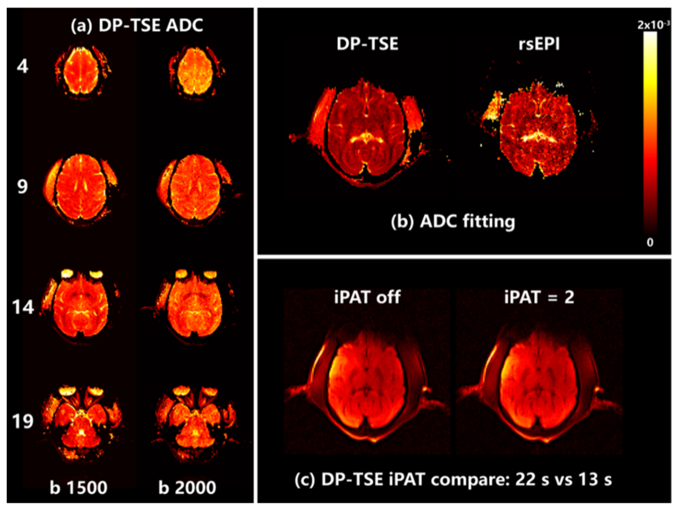
The ADC maps from DP-TSE with higher
b-values are presented in
Figure 4a, and the image from DW-rsEPI had a poor quality of ADC maps because
b > 1000 s/mm
2, which indicated that DP-TSE had the capability to provide high-quality dMRI with higher
b-values. The ADC curve fitting maps with the same TR (1500 ms) are shown in
Figure 4b. The ADC from the diffusion-prepared module are the same as those from the traditional diffusion-weighted method; moreover, DP-TSE appeared to be more adaptable to different measure parameters and more favorable to reduce the scan time, which is a major advantage of TSE-based sequence.
Figure 4c displays a slice of the diffusion image with parallel imaging acceleration (iPAT) on and off. The image qualities of iPAT 2 were almost the same as iPAT 1 (off) at
b = 500 s/mm
2, but with a significantly reduced scan time, which suggested that DP-TSE dMRI could improve the time efficiency without scarifying SNR.
4. Discussion
The present study has demonstrated an optimal TSE-based method for distortion-free, high-resolution, high-SNR and time-efficient brain dMRI over an anesthetized and well-stabilized macaque. The original and ultimate attempts of our implemented sequence were to achieve distortion-free, submillimeter-scale dMRI across species with improved SNR and time efficiency, which is heavily demanded in the early diagnosis and individualized treatment of brain diseases. We encountered some challenges during our preliminary experiments over human subjects, the most vital of which was in motion artifacts such as spontaneous head movements or respiratory motions, whereas the experiments with motion-free subjects as melons produced high-definition structures [
10]. Apart from common technique of employing EPI-based sequences with a number of average scans to enhance signals and using post-reconstructions to blur noise or correct distortions, the conventional compensation approach was to use motion-insensitive TSE-based sequences as readouts (e.g., the PROPELLER technique), although this requires scan time at least twice as high as the resolution increases. Instead of adding other redundant parts, we have proposed and implemented the DP-TSE approach for NHP brain dMRI
in vivo.
The dMRI study with an anesthetized macaque can contribute largely to mesoscale (submillimeter-scale) brain research such as white-matter-related disease. During the experiment, we used a 3D-printed stereotaxic head frame to stabilize the macaque skull and place the coil close enough to the brain, in order to eliminate motion-induced artifacts as much as possible. In addition, with TSE-based acquisition embedded, the dMRI suffered almost no extra distortion induced by the EPI readout other than the inherent inhomogeneous B0, whereas the diffusion preparation module suffered fewer high-order diffusion moments with spoilers applied. The multi-shot DW-EPI and DW-rsEPI sequences had similar advantages to address these issues by dividing EPI readouts into several segments, which have commonly been used for high-resolution dMRI and are ideal for control groups for this study; however, such fat effects caused by the wide bandwidth of RF pulses in DW-rsEPI for a shorter TE will not appear in DP-TSE instead, which therefor eliminates the signal loss by the RF inversion of fat saturation in DW-rsEPI.
In this study, we simply pushed the resolution to 1 mm in-plane and a 2 mm thickness to demonstrate the fair differences between EPI-based and TSE-based sequences without image correction at 3 T; promising results suggest that the TSE-based approach has promising potential in achieving submillimeter-scale resolution at 3 T or even higher field strengths of the MR system. Moreover, without disturbing effects from EPI, TSE-based dMRI could be expected to provide for more complex fiber geometries such as fiber crossings at the whole brain level. The results in
Figure 3 and
Figure 4 demonstrate that the implemented DP-TSE sequence is suitable for anesthetic macaque brain dMRI and is able to reveal the superiority of high image quality, especially with high
b-values, which paves the way to submillimeter dMRI study in NHPs. In additional, features such as no repetition scans and a parallel imaging capability may incite more possibilities for further research.


