Article

Gastrocnemius Activation throughout the Competitive Season in Athletes of Different Experience Levels

1
University of Coimbra, 3004-531 Coimbra, Portugal
2
Polytechnic of Coimbra, ESTeSC, 3046-854 Coimbra, Portugal
3
Department of Mechanical Engineering, University of Coimbra, CEMMPRE, 3030-788 Coimbra, Portugal
4
Polytechnic of Coimbra, ISEC, 3030-199 Coimbra, Portugal
5
Polytechnic of Coimbra, ESEC, 3030-329 Coimbra, Portugal
6
University of Coimbra, CIDAF, 3040-248 Coimbra, Portugal
*
Author to whom correspondence should be addressed.
Appl. Sci. 2021, 11(3), 984; https://doi.org/10.3390/app11030984
Submission received: 23 December 2020 / Revised: 13 January 2021 / Accepted: 18 January 2021 / Published: 22 January 2021

Abstract

:
Basketball is a team sport in which the players undergo rapid reactions, and running efforts are usually carried out in all directions. The main goal of the present study was to evaluate the muscle activity response in different basketball movements, considering two types of athletes with different levels of experience. Using a group of nine volunteers, female athletes from a senior basketball team, a data acquisition protocol was defined to identify the changes that occurred throughout the sports season. In this study, to assess the gastrocnemius muscle activity, intensity peak (%MC) and time to peak (TP) were evaluated for five typical basketball skills and movements involving running and jumping with and without the ball. Seven repetitions were executed for each movement, performed at two different time-points of the sports season: at the beginning of the teams’ competition, in November (T1), and at the season peak, in March (T2). TP presents greater values for T2 of the season when compared with T1. The results show that the level of experience of the athletes has a significant effect on the results of TP and %MC, particularly when comparing the two seasons.

1. Introduction

Regular sports activity improves skeletal muscle function and cardiovascular health, as well as prevents and reverses obesity, reduces anxiety and depression, and improves self-confidence and cognitive function [1,2]. Basketball involves frequent jumping, accelerating, decelerating, turning, and pivoting [3]. It is a team game during which players experience up to 105 high-intensity activities of short duration (2–6 s), occurring on average every 21 s. This suggests that, for basketball players, the ability to repeat high-intensity efforts such as sprints with and without a ball is very important [4]. Running efforts in basketball are usually carried out in all directions, with short and intense actions, with long and moderate activity and recovery, which can induce injuries [5,6,7]. Running while dribbling a ball is a fundamental basketball skill, and better players can distinguish themselves by experiencing less loss of contact with the ball while running [8]. A higher number of people participating in such a physically demanding sport can result in an increased risk of injury [9]. In recent years, the number of basketball players has increased, as well as the likelihood of injuries. To avoid the incidence of injuries, the use of strategies that include pre-season conditioning, functional training, and strength and balance programs should be considered, which must be continued and adjusted throughout the game season [10,11]. Several authors have evaluated the probability of injuries during basketball games [5,12,13,14]. The most common injuries that occur in basketball players are in lower extremities [5,15], posing a major risk for the ankle and knee [16]. According to Klyne et al. [17], gastrocnemius is one of three primary muscles spanning the ankle and the knee joint, which is why it becomes important to know how these muscles respond during a sports activity. Greater gastrocnemius forces during landing are associated with an increase in joint stiffness, protecting the joint from external loading and injury risk [18]. Various authors [19,20,21] have highlighted that ankle injuries are the most common injuries in basketball players because it is a sport that combines running and jumping. Longo et al. [22] proposed a program to prevent injuries in elite male players’ basketball. There are a lot of movements during a basketball game, like shots, free-throws, passes, dribbling, and rebounding. Recovering the ball after a shot, usually called rebound, is of particular interest since almost half of all basketball-related foot and ankle injuries occur during this movement [19]. As well as this, the effect of cutting promotes ankle inversion sprains, which represent the most common type of ankle injury in basketball [12,23]. Vieira et al. [24] performed a study in which they showed that a substantial medial gastrocnemius torque contribution tended to rotate up the medial aspect of the foot (ankle inversion). Although there are different methods for measuring muscle activation, such as visible light and infrared, the golden standard is electromyography [25]. In basketball players, surface electromyography (sEMG) is commonly used to measure the bioelectrical activity of muscles, especially for the upper limbs, during free throws. sEMG can contribute to the diagnosis of injuries because it makes it possible to obtain an electrical signal from a muscle during a body movement, and the degree of muscular activity shows the muscular level effort [26]. Henry et al. [27] used sEMG to assess gastrocnemius activity and verified a consistent medial gastrocnemius response to surface disturbances, which induces ankle flexion-inversion movements. Sanderson et al. [28] conducted a study using sEMG to assess the gastrocnemius response during a pedaling cadence and observed that the greatest changes in gastrocnemius length occurred during the recovery phase, when the muscles showed little excitement. The free throw is the principal activity evaluated by sEMG in basketball players to verify the relationship between the accuracy and the muscle activity [29,30,31]. SEMG is also used to evaluate the flexor carpi radialis (forearm) and the triceps brachii (upper arm) in the case of dribbling during basketball [32]. sEMG is also used in wheelchair basketball players to estimate the muscle activity in upper limbs, in the elbow flexor muscles, namely, biceps brachii longus, biceps brachii brevis, and brachioradialis [33,34]. During sEMG, the maximum muscle activation is used to identify the muscle contraction, which allows for the normalization of the signal [35]. sEMG is frequently used to evaluate the level of activation of core muscles during suspension exercises [36]. The knowledge of muscle activity during sports practice can help coaches to define appropriate exercises in different stages of the season.
Some studies have reported common injuries in basketball players. However, the literature does not present any work about the use of sEMG in evaluating the gastrocnemius activity during different basketball movements, considering different sports season times and athletes with two different experience levels. Therefore, this work intends to study the influence of different movements, season periods, and athletes’ levels on the gastrocnemius activity and contribute to developing knowledge that helps coaches and sport professional staff develop a training plan to avoid the occurrence of injuries.

2. Materials and Methods
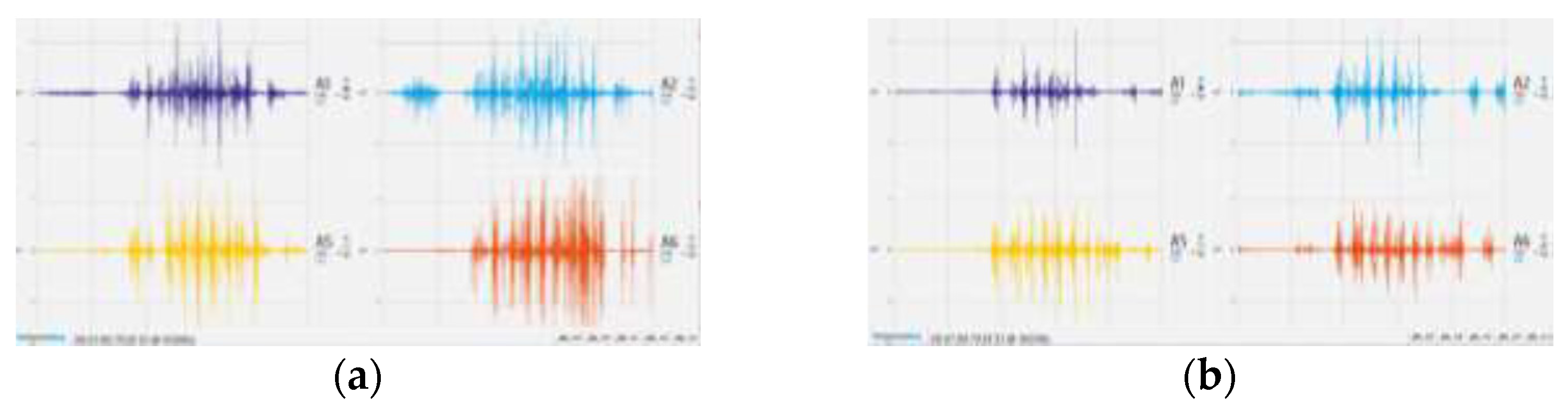
This study considered a group of volunteers, none of whom were professional female basketball athletes, divided into level 1 and level 2 according to their experience, which was defined based on the number of games played and whether the athlete belonged to a regional basketball elite team or not. Initially, the group was formed by 11 athletes; however, during the season, the group was reduced to 9 due to injury and scholarly reasons.
This study was approved by the local ethics committee and was performed according to the Declaration of Helsinki, with all the participants’ data considered and stored anonymously and confidentially. All subjects were fully informed of the nature of the study and gave written informed consent. Table 1 shows the sample characteristics and their experience level.
The data acquisitions were performed at two time-points of the sports season, the first one in November (T1), corresponding to the beginning of the season, and the second one in March (T2), corresponding to the peak of the season. Five different movements were considered, namely, running with the ball (RB), running without the ball (RNB), lay-up on the right side (LR), lay-up on the left side (LL), and rebound (R). The rebound was the only movement in these five without having an association with running; however, all these movements recruit the lower limb in different ways. Each athlete performed 7 repetitions of each movement for each test condition, which resulted in a total of 630 valid trial data. To avoid predisposition in the assessment, the test order was randomized. Between movements, the athletes had 1 min to rest, and between repetitions in the same movement, they had 30 s.
Surface electromyography data were collected via the BioSignalPlux system after skin preparation to decrease the impedance between the skin and the electrode. This telemetric system contains a data acquisition box and Bluetooth communication with the computer. The sensors used to record sEMG amplified signals with a bandwidth of 25-500 Hz, a common-mode rejection ratio of 100 dB, and an input impedance greater than 100 GOhm. The surface electrodes (Al/AgCl, AMBU BlueSensor N) were placed aligned with the muscle fiber orientation (center-to-center distance of 22 mm) at the most prominent part of the muscle bellies following SENIAM recommendations [37]. The same researcher performed electrode placement. The medial (MG) and lateral gastrocnemius (LG) muscles in both legs were identified through palpation of the muscle belly, with skin shaved, abraded, and cleaned using standard medical alcohol swabs for electrode placement. The reference electrode was placed in the tibial tuberosity (Figure 1). The sEMG data were sampled at 1000 Hz.
To analyze and observe the signal in the OpenSignals software, a frequency amplitude of the signal between 20 and 400 Hz was used. During the tests, athletes were equipped with sEMG apparatus, supported by a belt, which registered muscle activity. All acquisitions were achieved in the same gym, with the same ball (0.62 bar), and using the same basket table. All players used their normal basketball shoes, which were not changed between the two evaluation time-points of the season. In the present study, data normalization was performed in reference to the maximal contraction (%MC) during the activity [38,39].
Data were analyzed using IBM SPSS software (version 25.0) [40]. Descriptive statistics are presented using mean ± standard deviation. The normality test of the data was performed with the Shapiro–Wilk test. As the data lacked normality, the Wilcoxon test was used to compare the two assessment time-points (T1 and T2). The Friedman test was considered for multiple non-parametric comparisons between the different levels of athlete experience, muscles, and types of sports movements [41]. The significance level was set at 5%.

3. Results

Table 2 shows the average values ( X ¯ ) and the standard deviation (SD) of the time to peak of muscle activity (TP) and %MC at the two time-points of the season, considering each of the five different movements. The results of the statistical analysis (p) of the two season time-points for each muscle and sport movement are also present in Table 2, as well as the comparison between muscles at each season time-point.
An example of the sEMG raw signal of both legs’ lateral and medial gastrocnemius obtained at the two season time-points for one of the movements is illustrated in Figure 2.
For the comparisons performed, no statistical differences were found between the two season time-points nor between muscles. To verify if the athletes’ level of experience influences the time to peak muscle activity, the players were divided into two groups according to the number of games played and whether they belonged, or not, to the elite regional basketball team. The comparison between the group of more experienced athletes (level 1; n = 5) with the less experienced ones (level 2; n = 4) was performed for the five movements and the two season time-points (Table 3).
The level of experience of the athletes studied did not influence the time to peak muscle activity between the two season time-points for each sports movement and muscle. A similarity was found in both levels of athletes studied at each of the season time-points, which is graphically summarized in Figure 3, which shows the time to peak of all muscles’ activity for the two athlete levels considering the five basketball movements.
To verify any differences in the time to peak of muscle activity and the muscle intensity peak for all the muscles independently of the sport movement in each season moment (T1 and T2), the Friedman test was applied (Table 4).
Due to the observed differences in two muscles (MGL and LGR), a new comparison considering the five movements for the two mentioned muscles (Table 5) was performed.
Differences were found for LGR between RB and LR and for MGL between LR and R, LR and LL, and LR and R.

4. Discussion

The main objective of this study was to investigate the gastrocnemius activation during the competitive season in five different movements of the basketball game in women athletes with different levels of experience. For that purpose, surface electromyography was used to evaluate the time to peak of muscle activity (TP) and the muscle intensity peak (%MC). The obtained results (Table 2) show that rebound (R) is the movement for which the sEMG signal reaches the highest value for TP in T1 of the season, corresponding to the only movement that has no associated running in the five movements studied. The TP values obtained in the case of level 1 athletes when comparing T2 with T1 are higher for RB and RNB and lower for R, while the other two movements remain almost constant. For %MC, the T1 values are greater than the T2 ones, except in the case of R, for which they are lower. Statistical differences were observed for the medial gastrocnemius in the case of RNB, which agrees with Cibulka et al. [42], who observed that the sEMG activation was greater for the MG than the LG when standing and performing a heel raise. In T2, the RB and RNB show higher values of TP, since both are running movements over a long distance and require an effort of concentration and superior focus, which can lead to a performance that is closer to what happens in the real game and is therefore more natural, thus leading to higher TP values. As well as this, the movements associated with accelerations and decelerations require high muscle recruitment, which is in accordance with Hewit et al. [43], who stated that the primary muscles used for deceleration are the quadriceps and gastrocnemius.
Regarding the experience level of the athletes (Table 3) in relation to only the TP value, concerning the two season time-points, it can be observed that the level 1 athletes show higher values for T1 than the level 2 athletes for the five movements, and for both levels, R is the movement that presents a larger value. Nevertheless, while in the case of the level 1 athletes, a decrease in the TP value is present for T2 relative to T1, the opposite is observed for the level 2 athletes. Pakosz et al. [29] evaluated the arm during free throws and concluded that players of higher training experience have a lower time of muscle activation because the time necessary to promote the movement is shorter for advanced players [31]. In addition, Abe et al. [32] verified for basketball dribbling that experts showed lower sEMG arm signals than the beginners. In the present study, this is observed only at T2 of the season, which can be explained by the fact that, as the season progresses, more experienced athletes (1) improve their technique more easily, and, consequently, they need less time to perform the movement. Table 3 shows statistical differences between T1 and T2 when only RNB and LR were assessed, and no significant differences were evaluated for the different muscles analyzed.
The influence of all movements in TP and %MC for the four muscles under analysis (Table 4) presented statistically significant differences in TP for MGL in T1 and T2 and in %MC for LGR in T1. It can be verified from Table 5 that significant differences between movements for the TP variable occur only between LR and LL for T2 and between LR and R for both season time-points. %MC varies only between RB and LR for T1, while for T2, no statistically significant differences between movements are identified. Although the difference is almost non-existent, at T1, practically all p values are greater than 0.05, concluding that for this period of the season, in comparison with T2, the sEMG values are more homogeneous between all movements. At the beginning of the season, athletes are more concerned with recovering their physical form than improving their technique. As the season progresses, more experienced athletes move with greater precision, reducing muscle activation time, which is in agreement with Pakosz and Abe et al. [30,32].

5. Conclusions

Our main interest in this study was to examine possible differences in the muscular activity within gastrocnemius muscles between five principal movements in basketball practice, at two season time-points, performed by basketball players with different levels of experience. The analysis of the results obtained showed that no statistically significant differences occurred between the two season time-points. However, for TP, T2 of the season presents greater values when compared with T1, but %MC has an inverse trend, presenting higher values for T1 compared with T2. In addition, no statistically significant differences arose when comparing the four muscles (LGL, MGL, LGR, and MGR). Considering the five different movements, it is possible to conclude that in the case of T2, the results are more homogeneous than in the case of T1 for the more experienced athletes. At T1 and T2 of the season, rebound (R) shows greater TP values than the other four movements for level 1 athletes, but in the case of level 2 athletes, the greater value is obtained for R in T1 of the season, whereas for T2 of the season, the higher values are observed for RB and RNB. The level of experience of the athletes has an important role in TP, verifying that for T1, the athletes with more experience (level 1) present higher values when compared with those of level 2, but with the advance of the season (T2), this trend is inverted.
The obtained results can be used by coaches and athletes to help them to define drive- specific training programs adjusted to the time of the season and the performance level of athletes. According to the athletes’ characteristics, referring to the muscular activity, coaches can prepare specific exercises to optimize the muscular performance of each one. Future work will study a larger sample and professional athletes to establish differences between professional and amateur athletes in these and other basketball movements.

Author Contributions

M.A.C., L.R. and A.M.A. conceived and designed the study; C.M.A. performed the tests, analyzed the results, and helped to write the manuscript; M.A.C., L.R. and A.M.A. participated in the research work and helped to write the manuscript. C.M.A., M.A.C., L.R., R.M. and A.M.A. contributed to the acquisition, analysis, and interpretation of data and helped to write the manuscript. All authors have read and agreed to the published version of the manuscript.

Funding

This research was sponsored by FEDER funds through the program COMPETE—Programa Operacional Factores de Competitividade, and by national funds through FCT—Fundação para a Ciência e a Tecnologia, under Project UIDB/00285/2020 and Mais Centro Program, Center Region Coordination Committee of EU through European Regional Development Fund.

Institutional Review Board Statement

The study was conducted according to the guidelines of the Declaration of Helsinki, and approved by the Ethics Committee of the Polytechnic of Coimbra (CEPC 26/2018).

Informed Consent Statement

Informed consent was obtained from all subjects involved in the study.

Data Availability Statement

The data presented in this study are available on request from the corresponding author. The data are not publicly available due to restrictions eg privacy or ethics.

Conflicts of Interest

Authors declare no conflict of interest.

References

  1. Mickevičius, M.; Degens, H.; Kesminas, R.; Rutkauskas, S.; Satkunskiene, D.; Vadopalas, K.; Pukenas, K.; Jürimäe, J.; Skurvydas, A.; Kamandulis, S. Early anterior knee pain in male adolescent basketball players is related to body height and abnormal knee morphology. Phys. Ther. Sport 2018, 32, 273–281. [Google Scholar] [CrossRef] [PubMed]
  2. Haskell, W.L.; Lee, I.M.; Pate, R.R.; Powell, K.E.; Blair, S.N.; Franklin, B.A.; Macera, C.A.; Heath, G.W.; Thompson, P.D.; Bauman, A. Physical activity and public health: Updated recommendation for adults from the American College of Sports Medicine and the American Heart Association. Med. Sci. Sports Exerc. 2007, 39, 1423–1434. [Google Scholar] [CrossRef] [PubMed] [Green Version]
  3. Kong, Z.; Qi, F.; Shi, Q. The influence of basketball dribbling on repeated high-intensity intermittent runs. J. Exerc. Sci. Fit. 2015, 13, 117–122. [Google Scholar] [CrossRef] [PubMed] [Green Version]
  4. Castagna, C.; Abt, G.; Manzi, V.; Annino, G.; Padua, E.; D’Ottavio, S. Effect of recovery mode on repeated sprint ability in young basketball players. J. Strength Cond. Res. 2008, 22, 923–929. [Google Scholar] [CrossRef] [PubMed]
  5. Piedra, A.; Peña, J.; Ciavattini, V.; Caparrós, T. Relationship between injury risk, workload, and rate of perceived exertion in professional women’s basketball. Apunts Sports Med. 2020, 55, 71–79. [Google Scholar] [CrossRef]
  6. Abdelkrim, N.B.; El Fazaa, S.; El Ati, J. Time-motion analysis and physiological data of elite under-19-year-old basketball players during competition. Br. J. Sport Med. 2007, 41, 69–75. [Google Scholar] [CrossRef]
  7. Figueira, B.; Gonçalves, B.; Abade, E.A.; Paulauskas, R.; Masiulis, N.; Sampaio, J. Effects of a 4-week combined sloped training program in young basketball players’ physical performance. Sci. Sports 2019, 35, e1–e172. [Google Scholar] [CrossRef]
  8. Oliver, J.A. Basketball Fundamentals; Human Kinetics Publishers: Champaign, IL, USA, 2003; pp. 49–51. [Google Scholar]
  9. Borowski, L.A.; Yard, E.E.; Fields, S.K.; Comstock, R.D. The Epidemiology of US High School Basketball Injuries, 2005–2007. Epidemiol. US High Sch. Basketb. Inj. 2008, 36, 2328–2335. [Google Scholar] [CrossRef]
  10. Caine, D.; Maffulli, N.; Caine, C. Epidemiology of Injury in Child and Adolescent Sports: Injury Rates, Risk Factors, and Prevention. Clin. Sport Med. 2008, 27, 19–50. [Google Scholar] [CrossRef]
  11. Frisch, A.; Croisier, J.L.; Urhausen, A.; Seil, R.; Theisen, D. Injuries, risk factors and prevention initiatives in youth sport. Br. Med. Bull. 2009, 92, 95–121. [Google Scholar] [CrossRef] [Green Version]
  12. Cumps, E.; Verhagen, E.; Meeusen, R. Prospective epidemiological study of basketball injuries during one competitive season: Ankle sprains and overuse knee injuries. J. Sports Sci. Med. 2007, 6, 204–211. [Google Scholar] [PubMed]
  13. Baker, H.; Rizzi, A.; Athiviraham, A. Injury in the Women’s National Basketball Association (WNBA) From 2015 to 2019. Arthrosc. Sport Med. Rehabil. 2020, 2, e213–e217. [Google Scholar] [CrossRef] [PubMed]
  14. Siu, R.; Ling, S.K.K.; Fung, N.; Pak, N.; Yung, P.S.H. Prognosis of elite basketball players after an Achilles tendon rupture. Asia-Pac. J. Sport Med. Arthrosc. Rehabil. Technol. 2020, 21, 5–10. [Google Scholar]
  15. Deitch, J.R.; Starkey, C.; Walters, S.L.; Moseley, J.B. Injury risk in professional basketball players: A comparison of Women’s National Basketball Association and National Basketball Association athletes. Am. J. Sports Med. 2006, 34, 1077–1083. [Google Scholar] [CrossRef]
  16. Agel, J.; Arendt, E.A.; Bershadsky, B. Anterior cruciate ligament injury in national collegiate athletic association basketball and soccer: A 13-year review. Am. J. Sports Med. 2005, 33, 524–530. [Google Scholar] [CrossRef]
  17. Klyne, D.M.; Keays, S.L.; Bullock-Saxton, J.E.; Newcombe, P.A. The effect of anterior cruciate ligament rupture on the timing and amplitude of gastrocnemius muscle activation: A study of alterations in EMG measures and their relationship to knee joint stability. J. Electromyogr. Kinesiol. 2012, 22, 446–455. [Google Scholar] [CrossRef]
  18. Morgan, K.D.; Donnelly, C.J.; Reinbolt, J.A. Elevated gastrocnemius forces compensate for decreased hamstrings forces during the weight-acceptance phase of single-leg jump landing: Implications for anterior cruciate ligament injury risk. J. Biomech. 2014, 47, 3295–3302. [Google Scholar] [CrossRef]
  19. Dewar, R.A.; Arnold, G.P.; Wang, W.; Drew, T.S.; Abboud, R.J. Comparison of 3 ankle braces in reducing ankle inversion in a basketball rebounding task. Foot (Edinb.) 2019, 39, 129–135. [Google Scholar] [CrossRef]
  20. McKay, G.D.; Goldie, P.A.; Payne, W.R.; Oakes, B.W. Ankle injuries in basketball: Injury rate and risk factors. Br. J. Sports Med. 2001, 35, 103–108. [Google Scholar] [CrossRef]
  21. Fong, D.T.; Hong, Y.; Chan, L.K.; Yung, P.S.; Chan, K.M. A systematic review on ankle injury and ankle sprain in sports. Sports Med. 2007, 37, 73–94. [Google Scholar] [CrossRef] [Green Version]
  22. Longo, U.G.; Loppini, M.; Berton, A.; Marinozzi, A.; Maffulli, N.; Denaro, V. The FIFA 11+ program is effective in preventing injuries in elite male basketball players: A cluster randomized controlled trial. Am. J. Sports Med. 2012, 40, 996–1005. [Google Scholar] [CrossRef] [PubMed]
  23. Klem, N.R.; Wild, C.Y.; Williams, S.A.; Ng, L. Effect of External Ankle Support on Ankle and Knee Biomechanics During the Cutting Maneuver in Basketball Players. Am. J. Sports Med. 2017, 45, 685–691. [Google Scholar] [CrossRef] [PubMed]
  24. Vieira, T.M.M.; Minetto, M.A.; Hodson-tole, E.F.; Botter, A. Human Movement Science How much does the human medial gastrocnemius muscle contribute to ankle torques outside the sagittal plane? Hum. Mov. Sci. 2013, 32, 753–767. [Google Scholar] [CrossRef] [PubMed] [Green Version]
  25. Sikora, M.; Paszkiel, S. Muscle activity measurement using visible light and infrared. IFAC Pap. 2019, 52, 329–334. [Google Scholar] [CrossRef]
  26. Massó, N.; Rey, F.; Romero, D.; Gual, G.; Costa, L.; Germán, A. Surface electromyography applications in the sport. Apunts Med. Esport 2010, 45, 127–136. [Google Scholar] [CrossRef]
  27. Henry, S.M.; Fung, J.; Horak, F.B. EMG responses to maintain stance during multidirectional surface translations. J. Neurophysiol. 1998, 80, 1939–1950. [Google Scholar] [CrossRef] [Green Version]
  28. Sanderson, D.J.; Martin, P.E.; Honeyman, G.; Keefer, J. Gastrocnemius and soleus muscle length, velocity, and EMG responses to changes in pedalling cadence. J. Electromyogr. Kinesiol. 2006, 16, 642–649. [Google Scholar] [CrossRef]
  29. Pakosz, P.; Konieczny, B.E.M. Time Analysis of Muscle Activation during Basketball Free Throws. Cent. Eur. J. Sport Sci. Med. 2016, 15, 73–79. [Google Scholar] [CrossRef] [Green Version]
  30. Pakosz, P. EMG signal analysis of selected muscles during shots and passes in basketball. J. Health Promot. Recreat. 2011, 1, 9–14. [Google Scholar]
  31. Zachry, T.; Wulf, G.; Mercer, J.; Bezodis, N. Increased movement accuracy and reduced EMG activity as the result of adopting an external focus of attention. Brain Res. Bull. 2005, 30, 304–309. [Google Scholar] [CrossRef]
  32. Abe, S.; Nozawa, T.; Kondo, T. A Proposal of EMG-Based Training Support System for Basketball Dribbling. In Human Interface and the Management of Information. Designing Information Environments. Human Interface; Smith, M.J., Salvendy, G., Eds.; Lecture Notes in Computer Science; Springer: Berlin/Heidelberg, Germany, 2009; Volume 5617. [Google Scholar] [CrossRef]
  33. Hanks, M.; Oliver, G. Muscle Activation Patterns in Wheelchair Basketball Athletes with and without Physical Disability. Int. J. Physiatry 2018, 4, 013. [Google Scholar] [CrossRef] [Green Version]
  34. Uzun, S.; Pourmoghaddam, A.; Hieronymus, M.; Thrasher, T.A. Evaluation of muscle fatigue of wheelchair basketball players with spinal cord injury using recurrence quantification analysis of surface EMG. Eur. J. Appl. Physiol. 2012, 112, 3847–3857. [Google Scholar] [CrossRef] [PubMed]
  35. Roman-Liu, D.; Bartuzi, P. Influence of type of MVC test on electromyography measures of biceps brachii and triceps brachii. Int. J. Occup. Saf. Ergon. 2018, 24, 200–206. [Google Scholar] [CrossRef] [PubMed] [Green Version]
  36. Mok, N.W.; Yeung, E.W.; Cho, J.C.; Hui, S.C.; Liu, K.C.; Pang, C.H. Core muscle activity during suspension exercises. J. Sci. Med. Sport 2015, 18, 189–194. [Google Scholar] [CrossRef]
  37. Hermens, H.J.; Freriks, B.; Merletti, R.; Stegeman, D.; Blok, J.; Rau, G.; Disselhorst-Klug, C.; Hägg, G. European Recommendations for Surface ElectroMyoGraphy. Roessingh Res. Dev. 1999, 8, 13–54. [Google Scholar]
  38. Arendt-Nielsen, L.; Graven-Nielsen, T.; Svarrer, H.; Svensson, P. The influence of low back pain on muscle activity and coordination during gait: A clinical and experimental study. Pain 1996, 64, 231–240. [Google Scholar] [CrossRef]
  39. van Hedel, H.; Tomatis, L.; Muller, R. Modulation of leg muscle activity and gait kinematics by walking speed and bodyweight unloading. Gait Posture 2006, 24, 35–45. [Google Scholar] [CrossRef]
  40. Ghasemi, A.; Zahediasl, S. Normality tests for statistical analysis: A guide for non-statisticians. Int. J. Endocrinol. Metab. 2012, 10, 486–489. [Google Scholar] [CrossRef] [Green Version]
  41. Guerino, M.; Vicente, R.C.; Cesário, M.D.; Guirro, R. Cold-water immersion alters muscle recruitment and balance of basketball players during vertical jump landing. J. Sports Sci. 2015, 34, 1–10. [Google Scholar] [CrossRef]
  42. Cibulka, M.T.; Wenthe, A.; Boyle, Z.; Callier, D.; Schwerdt, A.; Jarman, D.; Strube, M.J. Variation in medial and lateral gastrocnemius muscle activity with foot position. Int. J. Sports Phys. Ther. 2017, 12, 233–241. [Google Scholar]
  43. Hewit, J.; Cronin, J.; Button, C.; Hume, P. Understanding Deceleration in Sport. Strength Cond. J. 2011, 33, 47–52. [Google Scholar] [CrossRef] [Green Version]
Figure 1. Surface electromyography (sEMG) equipment placed on one of the athletes.
Applsci 11 00984 g001
Figure 2. sEMG raw signal for one of the athletes in the basketball movement running with the ball at the two season time-points: (a) T1, (b) T2.
Applsci 11 00984 g002
Figure 3. Comparison between two experienced levels in both T1 and T2 for TP. TP1-T1—TP for the level 1 athlete in the season T1; TP1-T2—TP for the level 1 in the season T2; TP2-T1—TP for the level 2 in the season T1; TP2-T2—TP for the level 2 in the season T2; RB—running with the ball; RNB—running without the ball; LR—lay-up on the right side; LL—lay-up on the left side; R—rebound.
Applsci 11 00984 g003
Table 1. Volunteer characteristics.
Athletes Age (Years)Height (m)Mass (kg)BMI (kg/m2)Dominant HandYears of Practice Level of Experience
1191.7973.0522.8Right72
2191.6666.9624.3Right121
3191.7154.9718.8Right111
4221.6470.4726.2Left171
5231.6972.2625.3Right171
6211.5869.9028Right72
7181.6251.9619.8Right112
8191.6061.4424Right62
9191.6652.9119.2Left111
Mean ± SD19.9 ± 1.71.66 ± 0.0666.96 ± 8.6023.2 ± 3.3-11.0 ± 4-
Table 2. X ¯ (the average values) and SD (the standard deviation) of TP (the time to peak of muscle activity) and %MC (intensity peak) for five movements at two season time-points (T1 and T2).
Mov.Musc.TP (s)% MC
T1
X ¯ ± SD
T2
X ¯ ± SD
pT1
X ¯ ± SD
T2
X ¯ ± SD
p
RBLGL0.84 ± 0.420.92 ± 0.720.95383 ± 1587 ± 70.441
MGL0.71 ± 0.400.91 ± 0.710.37485 ± 1280 ± 220.401
LGR0.67 ± 0.300.88 ± 0.560.173103 ± 5687 ± 110.051
MGR0.89 ± 0.651.00 ± 0.630.59478 ± 1888 ± 100.214
P0.2690.586 0.6150.512
RNBLGL0.81 ± 0.280.96 ± 0.600.59480 ± 1886 ± 90.374
MGL0.77 ± 0.230.88 ± 0.550.76782 ± 1188 ± 110.025
LGR0.67 ± 0.200.95 ± 0.500.11077 ± 1886 ± 100.025
MGR0.79 ± 0.290.88 ± 0.550.85978 ± 1488 ± 90.173
p0.5060.145 0.8650.698
LRLGL0.66 ± 0.170.73 ± 0.370.67873 ± 2284 ± 70.441
MGL0.64 ± 0.170.70 ± 0.360.51582 ± 988 ± 90.123
LGR0.62 ± 0.080.69 ± 0.180.40777 ± 1384 ± 50.038
MGR0.76 ± 0.250.80 ± 0.190.51571 ± 1680 ± 190.173
p0.0530.129 0.1370.887
LLLGL0.78 ± 0.290.75 ± 0.300.67880 ± 1184 ± 80.678
MGL0.76 ± 0.240.83 ± 0.210.76784 ± 985 ± 100.953
LGR0.66 ± 0.230.74 ± 0.220.26084 ± 887 ± 50.214
MGR0.67 ± 0.170.81 ± 0.350.44185 ± 888 ± 100.767
p0.0680.706 0.9590.823
RLGL0.80 ± 0.950.71 ± 0.170.31481 ± 1173 ± 110.26
MGL0.91 ± 0.450.91 ± 0.360.95386 ± 786 ± 080.06
LGR0.86 ± 0.350.84 ± 0.250.59480 ± 1582 ± 100.594
MGR0.94 ± 0.320.84 ± 0.170.95381 ± 1778 ± 270.594
p0.2690.392 0.6440.512
Mov—movement; RB—running with the ball; RNB—running without the ball; LR—lay-up on the right side; LL—lay-up on the left side; R—rebound; Mus—muscle; LG—lateral gastrocnemius; MG—medial gastrocnemius; R—right leg; L—left leg.
Table 3. X ¯   and SD of time to peak of muscle activity for the two athlete levels considering the five movements.
Mov.Musc.T1T2
Level 1
X ¯ ± SD
Level 2
X ¯ ± SD
PLevel 1
X ¯ ± SD
Level2
X ¯ ± SD
p
RBLGL0.997 ± 0.540.657 ± 0.070.4650.683 ± 0.21.218 ± 1.060.465
MGL0.855 ± 0.500.519 ± 0.060.2730.737 ± 0.251.131 ± 1.060.715
LGR0.685 ± 0.310.645 ± 0.340.7150.669 ± 0.211.134 ± 0.790.068
MGR1.096 ± 0.820.627 ± 0.220.0680.861 ± 0.291.183 ± 0.930.715
RNBLGL0.851 ± 0.360.75 ± 0.180.2730.848 ± 0.331.099 ± 0.870.715
MGL0.894 ± 0.150.621 ± 0.240.0680.797 ± 0.30.975 ± 0.810.715
LGR0.78 ± 0.20.55 ± 0.130.1440.881 ± 0.181.039 ± 0.780.06
MGR0.985 ± 0.240.547 ± 0.090.0680.693 ± 0.151.114 ± 0.80.715
LRLGL0.746 ± 0.180.55 ± 0.080.0680.636 ± 0.040.837 ± 0.580.715
MGL0.745 ± 0.160.501 ± 0.050.0680.634 ± 0.090.781 ± 0.570.715
LGR0.671 ± 0.060.547 ± 0.050.0680.652 ± 0.130.727 ± 0.240.715
MGR0.89 ± 0.260.587 ± 0.050.0680.721 ± 0.120.713 ± 0.250.715
LLLGL0.897 ± 0.360.643 ± 0.050.4650.673 ± 0.060.838 ± 0.470.715
MGL0.864 ± 0.280.634 ± 0.110.2730.795 ± 0.130.863 ± 0.310.715
LGR0.773 ± 0.270.521 ± 0.050.2730.766 ± 0.270.713 ± 0.190.273
MGR0.724 ± 0.180.598 ± 0.160.4650.743 ± 0.310.885 ± 0.420.715
RLGL0.922 ± 0.40.652 ± 0.220.7150.793 ± 0.170.61 ± 0.140.068
MGL1.093 ± 0.550.69 ± 0.10.4651.025 ± 0.460.761 ± 0.110.715
LGR0.989 ± 0.430.706 ± 0.140.4650.876 ± 0.340.796 ± 0.090.465
MGR1.021 ± 0.420.846 ± 0.1050.7150.795 ± 0.220.902 ± 0.070.465
Mov.—movement; RB—running with the ball; RNB—running without the ball; LR—lay-up on the right side; LL—lay-up on the left side; R—rebound; Musc.—muscle; LG—lateral gastrocnemius; MG—medial gastrocnemius; R—right leg; L—left leg.
Table 4. Friedman test results for all movements to identify statistically significant differences for MA and %MC at the two season time-points.
MuscT1T2
TP% MCTP% MC
PppP
LGL0.7950.5540.1650.132
MGL0.029 *0.5750.004 *0.541
LGR0.0710.029 *0.2810.664
MGR0.2470.1440.3940.897
Musc.—muscle; LG—lateral gastrocnemius; MG—medial gastrocnemius; R—right leg; L—left leg; * p ≤ 0.05.
Table 5. Friedman test results to identify statistically significant differences for TP and %MC, for the two season time-points, and for the five movements of two muscles.
Mov. RNBLRLLR
PMA% MCMA% MCMA% MCMA% MC
T1RBMGL>0.05 >0.05 >0.05 0.073
LGR >0.05 0.046 * >0.05 0.171
RNBMGL >0.05 >0.05 >0.05
LGR >0.05 >0.05 >0.05
LRMGL >0.05 0.046 *
LGR 0.369 >0.05
LLMGL >0.05
LGR >0.05
T2RBMGL>0.05 0.526 >0.05 0.736
RNBMGL 0.526 >0.05 0.736
LRMGL 0.046 * 0.002 *
LLMGL >0.05
Mov—movement; RB—running with the ball; RNB—running without the ball; LR—lay-up on the right side; LL—lay-up on the left side; R—rebound; * p ≤ 0.05.
Publisher’s Note: MDPI stays neutral with regard to jurisdictional claims in published maps and institutional affiliations.

