Article

A New In Vitro Model to Evaluate Anti-Adhesive Effect against Fungal Nail Infections

Department of Drug Sciences, University of Pavia, Via Taramelli 12–14, 27100 Pavia, Italy
*
Author to whom correspondence should be addressed.
Appl. Sci. 2021, 11(5), 1977; https://doi.org/10.3390/app11051977
Submission received: 22 December 2020 / Revised: 31 January 2021 / Accepted: 16 February 2021 / Published: 24 February 2021

Abstract

:
Nail fungal infection is often mistakenly considered a minor issue or a purely esthetic problem that is not worth solving. However, onychomycosis has been demonstrated to have a negative impact on a patient’s social life. Therefore, given the poor efficacy of various therapy types, there is strong interest in exploring new methods for evaluating antifungal treatments. As such, the aim of this work was to develop a new protocol, using bovine hoof membranes as a model of the human nail to evaluate the capability of a product claiming to prevent fungal adhesion, which is the first step of the infection. In this work, two specific and representative fungal strains, Trichophyton rubrum and Candida albicans, were used. In order to evaluate the possible protective activity of a product against fungal contamination of the nail plate, it was first necessary to test the affinity of the hoof membranes to be contaminated by the fungi. Then, a pharmaceutical product and a base coat were tested as a positive and negative control, respectively, by introducing the membranes (anti-fungal, basic or no treatment and single vs. multiple treatments) into the fungal suspensions for three different contact times (15 min, 5 h and 24 h). The results showed that the more significant antiadhesive effect (AE) was obtained against Trichophyton rubrum than against Candida albicans. Furthermore, taking into account the results obtained at all testing times, 5 h appeared to be the best time for testing the antiadhesive activity. The results obtained after three treatments with drugs and on washed membranes, in comparison to one single application of antifungal product (AP), demonstrated clearly that the drug was able to penetrate deeper into the membranes to exert itself, even after washing and also after only 15 min of contact. Thus, hoof membrane has been shown to be a valuable in vitro model for this kind of product assessment.

Graphical Abstract

1. Introduction

Fungi are present in the air, soil, plants and water. There are approximately 1.5–5.0 million different species of fungi on Earth, some of which cohabitate with humans, but only a few hundred of them may induce diseases. Fungal infections are ignored by social and political communities, but they affect more than a billion people; they cause approximately 11.5 million life-threatening infections and more than 1.5 million deaths annually [1]. Fungal infections can be mainly associated with two types of microorganisms: molds and yeasts. Dermatophyte infections are broadly present among fungal diseases all over the world [2]. They are filamentous fungi predisposed to infect keratin-rich tissues and contain three genera: Trichophyton spp., Microsporum spp. and Epidermophython spp. [3]. Dermatophytes can also be subdivided into three groups based on the source of infection: the anthropophilic group, where infection is transmitted from one human to another through the direct contact; the zoophilic group, where transfer occurs from animals to humans or other animals; and the geophilic group, consisting of dermatophytes transmitted to humans after contact with contaminated soil [2].
Dermatophytes in particular are hyaline-settled molds. The hyphae of these mycelial organisms penetrate the stratum corneum of the skin and nails. Fungal cells produce keratinolytic proteases that provide them with entry into the host [4]. Some dermatophyte species, which are basically soil saprophytes and which have acquired the ability to digest keratin debris in the soil, have evolved to be able to parasitize animal keratin tissues [5].
The term dermatophytosis is used to describe infections by members of the genera Microsporum, Trichophyton and Epidermophyton. At present, it has been recognized that dermatophytosis includes the most common fungal infections worldwide, affecting 20–25% of the world population [3].
Until today, Trichophyton rubrum has been the leading pathogen for skin and nail fungal infections. In fact, this dermatophytic fungus is the one most commonly encountered in temperate zones and in about 80% of cases [6], while Microsporum canis, T. tonsurans and T. violaceum are present as the predominant dermatophytes involved in tinea capitis [3].
Even if dermatophytes are the most common etiologic agents, yeasts and non-dermatophyte molds similarly constitute a substantial number of cases, depending on the geographical area [7,8]. In fact, in temperate countries, Candida albicans infections have been shown to be recurrent since it can be isolated as frequently as dermatophytes [9].
Non-dermatophyte microorganisms can infect healthy skin and nails that have suffered some form of trauma, and they can aggravate existing dermatophyte infections. These include Candida, Aspergillus and Alternaria species. C. albicans infects fingernails, especially when people’s hands are immersed in water regularly. Moreover, C. albicans differs from other strains because it penetrates the nail plate only after the infection of the soft tissue around the nail has spread. The majority of these fungal infections are localized, but they could cause extensive necrosis of the involved tissue [10]. Non-dermatophytic molds cause 1.5–6% of cases of onychomycosis, mostly seen in the toenails of elderly individuals with a history of trauma (4 di cutis). Onychomycosis affects 5.5% of the world population and represents 20–40% of all onychopathies and approximately 30% of cutaneous mycotic infections [7]. Onychomycosis can be divided into three classes. The first class is represented by distal subungual onychomycosis (DSO), which is the most common form of onychomycosis. The fungus invades the nail bed and the lower part of the nail plate, starting from the hyponychium. The infected organism then migrates through the underlying nail matrix, resulting in mild inflammation, focal parakeratosis and subungual hyperkeratosis with two possible consequences: detachment of the nail plate from the nail bed and thickening of the sub-unitary region. This way, a subungual space is formed, which can serve as a reservoir for a superinfection of bacteria and molds, giving the plaque a yellowish-brown appearance. DSO is generally caused by T. rubrum [11]. It could be developed both in the fingernails and the toenails, although the latter is much more common.
Proximal subungual onychomycosis (PSO) constitutes the second class. It is a relatively uncommon subtype, and it occurs when the disease agent invades the proximal nail fold through the cuticle area. The possible consequences of this infection are subungual hyperkeratosis, proximal onycholysis, leukonia and destruction of the proximal nail plate. T. rubrum is the main cause. PSO is very common in AIDS patients, and it could be considered an early clinical marker of the infection [12].
Finally, there is white superficial onychomycosis (WSO). It is the least-common form of onychomycosis, and it occurs when the pathogenic agent invades the superficial layers of the nail plate. It can be recognized by the presence of well-defined so-called white islands on the nail plate surface, which gradually widen as the disease progresses. As a result, the nail becomes soft, rough and friable. The inflammation is minimal, and the main causative agent of this type of infection is Aspergillus niger.
Nail fungal infections are often mistakenly considered to be a minor issue and a purely aesthetic problem that is not worth solving [1,13]. However, onychomycosis has been demonstrated to have a negative impact on a patient’s social life, since patients are constantly afraid of passing on the infection to their relatives. They may feel uncomfortable showing their hands and feet, along with the strong impact that the infection may have on individuals at a higher risk of exposure such as immunocompromised patients, with whom the infection sticks more aggressively. Another issue with this pathology is the pharmacological treatment, requiring constant and long-term application, which can make the patient feel discouraged and stop the treatment [14]. Therefore, even if these infections are rarely life-threatening, they can have an important impact on public health and a patient’s life [5].
Despite the high frequency of these infections within the population, treatment options remain poor, and given the peculiarity of the affected area, new products are challenging to assess. Treatment is related to the classification and extent of the disease. There are some options to treat fungal nail infections, such as topical antifungals, systemic antifungals and partial or complete removal of the nail plate [15]. Topical treatments include ointments, creams and lacquers, usually containing an antifungal drug. Systemic treatments with antifungal agents are often selected as they are the most effective. However, they may also cause interactions with other medications or gastrointestinal side-effects. Finally, partial nail avulsion drastically reduces the fungal load and may increase the penetration of topical treatments. Complete nail avulsion is necessary for the most persistent and serious cases as a last resort when other treatments have failed [16].
Clearly, onychomycosis is difficult to treat. Achieving complete treatment can take as long as 18 months, and a cure is not achieved at all in 20–25% of treated patients [14]. Furthermore, the disease is associated with very high recurrence rates due to the presence of residual fungal spores or hyphae, with relapses occurring in 6.5–53% of patients [17]. The efficacy of current treatments is limited by the slow growth of toenails, nail keratin thickness preventing the penetration of topical and systemic drugs and the survival of fungi in surrounding environments (such as footwear) for long periods. Because of their lack of an intrinsic immune function and impenetrable nature, nails are a particularly challenging tissue to treat. Individuals with onychomycosis can experience very long-lasting disease, especially in the absence of effective treatment [18]. Therefore, given the poor efficacy of various forms of therapy, there is strong interest in exploring new methods for evaluating antifungal treatments.
The first tests focused on the evaluation of activity against dermal infections dating back to the 1960s, where guinea pigs were used to induce dermatophytosis through occlusion of an established area [19,20]. However, recent studies demonstrated that occlusion does not offer any advantages in the establishment of skin infections [21]. Among the most-used methods to evaluate the antimicrobial efficacy against fungal growth is the agar zone of inhibition. It is similar to the disk diffusion susceptibility test, though it does not quantify the antimicrobial efficacy [22,23]. To obtain a quantification of the efficacy on a small surface, the best experimental designs are based on a microbial suspension, in which an inoculum of microorganisms suspended in a nutrient medium is exposed to a test sample and incubated following specific parameters [24]. Tatsumi et al., in trying out a topical triazole, produced experimental tinea unguium and tinea pedis through the inoculation of Trichophyton mentagrophyte between the toes of the hind paws of guinea pigs [25]. Schaller et al. [26] tested the effectiveness of amorolfine against dermatophyte nail infections using an in vitro model based on the employment of a nail powder and a drug incubation period of 4 weeks.
Therefore, the aim of this work was to develop a new protocol, using hoof membranes as an in vitro model to evaluate the capability of a product claiming to prevent fungal adhesion, which is the first step of the infection. Bovine hoof keratin membranes have been chosen as the in vitro model since they are widely used in several fields of nail-related research [27,28].
The novelty of this work is that of seeing not only the antifungal effect, which can be evaluated with a more traditional assay to quantify the minimum inhibitory concentrations (MICs) of the tested drugs, in addition to the antiadhesive effect (AE) of a specific substance on the nail, using hoof membranes as a model of the human nail.
In this work, two specific and representative fungal strains, Trichophyton rubrum and Candida albicans, were used, and a pharmaceutical product and a base coat were tested as positive and negative controls to set up the in vitro model.

2. Materials and Methods

2.1. Materials

Candida albicans ATCC 10231 and Trichophyton rubrum ATCC 28188 (Thermo Fisher Diagnostic S.P.A., Milano, Italy) were acquired, in addition to Sabouraud dextrose agar (SDA), potato dextrose agar (PDA), Iso-Sensitest broth (ISB), potato dextrose broth (PDB) and tryptone soy agar (TSA) obtained from Oxoid, Basingstoke, UK.
Benzalkonium chloride (Farmalabor, Assago, Italy) and isopropyl alcohol (Carlo Erba, Cornaredo, Italy) were acquired, and a commercially available antifungal product (AP) containing a 5% solution of the antifungal active amorolfine (Galderma, Agrate Brianza, Italy) and a commercially available water-repellent base coat (BC) (OPI Italia, Sifarma, Italy) were used.

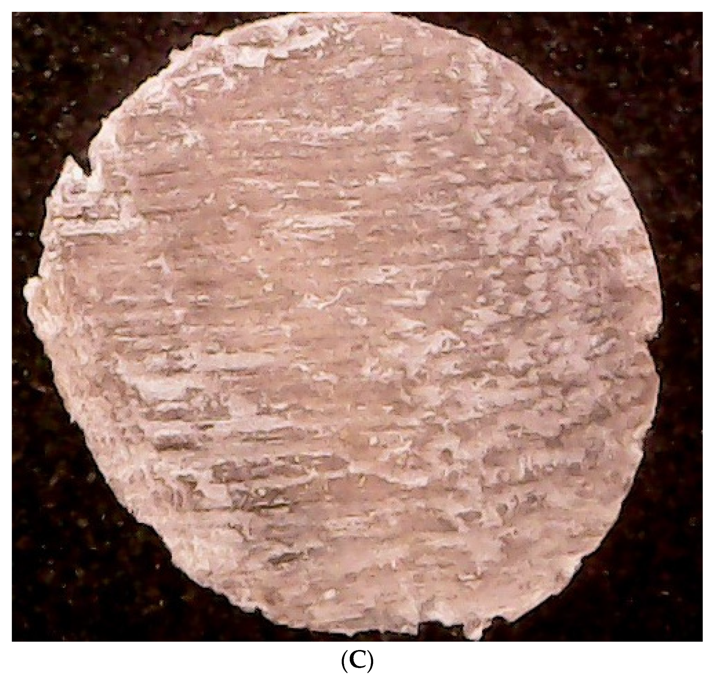
2.2. Bovine Membrane Production and Selection

2.2.1. Bovine Membrane Production

All hoof membranes used for these experiments were obtained from freshly slaughtered 3-year-old cattle (Azienda Agricola Pluderi Marcellino, San Colombano al Lambro, Italy).
The freshly slaughtered bovine hooves were dipped in liquid nitrogen, cored using a Rolson® plug cutter with a 16 mm diameter from Rolson Tools Ltd (Twyford, UK) and cut into slices of about 300 µm with a Graziano SAG 12 precision lathe (Tortona, Alessandria, Italy). The liquid nitrogen was essential to prevent mechanical deformation during core drilling and subsequent cutting.
Before every experiment and evaluation, the membranes were sanitized with a sequence that consisted of washing with ethanol 70% v/v and a benzalkonium chloride mixture (0.4 g benzalkonium chloride, 70 g isopropyl alcohol and distilled water up to 100 g) and being maintained in a climatic chamber at 25 °C and 40% RH (Relative Humidity) (ClimaCell 111, MMM Medcenter Einrichtungen GmbH, Munchen, Germany).

2.2.2. Bovine Membrane Morphology Evaluation

The membrane morphology analysis was performed with a model BW 1008 digital microscope (Brightwell Technology Limited, Shenzhen, China). Since the degree of surface roughness may affect the degree of fungal adhesion, the membranes were first classified based on their morphological superficial properties into smooth, semi-flaky and flaky samples. Then, the flaky or semi-flaky membranes were selected for the experiments in order to have homogeneous groups and mimic a situation with a high probability of infection. Figure 1 shows examples of the smooth, semi-flaky and flaky membranes.

2.3. Preliminary Microbiological Experiment

2.3.1. Fungal Strains, Culture Conditions and Sample Preparation

The strains used in this study were Candida albicans ATCC 1023 and Trichophyton rubrum ATCC 28,188. The C. albicans was grown at 37 °C for 24 h in an Iso-Sensitest broth, and its dilutions were plated in Sabouraud dextrose agar.
The T. rubrum was grown at 21–24 °C for 6 days in a potato dextrose broth, and its dilutions were plated in potato dextrose agar.
Before evaluation of the possible antiadhesive activity, the membranes were immersed for 15 min in 0.4% benzalkonium chloride in order to eliminate possible pre-existing contaminations. The membranes were then immersed in 1 mL of sterile water, which was then diluted and plated in TSA, PDA and SDA. No bacterial or fungal growth was shown.

2.3.2. Contamination of Membranes with the Chosen Microorganism

A test on the membranes to assess their capability to be contaminated with the chosen microorganisms was performed. For this purpose, six membranes were cut into sections and used. The membrane sections were then soaked in the fungal solution overnight. For both pathogenic agents, one section at a time for each contact time (15 min, 5 h and 24 h) was used. The experiment was carried out in triplicate.
At the end of each contact time, the membranes were extracted, shaken and placed in a Petri dish to dry for a maximum of 20 min at 37 °C. Afterward, the membranes were put into 2 mL of sterile water. After 30 min, three serial dilutions from that solution were made (1:100, 1:10,000 and 1:1,000,000). Then, all three dilutions were plated using SDA for C. albicans and PDA for T. rubrum. They were then incubated in aerobic conditions for 24 h at 35–36 °C (C. albicans) and for 72 h at 21–26 °C (T. rubrum). A microbial count was performed in order to assess the membrane contamination.

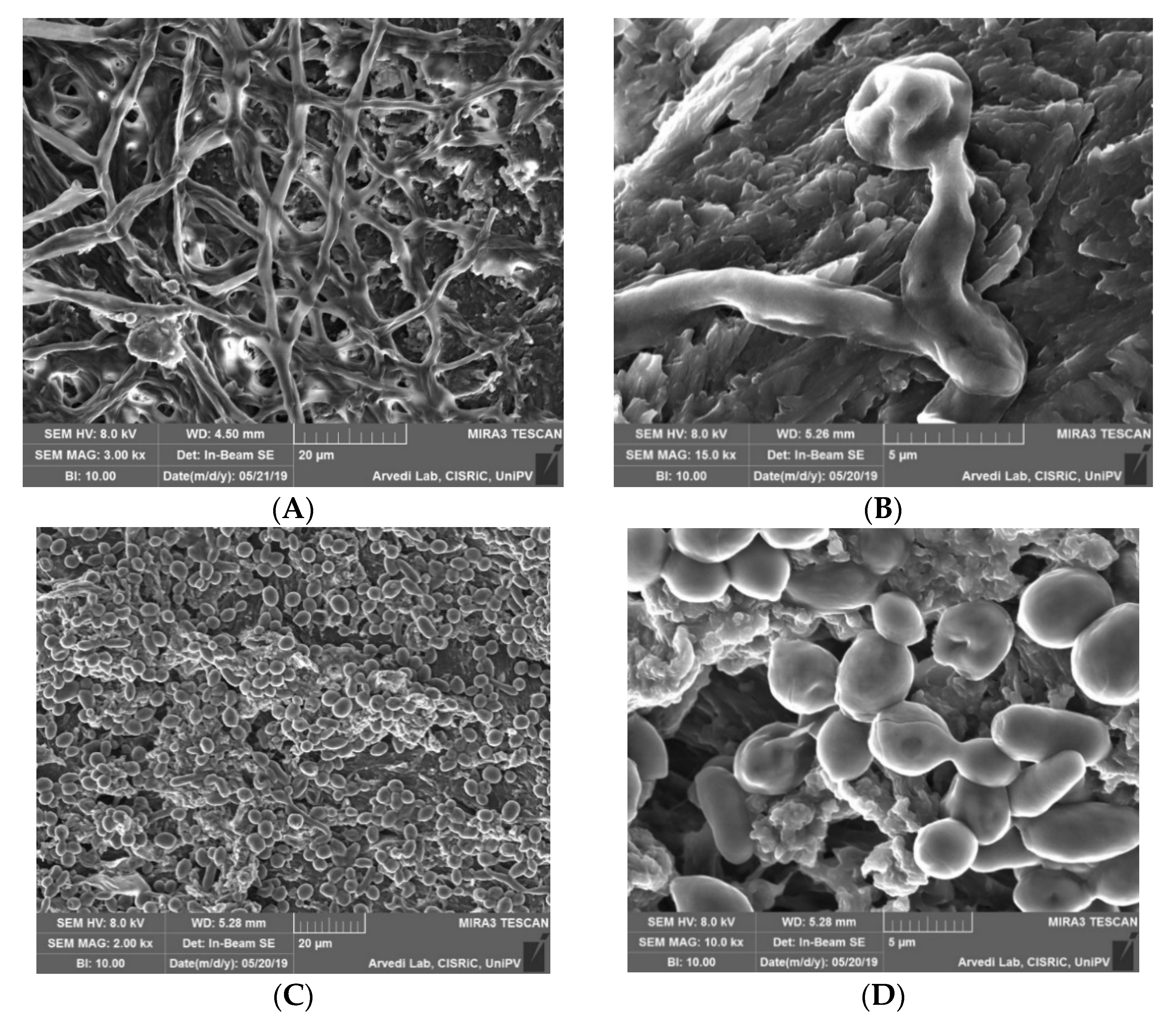
2.3.3. Morphological Analysis of Contaminated Membranes

Morphological analysis of the contaminated membranes was performed with a high-resolution scanning electron microscope (SEM) (TESCAN, Mira 3 XMU, Brno, Czech Republic).
The membrane sections were directly mounted on aluminum pin stubs by means of graphite tape and coated with carbon using a Cressington 208 C (Cressington Scientific Instrument Ltd., Watford, UK) prior to observation. SEM analysis was performed while operating at 20 kV.

2.4. In Vitro Evaluation of Reducing Fungal Adhesion to the Nail Plate

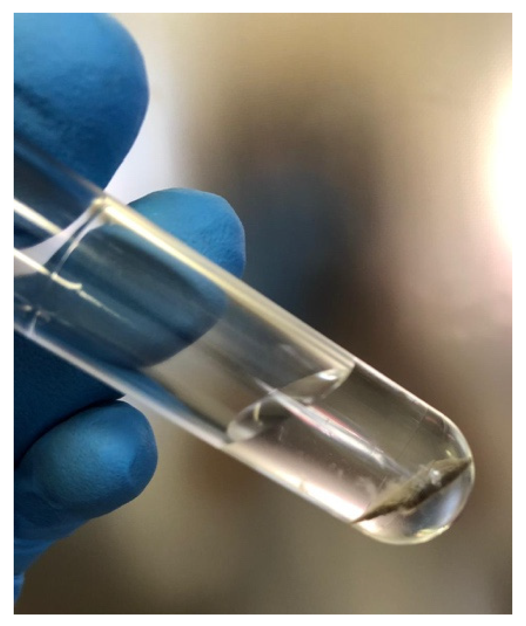
2.4.1. Sample Preparation

For this experiment, a commercially available antifungal product (AP) and a commercially available water-repellent base coat (BC) were used.
The AP was a 5% solution of amorolfine, an antifungal belonging to the class of morpholines. It is a well-known drug used for the treatment and prevention of dermatomycosis and onychomycosis [29].
The BC was a very common product used in nail care practice. It creates a barrier, helping shield nails from the damaging effects of nail polish. Furthermore, it prevents staining and peeling.
For this test, a BC was chosen as a negative control—in comparison with an AP as a positive control—to understand if the water-repellent physical barrier created by the products contributed to avoiding fungal adhesion on the nail. In this work, the contact angle measurement was used to test the water-repellent capability of the chosen base coat. Briefly, the contact angle is a measure of the wettability of a solid by a liquid and, in this case, the membranes treated with a base coat and water [30]. Generally speaking, high angle values were indicative of a poor interaction between the surface and the liquid, and a low value indicated high affinity between them. Three membranes with the same morphological properties were analyzed using a DMe-211Plus contact angle meter (KYOWA Interface Science Co., Ltd., Nobitome, Saitam, Japan) before and after water-repellent treatment application.

2.4.2. Test Procedure

For the experiment, bovine membranes were cut into sections, immersed for 15 min in 0.4% benzalkonium chloride and divided into groups (at least three sections for the groups). Two different procedures were carried out: multi-treatment and single-treatment, as explained below.
For the multi-treatment procedure, membranes were divided into three groups: treated with an AP (group 1); treated with a BC (group 2); and a control group with no treatment (group 3).
The duration of the experiment was about 60 h. The membranes in groups 1 and 2 were subjected to three cycles of treatment (at least 18 h of persistence of the product on the membranes) and washed between each cycle with a mixture of warm water and hand soap to mimic the real-life situation of handwashing. The control group (group 3) only underwent three cycles of the washing procedure. After the last washing cycle, the membranes were subjected to microbiological assessment.
For all durations, the experiment membranes were maintained in a climatic chamber at 25 °C and 40% RH (ClimaCell 111 MMM, Medcenter Einrichtungen GmbH, Munchen, Germany).
For the single-treatment procedure, the membranes were divided into two groups: a single application of an AP (group 4) and a single application of a BC (group 5).
After 18 h of persistence of the product on the membranes, the samples were subjected to microbiological assessment without any washing cycles.

2.4.3. Microbiological Assessment

The fungal cultures were prepared in potato dextrose broth for each type of fungal strain with different incubation times (24 h for C. albicans and 72 h for T. rubrum). The microbial suspensions had initial inoculums of approximately 2–3 × 10⁶ CFU/mL (colony-forming unit/mL) for C. albicans and 2–3 × 10⁵ CFU/mL for T. rubrum.
The treatment groups (1–5) were introduced within the cultures, one for each contact time (15 min, 5 h and 24 h), for both fungal strains. At the end of each contact time, the membranes were extracted, shaken and placed in a Petri dish to dry for a maximum of 20 min at 37 °C. Then, the membranes were put into 2 mL of sterile water, gently mixed and, after 30 min, three dilutions were made from that solution (1:100, 1:10,000 and 1:1,000,000). The fungal dilutions were collected on SDA and PDA for the different strains. The plates were incubated in aerobic conditions for 24 h at 35–36 °C (C. albicans) and for 72 h at 21–26 °C (T. rubrum).
After the exposure time, the antiadhesive effect (AE) of the product was calculated by applying the logarithmic reduction rate equation given below:
AE = log(Nc) − log(Nd)
where Nc is the colony-forming units (CFU) measurement of the control membrane suspension (sample 3) and Nd is the colony-forming units (CFU) measurement of the treated sample suspension (after application of groups 1–5). For each microorganism, three replicates were analyzed for each test.
Sterility and growth controls were also included for each time and fungal strain. The results from all membranes over time were expressed as the mean ± SD (standard deviation) of the treatment group. This test was carried out following suitably modified USA and European Standards (EN) applicable for testing the efficacy of disinfectant (bactericidal or fungicidal activity) used in food, industrial, domestic and institutional areas [31,32,33].

2.4.4. Statistical Analyses

One-way analysis of variance (ANOVA) was performed to compare multiple groups. All analyses were run using Prism 8 for Windows, and differences were considered to be significant at a level of p < 0.05.

3. Results and Discussion

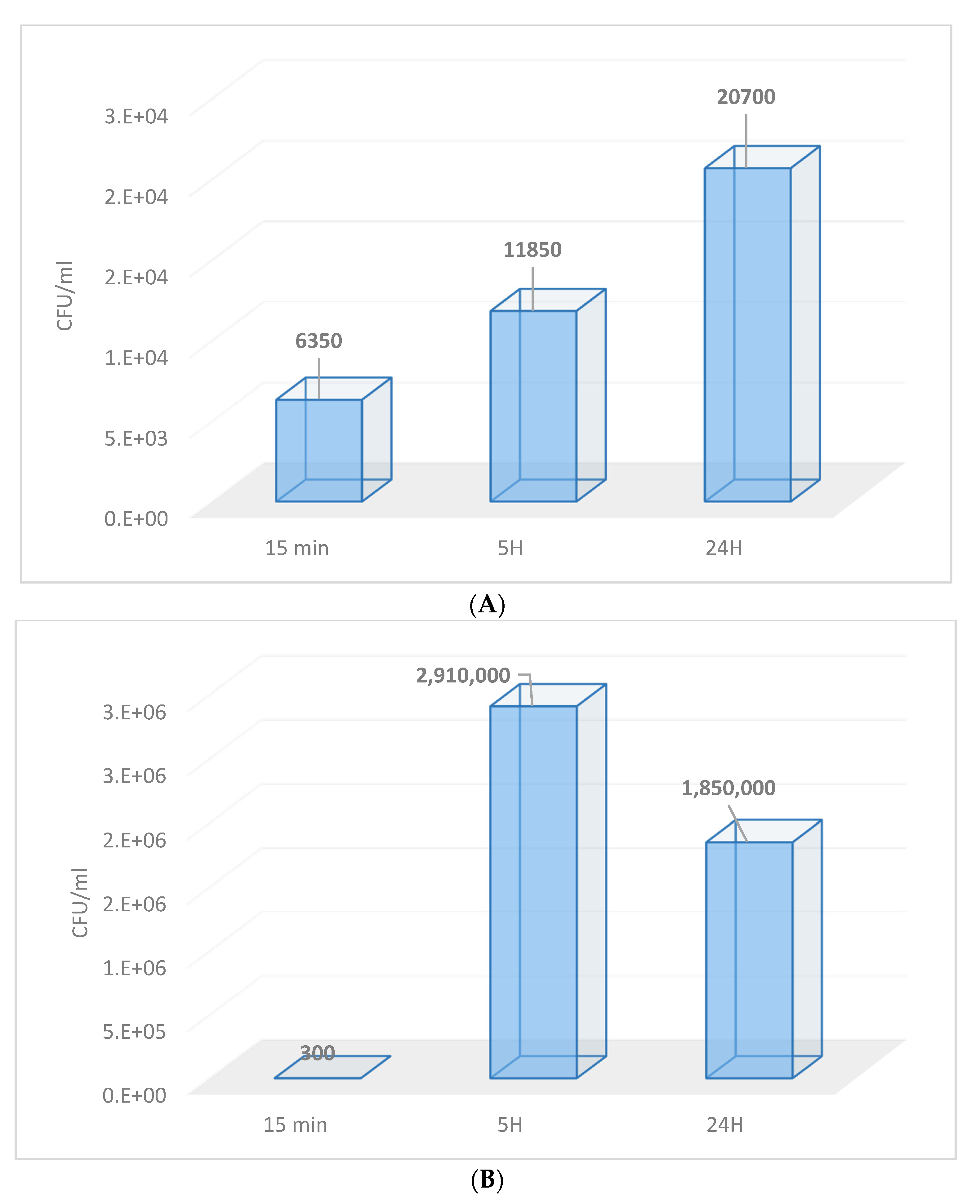
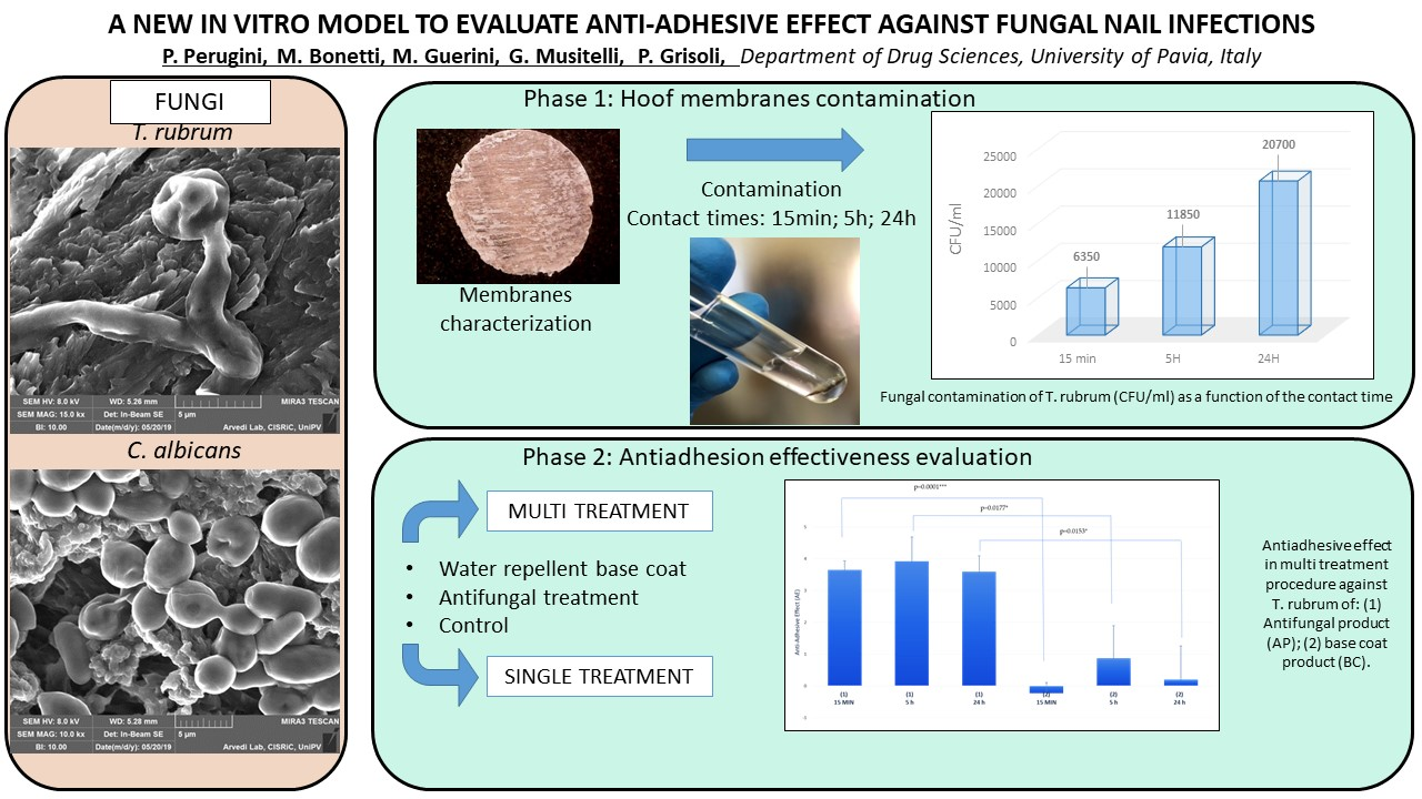
3.1. Contamination of Membranes by Fungi

The aim of this work was to set up a new in vitro model to evaluate the protective effects of cosmetic or pharmaceutical products against fungal adhesion on nails.
For this purpose, the first step was to test the affinity of the hoof membranes, used as an in vitro model for human nails, to be contaminated by fungi. In this work, two specific and main representative fungal strains, Trichophyton rubrum and Candida albicans, were used.
These fungi present different growth characteristics. C. albicans has a shorter generation time because it has an optimal growth period of 24 h. On the contrary, T. rubrum has a longer generation time [31].
Figure 2 shows the results of the nail fungal contamination by T. rubrum and C. albicans, respectively, as a function of the contact time.
As can be seen in Figure 2, the membranes were contaminated differently by the two fungal species. The contamination, in the case of T. rubrum, was proportional to the contact time, whereas the C. albicans contamination presented a peak of growth at 5 h and then showed a modest decline toward 24 h. That could be explained by the fact that the optimal growth period of C. albicans is 48 h, and after prolonged exposure, the fungal cells are stressed. Therefore, considering the initial growth period of the culture (48 h) and the subsequent contact time with the membranes (24 h) for a total of 72 h, a decline in the microbial content after the second exposure time was understandable.
The fact that there was less growth for T. rubrum depended on the nature of the fungus itself, as candida is a yeast with a faster duplication time. Therefore, it first colonizes the surface. T. rubrum is a multicellular microorganism, so it has a longer reproduction time and therefore a slower colonization time. The different values depended on the microbial title of the starting culture, as can be seen from the control values of approximately 2–3 × 10⁶ CFU/mL (colony forming unit/mL) for C. albicans and 2–3 × 10⁵ CFU/mL for T. rubrum.
Figure 3 shows an example of one membrane section after 24 h of contact with T. rubrum, in which contamination is clearly evident.
The SEM analysis confirmed successful contamination, as is shown in Figure 4.

3.2. In Vitro Evaluation of Fungal Adhesion to the Nail Plate

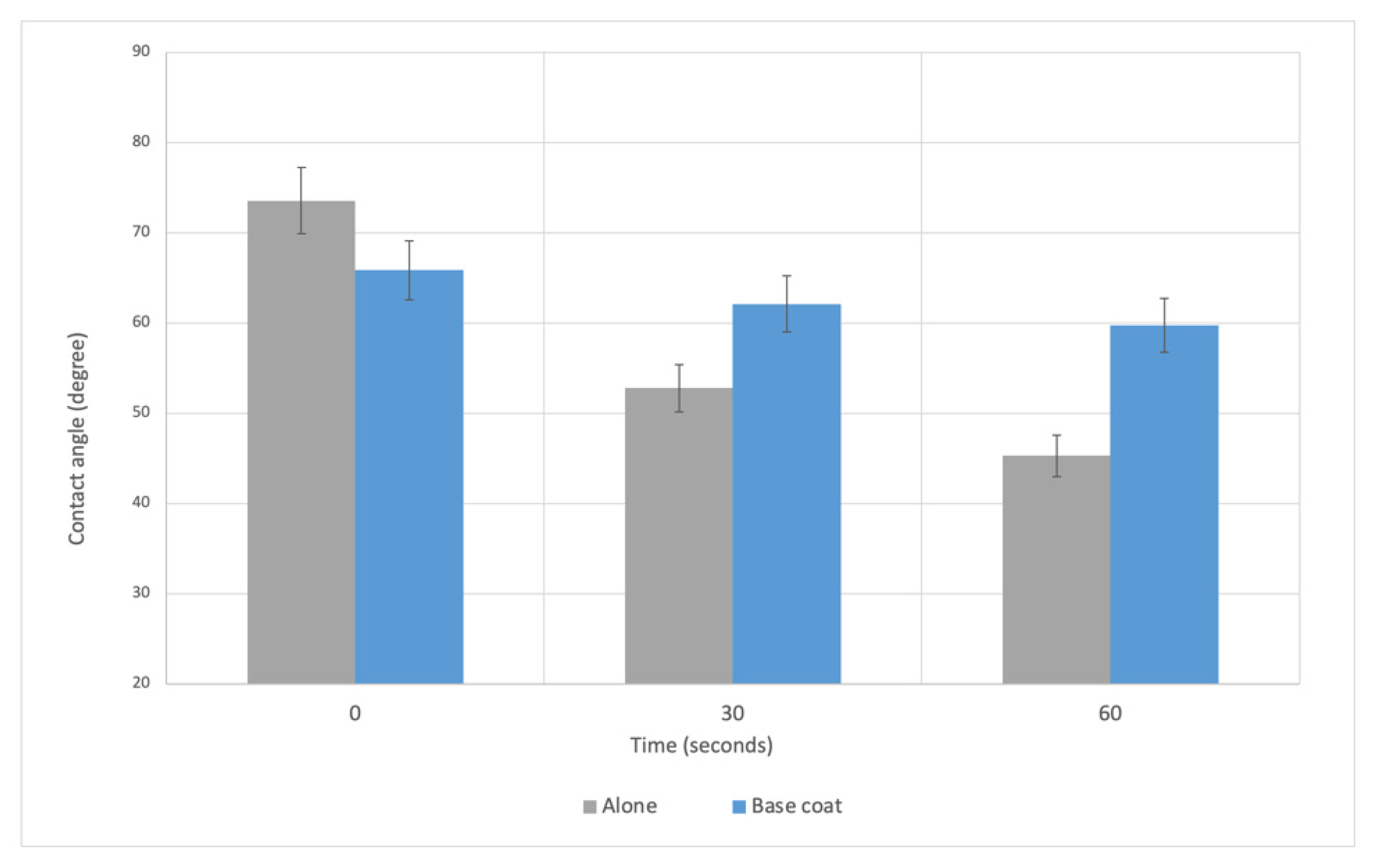
3.2.1. Sample Preparation

Sample choice was a critical step in order to assess the validity of the procedure for evaluating the antiadhesive properties of different products and helping understand the mechanism behind the efficacy. The antifungal lacquer containing amorolfine (AP) was the best choice as a positive control, since the compound is well-known for its antifungal activity [29]. However, it was not so easy to find a suitable product which could act only as a physical barrier on the membrane (negative control). The negative control was necessary to understand the role of the physical barrier created by the products on the nails in avoiding fungal adhesion.
For this purpose, a water-repellent base coat (BC) was chosen.
Measurement of the contact angle was the technique applied in this work to verify the water-repellent properties of the BC on the membranes.
Figure 5 shows the results of the contact angle measurements on the membrane alone and after treatment with a BC at three different time points (0 s, 30 s and 60 s) after water drop deposition. The results are expressed as the mean ± SD (standard deviation) of the angle (°) between the surface and the water drop.
Looking at the data reported in Figure 5, it is possible to appreciate the effect of the water-repellent barrier on the membranes. For the membranes alone, there was a reduction in the angle values over time, indicating an increase in the affinity of the membrane surface for the liquid over time. On the other hand, for the membranes treated with a BC, the angle value at the beginning (time 0) was 66°, lower than that obtained from the untreated membranes (74°). That result was coherent, with a smoother and more slippery surface after the treatment. After that, the contact angle remained quite constant, with very low variations from 0–60 s, showing real water–repellent activity.

3.2.2. Microbiological Assessment

In order to define the correct testing step for evaluating the effectiveness and mode of action of different products, two different procedures for microbiological assessment were carried out. The results are presented separately below according to the procedure used.

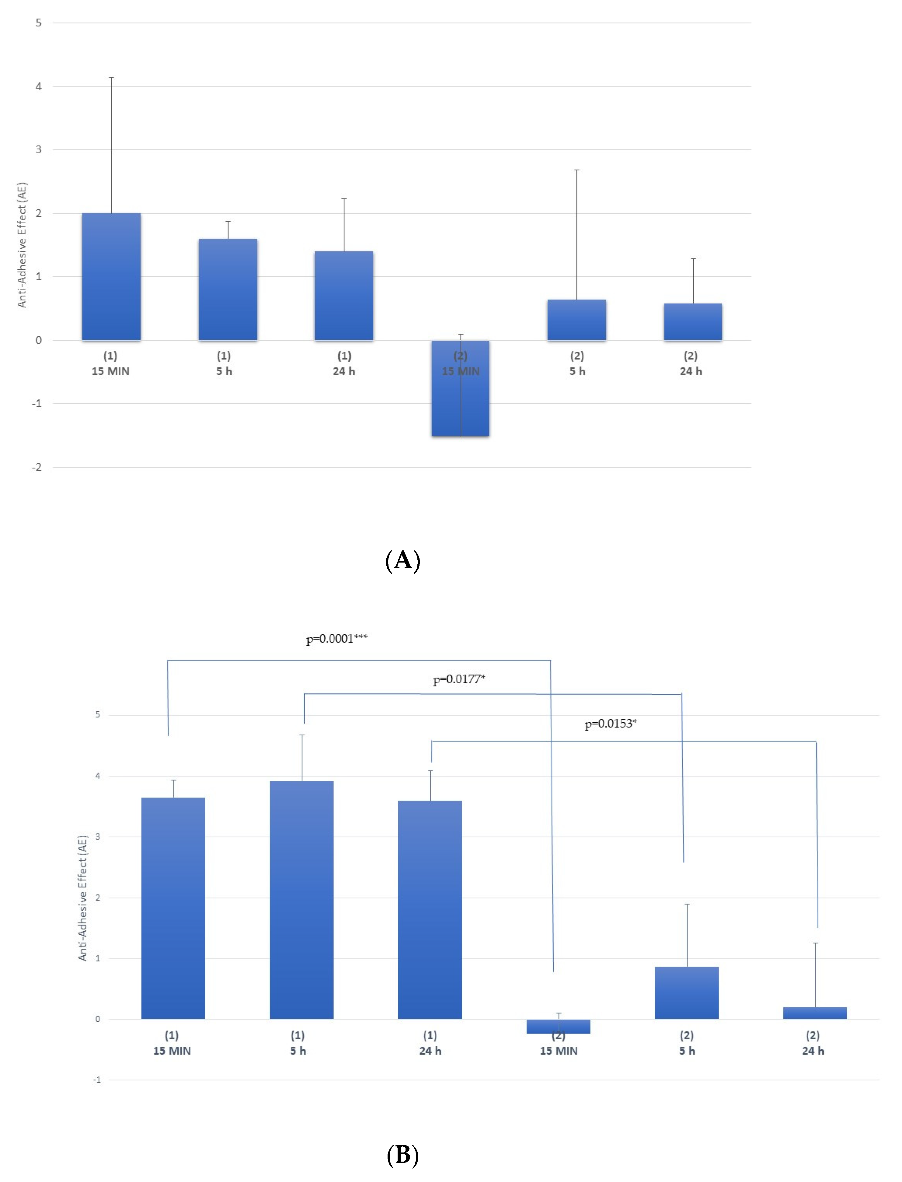
Multi-Treatment Procedure

The results obtained from the antifungal product (AP) and the base coat (BC) in the multi-treatment procedure against Candida albicans and Trichophyton rubrum in Figure 6A,B, respectively. In both groups, the membranes were treated three times and washed both between applications and at the end of the study before microbiological assessment. The control group membranes only underwent the washing procedure.
The antiadhesive effect (AE) was expressed as the quotient of the difference of the logarithms of the colony-forming units (CFU) between the control membrane suspension (sample 3) and the colony-forming units (CFU) of the treated sample suspension in each sampling time. The higher the AE was, the higher the effectiveness of the product against fungal adhesion was. Following the international standards [33], the minimum logarithmic reduction acceptable to indicate suitable prevention of surface contamination was two. Thus, despite the fact that no study concerning the possible effect on nail fungal contamination has been published yet, in this work, an AE value of two or higher was chosen as the cut-off value to consider the product able to prevent fungal infection.
Starting from this data, the AP seemed to be able to prevent fungal infection, particularly against Trichophyton rubrum. In fact, as is shown in the graphs in Figure 6, at each time, the antiadhesive activity of the AP against Trichophyton rubrum was nearly double that against Candida albicans; specifically, after 5 h, the activity was 3.92 ± 0.76 and 1.60 ± 0.28, respectively. This result agrees with the literature, in which the efficacy of amorolfine evaluated by its minimum inhibitory concentration (MIC) values, which represent the lower concentration that prevents microbial growth, was reported to be stronger against T. rubrum than against C. albicans [26,34,35].
The statistical analyses carried out on each group by applying a one-way ANOVA test revealed no statistical differences over time. On the contrary, significant statistical differences were obtained using a t-test between group 1 and 2 at each time against T. rubrum infection. The results of the statistical analysis are evident in Figure 6B.
In addition, these results, obtained after three treatments with drugs and on washed membranes, demonstrated clearly that the drug was able to penetrate deeper into the membranes to exert its action, both after washing and after only 15 min of contact.
On the other hand, the AE values obtained after treatment with a water-repellent base coat showed no antiadhesive effect, since they were much lower and not significantly different at each testing time and for both fungal species. This result highlighted that a simple physical barrier cannot exert a protective effect against fungal infection.

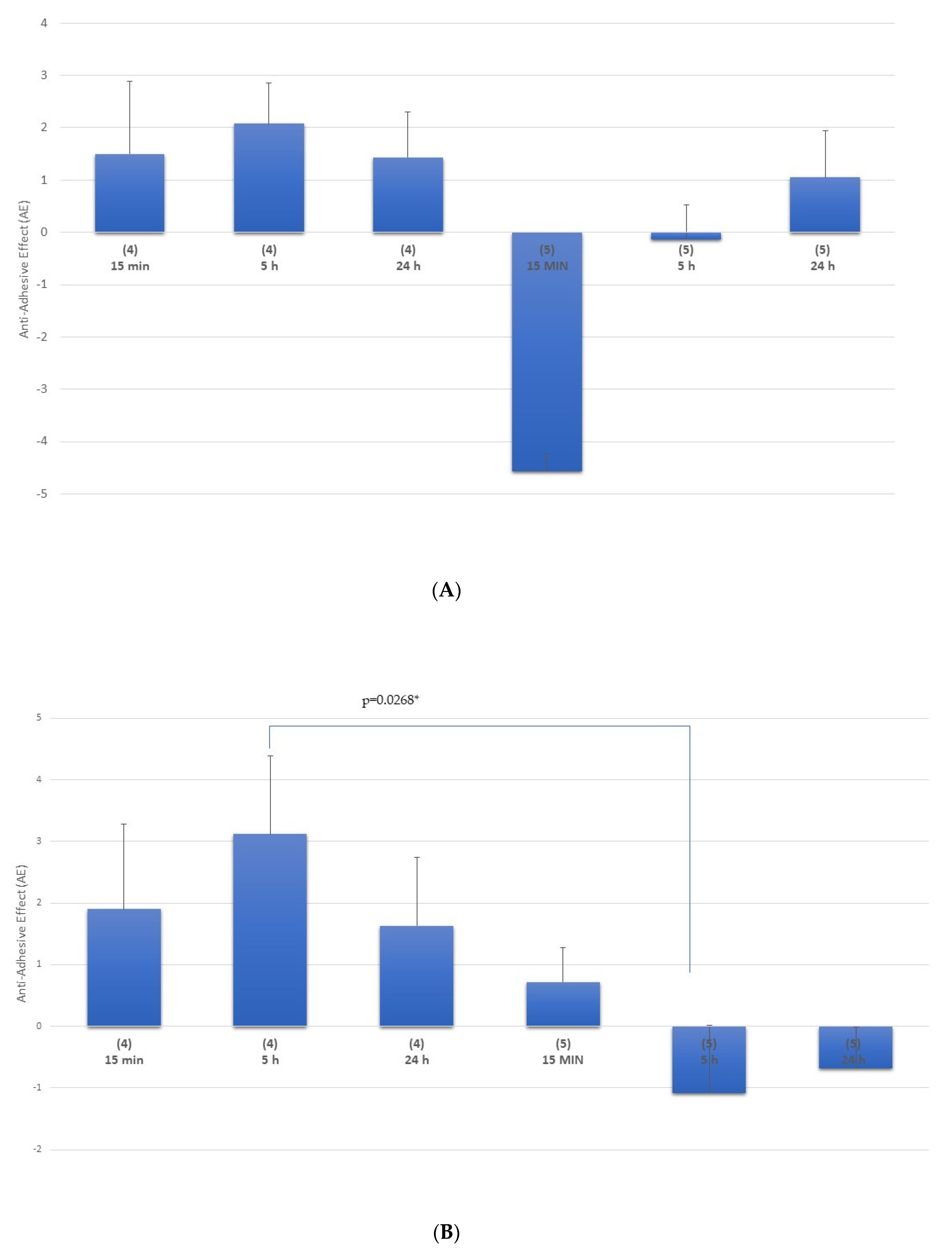
Single-Treatment Procedure

This procedure aimed to highlight the effectiveness of the product after just a single application. Figure 7 shows the results obtained from the antifungal product (AP) and the base coat (BC) in the single-treatment procedure for group 4 and 5, respectively, in which the membranes were treated once with the products and not washed. This way, the product remained on the membrane surface before microbiological assessment.
As was seen in the multiple-treatment procedure, the antiadhesive activity of the AP after only one treatment was higher at each time against Trichophyton rubrum than against Candida albicans; the higher activity seemed to be after 5 h of contact, being 3.12 ± 1.27 and 2.08 ± 0.78, respectively. Moreover, considering the obtained values of the BC, different behavior was noted between the two fungal species, with negative AE values in the first 5 h for C. albicans and weakly positive values for T. rubrum. This was due to the different metabolisms of the two fungi. In fact, the great adaptability of C. albicans translates into an ability to rapidly respond to stress factors, take up nutrients or multiply under different conditions. As such, this yeast showed a greater rapidity of growth and adhesion compared with T. rubrum, which has a slower cell duplication time [36].
The AE values obtained from a single treatment with the BC confirmed that this product did not show antiadhesive effects.
Furthermore, taking into account the results obtained at all testing times, 5 h appeared to be the best time to test antiadhesive activity. Therefore, in order to verify the potency of an AP, it could be interesting to compare the activity detected after multiple treatments with the activity after a single application.
Table 1 reports the summary of the statistical analysis carried out on all antiadhesive effects (AEs) obtained from all groups after multiple treatments of an AP compared with the one obtained after just a single application of an antifungal product.
The results clearly show that the prevention activity changed dramatically in relation to the fungal species. In fact, amorolfine could exert statistically significant antiadhesion activity only against T. rubrum.
Furthermore, the lack of a statistically significant difference between multiple treatments and one single application of an AP highlights that the drug could penetrate into the membrane to maintain its activity against T. rubrum even when washed.

4. Conclusions

The applied in vitro method for membrane contamination showed itself to be effective for the fungal species used. Thus, hoof membranes were shown to be a valuable in vitro model to consider for this kind of product assessment. Hoof membranes can be produced with specific characteristics to mimic a wide range of nail conditions (e.g., healthy or dystrophic). Moreover, the selection of flaky samples through morphological assessment, as well as the inner higher permeabilities of the membranes, allow for the set-up of an efficacy test within extreme conditions highly favoring fungal contamination.
However, some challenges were faced with the model, and they need to be taken into account, such as the flaking of the membrane itself, which can become more permeable in an aqueous environment.
The preliminary sanitization procedure carried out on the membranes permitted the obtaining of the substrate with very low microbial contamination, and thus it was very useful for the aim of this test.
Despite the fact that no study concerning the possible effect on nail fungal contamination has been published yet, in this work, an AE value of two or higher, actually used in international guidelines for surface contamination, was chosen as the cut-off value to consider a product able to prevent fungal infection.
The protocol used in this work permitted identifying a suitable negative control in a water-repellent base coat, which can be used in all further studies as a reference.
The results obtained from microbiological experiments evidenced clearly that the preventing activity of a product changes dramatically in relation to fungal species. In fact, even an antifungal drug like amorolfine could exert statistically significant antiadhesion activity against only one fungus; its activity was particularly high against T. rubrum.
Furthermore, taking into account the results obtained at all testing times, 5 h appeared to be the best time to test antiadhesive activity.
Finally, statistical analysis permitted concluding that the antiadhesive activity found between multiple treatments and one single application of an AP was the same, highlighting that the drug could penetrate into the membrane to maintain its activity against T. rubrum even when washed.
This work represents a starting point for the development of a new method to evaluate the protective effects of drugs, medical devices or cosmetics against fungal adhesion, using hoof membranes as a model.
Further studies should be carried out to standardize this test, especially regarding the use of different fungal species alone or mixed together as well as in different conditions to simulate real-life situations.

Author Contributions

Conceptualization, P.P. and P.G.; methodology, P.G., M.G., M.B. and P.P.; investigation, M.G., M.B. and G.M.; data curation, P.P. and P.G.; first draft preparation, M.G. and M.B.; writing—review and editing, M.G., P.P., M.B., G.M. and P.G.; supervision, P.P.; project administration, P.P. All authors have read and agreed to the published version of the manuscript.

Funding

This research received no external funding. The project was self-funded by the research group at the Department of Drug Sciences coordinated by Paola Perugini.

Data Availability Statement

The data presented in this study are available in this article.

Acknowledgments

The authors are grateful to the Arvedi Laboratory, CISRiC (Centro Interdipartimentale di Studi e Ricerche per la Conservazione del Patrimonio Culturale), University of Pavia in Pavia, Italy for allowing the use of their scanning electron microscope.

Conflicts of Interest

The authors declare no conflict of interest.

References

  1. Tudela, J.L.R.; Denning, D.W. Recovery from serious fungal infections should be realisable for everyone. Lancet Infect. Dis. 2017, 17, 1111–1113. [Google Scholar] [CrossRef]
  2. Al-Khikhani, F.H. Dermatophytosis a Worldwide Contiguous Fungal Infection: Growing Challenge and Few Solutions. Biomed. Biotechnol. Res. J. 2020, 4, 117–122. [Google Scholar]
  3. Zhan, P.; Liu, W. The changing face of dermatophytic infections worldwide. Mycopathologia 2017, 182, 77–86. [Google Scholar] [CrossRef]
  4. Hay, R. Fungal skin infections. Arch. Dis. Child. 1992, 67, 1065–1067. [Google Scholar] [CrossRef] [PubMed] [Green Version]
  5. Aly, R. Ecology and epidemiology of dermatophyte infections. J. Am. Acad. Dermatol. 1994, 31, 21–25. [Google Scholar] [CrossRef]
  6. Evans, E. Causative pathogens in onychomycosis and the possibility of treatment resistance: A review. J. Am. Acad. Dermatol. 1998, 38, 32–36. [Google Scholar] [CrossRef]
  7. Adeklandi, S.; Pal, S.; Sharma, N.; Juyal, D.; Sharma, M.; Dimri, D. Incidence and epidemiology of onychomycosis in patients visiting a tertiary care hospital in India. Cutis 2015, 95, E20–E25. [Google Scholar]
  8. Clayton, Y. Clinical and mycological diagnostic aspects of onychomycoses and dermatomycoses. Clin. Exp. Dermatol. 1992, 17, 37–40. [Google Scholar] [CrossRef] [PubMed]
  9. Midgley, G.; Moore, M.K.; Cook, J.C.; Phan, Q.G. Mycology of nail disorders. J. Am. Acad. Dermatol. 1994, 31, 68–74. [Google Scholar] [CrossRef]
  10. Public Health England. UK standards for microbiology investigations B 39. In Investigations of Dermatological Specimens for Superificial Mycoses; Public Health English: London, UK, 2006; pp. 1–26. [Google Scholar]
  11. Elewsky, B.E. Large scale epidemiological study of the causal agents of onychomycosis: Mycological finding from the multicenter onychomycosis study of terbinafine. Arch. Dermatol. 1997, 133, 1317–1318. [Google Scholar] [CrossRef]
  12. Dompmartin, D.; Dompmartin, A.; Deluol, A.M.; Grosshans, E.; Coulaud, J.P. Onychomycosis and AIDS: Clinical laboratory findings in 62 patients. Int. J. Dermatol. 1990, 29, 337–339. [Google Scholar] [CrossRef]
  13. Elewsky, B.E. Onychomycosis: Pathogenesis, Diagnosis, and Management. Clin. Microbiol. Rev. 1998, 11, 415–429. [Google Scholar] [CrossRef] [Green Version]
  14. Scher, R.K.; Baran, R. Onychomycosis in clinical practice. Factors contributing to recurrence. Br. J. Dermatol. 2003, 149, 5–9. [Google Scholar] [CrossRef] [PubMed]
  15. Shemer, A. Update medical treatment of onychomycosis. Dermatol. Ther. 2012, 25, 582–593. [Google Scholar] [CrossRef] [PubMed]
  16. Chan, H.L.H.; Wong, T.E.; Yeung, K.C. Psychosocial perception of adults with onychomycosis: A blinded, controlled comparison of 1017 adult Hong Kong residents with or without onychomycosis. Biopsychosoc. Med. 2014, 15, 1–9. [Google Scholar]
  17. Tosti, A.; Elewski, B.E. Onychomycosis: Practical approaches to minimize relapse and recurrence. Skin Appendage Disord. 2016, 2, 83–87. [Google Scholar] [CrossRef] [Green Version]
  18. Christenson, J.K.; Peterson, G.M.; Naunton, M.; Bushell, M.; Kosari, S.; Baby, K.E.; Thomas, J. Challenges and Opportunities in the Management of Onychomycosis. J. Fungi 2018, 4, 87. [Google Scholar] [CrossRef] [Green Version]
  19. Bonk, A.F.; Friedman, L.; Derbes, V.J. Experimental dermatophytosis. J. Investig. Derm. 1962, 39, 281–286. [Google Scholar] [CrossRef] [PubMed] [Green Version]
  20. Greenberg, J.H.; King, R.D.; Krebs, S.; Field, R. A quantitative dermatophyte infection model in the guinea pig—A parallel to the quantitated human infection model. J. Investig. Derm. 1967, 67, 704–708. [Google Scholar] [CrossRef] [PubMed] [Green Version]
  21. Saunte, D.M.; Hasselby, J.P.; Brillowska-Dabrowska, A.; Frimodt-Møller, N.; Svejgaard, E.L.; Linnemann, D.; Nielsen, S.S.; Haedersal, M.; Arendrup, M.C. Experimental guinea pig model of dermatophytosis: A simple and useful tool for the evaluation of new diagnostic. Med. Mycol. 2008, 46, 303–313. [Google Scholar] [CrossRef] [Green Version]
  22. Klink, C.D.; Binnebosel, M.; Lambertz, A.; Alizai, H.P.; Roeth, A.; Otto, J.; Klinge, U.; Neumann, U.P.; Junge, K. In vitro and in vivo characteristics of gentamicin-supplemented polyvinylidenfluoride mesh materials. J. Biomed. Mater. Res. 2012, 100, 1195–1202. [Google Scholar] [CrossRef] [PubMed]
  23. Wong, S.Y.; Moskowitz, J.S.; Veselinovic, J.; Rosario, R.A.; Timachova, K.; Blaisse, M.R.; Fuller, R.C.; Klibanov, A.M.; Hammond, P.T. Dual functional polyelectrolyte multilayer coatings for implants: Permanent microbicidal base with controlled release of therapeutic. J. Am. Chem. Soc. 2010, 132, 17840–17848. [Google Scholar] [CrossRef] [Green Version]
  24. Dijck, P.V.; Sjollema, J.; Cammue, B.P.A.; Lagrou, K.; Berman, J.; d’Enfert, C.; Andes, D.R.; Arendrup, M.C.; Brakhage, A.A.; Calderone, R.; et al. Methodologies for in vitro and in vivo evaluation of efficacy of antifungal and antibiofilm agents and surface coatings against fungal biofilms. Microb. Cell 2018, 5, 300–326. [Google Scholar] [CrossRef]
  25. Tatsumi, Y.; Yokoo, M.; Senda, H.; Kakehi, K. Therapeutic efficacy of topically applied KP-103 against experimental tinea unguium in guinea pigs in comparison with amorolfine and terbinafine. Antimicrob. Agents Chemother. 2002, 46, 3797–3801. [Google Scholar] [CrossRef] [Green Version]
  26. Schaller, M.; Borelli, C.; Berger, U.; Walker, B.; Schmidt, S.; Weindl, G.; Jackel, A. Susceptibility testing of amorolfine, bifonazole and ciclopiroxolamine against Trichophyton rubrum in an in vitro model of dermatophyte nail infection. Med. Mycol. 2009, 47, 753–758. [Google Scholar] [CrossRef] [Green Version]
  27. Monti, D.; Mazzantini, D.; Tampucci, S.; Vecchione, A.; Celandroni, F.; Burgalassi, S. Ghelardi, Ciclopirox and Efinaconazole Transungual Permeation, Antifungal Activity, and Proficiency to Induce Resistance in Trichophyton rubrum. Antimicrob. Agents Chemother. 2019, 63, 1–11. [Google Scholar] [CrossRef] [PubMed] [Green Version]
  28. Nogueiras-Nieto, L. Hydration and N-acetyl-l-cysteine alter the microstructure of human nail and bovine hoof: Implication for drug delivery. J. Control. Release 2011, 156, 337–344. [Google Scholar] [CrossRef] [Green Version]
  29. Sigurgeirsson, B.; Olafsson, J.H.; Steinsson, J.T.; Kerrouche, N.; Sidou, F. Efficacy of amorolfine nail lacquer for the prophylaxis of onychomycosis over 3 years. J. Eur. Acad. Dermatol. Venereol. 2010, 24, 910–915. [Google Scholar] [CrossRef] [PubMed]
  30. Yuan, Y.; Randall Lee, T. Contact Angle and Wetting Properties. Surf. Sci. Tech. Springer 2013, 51, 3–34. [Google Scholar]
  31. Russel, A.D.; Hugo, W.B.; Ayliffe, G.A.J. Principles and Practice of Disinfection, Preservation and Sterilization; Blackwell Publishing Ltd.: Hoboken, NJ, USA, 2004. [Google Scholar]
  32. 32 EN 13697:2019 Chemical Disinfectants and Antiseptics—Quantitative Non-Porous Surface Test for the Evaluation of Bactericidal and/or Fungicidal Activity of Chemical Disinfectants Used in Food, Industrial, Domestic and Institutional Areas—Test Method and Requirements without Mechanical Action; CEN-CENELEC Management Centre: Brussels, Belgium, 2019.
  33. Guidance on the Biocidal Products Regulation Volume II Efficacy—Assessment and Evaluation (Parts B+C) Version 3.0; ECHA (European Chemical Agency): Helsinki, Finland, 2018.
  34. Li, R.; Wan, Z.; Wang, A.P.; Shen, Y.N.; Lu, C.M.; Li, M.; Xi, L.; Liu, W.D.; Zeng, F.K. In vitro susceptibility testing of amorolfine in pathogenic fungi isolated from dermatomycosis patients in China. Mycoses 2004, 47, 402–406. [Google Scholar] [CrossRef] [PubMed]
  35. Jo Siu, W.; Tatsumi, Y.; Senda, H.; Pillai, R.; Nakamura, T.; Sone, D.; Fothergill, A. Comparison of In vitro Antifungal Activities of Efinaconazole and Currently Available Antifungal Agents against a Variety of Pathogenic Fungi Associated with Onychomycosis. Antimicrob. Agents Chemother. 2013, 57, 1610–1616. [Google Scholar] [CrossRef] [PubMed] [Green Version]
  36. Ciurea, C.N.; Kosovski, I.N.; Mare, A.D.; Toma, F.; Pintea-Simon, I.A.; Man, A. Candida and Candidiasis—Opportunism Versus Pathogenicity: A Review of the Virulence Traits. Microorganisms 2020, 8, 857. [Google Scholar] [CrossRef] [PubMed]
Figure 1. Example of a (A) smooth membrane, (B) semi-flaky membrane and (C) flaky membrane.
Applsci 11 01977 g001aApplsci 11 01977 g001b
Figure 2. Fungal contamination of (A) T. rubrum (CFU/mL) and (B) C. albicans (CFU/mL) as a function of the contact time.
Applsci 11 01977 g002
Figure 3. Membrane section after 24 h of contamination with T. rubrum.
Applsci 11 01977 g003
Figure 4. Scanning electron microscope (SEM) images of a membrane contaminated with T. rubrum at (A) a lower magnification and (B) a higher magnification, and with C. albicans at (C) a lower magnification and (D) a higher magnification.
Applsci 11 01977 g004
Figure 5. Contact angle measurements of the base coat treatment with respect to the membrane alone.
Applsci 11 01977 g005
Figure 6. Antiadhesive effect of the antifungal product (AP) in group 1 and the base coat (BC) in group 2 for the multi-treatment procedure against (A) C. albicans and (B) T. rubrum. Statistical significance: *, p < 0.05 is significant; **, p < 0.01 is strongly significant; *** p < 0.001 is very strongly significant.
Applsci 11 01977 g006
Figure 7. Antiadhesive effect in the single-treatment procedure of group 4 with an antifungal (AP) product and group 5 with a base coat (BC) product against (A) C. albicans and (B) T. rubrum. Statistical significance: *, p < 0.05 is significant; **, p < 0.01 is strongly significant; *** p < 0.001 is very strongly significant.
Applsci 11 01977 g007
Table 1. Statistical analysis performed on all groups by a one-way analysis of variance (ANOVA) multiple comparisons test. Statistical significance: *, p < 0.05 is significant; **, p < 0.01 is strongly significant; *** p < 0.001 is very strongly significant.
Multiple Comparisons Test against Tricophytum rubrum
Comparison GroupsMean Difference95.00% CI of Diff.Significantp Value
1 vs. 41.503−0.2805 to 3.287No0.1018
1 vs. 23.4401.656 to 5.224Yes0.0012 **
1 vs. 54.0672.283 to 5.851Yes0.0004 ***
4 vs. 21.9370.1528 to 3.721Yes0.0341 *
4 vs. 52.5630.7795 to 4.347Yes0.0076 **
2 vs. 50.6267−1.157 to 2.411No0.8313
Multiple Comparisons Test against Candida albicans
Comparison GroupsMean Difference95.00% CI of Diff.Significantp Value
1 vs. 4−0.003333−4.240 to 4.233No>0.9999
1 vs. 21.763−2.473 to 6.000No0.5695
1 vs. 52.883−1.353 to 7.120No0.2085
4 vs. 21.767−2.470 to 6.003No0.5682
4 vs. 52.887−1.350 to 7.123No0.2077
2 vs. 51.120−3.117 to 5.357No0.8313
Publisher’s Note: MDPI stays neutral with regard to jurisdictional claims in published maps and institutional affiliations.

