Article

An RNA Triangle with Six Ribozyme Units Can Promote a Trans-Splicing Reaction through Trimerization of Unit Ribozyme Dimers

1
Department of Chemistry, Graduate School of Science and Engineering, University of Toyama, Gofuku 3190, Toyama 930-8555, Japan
2
Department of Chemistry, Graduate School of Science, Kyoto University, Kyoto 606-8502, Japan
3
Department of Life Science Frontiers, Center for iPS Cell Research and Application (CiRA), Kyoto University, Kyoto 606-8502, Japan
4
Institute for Integrated Cell-Material Sciences, Kyoto University, Kyoto 606-8502, Japan
*
Author to whom correspondence should be addressed.
Appl. Sci. 2021, 11(6), 2583; https://doi.org/10.3390/app11062583
Submission received: 29 November 2020 / Revised: 5 March 2021 / Accepted: 11 March 2021 / Published: 14 March 2021
(This article belongs to the Special Issue Nucleic Acids Conjugates for Biotechnological Applications)

Abstract

:
Ribozymes are catalytic RNAs that are attractive platforms for the construction of nanoscale objects with biological functions. We designed a dimeric form of the Tetrahymena group I ribozyme as a unit structure in which two ribozymes were connected in a tail-to-tail manner with a linker element. We introduced a kink-turn motif as a bent linker element of the ribozyme dimer to design a closed trimer with a triangular shape. The oligomeric states of the resulting ribozyme dimers (kUrds) were analyzed biochemically and observed directly by atomic force microscopy (AFM). Formation of kUrd oligomers also triggered trans-splicing reactions, which could be monitored with a reporter system to yield a fluorescent RNA aptamer as the trans-splicing product.

1. Introduction

Folded polypeptides work not only in monomeric states but also in assembled states, including homo- and heterooligomers [1,2,3]. In the course of enzyme evolution, symmetrical protein homooligomers with polygonal and polyhedral shapes emerged from their monomeric ancestors, presumably due to the advantages in their biological properties over the respective monomeric states, including enzymatic activities and structural stability [1,2,3]. Interfaces between monomer units play a key role in the assembly of protein oligomers. The artificial design of protein oligomers with symmetrical shapes has been explored with the use of protein–protein interaction interfaces extracted from naturally occurring protein oligomers, generating the field of protein nanostructure research [4,5,6]. Recent challenges in this field include de novo design of artificial protein–protein interaction interfaces that can serve as modular structural units for versatile protein nanostructure design [7,8]. Among the various forms of symmetrical protein homooligomers, symmetrical protein homodimers can be regarded as the simplest molecular parts for the rational design and assembly of protein structures [9,10,11].
RNA is a biopolymer that can fold into defined 3D structures with biological functions. Structured RNAs are known to perform many types of protein-like functions, including roles as catalysts and receptors [12,13,14]. Large and complex 3D RNA structures frequently formed through noncovalent assembly of structural modules, which can fold locally within a single polynucleotide chain [15]. Intramolecular assembly of RNA structural modules is supported and maintained by RNA–RNA interaction interfaces [15,16]. Their association is usually strong enough to reconstitute the catalytic activity in a multimolecular format, in which separately prepared modular RNAs are assembled noncovalently by RNA–RNA interaction interfaces [17,18,19,20,21,22,23,24,25].
We recently engineered an RNA–RNA interaction interface of the Tetrahymena group I ribozyme to design a prototype of ribozyme modules (abbreviated as RzM-0, Figure 1) with self-dimerizing capability [26,27]. The RNA–RNA interface in the RzM-0 homodimer is supported by two sets of tertiary interactions between the ∆P5 core ribozyme module and P5abc module, which are connected by flexible linker elements (P5-P5a linker) in the parent ribozyme and form strong intramolecular associations (WT group I ribozyme, Figure 1). We rationally replaced the flexible P5-P5a with a rigid duplex to prevent intramolecular ∆P5-P5abc assembly (Figure S1C), resulting in homodimerization of the resulting variant (RzM-0 in Figure 1) in a head-to-head manner by forming RzM:RzM interface 0:0 [26,27]. A pair of symmetrical ∆P5-P5abc interfaces yielding RzM:RzM homodimer was then engineered to asymmetrical recognition interfaces, such as RzM:RzM interface 1:1′, yielding RzM:RzM heterodimers (Figure 1). We then connected a pair of RzM monomers in a tail-to-tail manner at their P6b region, enabling the resulting covalent dimer (termed unit ribozyme dimer and abbreviated as Urd) to homooligomerize in a directional manner (Figure 1) [28]. Unit ribozyme dimers (Urds) homooligomerized to form an open chain assembly (Figure 1). The extents of oligomerization were programmable by designing the specificity of the assembly interfaces between two ribozyme modules (RzMs) in Urds [28]. We also expected that the parent Urds could be modified to produce closed forms of symmetrical oligomers if we used a bent P6b-P6b linker. In this study, we constructed and examined a new series of Urds, which were designed to form closed trimers with a triangular shape.

2. Materials and Methods

2.1. Molecular Design

Three-dimensional structural models of kink-turn unit ribozyme dimer (abbreviated as kUrd) 1-K-1′ and its closed homotrimer were constructed using the crystal structure of a shortened form of the Tetrahymena group I ribozyme (PDB ID: 1X8W) [29], a model 3D structure of its full-length form [27] and the KT-15 kink-turn motif (PDB ID: 1JJ2) [30]. Molecular modeling was performed using Discovery Studio (BIOVIA, San Diego, CA, USA), protocol of which has been described with an example to construct a 3D model structure of the RzM:RzM dimer [27]. In this study, the modeling was continued to connect three RzM:RzM dimers and three KT-15 motifs by pairs of A-form RNA duplexes (15 bp/10 bp).

2.2. Plasmid Construction and RNA Preparation

Plasmids encoding the sequences of α- and β-chains of kUrd RNAs were derived in two steps from plasmids encoding chimeric constructs of the wild-type Tetrahymena group I intron and its M1 variant-type intron [31]. PCR-based mutagenesis was used in each step of the stepwise plasmid construction. We first changed the sequences of P5-P5a linker elements of the starting plasmids to form the rigid P5-P5a duplex to yield RzM:RzM interfaces. The resulting plasmids were further modified by replacing the sequences of P6b elements to introduce the KT-15 kink-turn as the bent linker element. The resulting plasmids were used as templates for PCR. For each PCR, the sense primer contained the T7 promoter sequence. PCR-amplified DNA templates were used for synthesis of α- and β-chains of kUrd RNAs by in vitro transcription with T7 RNA polymerase. Transcription reactions were performed for 4.5 h at 37 °C in the presence of nucleotide triphosphates (1 mM each), 15 mM Mg2+, 40 mM Tris-Cl (pH 7.5), 10 mM dithiothreitol and 2 mM spermidine. The DNA template in the reaction mixture was removed by DNase treatment for 30 min. The transcribed RNA was purified on 4% denaturing polyacrylamide gels. 3′-End labeling of RNAs with BODIPY fluorophore was performed according to the published protocol in which the diol moiety in the 3′ end ribose was oxidized by NaIO4 to produce two aldehyde moieties, which were then connected with a primary amino group in the BODIPY derivative in the presence of NaBH3CN [32].

2.3. Preparation of L7Ae Protein and L7Ae–EGFP Fusion Protein

L7Ae protein and L7Ae–EGFP fusion protein were prepared according to the published protocol [33,34]. Briefly, the pET 28-b vector was used for cloning and construction of the recombinant protein L7Ae from Archaeoglobus fulgidus and its EGFP fusion protein. Escherichia coli BL-21 (DE3) (pLysS) cells were then used for protein production.

2.4. Electrophoretic Mobility Shift Assay (EMSA) of kUrd Oligomers

To analyze homooligomerization of kUrd 1-K-1′, aqueous solution (18 μL) containing its α- and β-chain RNAs (0.14 μM) was heated at 80 °C for 2.5 min and then cooled to 37 °C. The 10× concentrated buffer (2 μL) containing 300 mM Tris-Cl (pH 7.5) and 100–200 mM Mg(OAc)2 was added to the RNA solution. The resulting solution contained 30 mM Tris-Cl (pH 7.5), 10–20 mM Mg(OAc)2 and 0.125 μM each α- and β-chain RNA to form 0.125 μM 1-K-1′. To analyze heterotrimers, three kUrd solutions (6.67 μL each), each of which contained 0.375 μM each α- and β-chain RNA, were prepared separately and their mixed solution (20 μL) was then prepared. To analyze heterodimers, two kUrd solutions (10 μL each), each of which contained 0.25 μM each α- and β-chain RNA, were prepared separately and their mixed solution (20 μL) was then prepared. The resulting solution (20 μL) containing kUrd homo- or heterooligomers was incubated at 37 °C for 30 min and then 4 °C for 30 min and 6× loading buffer containing 50% glycerol and 0.1% xylene cyanol (4 μL) were added. The samples (24 μL) were loaded onto a 5% non-denaturing polyacrylamide gel (29:1 acrylamide:bisacrylamide) containing 50 mM Tris-acetate (pH 7.5) and 5–25 mM Mg(OAc)2. Electrophoresis was performed at 4 °C, 200 V for the initial 5 min, followed by 75 V for 5 or 12 h. Gels were analyzed using a Pharos FX fluoroimager (BioRad, Hercules, CA, USA). Each RzM:RzM interface consists of a pair of ∆P5/P5abc interfaces. The binding affinity between ∆P5 core module and P5abc module varied considerably depending on the identity of the ∆P5/P5abc interface [35,36]. Stability of the interface is improved by increasing Mg2+ concentration [35,36]. In each EMSA, we therefore tuned the concentration of Mg2+.

2.5. Atomic Force Microscopy

Atomic force microscopy (AFM) was performed on a high-speed AFM (Nano Live Vision; RIBM, Tsukuba, Japan) according to the protocol reported previously [37]. The sample solutions containing kUrd RNAs were diluted to a final concentration of ~80 nM in folding buffer containing 17.5 mM Mg2+ and 2 μL of the sample was deposited onto the mica surface of the AFM.

2.6. Electrophoretic Mobility Shift Assay (EMSA) of the kUrd Trimer with L7Ae Protein

Three kUrd solutions (4.1 μL each), each of which contained 30 mM Tris-Cl (pH 7.5), 17.5 mM Mg(OAc)2 and 0.61 μM each α- and β-chain RNA, were prepared separately according to the protocol for kUrd oligomer formation. To each kUrd solution (4.1 μL) was added 5× concentrated binding buffer (1.33 μL) containing 50 mM HEPES-KOH (pH 7.5), 750 mM KCl and 7.5 mM MgCl2. To this solution, was added a solution (1.25 μL) of L7Ae (or L7Ae–EGFP) protein with appropriate concentration and the resulting solution (6.68 μL) was incubated at 37 °C for 30 min. Three kUrd+L7Ae solutions were then mixed. The resulting solution (20 μL) containing three kUrds (0.125 μM each) and L7Ae was incubated at 37 °C for 30 min and then 4 °C for 30 min and 6× loading buffer containing 50% glycerol and 0.1% xylene cyanol (4 μL) was added. The samples (24 μL) were analyzed in a same manner to EMSA of kUrd oligomers.

2.7. Substrate Cleavage Reaction

To prepare a set of kUrd monomer solutions, aqueous solutions containing appropriate pairs of α- and β-chain RNAs (final concentration of each kUrd: 0.25 μM) were heated at 80 °C for 5 min and then cooled to 37 °C. Then, 10× concentrated reaction buffer (final concentration: 30 mM Tris-Cl, pH 7.5 and 3 mM or 5 mM MgCl2) and 2 mM guanosine triphosphate (final concentration: 0.2 mM) were added to the RNA solution and incubated at 37 °C for 30 min to form a given kUrd monomer. The resulting kUrd monomer solutions were then mixed to afford a kUrd oligomer solution, which was additionally incubated at 37 °C for 30 min. Ribozyme reactions were started by adding substrate-a (5′-FAM-GGCCCUCUAAAAA-3′, final concentration: 0.25 μM) conjugated with carboxyfluorescein (FAM) at its 5′ end [37]. The resulting solution containing 0.25 μM each kUrd and 0.25 μM substrate-a was kept at 37 °C. Aliquots were taken at given time points and the mixtures were electrophoresed on 15% denaturing polyacrylamide gels. The substrate and reaction products were analyzed using a Pharos FX fluoroimager. All of the ribozyme activity assays were repeated at least twice and representative gel images are shown.

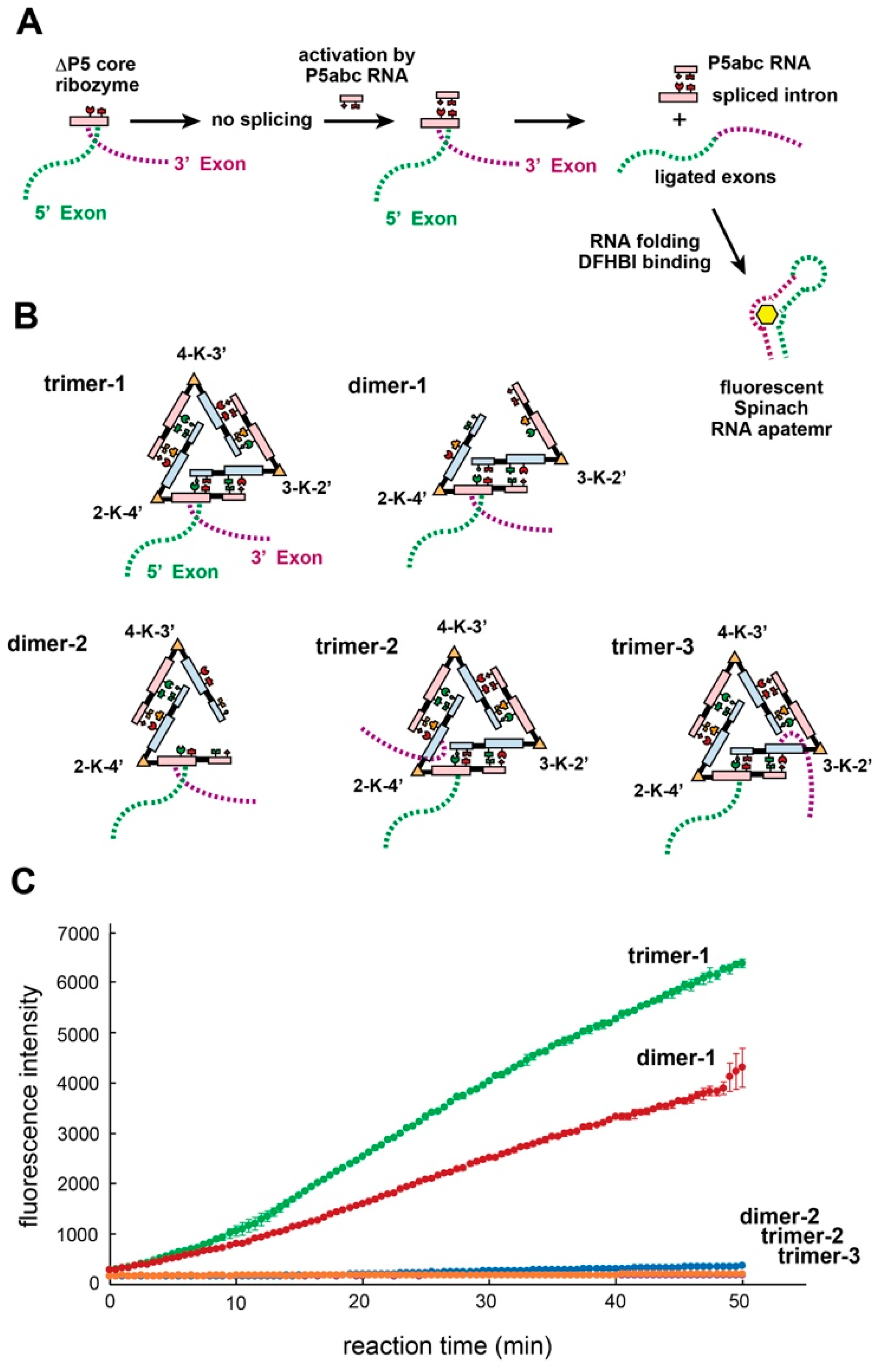
2.8. Trans-Splicing Monitored by Fluorescence of Spinach RNA–DFHBI

To prepare three kUrd monomer solutions, aqueous solutions (17 μL) containing appropriate pairs of α- and β-chain RNAs (final concentration of each kUrd: 0.25 μM) were heated at 80 °C for 5 min and then cooled to 37 °C. Then, 10× concentrated reaction buffer (2 μL, final concentration: 40 mM Tris-Cl, pH 7.5, 125 mM KCl, 5 mM MgCl2 and 10 μM DFHBI) and 2 mM guanosine triphosphate (1 μL, final concentration: 0.2 mM) were added to the RNA solution and incubated at 37 °C for 30 min to form a given kUrd monomer. The resulting kUrd monomer solutions were then mixed to afford a kUrd oligomer solution (60 μL) containing 0.25 μM each kUrd and initiate the trans-splicing reaction. The solution was transferred quickly to the wells of a microplate reader with a mineral oil overlay. The plate was then incubated at 37 °C in a plate reader (Infinite F200 Pro; Tecan, Männedorf, Switzerland), which was pre-warmed at 37 °C and recorded fluorescence intensity of the sample solution.

3. Results

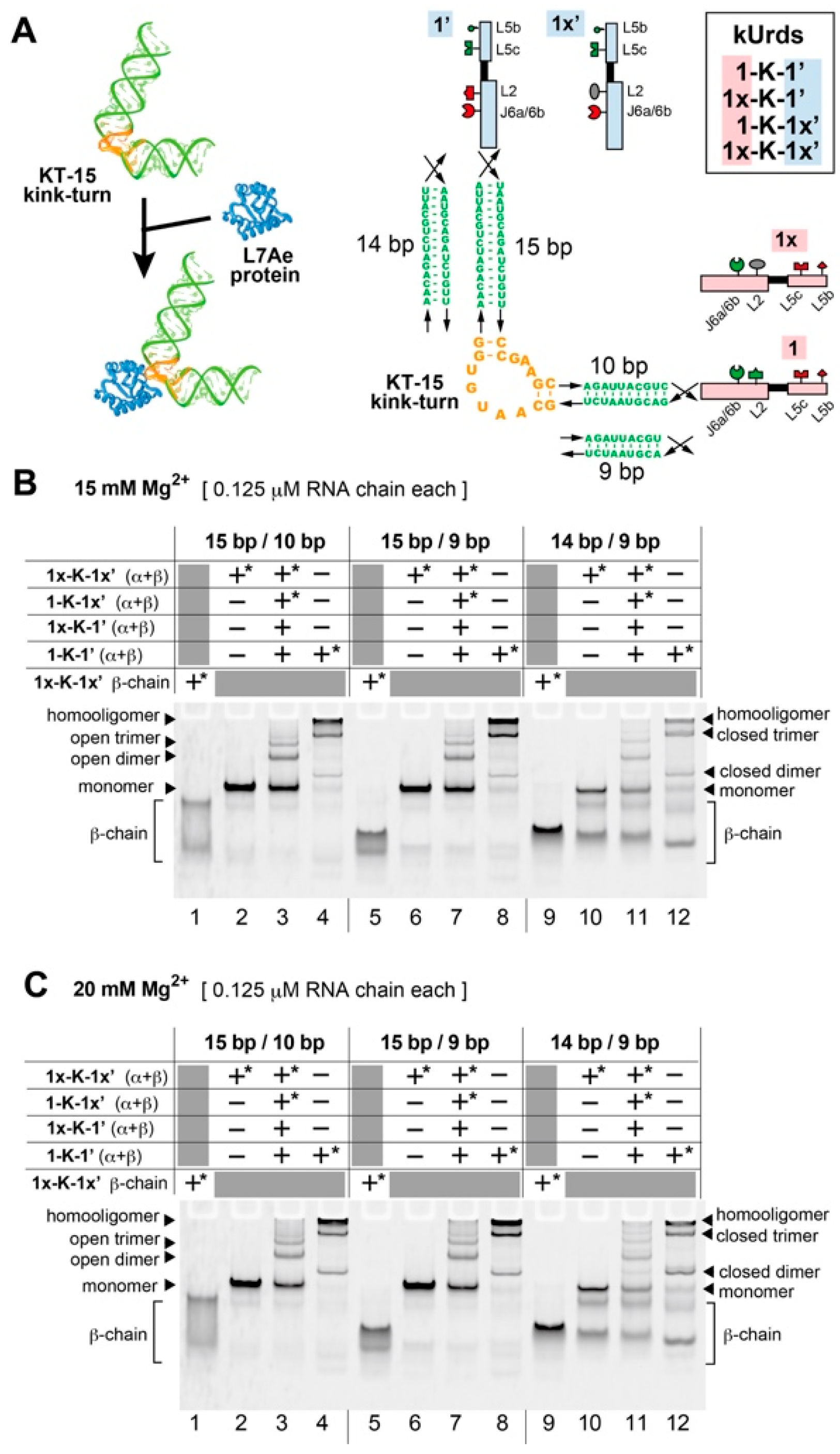
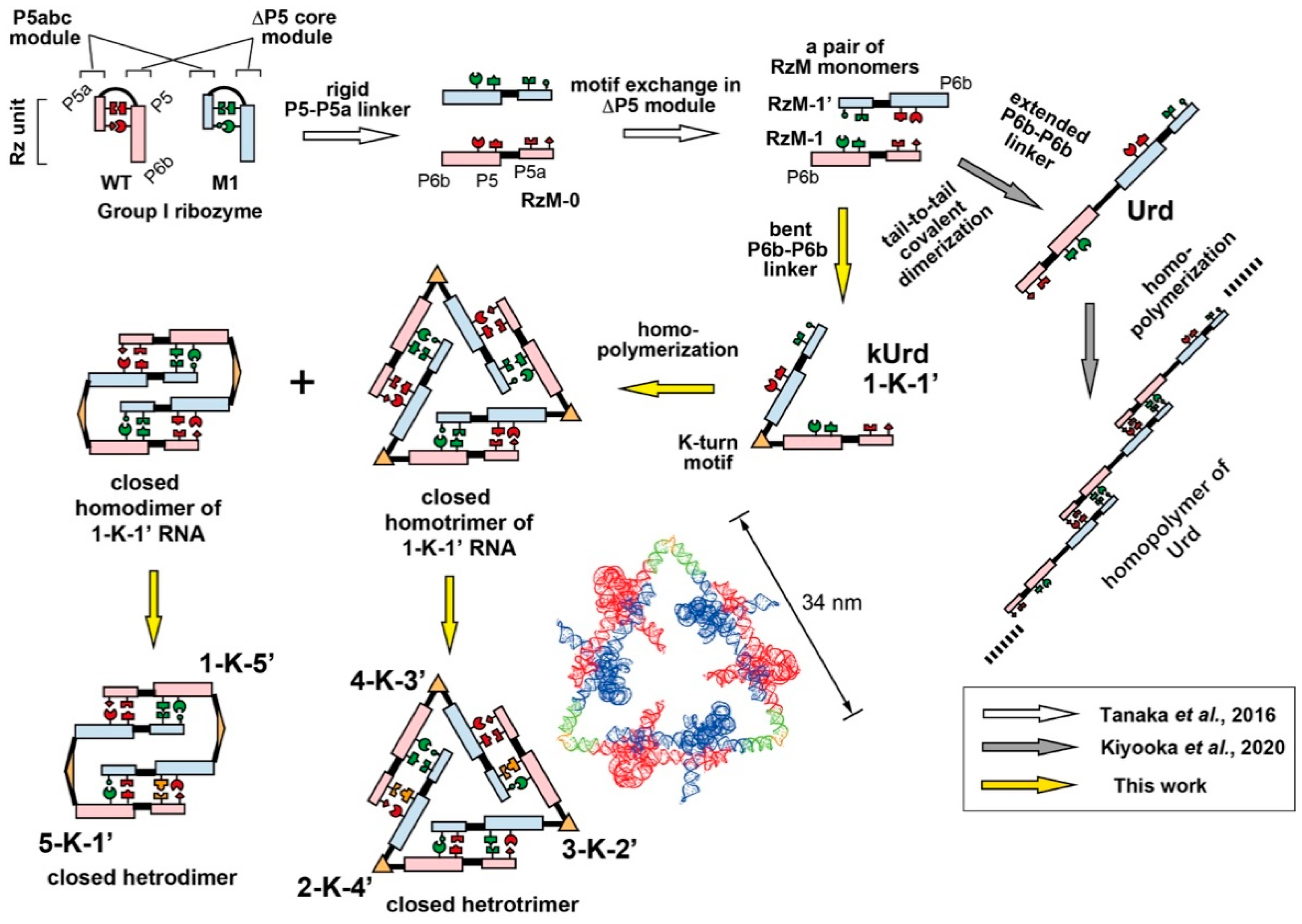
3.1. Design of Unit Ribozyme Dimers (Urds) with a Bent Linker

To design a new series of Urds to form closed trimers, we needed a suitable RNA motif consisting of two helices with fixed (~60°) angles as a bent linker unit. We selected the kink-turn RNA motif (Figure 1 and Figure S2) [30], which is one of the most extensively studied RNA motif families and provides a sharp (~60°) bend to the helical axis (Figure 2A). Kink-turn motifs have been identified in a diverse range of naturally occurring RNA structures with various functions [30]. Here, we chose the KT-15 kink-turn motif (Figure 2A), which not only serves as an RNA structural element but also acts as a recognition element for the ribosomal protein, L7Ae (Figure 2A) [30]. This RNA–protein complex has also been used as a module in synthetic RNA–protein nanostructures [33,34,38,39] and also synthetic RNA switches [40,41,42,43].

3.2. Homooligomerization of kUrds

A new class of Urd variants bearing the KT-15 motif was designated as kink-turn unit ribozyme dimer (abbreviated as kUrd, Figure 1). To optimize kUrd linkers, we compared three linker elements, each of which had the KT-15 motif flanked by a pair of duplexes with 15 bp/10 bp, 15 bp/9 bp, or 14 bp/9 bp (Figure 2A). The three distinct kUrd linkers were used to connect two RzM monomers (RzM-1 and RzM-1′) in a tail-to-tail manner, yielding three 1-K-1′ RNAs (Figure 2A and Figure S2). The three 1-K-1′ were designed to homotrimerize to form closed trimers (Figure 1). For each 1-K-1′, we prepared 1x-K-1′ and 1-K-1x′ to eliminate assembly ability of the RzM on one side by introducing a UUCG tetraloop [44,45] into the L2 element (where x designates L2 UUCG, see Figures S1D and S3). We also prepared 1x-K-1x′ mutant for each 1-K-1′ to eliminate assembly ability of RzMs on both sides (Figure S3). We analyzed three 1-K-1′ kUrds with distinct pairs of helices by electrophoretic mobility shift assay (EMSA) to characterize their assembly properties. Each 1-K-1′ kUrd was composed of two RNA strands (α- and β-RNA chains) (Figures S2B and S3, Table S1). Preliminary experiment showed that the two RNA strands assembled efficiently to form the kUrd monomer (Figure S4). We first labeled the 3′-end of the β-chain RNA with the BODIPY fluorophore [32] to visualize kUrd and its assembly in native gels in the presence of 10 mM Mg2+ (Figure S5A), 15 mM Mg2+ (Figure 2B) and 20 mM Mg2+ (Figure 2C).
BODIPY-labeled β-chain RNA of 1x-K-1x′, which can also be used as β-chain RNA of 1-K-1x′, showed broad and multiple bands (lanes 1, 5 and 9 in Figure 2B,C). The β-chain RNA, however, showed a sharp and retarded band in the presence of an equimolar amount of the partner α-chain RNA (lanes 2, 6 and 10 in Figure 2B,C), indicating assembly of the α- and β-chains RNAs to form a structured kUrd 1x-K-1x′ monomer. Introduction of two L2-UUCG loops into both of the RzMs in kUrd afforded 1x-K-1x′ to selectively form a monomeric state (lanes 2, 6 and 10 in Figure 2B,C). We then confirmed the assembly state of 1-K-1′ and its mutants by preparing a solution containing equal amounts of four kUrds (1x-K-1x′, 1x-K-1′, 1-K-1x′ and 1-K-1′), prepared by mixing two α-chain RNAs and two β-chain RNAs in a single tube (lanes 3, 7 and 11 in Figure 2B,C). BODIPY-labeled β-chain RNAs for 1x-K-1x′ and 1-K-1x′ selectively visualized kUrd monomer (1x-K-1x′), kUrd dimer (1x-K-1′:1-K-1x′) and kUrd open trimer (1x-K-1′:1-K-1′:1-K-1x′). In addition, to kUrd monomers, two new bands with lower mobilities were observed (lanes 3, 7 and 11 in Figure 2B,C), which were expected to be a kUrd dimer consisting mainly of 1x-K-1′:1-K-1x′ and a kUrd open trimer consisting mainly of 1x-K-1′:1-K-1′:1-K-1x′.
We then analyzed the solution containing only 1-K-1′ (lanes 4, 8 and 12 in Figure 2B,C), which was expected to form homooligomers, including a closed form homotrimer. The dominant band was retained in the gel slot, suggesting the formation of high-molecular weight oligomers. Two new bands, which were not seen in lanes 3, 7 and 11, were also observed. The lower mobility band would correspond to the desired closed trimer containing six ribozyme units (lanes 4, 8 and 12 in Figure 2B,C). The higher mobility band could be a closed dimer of Urds, which was not predicted in molecular modeling of 1-K-1′. In the presence of 10 mM Mg2+, the unit kUrd appeared to form efficiently (lanes 2, 6 and 10 in Figure S5A, see also Figure S5B,C) but oligomeric states of kUrds were formed less efficiently than those with 15 mM Mg2+ (Figure S5B,C). Formation of oligomeric states of kUrds seemed enhanced in the presence of 20 mM Mg2+ compared to 15 mM Mg2+ (Figure S5B). Among the three kUrds with distinct pairs of duplexes (15 bp/10 bp, 15 bp/9 bp and 14bp/9 bp) in the kink-turn linker elements, the 15 bp/10 bp and 15 bp/9 bp linkers worked better than the 14 bp/9 bp linker, which produced multiple bands in the presence of 20 mM Mg2+ (Figure 2C). In the following experiments, we used the 15 bp/10 bp pair as the kink-turn linker element.

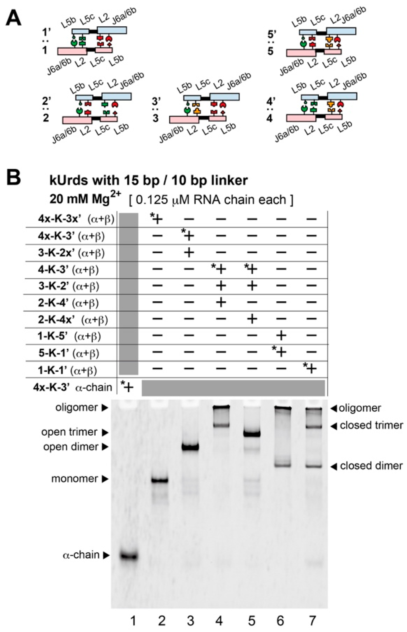
3.3. Block Copolymerization of kUrds

Based on the homooligomerization of 1-K-1′ forming the desired closed trimer and unexpected closed dimer, we then examined selective formation of a closed trimer and a closed dimer through copolymerization of two or three different kUrds (Figure 1). We substituted two RzMs in 1-K-1′ to prepare a trio of Urds (3-K-2′, 2-K-4′ and 4-K-3′) for triblock-type kUrd copolymer. For this purpose, we introduced three RzM:RzM interactions 2:2′, 3:3′ and 4:4′ (Figure 3A and Figure S1D), which were designed to be orthogonal to one another in their recognition specificity [28]. In a similar manner, a pair of Urds (1-K-5′ and 5-K-1′) were also designed for diblock-type kUrd copolymer (Figure 3A and Figure S1D), which would selectively yield a closed dimer. In block copolymerization by two or three distinct kUrds, we first prepared each kUrd separately by assembling its α- and β-RNA chain RNAs (Figures S2B and S4). Solutions of preformed kUrds were then mixed to form block copolymers of kUrds.
We first examined copolymerization of 3-K-2′, 2-K-4′ and 4-K-3′ to obtain the selective formation of a closed trimer (Figure 3B). To distinguish the closed trimer from the open trimer, we also examined 4-K-3x′, with which ring closure would be blocked by disruption of the 3:3′ interface. As seen in lane 4 in Figure 3B, assembly of three kUrds provided a migrating band the mobility of which was close to that of the band corresponding to the possible closed trimer in homopolymerization of 1-K-1′ (lane 7 in Figure 3B). Higher oligomerization of kUrds seen in 1-K-1′ homopolymers (lane 7 in Figure 3B) was still seen in triblock copolymers (lane 4 in Figure 3B). In the presence of 4-K-3x′ in place of 4-K-3′, no band was retained in the sample well (lane 5 in Figure 3B), consistent with the molecular design to form the open block trimer (3-K-2′:2-K-4′:4-K-3x′) lacking further oligomerization ability. Selective formation of the open form of kUrd trimer was suggested by the single migrating band (lane 5 in Figure 3B), which migrated slower than the open kUrd dimer (4x-K-3:3-K-2x′, see lane 3 in Figure 3B) but faster than the possible closed trimers (lanes 5 and 7 in Figure 3B). Importantly, in the trio of kUrd designed for triblock copolymer (lane 4 in Figure 3B), no band corresponding to the possible closed dimer of 1-K-1′ (see lane 7 in Figure 3B) was observed. Bands corresponding to the open dimer and monomer were not seen in lane 4 (Figure 3B), suggesting that triblock oligomerization proceeded efficiently. Comparison between two trios of kUrds with/without ring closure ability (lanes 4 and 5 in Figure 3B) suggested that the closed trimer is thermodynamically stable and the dissociation of RzM interfaces in the closed trimer occurred poorly.
The pair of kUrds for diblock copolymer also formed oligomers with no migration (lane 6 in Figure 3B). This mixture also yielded a band the mobility of which corresponded to the possible closed dimer of 1-K-1′ (lanes 6 and 7 in Figure 3B). No band was seen in lane 6 at the position corresponding to that of the possible closed trimer. In the closed heterotrimer consisting of three distinct kUrds, each kUrd was formed by α- and β-chain RNAs. As the kUrd closed heterotrimer contained six distinct RNA chains, we labeled one of six RNA chains with BODIPY fluorophore and monitored the mobility of the closed trimer on native gels (Figure S6). Regardless of the identity of the BODIPY-labeled RNA chain, all EMSA showed similar behavior, supporting the formation of the closed heterotrimer by assembly of three kUrds.

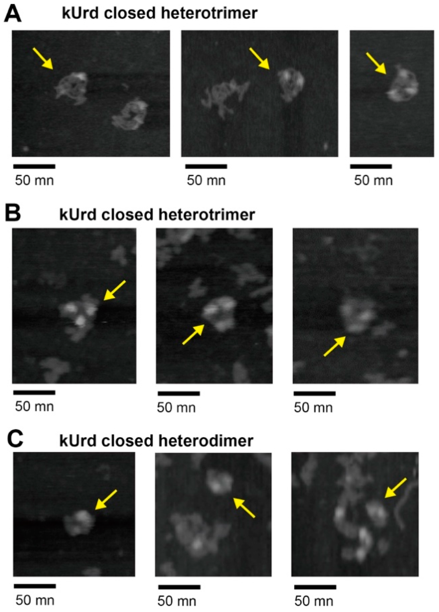
3.4. Atomic Force Microscopy Analysis of kUrd Heterotrimer and Heterodimer

Based on the observation that three kUrds are likely to form closed trimers in both homopolymerization and triblock copolymerization, we visualized the molecular structures of the heterotrimer (3-K-2′:2-K-4′:4-K-3′) and heterodimer (1-K-5′:5-K-1′) by atomic force microscopy (AFM) [33,37,38,39]. In the presence of 17.5 mM Mg2+, a sample containing three kUrds gave AFM images containing objects with triangular shapes (Figure 4A,B). The triangular objects were observed with limited frequency. We measured lengths of 21 sides of seven triangular objects observed in AFM images. While their lengths varied between 28.1 nm 39.6 nm, their average value was 34.5 nm. This value was closely similar to that predicted by molecular modeling of the closed trimer (approximately 34 nm, Figure 1). In some of the AFM images of kUrd triangles, we observed triangular shapes as three bright regions (Figure 4B), heights of which were 6~8 nm (Figure S7A).
We then analyzed AFM images of a solution containing 1-K-5′ and 5-K-1′ that formed a closed heterodimer, 1-K-5′:5-K-1′ (Figure 4C). Due to the limited efficacy of the closed dimer formation (Figure 3B), AFM provided the corresponding images inefficiently. A few RNA objects, however, were found to reflect closed dimers (Figure 4C), in which two bright spots were observed in each object. Heights of bright spots were 5~7 nm (Figure S7B). We also analyzed AFM images of two mutant kUrds with disrupted L2 interacting motifs (Figure S8). They retained some of RNA interacting motifs but were not able to form kUrd dimers (Figure S8A). The two mutant kUrds formed some aggregates on mica surfaces but gave no bright spots in the presence of 17.5 mM Mg2+ (Figure S8C,D).

3.5. Decoration of the kUrd Closed Trimer with Kink-Turn Binding Protein L7Ae

We then decorated the kUrd closed trimer with the RNA binding protein, L7Ae, for which the KT-15 kink-turn motif serves as a binding element (Figure 2A) [30]. We performed EMSA of the heterotrimer (3-K-2′:2-K-4′:4-K-3′) in the absence and presence of L7Ae protein. In the presence of each RNA chain at 0.125 μM (2.5 pmol each in 20 μL of solution), theoretical amounts of the KT-15 motif in the resulting three distinct kUrd units (7.5 pmol in 20 μL of solution) was also 0.375 μM (7.5 pmol in 20 μL of solution).
In the presence of 0.375 μM L7Ae protein, slight but distinct retardation of the closed trimer was observed (lane 6 in Figure 5A). A twofold molar excess amount of L7Ae protein (0.75 μM) over the theoretical amount of kink-turn motif (0.375 μM) did not cause further retardation of the band (lane 7 in Figure 5A). The mobility of the trimer band did not change in the presence of a greater excess of L7Ae protein (Figure S9). These observations suggested that the retarded band corresponded to the RNA–protein complex consisting of the kUrd closed trimer possessing three KT-15 kink-turns and three L7Ae protein molecules. In the kUrd trimer–L7Ae complex, fluorescent visualization of L7Ae proteins was also examined using an engineered L7Ae protein fused to enhanced green fluorescent protein (EGFP) (Figure 5B) [46].
L7Ae–EGFP fusion protein, which showed a broad band in free form (lane 4 in Figure 5B), shifted significantly in the presence of the kUrd heterotrimer (lane 3 in Figure 5B). In lane 3, the major band migrated more slowly than the free kUrd heterotrimer (lane 1) and also more slowly than the complex of closed trimer and L7Ae protein (lane 2). The major band in lane 3 corresponded to a complex of the closed trimer and L7Ae–EGFP fusion protein. The faster migrating band in lane 3 may correspond to an RNA–protein complex containing an open trimer or an open dimer because it was observed very weakly as a byproduct in assembly of the closed trimer (lane 4 in Figure S9B).

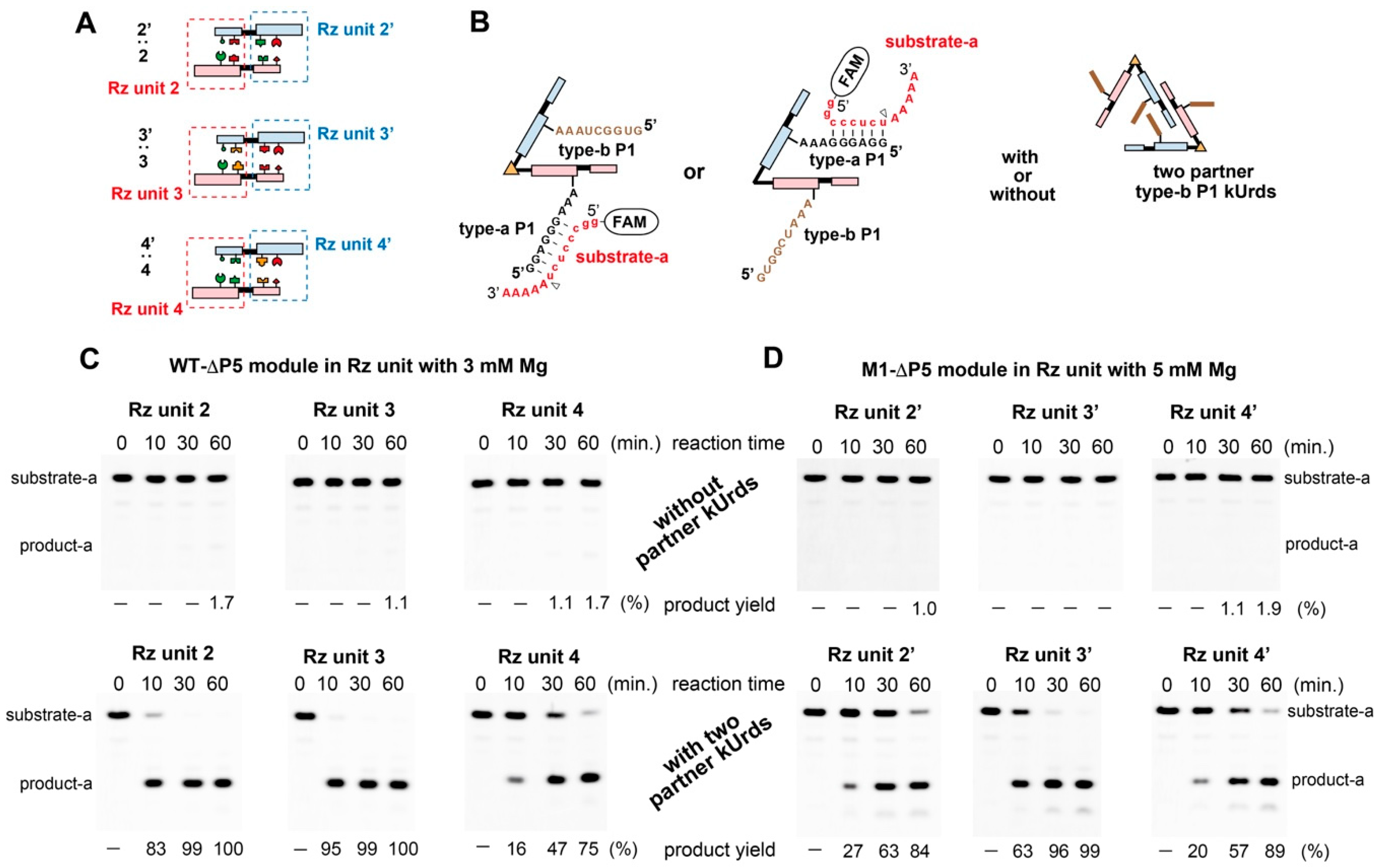
3.6. Catalytic Activities of kUrds and Their Oligomers

Although closed trimers and closed dimers were not formed exclusively without higher oligomers, it was interesting to see the catalytic abilities of ribozyme units in kUrds because their assembly would produce an active ribozyme unit consisting of the ∆P5 core ribozyme module and P5abc activator module (Figure 1 and Figure 6A) [18].
As the ∆P5 core ribozyme can be activated by transient (weak) association of the P5abc module, activity assay required a lower Mg2+ concentration than EMSA [36]. In the substrate cleavage reactions by the wild-type ∆P5 module (Figure S1A) and M1 type ∆P5 module (Figure S1B), the efficiencies of P5abc-depedent activation were optimum in the presence of 3 mM Mg2+ and 5 mM Mg2+, respectively. In the closed heterotrimer, six ∆P5-P5abc ribozyme units were formed, in which three had the wild-type ∆P5 modules (units 2, 3 and 4) with the remaining three having the M1 type ∆P5 module (units 2′, 3′ and 4′) (Figure 6A). We examined activation of the ∆P5 core ribozyme by the P5abc module upon formation of kUrd oligomers including the closed trimers.
We developed an assay system with which each ∆P5-P5abc ribozyme unit in kUrd oligomers could be analyzed separately. In the ∆P5-P5abc ribozyme unit, the P1 substrate recognition element for the cleavage reaction could be introduced in a modular manner. In EMSA and AFM analysis, α- and β-RNA chain RNAs commonly possessed a P1 recognition element for type-a substrate. To analyze the single ribozyme units in kUrds and in their oligomers selectively, we employed the second P1 element recognizing type-b substrate (Figure 6B) [47]. The substrate recognition of type-a and type-b P1 elements are highly orthogonal to each other [37]. The type-a substrate-P1 pair was introduced selectively to one of six ribozyme units, while the remaining five units were designed to possess type-b P1 elements (Figure 6B). The cleavage reaction of type-a substrate thus, enabled us to selectively monitor the catalytic ability of a ribozyme unit with a type-a P1 element (Figure 6B).
In the isolated state of each kUrd, its wild-type ∆P5 ribozyme module possessing the type-a P1 element showed no activity in the presence of 3 mM Mg2+ (Figure 6C, top). Addition of the partner kUrds in which the ∆P5 ribozyme module possessed only type-b P1 elements, however, induced the catalytic ability to cleave the type-a substrate (Figure 6C, bottom). In a similar manner, the M1 type ribozyme unit in each kUrd was also inactive with 5 mM Mg2+ (Figure 6D, top) but activated by the addition of partner kUrds (Figure 6D, bottom). Such activation behavior was primarily conducted by the association of ∆P5 catalytic modules in each kUrd with P5abc activator modules with the partner kUrds. It should be noted that moderate differences were observed in the catalytic activities among the three ribozyme units sharing the wild-type (unit 2, 3 and 4) or M1 type (unit 2′, 3′ and 4′) ∆P5 module (Figure 6C,D). The catalytic activity of the ∆P5-P5abc ribozyme unit was governed by the identity of the ∆P5 module and also by the identity of the RNA–RNA interface between the ∆P5 module and the P5abc module (Figure 6A) [35,36].

3.7. Trans-Splicing Reaction Promoted by kUrd Oligomer Formation

The intron form of ∆P5 core ribozyme module can perform the self-splicing reaction to yield ligated exons in the presence of the P5abc activator module (Figure 7A) [18]. The pair of ∆P5 core ribozyme modules in each kUrd were composed of α- and β-chain RNAs. In the intron form of wild-type ∆P5 core ribozyme module on one side of kUrd, 5′ and 3′ exons were provided by α- and β-chain RNAs, respectively (Figure S10A). In the intron form of the M1 type ∆P5 core ribozyme module on the opposite side of kUrd, 5′ and 3′ exons were provided by β- and α-chain RNAs, respectively (Figure S6B). The organization of α- and β-chain RNAs in the intron form of the ∆P5 core ribozyme modules would conduct trans-splicing reaction joining 5′ and 3′ exons, which were provided by two separate RNA chains, to produce a mature exon sequence (Figure S10A,B) [31,48]. Trans-splicing promoted by group I ribozyme has been recognized as a promising strategy to repair mRNAs of disease-related genes bearing undesirable mutations [48,49,50]. Therefore, we examined trans-splicing reaction catalyzed by a kUrd in triblock copolymers (3-K-2′:2-K-4′:4-K-3′) including the closed trimer.
We installed 5′ and 3′ exon sequences and their recognition elements into the α- and β- chain RNAs of 2-K-4′ kUrd, respectively (Figure S10C and Table S2). Exon sequences were appended to the ribozyme module consisting of the wild-type ∆P5 ribozyme module activated by the P5abc module provided by 3-K-2′ kUrd through the 2:2′ interface (Figure 7B and Figure S10C). We used a shortened derivative of the Spinach RNA aptamer as a ligated exon sequence dissected in precursor form (Figure 7A) [51]. Spinach RNA is an RNA receptor for DFHBI and its fluorescence is induced by restricting the molecular motion of DFHBI in the complex form [52]. The progression of trans-splicing reaction can be monitored in the presence of DFHBI [51,53]. To evaluate the effects of kUrd oligomer formation on the trans-splicing reaction, we prepared a set of kUrd variants some of which possessed 5′ and 3′ exon sequences (Figure 7B and Figure S10, see also Table S2). A trio of kUrds (3-K-2′:2-K-4′:4-K-3′) to form a triblock oligomer (trimer-1) exhibited an increase in emission (green plot in Figure 7C). We also examined two sets of triblock oligomers (trimer-2 and trimer-3) in which the 5′ exon and 3′ exon were installed to incorrect RNA chains and the two exons were unable to meet in one ∆P5 ribozyme unit. The two sets of kUrds showed no increase in fluorescence from Spinach RNA complexed with DFHBI (purple plot and blue plot in Figure 7C). These data indicated that the correct assignments of 5′ and 3′ exons in six RNA chains are crucial in the oligomeric state of kUrds.
To evaluate the effects of triblock oligomerization (3-K-2′:2-K-4′:4-K-3′) in the splicing proficient trimer (trimer-1), we assayed a solution containing 2-K-4′ and 3-K-2′, which form dimer-1, in which the intron form of the ribozyme unit in 2-K-4′ was formed correctly and 2-K-4′ was correctly activated by the P5abc module in 3-K-2′ (Figure 7B). The difference in activity between the presence and absence of the third kUrd unit (4-K-3′) mainly reflected the oligomerization ability. The solution of dimer-1 exhibited emission (red plot in Figure 7C), which was, however, about two thirds that of the solution containing three kUrds. This observation suggested that 4-K-3′ improved the activity of the splicing ribozyme unit through the formation of oligomeric states. To examine the contribution of the P5abc module provided by 3-K-2′, we depleted the 3-K-2′ unit from the three kUrd components. The sample solution containing 2-K-4′ and 4-K-3′ to form dimer-2 showed no increase in emission (orange plot in Figure 7C), indicating the importance of activation by the P5abc module to conduct full splicing ability of the ∆P5 ribozyme module possessing 5′ and 3′ exons.

4. Discussion

In this study, we performed molecular modeling of kUrd to design a closed trimer, the formation of which was confirmed by EMSA and AFM analyses. On the other hand, significant formation of higher oligomers indicated that the current structural design was insufficient partly due to the 3D structure of the full-length Tetrahymena ribozyme used for the molecular design in this study. In molecular modeling of kUrds (a dimeric form of an engineered Tetrahymena ribozyme connected by the kink-turn motif), we used a model 3D structure of the full-length ribozyme and the X-ray crystallographic structure of a shortened form of the ribozyme lacking some peripheral elements [29]. There may have been some deviations from the full-length Tetrahymena ribozyme 3D structure, which has yet to be solved by X-ray crystallography. Improvement of 3D structural design, however, may be possible because a new 3D model structure of the full-length ribozyme was constructed recently by combining cryo-EM data, chemical probing data and molecular modeling [54]. Although the resolution of this new model is modest (6.8 Å), it will be useful to refine the design of ribozyme-based nanostructures.
The formation of the closed dimer is also an issue to be resolved for precise design of kUrd-based RNA nanostructures. Formation of the closed kUrd dimer, which was unexpected from molecular modeling, suggested the structural flexibility of kUrd RNAs and entropic effects in RNA assembly. The flexible nature of the kink-turn RNA motifs has been analyzed and reported previously [55,56,57]. Therefore, we hypothesized that the formation of the closed dimer may also be supported by the structural flexibility of the KT-15 kink-turn motif. Distinct from our previous Urd with an extended linker, each kUrd can accept L7Ae protein at its linker element bearing the KT-15 kink-turn motif. EMSA of the heterotrimer in the presence of L7Ae suggested the formation of KT-15–L7Ae complex at each corner of the triangle-shaped closed kUrd trimer. We also performed AFM analysis of the heterotrimer in the presence of L7Ae but no significant differences were observed between images of kUrds with and without L7Ae. This was conceivable because L7Ae is much smaller than kUrd RNA. We also found that L7Ae protein strongly inhibited the catalytic ability of kUrds. Collaboration between L7Ae protein and the Tetrahymena ribozyme unit in catalysis is still an open issue.
In this study, we mainly analyzed the closed trimer and closed dimer of kUrds. The closed trimer contained six ribozyme units and three kink-turn motifs that interacted with L7Ae proteins. In reactions promoted by the ribozyme unit in kUrd oligomers, trans-splicing reaction proceeded more efficiently in the presence of three kUrds forming a heterotrimer than two kUrds providing the core ∆P5 ribozyme and P5abc activator. Although the extent of improvement was moderate, this was a promising result as an initial step toward RNA processing in vivo controlled by ribozyme-based nanostructures. The kUrd trimer containing six group I ribozyme units consists of nearly 2400 nucleotides, which is comparable to bacterial 16S and 23S ribosomal RNAs. The size of the triangle shape (with an edge of approximately 34 nm) was also comparable to the bacterial ribosome (approximately 20 nm in diameter). Although the structure and function of kUrd-based nanostructures are still primitive, this study represents an initial step toward artificial construction of complex RNA nanostructures capable of playing crucial roles in naturally occurring biological systems. Trans-splicing ribozymes have been investigated as a promising tool to repair mutated mRNAs causing genetic disorders [49,50]. Ribozyme dimers employed in this study may be used to repair two distinct mutated mRNA simultaneously and cooperatively. Two ribozyme units in a dimer also target the same mRNA sequence, to which two trans-splicing ribozyme units perform distinct splicing reactions to yield different products. For example, one ribozyme unit repairs the mutation of the target mRNA whereas the other ribozyme unit attaches a fluorescent RNA aptamer to the target mRNA for its fluorescent imaging. Fabrication of closed trimers using ribozyme dimers and KT-15 kink-turn would enable us to attach various protein components through L7Ae protein that binds to KT-15 [38,39]. Such ribozyme-based RNA-protein nanoparticle can be further decorated by RNA-based sensors and therapeutic RNA modules such as RNA aptamers and siRNAs, with which next generation of RNA-based nanomedicines may be developed.

Supplementary Materials

The following are available online at https://www.mdpi.com/2076-3417/11/6/2583/s1, Figure S1: Sequences and secondary structures of the Tetrahymena group I ribozymes and their structural elements used for modular engineering in this study, Figure S2: Scheme of rational redesign of unimolecular Tetrahymena ribozymes to generate kUrd 1-K-1′, Figure S3: Kink-turn unit ribozyme dimers (kUrds) employed in this study, Figure S4: Formation of kUrds through the assembly of their α-chain and β-chain RNAs, Figure S5: Oligomerization of 1-K-1′and its mutants, Figure S6: EMSA of triblock copolymers formed in the presence of 20 mM Mg2+, Figure S7: AFM images of the closed heterotrimer and heterodimer and their cross sections, Figure S8: AFM images of the closed heterodimer and its mutant monomers, Figure S9: Complex formation of L7Ae proteins with KT-15 kink-turn motifs in a closed kUrd heterotrimer, Figure S10: Closed kUrd trimers and their partial dimers used for analysis of the trans-splicing reaction.

Author Contributions

Conceptualization, Y.I.; methodology, Y.I., H.S. (Hirohide Saito), H.S. (Hiroshi Sugiyama), M.E. and S.M.; investigation, J.A., K.H. and Y.F.; writing—original draft preparation, Y.I. and T.Y.; writing—review and editing, H.S. (Hirohide Saito), H.S. (Hiroshi Sugiyama), M.E. and S.M.; supervision, Y.I., H.S. (Hirohide Saito), H.S. (Hiroshi Sugiyama) and M.E. All authors have read and agreed to the published version of the manuscript.

Funding

This research was funded by University of Toyama Discretionary Funds of the President “Toyama RNA Collaborative Research” (to Y.I. and S.M.).

Institutional Review Board Statement

Not applicable.

Informed Consent Statement

Not applicable.

Data Availability Statement

Not applicable.

Conflicts of Interest

The authors declare no conflict of interest.

References

  1. Goodsell, D.S.; Olson, A.J. Structural symmetry and protein function. Annu. Rev. Biophys. Biomol. Struct. 2000, 29, 105–153. [Google Scholar] [CrossRef]
  2. Lesieur, C. The assembly of protein oligomers: Old stories and new perspectives with graph theory. In Oligomerization of Chemical and Biological Compounds; Lesieur, C., Ed.; IntecOpen: London, UK, 2014. [Google Scholar] [CrossRef] [Green Version]
  3. Jones, S.; Thornton, J.M. Protein-protein interactions: A review of protein dimer structures. Prog. Biophys. Mol. Biol. 1995, 63, 31–65. [Google Scholar] [CrossRef]
  4. Glover, D.J.; Clark, D.S. Protein calligraphy: A new concept begins to take shape. ACS Cent. Sci. 2016, 2, 438–444. [Google Scholar] [CrossRef] [Green Version]
  5. Sun, H.; Luo, Q.; Hou, C.; Liu, J. Nanostructures based on protein self-assembly: From hierarchical construction to bioinspired materials. Nano Today 2017, 14, 16–41. [Google Scholar] [CrossRef]
  6. Kuan, S.L.; Bergamini, F.R.G.; Weil, T. Functional protein nanostructures: A chemical toolbox. Chem. Soc. Rev. 2018, 47, 9069–9105. [Google Scholar] [CrossRef] [PubMed] [Green Version]
  7. Boyken, S.E.; Chen, Z.; Groves, B.; Langan, R.A.; Oberdorfer, G.; Ford, A.; Gilmore, J.M.; Xu, C.; DiMaio, F.; Pereira, J.H.; et al. De novo design of protein homo-oligomers with modular hydrogen-bond network-mediated specificity. Science 2016, 352, 680–687. [Google Scholar] [CrossRef] [Green Version]
  8. Fallas, J.A.; Ueda, G.; Sheffler, W.; Nguyen, V.; McNamara, D.E.; Sankaran, B.; Pereira, J.H.; Parmeggiani, F.; Brunette, T.J.; Cascio, D.; et al. Computational design of self-assembling cyclic protein homo-oligomers. Nat. Chem. 2017, 9, 353–360. [Google Scholar] [CrossRef] [Green Version]
  9. Kuhlman, B.; O’Neill, J.W.; Kim, D.E.; Zhang, K.Y.; Baker, D. Conversion of monomeric protein L to an obligate dimer by computational protein design. Proc. Natl. Acad. Sci. USA 2001, 98, 10687–10691. [Google Scholar] [CrossRef] [Green Version]
  10. Stranges, P.B.; Machius, M.; Miley, M.J.; Tripathy, A.; Kuhlman, B. Computational design of a symmetric homodimer using beta-strand assembly. Proc. Natl. Acad. Sci. USA 2011, 108, 20562–20567. [Google Scholar] [CrossRef] [PubMed] [Green Version]
  11. Mou, Y.; Huang, P.S.; Hsu, F.C.; Huang, S.J.; Mayo, S.L. Computational design and experimental verification of a symmetric protein homodimer. Proc. Natl. Acad. Sci. USA 2015, 112, 10714–10719. [Google Scholar] [CrossRef] [Green Version]
  12. Fedor, M.J.; Williamson, J.R. The catalytic diversity of RNAs. Nat. Rev. Mol. Cell Biol. 2005, 6, 399–412. [Google Scholar] [CrossRef]
  13. Weinberg, C.E.; Weinberg, Z.; Hammann, C. Novel ribozymes: Discovery, catalytic mechanisms, and the quest to understand biological function. Nucleic Acids Res. 2019, 47, 9480–9494. [Google Scholar] [CrossRef] [PubMed]
  14. Breaker, R.R. Prospects for riboswitch discovery and analysis. Mol. Cell 2011, 4, 867–879. [Google Scholar] [CrossRef] [Green Version]
  15. Butcher, S.E.; Pyle, A.M. The molecular interactions that stabilize RNA tertiary structure: RNA motifs, patterns, and networks. Acc. Chem. Res. 2011, 44, 1302–1311. [Google Scholar] [CrossRef] [PubMed]
  16. Ishikawa, J.; Fujita, Y.; Maeda, Y.; Furuta, H.; Ikawa, Y. GNRA/receptor interacting modules: Versatile modular units for natural and artificial RNA architectures. Methods 2011, 54, 226–238. [Google Scholar] [CrossRef]
  17. Jarrell, K.A.; Dietrich, R.C.; Perlman, P.S. Group II intron domain 5 facilitates a trans-splicing reaction. Mol. Cell. Biol. 1988, 8, 2361–2366. [Google Scholar] [CrossRef] [Green Version]
  18. van der Horst, G.; Christian, A.; Inoue, T. Reconstitution of a group I intron self-splicing reaction with an activator RNA. Proc. Natl. Acad. Sci. USA 1991, 88, 184–188. [Google Scholar] [CrossRef] [Green Version]
  19. Engelhardt, M.A.; Doherty, E.A.; Knitt, D.S.; Doudna, J.A.; Herschlag, D. The P5abc peripheral element facilitates preorganization of the Tetrahymena group I ribozyme for catalysis. Biochemistry 2000, 39, 2639–2651. [Google Scholar] [CrossRef]
  20. Doudna, J.A.; Cech, T.R. Self-assembly of a group I intron active site from its component tertiary structural domains. RNA 1995, 1, 36–45. [Google Scholar]
  21. Ikawa, Y.; Shiraishi, H.; Inoue, T. Trans-activation of the Tetrahymena ribozyme by its P2-2.1 domains. J. Biochem. 1998, 123, 528–533. [Google Scholar] [CrossRef]
  22. Rahman, M.M.; Matsumura, S.; Ikawa, Y. Effects of molecular crowding on a bimolecular group I ribozyme and its derivative that self-assembles to form ribozyme oligomers. Biochem. Biophys. Res. Commun. 2018, 507, 136–141. [Google Scholar] [CrossRef]
  23. Pan, T. Higher order folding and domain analysis of the ribozyme from Bacillus subtilis ribonuclease P. Biochemistry 1995, 34, 902–909. [Google Scholar] [CrossRef]
  24. Loria, A.; Pan, T. Domain structure of the ribozyme from eubacterial ribonuclease P. RNA 1996, 2, 551–563. [Google Scholar]
  25. Kim, H.; Poelling, R.R.; Leeper, T.C.; Meyer, M.A.; Schmidt, F.J. In vitro transactivation of Bacillus subtilis RNase P RNA. FEBS Lett. 2001, 506, 235–238. [Google Scholar] [CrossRef] [Green Version]
  26. Tanaka, T.; Matsumura, S.; Furuta, H.; Ikawa, Y. Tecto-GIRz: Engineered group I ribozyme the catalytic ability of which can be controlled by self-dimerization. ChemBioChem 2016, 17, 1448–1455. [Google Scholar] [CrossRef] [PubMed]
  27. Tanaka, T.; Ikawa, Y.; Matsumura, S. Rational engineering of a modular group I ribozyme to control its activity by self-dimerization. Methods Mol. Biol. 2017, 1632, 325–340. [Google Scholar] [PubMed]
  28. Kiyooka, R.; Akagi, J.; Hidaka, K.; Sugiyama, H.; Endo, M.; Matsumura, S.; Ikawa, Y. Catalytic RNA nano-objects formed by self-assembly of group I ribozyme dimers serving as unit structures. J. Biosci. Bioeng. 2020, 130, 253–259. [Google Scholar] [CrossRef] [PubMed]
  29. Guo, F.; Gooding, A.R.; Cech, T.R. Structure of the Tetrahymena ribozyme: Base triple sandwich and metal ion at the active site. Mol. Cell 2004, 16, 351–362. [Google Scholar]
  30. Klein, D.J.; Schmeing, T.M.; Moore, P.B.; Steitz, T.A. The kink-turn: A new RNA secondary structure motif. EMBO J. 2001, 20, 4214–4221. [Google Scholar] [CrossRef] [PubMed]
  31. Tanaka, T.; Hirata, Y.; Tominaga, Y.; Furuta, H.; Matsumura, S.; Ikawa, Y. Heterodimerization of group I ribozymes enabling exon recombination through pairs of cooperative trans-splicing reactions. ChemBioChem 2017, 18, 1659–1667. [Google Scholar] [CrossRef]
  32. Ikawa, Y.; Moriyama, S.; Furuta, H. Facile syntheses of BODIPY derivatives for fluorescent labeling of the 3’ and 5’ ends of RNAs. Anal. Biochem. 2008, 378, 166–170. [Google Scholar] [CrossRef]
  33. Ohno, H.; Kobayashi, T.; Kabata, R.; Endo, K.; Iwasa, T.; Yoshimura, S.H.; Takeyasu, K.; Inoue, T.; Saito, H. Synthetic RNA-protein complex shaped like an equilateral triangle. Nat. Nanotechnol. 2011, 6, 116–120. [Google Scholar] [CrossRef] [PubMed] [Green Version]
  34. Ohuchi, S.J.; Sagawa, F.; Sakamoto, T.; Inoue, T. A trifunctional, triangular RNA-protein complex. FEBS Lett. 2015, 589, 2424–2428. [Google Scholar] [CrossRef] [PubMed] [Green Version]
  35. Tanaka, T.; Furuta, H.; Ikawa, Y. Installation of orthogonality to the interface that assembles two modular domains in the Tetrahymena group I ribozyme. J. Biosci. Bioeng. 2014, 117, 407–412. [Google Scholar] [CrossRef]
  36. Rahman, M.M.; Matsumura, S.; Ikawa, Y. Artificial RNA motifs expand the programmable assembly between RNA modules of a bimolecular ribozyme leading to application to RNA nanostructure design. Biology 2017, 6, 37. [Google Scholar] [CrossRef] [PubMed] [Green Version]
  37. Oi, H.; Fujita, D.; Suzuki, Y.; Sugiyama, H.; Endo, M.; Matsumura, S.; Ikawa, Y. Programmable formation of catalytic RNA triangles and squares by assembling modular RNA enzymes. J. Biochem. 2017, 161, 451–462. [Google Scholar] [CrossRef]
  38. Fujita, Y.; Furushima, R.; Ohno, H.; Sagawa, F.; Inoue, T. Cell-surface receptor control that depends on the size of a synthetic equilateral-triangular RNA-protein complex. Sci. Rep. 2014, 4, 6422. [Google Scholar] [CrossRef] [Green Version]
  39. Osada, E.; Suzuki, Y.; Hidaka, K.; Ohno, H.; Sugiyama, H.; Endo, M.; Saito, H. Engineering RNA-protein complexes with different shapes for imaging and therapeutic applications. ACS Nano. 2014, 8, 8130–8140. [Google Scholar] [CrossRef] [PubMed]
  40. Saito, H.; Kobayashi, T.; Hara, T.; Fujita, Y.; Hayashi, K.; Furushima, R.; Inoue, T. Synthetic translational regulation by an L7Ae-kink-turn RNP switch. Nat. Chem. Biol. 2010, 6, 71–78. [Google Scholar] [CrossRef]
  41. Wroblewska, L.; Kitada, T.; Endo, K.; Siciliano, V.; Stillo, B.; Saito, H.; Weiss, R. Mammalian synthetic circuits with RNA binding proteins for RNA-only delivery. Nat. Biotechnol. 2015, 33, 839–841. [Google Scholar] [CrossRef] [Green Version]
  42. Matsuura, S.; Ono, H.; Kawasaki, S.; Kuang, Y.; Fujita, Y.; Saito, H. Synthetic RNA-based logic computation in mammalian cells. Nat. Commun. 2018, 9, 4847. [Google Scholar] [CrossRef]
  43. Ohno, H.; Akamine, S.; Saito, H. Synthetic mRNA-based systems in mammalian cells. Adv. Biosyst. 2020, 4, e1900247. [Google Scholar] [CrossRef] [PubMed]
  44. Ennifar, E.; Nikulin, A.; Tishchenko, S.; Serganov, A.; Nevskaya, N.; Garber, M.; Ehresmann, B.; Ehresmann, C.; Nikonov, S.; Dumas, P. The crystal structure of UUCG tetraloop. J. Mol. Biol. 2000, 304, 35–42. [Google Scholar] [CrossRef]
  45. Hall, K.B. Mighty tiny. RNA 2015, 21, 630–631. [Google Scholar] [CrossRef] [PubMed]
  46. Zhang, G.; Gurtu, V.; Kain, S.R. An enhanced green fluorescent protein allows sensitive detection of gene transfer in mammalian cells. Biochem. Biophys. Res. Commun. 1996, 227, 707–711. [Google Scholar] [CrossRef]
  47. Campbell, T.B.; Cech, T.R. Mutations in the Tetrahymena ribozyme internal guide sequence: Effects on docking of the P1 helix into the catalytic core and correlation with catalytic activity. Biochemistry 1996, 35, 11493–11502. [Google Scholar] [CrossRef]
  48. Ikawa, Y.; Matsumura, S. Engineered group I ribozymes as RNA-based modular tools to control gene expression. In Applied RNA Bioscience; Masuda, S., Izawa., S., Eds.; Springer: Berlin, German, 2018; pp. 203–220. [Google Scholar]
  49. Müller, U.F. Design and experimental evolution of trans-splicing group I intron ribozymes. Molecules 2017, 22, 75. [Google Scholar] [CrossRef] [Green Version]
  50. Lee, C.H.; Han, S.R.; Lee, S.W. Therapeutic applications of group I intron-based trans-splicing ribozymes. Wiley Interdiscip. Rev. RNA 2018, 9, e1466. [Google Scholar] [CrossRef]
  51. Furukawa, A.; Maejima, T.; Matsumura, S.; Ikawa, Y. Characterization of an RNA receptor motif that recognizes a GCGA tetraloop. Biosci. Biotechnol. Biochem. 2016, 80, 1386–1389. [Google Scholar] [CrossRef] [PubMed] [Green Version]
  52. Paige, J.S.; Wu, K.Y.; Jaffrey, S.R. RNA mimics of green fluorescent protein. Science 2011, 333, 642–646. [Google Scholar] [CrossRef]
  53. Furukawa, A.; Tanaka, T.; Furuta, H.; Matsumura, S.; Ikawa, Y. Use of a fluorescent aptamer RNA as an exonic sequence to analyze self-splicing ability of a group I intron from structured RNAs. Biology 2016, 5, 43. [Google Scholar] [CrossRef] [PubMed]
  54. Kappel, K.; Zhang, K.; Su, Z.; Watkins, A.M.; Kladwang, W.; Li, S.; Pintilie, G.; Topkar, V.V.; Rangan, R.; Zheludev, I.N.; et al. Accelerated cryo-EM-guided determination of three-dimensional RNA-only structures. Nat. Methods 2020, 17, 699–707. [Google Scholar] [CrossRef] [PubMed]
  55. Matsumura, S.; Ikawa, Y.; Inoue, T. Biochemical characterization of the kink-turn RNA motif. Nucleic Acids Res. 2003, 31, 5544–5551. [Google Scholar] [CrossRef] [Green Version]
  56. Goody, T.A.; Melcher, S.E.; Norman, D.G.; Lilley, D.M. The kink-turn motif in RNA is dimorphic, and metal ion-dependent. RNA 2004, 10, 254–264. [Google Scholar] [CrossRef] [PubMed] [Green Version]
  57. Shi, X.; Huang, L.; Lilley, D.M.; Harbury, P.B.; Herschlag, D. The solution structural ensembles of RNA kink-turn motifs and their protein complexes. Nat. Chem. Biol. 2016, 12, 146–152. [Google Scholar] [CrossRef] [Green Version]
Figure 1. Design of closed oligomers based on engineered group I ribozyme dimers. Scheme of modular redesign to generate unit ribozyme dimers (kUrds) and their oligomerization. White arrows indicate structural redesign to prepare RzM RNAs and their noncovalent dimerization as reported by Tanaka et al. in 2016 [26]. Gray arrows indicate rational redesign for covalent dimerization of RzM RNAs reported by Kiyooka et al. in 2020 [28]. Yellow arrows indicate structural redesign to construct kUrd 1-K-1′ in this study. Sequences and secondary structures of wild-type ribozyme, M1 variant ribozyme and their structural elements are shown in Figure S1.
Applsci 11 02583 g001
Figure 2. Structures of kUrds and their assembly analyzed by EMSA. (A) 3D structures of KT-15 kink-turn motif (orange) with a pair of duplexes (green) and its binding with L7Ae protein (blue). Sequence and secondary structures of the linker elements of kUrds with three distinct pairs of duplexes. Small red and green icons indicate RNA motifs. Mutations in L2 elements to disrupt RzM:RzM interaction are also shown as elliptic gray icons. Sequences and secondary structures of these RNA motifs are shown in Figure S1. (B) EMSA of 1-K-1′ homopolymers formed in the presence of 15 mM Mg2+. Asterisks indicate RNA chains labeled with BODIPY fluorophore. (C) EMSA of 1-K-1′ homopolymers formed in the presence of 20 mM Mg2+. Asterisks indicate RNA chains labeled with BODIPY fluorophore.
Applsci 11 02583 g002
Figure 3. Assembly of a trio of kUrds to form a closed heterotrimer and a pair of kUrds to form a closed heterodimer. (A) Five distinct pairs of RzM:RzM interfaces, three of which were used in a closed heterotrimer configuration and the remaining two of which were used in a closed heterodimer configuration. Sequences and secondary structures of RNA motifs specifying RzM:RzM interfaces are shown in Figure S3. (B) EMSA of triblock and diblock kUrd copolymers formed in the presence of 20 mM Mg2+. Asterisks indicate RNA chains labeled with BODIPY fluorophore.
Applsci 11 02583 g003
Figure 4. AFM imaging of closed kUrd heterotrimers and closed heterodimers. (A,B) AFM of closed kUrd trimers in the presence of 17.5 mM Mg2+. Yellow arrows indicate closed kUrd trimers. (C) AFM of closed kUrd dimers in the presence of 17.5 mM Mg2+. Yellow arrows indicate closed kUrd dimers.
Applsci 11 02583 g004
Figure 5. Complex formation of L7Ae protein with KT-15 kink-turn motifs in a closed kUrd heterotrimer. (A) EMSA of a closed kUrd heterotrimer in the absence and presence of L7Ae protein. (B) EMSA of a closed kUrd heterotrimer in the absence and presence of L7Ae protein and L7Ae-EGFP fusion protein.
Applsci 11 02583 g005
Figure 6. Activation of ribozyme unit upon assembly of kUrds. (A) Formation of two ribozyme units in each RzM:RzM interface. One ribozyme unit (2, 3, or 4) formed the wild-type ribozyme and the other ribozyme unit (2′, 3′, or 4′) formed the M1 type ribozyme. (B) Sequences of substrate-a and two types of P1 binding elements. Location of P1 element in the secondary structure of the Tetrahymena group I ribozyme are shown in Figure S1. (C) Cleavage of substrate-a by the wild-type ribozyme unit in a given kUrd without (top) or with (bottom) its partner kUrds in the presence of 3 mM Mg2+. (D) Cleavage of substrate-a by the M1 type ribozyme unit in a given kUrd without (top) or with (bottom) its partner kUrds in the presence of 5 mM Mg2+.
Applsci 11 02583 g006
Figure 7. Trans-splicing reaction by the ribozyme unit 2 formed in the closed kUrd heterotrimer. (A) Parent cis-splicing reaction of the bimolecular group I ribozyme. 5′ and 3′ exons were both provided by the unimolecular ∆P5 ribozyme RNA, the activity of which is repressed in the absence of the P5abc RNA. Splicing reaction produced Spinach RNA as the ligated exons. Spinach RNA captured DFHBI and induced its fluorescence. (B) Closed kUrd trimers and their partial dimers used for analysis of the trans-splicing reaction. (C) Time-dependent increases in fluorescence of Spinach RNA–DFHBI complex. Splicing reactions were carried out at 37 °C.
Applsci 11 02583 g007
Publisher’s Note: MDPI stays neutral with regard to jurisdictional claims in published maps and institutional affiliations.

