Article

Influence of Silver Nanoparticles, Laser Light and Electromagnetic Stimulation of Seeds on Germination Rate and Photosynthetic Parameters in Pumpkin (Cucurbita pepo L.) Leaves

by
Agata Dziwulska-Hunek
1,
Magdalena Kachel
2,
Mariusz Gagoś
3 and
Mariusz Szymanek
4,*
1
Department of Biophysics, University of Life Sciences in Lublin, Akademicka 13, 20-950 Lublin, Poland
2
Department of Machinery Exploitation and Management of Production Processes, University of Life Sciences in Lublin, Głęboka 28, 20-612 Lublin, Poland
3
Department of Cell Biology, Maria Curie Skłodowska University, Akademicka 19, 20-031 Lublin, Poland
4
Department of Agricultural, Forest and Transport Machinery, University of Life Sciences in Lublin, Głęboka 28, 20-612 Lublin, Poland
*
Author to whom correspondence should be addressed.
Appl. Sci. 2021, 11(6), 2780; https://doi.org/10.3390/app11062780
Submission received: 8 February 2021 / Revised: 12 March 2021 / Accepted: 16 March 2021 / Published: 19 March 2021
(This article belongs to the Section Food Science and Technology)

Abstract

:
The study aimed to determine the impact of laser light (L), magnetic stimulation (p) and silver nanocolloid (AgNC) on pumpkin seeds on the germination rate and content of photosynthetic pigments as well as the efficiency of photosynthesis and greenness index in the plant’s development stages. Seeds germinated after the use of various combinations of different refining techniques. The best results were observed for the alternating magnetic field, where the germination energy increased significantly by 20% relative to the control. A similar effect was observed in terms of germination capacity which increased by 4%. A decrease in terms of emergence rate was observed in all study groups. Leaves grown from seeds soaked in nanocolloidal silver on platters were characterised by a significantly higher content of chlorophyll a and b by, respectively, 53 and 11%, as well as 79% higher carotenoid content. The leaves of potted plants contained 42 and 43% more chlorophyll a in groups p and AgNC. In addition, 66 and 81% more carotenoids in groups L and p. At the stage of the onset of flowering of pumpkin, an improvement in terms of photosynthetic efficiency and greenness index was observed in all study groups. The highest improvement was recorded for seeds soaked in silver and reached 23% (intensity of photosynthesis) and 11% (greenness index SPAD).

1. Introduction

Pumpkin (Cucurbita pepo L.) is a member of the Cucurbitaceae family. It is cultivated in many parts of the world. It is nutritious and offers a range of health and beauty benefits, for instance due to its content of carotenoids which are known for their anti-cancer and anti-oxidative qualities [1,2,3]. Cucurbita seeds contain approximately 27% protein and 54% fat, which makes them a valuable source of nutrients for both humans and animals [4,5]. They are also rich in vitamins, mineral salts, and pectin [6,7]. The edible parts of Cucurbita include its: pulp, seeds, leaves, and flowers [1]. It finds many culinary applications, particularly as an ingredient of soups, salads, sauces, cakes and desserts [8]. A very important yielding factor is the adequate preparation of seed lot, whose aim is to increase the germination capacity of seeds and then improve the vigor of seedlings [9]. In recent years, the intensification of crop production in farm practices began to actively introduce electrophysical methods of affecting plants and seeds of cereals, vegetables, and legumes [10]. Pre-sowing laser and magnetic bio-stimulation of seeds beneficial effect on germination, seedling growth, and yield have attracted the attention agriculturists [11,12]. A lot of studies which have been conducted so far showed a positive influence of a pre-sowing treatment of seeds with physical stimulants (laser light, magnetic and electric fields, gamma radiation, etc.) on the stimulation of germination, initial development and yielding of selected cereals [11,12,13,14,15,16]. Reports available in literature also mention increased content of photosynthetic pigments [17,18,19,20,21] and increased protein concentration [17,20] in crops and vegetables grown from seeds subjected to electromagnetic stimulation prior to sowing.
Laser activation of plants results in an increase of their bioenergetic potential, leading to higher activation at fitochrome, fitohormone and fermentative systems, as a stimulation of their biochemical and physiological processes [21]. A number of the most recent studies have also turned their attention to nanotechnological techniques offering many potential applications in scientific, technological, and agricultural contexts [22,23,24,25] due to their antibacterial and antifungal benefits [26]. Nanoparticles are now being used in the manufacture household goods, food packaging, feed production, as well as textile and cosmetic products [27,28,29]. The renewed interest in nanoparticles and nanomaterials stems from their unique and improved qualities related to their size, distribution, and morphological structure. A growing number of researchers are looking into their potential due to the observed increasing microbiological resistance to metal ions, antibiotics, and development of immune bacterial strains [30]. In recent years, numerous studies in the field of agriculture have considered the use of silver nanoparticles (AgNPs) as possible pre-sowing treatment or nano-fertilizer. The researchers considered the effects of silver nano-particles on the height of the developing plant [31], amount of the harvestable biomass [32,33], germination capacity [34,35], seed harvest, and fruit quality [33,34,35,36]. The studied uses of silver nano-particles in plant production have not explicitly confirmed the intensification or inhibition of plant growth as a result of the same; instead, it has been observed that the dosage of AgNP to considerable extent determines its harmful or beneficial effects [37,38]. The potential applicability of silver nanoparticles (AgNPs) in agriculture remains a matter of some debate [39,40]. The influence of AgNPs seems to depend on the species and age of the given plant, the size and concentrations of the nanoparticles themselves, as well as the experimental conditions, including: temperature and duration as well as the method of applying the preparation. It is generally accepted that the impact of nanoparticles on plant growth and development can be both positive and negative [41,42]. AgNPs increased the growth rate and biochemical characteristics (chlorophyll, carbohydrate and protein content, anti-oxidative enzymes) of Brassica juncea, common bean, and corn [43,44]. Krishnaraj et al. [41] studied the influence of biologically synthesized AgNPs in a hydroponic cultivation on the increase of Bacopa monnieri and discovered that biosynthesised AgNPs have a significant impact on the germination capacity of seeds. Furthermore, AgNPs contributed to the intensification of seed germination and seedling of Boswellia ovaliofoliolata trees [45]. Crop, vegetable, and leguminous plantations around the world are affected by a variety of stress conditions (draught, water deficits, salination, etc.) which can significantly influence seed germination [46,47]. To answer the same, constant efforts are being made with the view of finding new methods of improving the seed material [48]. Among these, nanotechnology remains a relative novelty. We still do not know the full extent of its potential side effects, i.e., the related mechanisms of toxicity or the consequences of releasing the material into the ecosystem [49]. The study described below focused on verifying various techniques with the potential of improving the germination and emergence rate of pumpkin seeds, determining the respective content of chlorophylls and carotenoids, and photochemical intensity.
The aim of the study was to analyze the effects of seed stimulation using laser light, electromagnetic fields and silver nanocolloid on the germination, emergence rate, content of photosynthetic pigments, and photosynthetic efficiency in the respective stages of the plants’ development.

2. Material and Methods

2.1. Plant Material and Seed Treatment

In the experiment, non-encapsulated seeds of Polish pumpkin cultivar ‘Miranda’ (Torseed, Toruń, Poland) were used. A pumpkin cultivar that does not have a hard cover (seeds without shell); therefore, its cultivation is not easy. Pumpkin tolerates drought badly, it is sensitive to frosts, both in spring and autumn. The seeds are characterized by a high oil content, so they are sometimes used in extruders. Improving the quality of seed material and, as a result, increasing yields is the most important task of the Polish farm industry.
The seeds were stimulated prior to sowing using the following methods: He-Ne laser light (Figure 1) at the wavelength of 632.8 nm; surface power density of 6 mW∙cm−2, with the exposure time of 1 min (L); alternating magnetic field with 30 mT induction with the exposure time of 1 min (p); silver nanoparticles (AgNPs)—by way of soaking the seeds in silver nanocolloid (AgNC) and non-stimulated samples C (control).
The surface power density was measured with the use of a power meter laser (Model CTL-2001, LaserInstruments, Warszawa, Poland). Stimulation using alternating magnetic field was provided using Petruszewski’s magnet [50]. Its design and principle of operation has been described in the relevant paper by Muszyński et al. [51].
The nanomaterial used for laboratory tests was obtained in the form of commercially available nanocolloid concentrated at ≥0.1% silver (ITP-1KAg PO) (1000 ppm AgNC) (by ITP-SYSTEM Sp. z o.o. in Dąbrowa Górnicza, Poland).
Measured proportions of nanocolloid were added in addition to 1 L of sterile distilled water and stirred at room temperature for 15 min 2 mL of the pre-prepared solution of the AgNC concentrated at 50 mL·L−1 was poured over groups of seeds intended for the AgNC bath, which were subsequently left to soak for 1 h at the temperature of 20 °C. After one hour, the seeds were strained and sown.

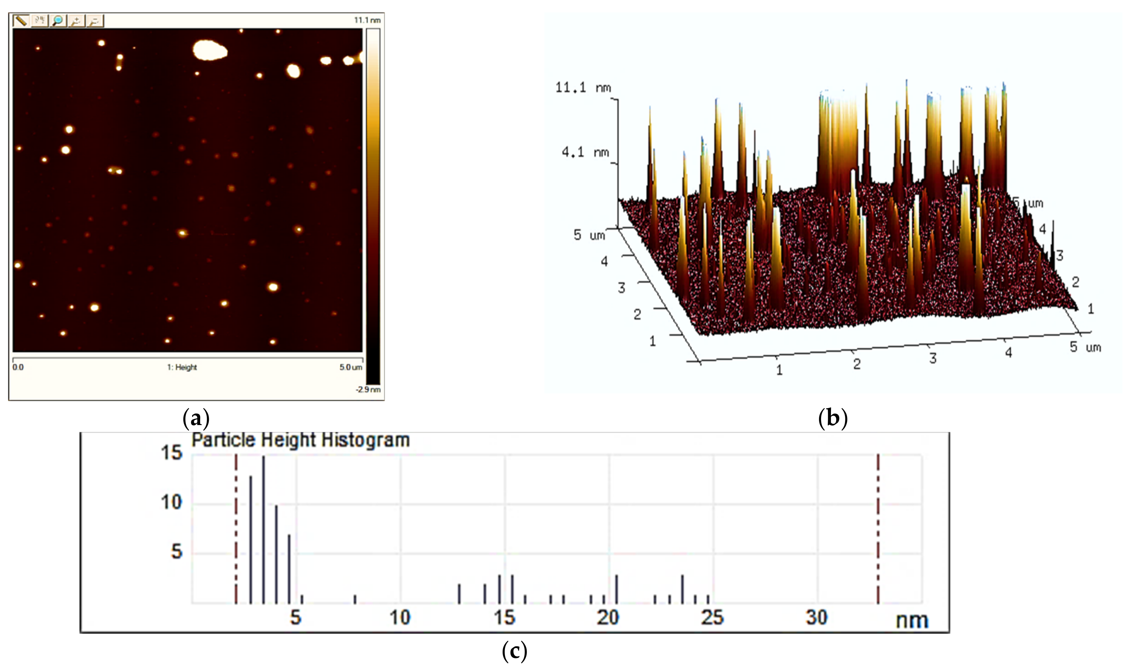
2.2. Atomic Force Microscopy (AFM) Analysis

The analysis entailed confirmation of the nanoparticle content in the preparations.
It was performed with the use of an atomic force microscope NanoScope V (Veeco, New York, NY, USA), which allows magnified and three-dimensional imaging of solid surfaces with the magnification rage from 2000 to 500,000 times. The measurement accuracy was consistent with the PN-EN ISO/IEC 17,025 standard. The AFM preparation was obtained by placing a drop of the solution into a mica plate glued to a metal disc and evaporating it. Measurements were conducted at room temperature [52]. The obtained AFM images were processed using nanoscope analysis software (Bruker, Billerica, MA, USA).

2.3. The Experiment of Pumpkin Germination

The experiment entailing pumpkin germination on laboratory Petri dishes and in pots in the greenhouse (Table 1). The mean temperature inside the greenhouse was 19 °C in May, 25 °C in June, and 35 °C in July, at the relative humidity of 40%.
The experimental germination in Petri dishes took place at room temperature of 20 ± 1 °C, with 12/12 h insolation. Seeds were sown of 25 per onto Petri dishes (with dimensions of 120 × 15 mm) lined with three layers of blotting paper moistened with distilled water (5 mL) in each sample. In the greenhouse, seeds were sown of 6 per pot containing universal soil with the addition of humidifier and NPK fertilizer, in four replicates per each study group. Each sample included four replications. The pots were 25 cm in diameter and 7.5 liters in capacity.
Energy (GE) and capacity (GC) germination in laboratory tests were calculated from the formulas:
G E = N g 4 N   × 100 %
G C = N g 8 N   × 100 %
where:
  • Ng4, g8—number of seeds germination after 4 and 8 days.
  • N—number of seeds sown.
In the greenhouse tests, the number of plants after 8 days (emergence) (Ne8) and 14 days (Ne14) was given. The measurements were conducted on 10 randomly selected leaves for each of the study groups, in four various stages of the plants’ development. The BBCH scale was used to identify the phenological development stages of plants [53].

2.4. Spectrophotometric Methods

Spectrophotometric methods were used to analyze photosynthetic pigments in seed leaves from the dishes (after 8 days) and pumpkin leaves from the pots (after 14 days), collected at the stage of emergence. The analysis entailed isolating chlorophylls and carotenoids from the leaves in acetone containing 0.01% BHT—butylated hydroxytoluene (in darkness) to avoid oxidation. Carry Bio 300 dual beam spectrophotometer (Agilent Technologies, CA, United States) was used to measure UV-Vis spectra. Pigment concentrations were calculated in accordance with the Lichtenhaler and Buschmann [54] procedure. The content of chlorophyll was determined by spectrophotometric method in three replications for each research object.

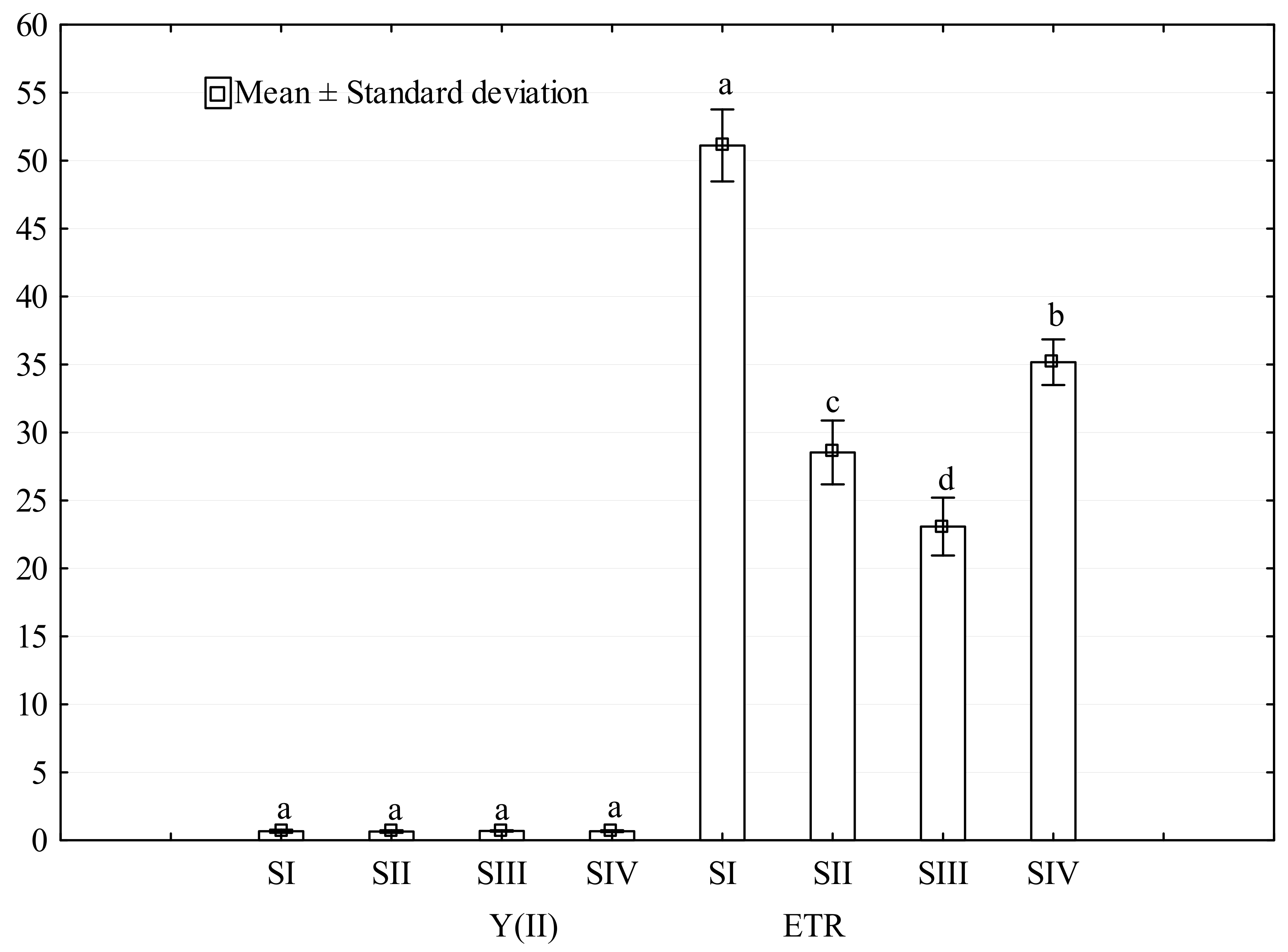
2.5. Photosynthetic Activity of Plants

To estimate the actual photochemical activity of PSII in situ, chlorophyll a fluorescence in plants was measured using a pulse amplitude-modulated (PAM) fluorimeter (Mini PAM, Walz, Germany) with a light emitting diode at 650 nm and a standard intensity 0.15 μmol m−2 s−1 PAR (photosynthetically active radiation). The device’s built-in sensor projects light impulses (triggered on and off at a very high frequency) onto a leaf placed in a metal clasp, and registers the fluorescence signal from excited chlorophyll a molecules. The following fluorescence parameters were measured: F-the present fluorescence yield, M-fluorescence after saturation pulse was applied, Y(II) = (M − F)/M, PAR-photosynthetically active radiation, ETR-electron transfer rate calculated as: ETR = Y(II) × PAR × ETR-factor; i.e., ETR = Y(II) × PAR × 0.5 × 0.84. The standard factor 0.84 corresponds to the fraction of incident light absorbed by a leaf [20,55]. The study included measurements of the greenness index (SPAD) with the use of chlorophyll meter (model SPAD-502, Konica Minolta, City, Japan) in which a leaf is placed in a clasp and 2 s later the result appears on the screen and the device is ready to scan another leaf. SPAD greenness index and photosynthesis efficiency using Mini-PAM were determined on 10 randomly selected leaves. Moreover, the measurement of the greenness and efficiency of photosynthesis takes place in four phases of plant development BBCH scale (the scale used in the UE to identify the various stages of plants) (Table 1).

2.6. Statistical Analysis

The research data were processed by analysis of variance (ANOVA) using the computer program Statistica. The analysis of variance was preceded by checking whether the results are normally distributed with the Shapiro-Wilk test and the homogeneity of variance with the Levene’s test. Arithmetical means and standard deviation of the experimental data were calculated. Significant differences at p ≤ 0.05 among the data were evaluated according to Tukey’s LSD test. Correlation analysis was performed to determine the strength and character of the relationships between variables. The correlation was statistically significant when p ≤ 0.05.

3. Results and Discussion

3.1. AFM Analysis of AgNC

The analysis consisted in confirming the content of nanoparticles in the preparations.
In order to assess the surface properties and imaging in nanoscale resolutions, the analyzed samples of AgNC nanocolloids were subjected to a morphological analysis with AFM. Images were taken in the two-and three-dimensional AgNC topography format, as presented in Figure 2. The paraemters adopted during analysis enabled the identification of minimum and maximum height values for the AgNC nanoparticles.
Based on the obtained results (Figure 2), it was concluded that the mean analyzed surface area 16,314.2 nm2, 75 AgNC were observed. The minimum and maximum height of the same was, respectively, 2.136 nm and 32.91 nm.

3.2. Germination Seeds

The germination energy was higher in seeds subjected to laser light and magnetic field stimulation and was smaller for silver nanocolloid, relative to the control (Table 2).
The conducted analysis showed that a significant increase was observed in relation to the controls for GE by about 37% for p. In turn, with regard to Ne8, a significant decrease was observed for AgNC by about 83%, and, for Ne14 for p and AgNC by about 50% and 66%, respectively. A significantly positive effect of laser radiation on the germination of seeds was found in white lupine [56], white clover [57], radish [58], cucumber [59] and tomato [60]. No positive effect was observed in oats [61], sugar beet [62], cucumber and tomato [60], and pepper [61]. Similarly to the studies presented here, many authors examined the effect of an He-Ne laser and alternating magnetic field on the germination of amaranth seeds. Dziwulska-Hunek et al. [63] used the laser beam stimulation of the power density of 6 mW/cm2, the alternating magnetic field with the induction 30 mT, the frequency 50 Hz for t = 30 s as well as the combination of both factors for amaranth seeds cvs Aztek and Rawa. The effects on the germination of the seeds were examined on Petri dishes in darkness at temperatures from 10 to 55 °C. The most pronounced effects important for the germination process were registered at the temperatures of 20 and 25 °C. When the above parameters of the stimulation of amaranth seeds cv. Rawa were present, the increase in dry matter, raw protein, crude fiber, crude ash and in the final yield was observed. An increase in the levels of Leu, Val, Lys, and Phe + Tyr amino acids as well as decreased levels of Arg, Glu, and Ala. The levels of Cys, Tr, Ile, His, and Pro remained unchanged. Sujak and Dziwulska-Hunek [64] applied a laser beam of the power density of 6 mW/cm2, alternating magnetic field with an induction of B = 30 mT and frequency of 50 Hz for t = 30 s, and the combination of these factors to stimulate amaranth seeds. A magnetic field applied to dormant seeds was found to increase the rate of subsequent seedling growth of barley, corn, beans, wheat, certain tree fruits, and other tree species. Moreover, a low frequency magnetic field (16 Hz) can be used as a method of post-harvest seed improvement for different plant species, especially for seeds of temperature sensitive species germinating at low temperatures [65]. Studies conducted by Gubbins et al. [66] and El-Temsah and Joner [67] demonstrated that the use of AgNPs in the concentration of 10 mg·L−1 AgNPs inhibited the germinating capacity of Hordeum vulgare seeds and reduced the length of sprouts in flax (Linum usitatissimum) and barley (Hordeum vulgare). Amooaghaiea et al. [68] observed that the use of silver nanocolloid concentrated at 0.2 to 1.6 mg·L−1 contributed to the inhibition of seed germination and lipase activity in germinating seeds and seedlings of Brassica nigra. In the present study, seeds soaked in silver nanocolloid showed a decrease in germination. Stmapoulis et al. [69] reported that the use of a preparation containing nanoparticles at the size of 100 nm AgNPs at 100 and 500 mg·L−1 resulted in a 41 and 57% biomass decrease in a cultivation of Cucurbita pepo, relative to the control, whereas, in a study by Abdel-Azeem and Elsayed [70], where silver nanoparticles of varying size were used (65, 50, and 20 nm) to test their impact on the germination capacity of Vicia faba, the results clearly indicated that the varying AgNP diameters had only a minor bearing on the process of germination when compared to the control. Similarly, the results obtained by Barren et al. [71] did not demonstrate a significant impact of AgNPs on the germination capacity of Cucumis sativus or Lactuca sativa seeds. Yasur and Rani [72] also confirmed in their study that the application of AgNC did not have significant influence on the germination capacity of seeds, length of roots, or overall growth of castor bean, Ricinus communis L., even at higher concentrations. In a study conducted by Mehrian et al. [73], it was observed that the use of silver nanoparticles in high concentrations of 75 and 100 mg·L−1 and sized at 20 nm contributed to a significant increase of stress suffered by plants, and, consequently, to their reduced vitality and lower number of tomato seedlings. In another experiment [23], it was observed that the germination capacity of barley seeds was reduced after the application of AgNPs in various concentrations. On the other hand, results reported by Hojjat and Hojjat [34] suggest that increased salinity of the environment of plant development resulted in reduced germination while application of silver nanoparticles actually improved plant germination parameters.

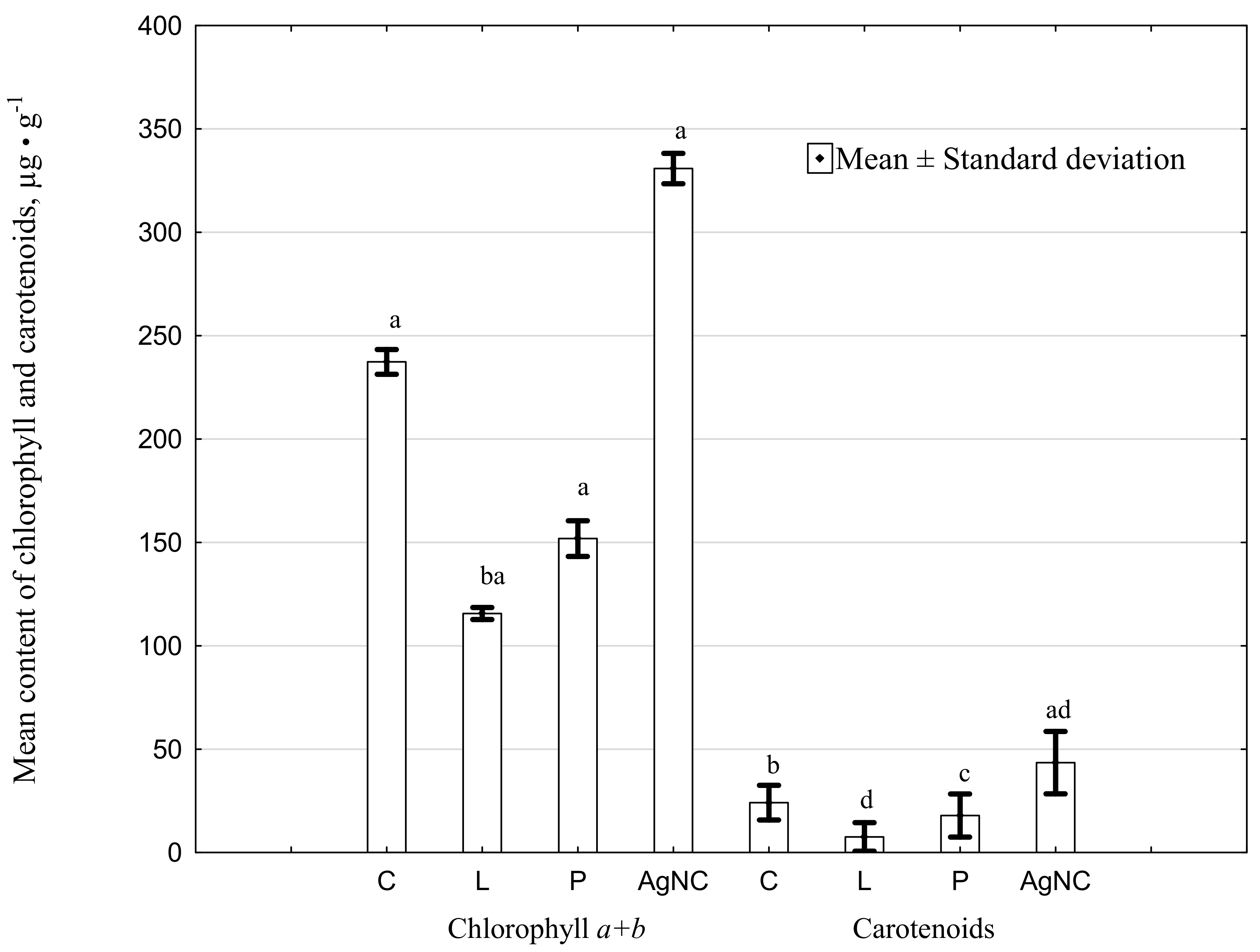
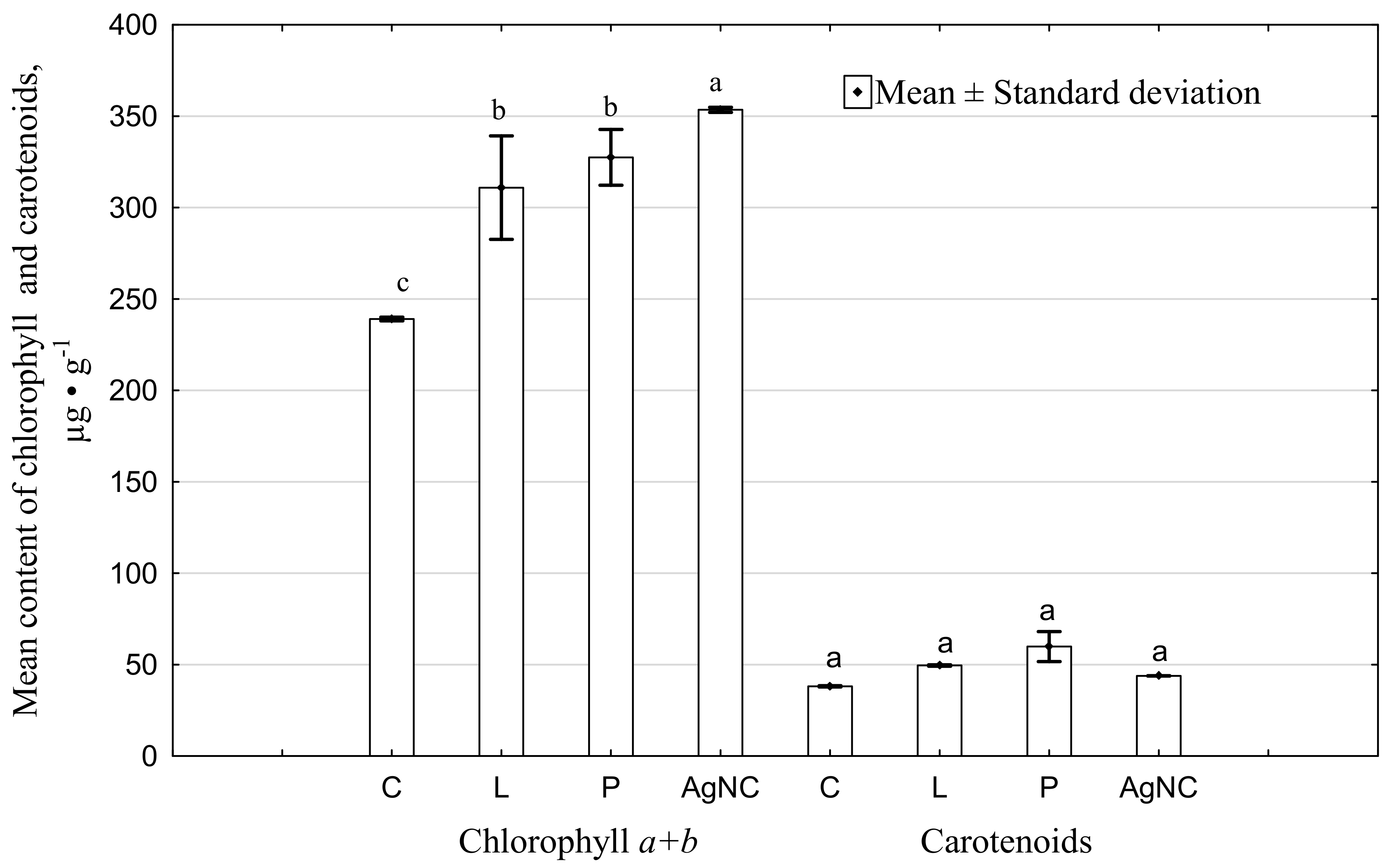
3.3. Elements Concentrations in Leaves of Pumpkin

In the leaves of sprouts grown in Petri dishes from seeds soaked in silver, a significant increase was observed in terms of chlorophyll a and b content, of respectively: 54% and 11% compared to the control. In all other cases, a decrease in the pigment’s content was observed (Figure 3a).
The concentration of carotenoids in the leaves of sprouts grown in the dishes, in the sample treated with AgNC, was significantly higher, by approximately 79%, relative to the control (C). A reduction in the pigment’s concentration was observed in samples stimulated with laser light and magnetic field. Leaves from plants grown in pots (Figure 3b) revealed increased chlorophyll a, b and carotenoid content in all study groups. The highest significant values of chlorophyll a were recorded for the p and AgNC samples, which amounted to 42 and 43%, respectively, compared to the control. Moreover, a significant content of chlorophyll b was found only in trial p, where it was 25% more than in relation to C. Stimulation with laser light and alternating magnetic field significantly influenced the content of carotenoids, with an increase of 66 and 81%, respectively. In another study conducted by Liu et al. [74], the use of green LED light resulted in a decrease in chlorophyll and carotenoid content in lettuce. In a study by Liu et al. [74] conducted on cucumber plants, saline stress caused a reduction in photosynthetic efficiency. The content of chlorophyll depended on the development phase and the method used. Nanotechnology can have a beneficial effect on the increase in photosynthetic pigments concentration in chickpea according to the results reported by Anusuya and Banu [49]. In a study by Fayez et al. [22] it was observed that silver nanoparticles applied in various concentrations caused a decrease in chlorophyll a, b and carotenoid content in the leaves of barley, relative to the control. According to Kachel et al. [75], the content of photosynthetic pigments in the leaves of pumpkin with nano silver (concentration of 50 mL L−1) decreased after 22 days, and, after 29 days, there were more of them than in the control group. The increase was 5% (Chl a and Carotenoids) and 10% (Chl b). In our study, at the same concentration of Ag nanoparticles for pumpkin leaves after 8 (Petrry dishes) and 14 days (pots), the concentrations of chlorophyll a (43–54%), chlorophyll b (11–19%), and carotenoids (25–79%) compared to the control. In other studies on rapeseed oil produced from seeds treated with nanoparticles, regardless of the harvest year or the methods used, the content of chlorophylls and carotenoids was significantly higher than the control samples chlorophyll content in plants was also analysed by Mazumdar [76]. Based on the obtained results, he concluded that silver nanoparticles in B. campestris significantly reduced the total chlorophyll content relative to the control when applied in the concentrations of 500 µg·L−1 and 1000 µg·L−1. Moreover, negative effects were also observed at the dose of 1000 µg·L−1 Ag nanoparticles applied in V. radiata and B. campestris with regard to total chlorophyll content compared to the control. In a study conducted by Asghar et al. [12], the researchers reported increased chlorophyll content in soya plants grown from seeds stimulated with He-Ne laser light and alternating magnetic fields. As observed by Winkelmann et al. [77], silver nanoparticles can interfere with cells’ biological functions due to two distinct mechanisms. A nanoparticle may be absorbed by a plant cell and start to release silver cations as products of various redox reactions. Cells can also fully absorb nanoparticles in a process known as endocytosis. It is less common in the case of larger nanoparticles due to their size. Once inside a cell, a nanoparticle may release Ag+ or silver atoms which then bond with proteins and hinder their functional capacity. One of the signs of efficiently working defence mechanisms against oxidative stress is a plant’s ability to produce chlorophylls and carotenoids. Chlorophyll particles undergo degradation when a plant is under conditions of abiotic stress caused by absorbing excessive amounts of metals with toxic effects on the plant. In their study, Qian et al. [38] demonstrated that two-week-long exposition of A. thaliana cuttings to AgNPs resulted in a decrease in their Chl a and Chl b content. AgNPs applied in concentrations of 0.5 and 3.0 mg·L−1 significantly reduced the plant’s Chl a content to the level of 60.9% compared to the control in both cases. Moreover, the above concentration of AgNPs (0.5 and 3.0 mg·L−1) also triggered a decrease in Chl b levels.
The correlations could be observed between the particular groups: C × C (r = −0.360), L × L (r = 0.856), p × p (r = 0.982), AgNC × AgNC (r = −0.967) for pots and C × C (r = −0.152), L × L (r = 0.971), p × p (r = 0.353), AgNC × AgNC (r = −0.086) for dishes (Table 3).
Calculations of the correlation coefficients (Table 3) between the values of total chlorophyll and carotenoids revealed no correlation between the Petri dish and pots study groups. As one of the characteristics increased, so did the other, which may suggest that the two are correlated with each other. In the case of seed leaves grown on dishes, no correlation between total chlorophyll a + b and carotenoids was observed.
Compared to the controls, a reduction of mean content of chlorophyll a + b in dishes (Figure 4) was observed for L by about 50% and for P by about 35% an increase of AgNC by about 38%. With regard to the carotenoids, the decrease was for L and P and increase by AgNc by about 68%, 27% and 79% respectively.
Compared to the controls, an increase of mean content of chlorophyll a + b in pots (Figure 5) was observed for L by about 29%, for P by about 28% and for AgNC by about 47%. With regard to the carotenoids, the increase was observed for L, P and AgNc by about 29%, 55% and 15% respectively.
PAR radiation was low (Table 4), ranging from 57 (L, SIII) to 302 μ mol of photons m−2 s−1 (AgNC, SIV). The value of photosynthesis intensity for the low PAR was, respectively, 0.713 and 0.646. Light intensity in all study samples was reduced at the SII and increased at the stage of SIV when compared to the control. Photosynthetic efficiency at the onset of SII was increased in all study groups compared to the control. The highest increase was observed in the group treated with AgNC and reached 29%. Low intensity of PAR means that a large dose of energy was absorbed, which had a positive effect on photosynthesis, which, in plants soaked in silver, reached 0.728. Low PAR intensity resulted in increased photosynthetic activity at SII while reducing the same at the SIV. The described studies indicated an increase in terms of photochemical efficiency and greenness index at the SII in all study groups when compared to the control. The highest values of photochemical efficiency and greenness index were observed in leaves grown from seeds soaked in AgNC. The same were respectively 26 and 11% higher than in the control. According to a study by Kumar et al. [78] on safflower plants (Carthamus tinctorius L.) treated with laser beams (wavelength 632.8 nm) at different times (0.5 h, 1 h, 1.5 h) showed a differentiation in photosynthetic activity, but its increase was noticed compared to the control. In our research, laser light had a variable effect on the photosynthesis efficiency in pumpkin leaves depending on the stage of development. It was the highest in the SII (14%) and SIII (4%) phases. In other studies on long-term laser beam irradiation (wavelength 632.8 nm), it was noticed that the photosynthetic activity did not differ significantly between the research objects [79]. According to research study by Li et al. [80] regarding the effect of red (R), white (B) and mixed light (RB) on the photosynthesis efficiency in red pepper leaves, it was noticed that the leaves were thicker and the ability to transpose electrons in the process of photosynthesis and its speed increased by lights B and RB.
The mean value of Y(II) was within the range from 0.644 (SII) to 0.685 (SIII) and the ETR value from 24.11 (SIII) to 51.37 (SI) (Figure 6). In the present study, the application of silver nanocolloid produced an improvement in terms of photochemical efficiency, particularly at the stages of: budding and onset of flowering.
Based on the data presented in Table 5, no significant correlation between the Y(II) and ETR at the respective stages can be identified.
Average correlation (significance at the level of α = 0.05) could only be observed between the SII and SIII (r = 0.408) and between the SII and SIV (r = 0.465).
To recapitulate the presented study, it can be observed that the use of various techniques intended as a way to improve germination as well as photosynthetic pigment content and photosynthetic efficiency, yielded varying results. The greenness index at the stages of: budding, onset of flowering, and flowering was higher in all plants grown from seeds treated with laser light and alternating magnetic fields as well as soaked in silver nanocolloid, when compared to the control. On the other hand, the same was lower at the stage of fructification due to the aging of leaves and their change to a more yellowish-orange color.

4. Conclusions

The best effects in terms of increase of the germination energy and capacity were observed in the case of seeds stimulated with magnetic fields, where the same improved, respectively, by 20% and 4% relative to the control. The emergence and density of plants deteriorated in all study samples. At the stage of the onset of flowering pumpkin, an improvement in terms of photosynthetic efficiency and greenness index was observed in all study groups. The highest improvement was recorded for seeds soaked in nanocolloid of silver and reached 23% (intensity of photosynthesis) and 11% (greenness index).
The highest chlorophyll a, b and carotenoid content was recorded in leaves grown on Petri dishes from seeds soaked in nanocolloid of silver. The increase relative to the control was, respectively, 53, 11, and 79%. In turn, leaves of pumpkin grown in pots displayed the highest content of chlorophyll a—42 and 43% increase (p and AgNC), chlorophyll b by 25% (p), and carotenoids by 66 and 81% (L and p) as compared to the control.

Author Contributions

A.D.-H., M.G. Conceptualization, Methodology, Formal Analysis, Data Curation, Writing—Original Draft; M.K., M.G., M.S. Investigation, Resources, Writing—Original Draft. All authors have read and agreed to the published version of the manuscript.

Funding

This research received no external funding.

Data Availability Statement

Data contained within the article.

Conflicts of Interest

The authors declare no conflict of interest.

References

  1. Borthakur, P.R.; Barua, A.G. Fluorescence Studies of the Seeds of the Pumpkin (Cucurbita pepo L.). Natl. Acad. Sci. Lett. 2014, 37, 275–279. [Google Scholar] [CrossRef]
  2. Kadzińska, J.; Janowicz, M.; Kalisz, S.; Sitkiewicz, I.; Mika, M. The influence of edible coatings on the change of the properties of pumpkin fruits during storage. Adv. Food Process. Technol. 2017, 2, 37–45. [Google Scholar]
  3. Wu, H.; Zhu, J.; Diao, W.; Wang, C. Ultrasound-assisted enzymatic extraction and antioxidant activity of polysaccharides from pumpkin (Cucurbita moschata). Carbohydr. Polym. 2014, 113, 314–324. [Google Scholar] [CrossRef]
  4. Giami, S.; Chibor, B.; Edebiri, K.; Achinewhu, S. Changes in nitrogenous and other chemical constituents, protein fractions and in vitro protein digestibility of germinating fluted pumpkin (Telfairia occidentalis Hook) seed. Plant Foods Hum. Nutr. 1999, 53, 333–342. [Google Scholar] [CrossRef] [PubMed]
  5. Zhang, Y.; Zhou, J.; Wu, T.; Cao, J. Shoot regeneration and the relationship between organogenic capacity and endogenous hormonal contents in pumpkin. Plant Cell Tissue Organ. Cult. 2008, 93, 323–331. [Google Scholar] [CrossRef]
  6. DeRossi, A.; De Pilli, T.; Giuliani, R.; Orlando, I.; Palmieri, L.; Severini, C. Study on prestabilization of pumpkin (cucurbita moschata) by osmotic dehydration in quaternary complex solution. J. Food Process. Eng. 2011, 34, 398–413. [Google Scholar] [CrossRef]
  7. Kaźmińska, K.; Sobieszek, K.; Targońska-Karasek, M.; Korzeniewska, A.; Niemirowicz-Szczytt, K.; Bartoszewski, G. Genetic diversity assessment of a winter squash and pumpkin (Cucurbita maxima Duchesne) germplasm collection based on genomic Cucurbita -conserved SSR markers. Sci. Hortic. 2017, 219, 37–44. [Google Scholar] [CrossRef]
  8. Katsoufi, S.; Lazou, A.E.; Giannakourou, M.C.; Krokida, M.K. Mass transfer kinetics and quality attributes of osmo-dehydrated candied pumpkins using nutritious sweeteners. J. Food Sci. Technol. 2017, 54, 3338–3348. [Google Scholar] [CrossRef] [PubMed]
  9. Podleśny, J.; Stochmal, A.; Podleśna, A.; Misiak, L.E. Effect of laser light treatment on some biochemical and physiological processes in seeds and seedlings of white lupine and faba bean. Plant Growth Regul. 2012, 67, 227–233. [Google Scholar] [CrossRef]
  10. Levina, N.S.; Tertyshnaya, Y.V.; Bidey, I.A.; Elizarova, O.V. Presowing treatment of seeds. Agri. Mach. Technol. 2018, 12, 13–18. [Google Scholar]
  11. Pietruszewski, S.; Muszyński, S.; Dziwulska, A. Electromagnetic fields and electromagnetic radiation as non-invasive extermal stimulants for seeds (selected methods and responses). Int. Agrophys. 2007, 21, 95–100. [Google Scholar]
  12. Asghar, T.; Jamil, Y.; Iqbal, M.; Abbas, M. Laser light and magnetic field stimulation effect on biochemical, en-zymes activities and chlorophyll contents in soybean seeds and seedlings during early growth stages. J. Photochem. Photobiol. B Biol. 2016, 165, 283–290. [Google Scholar] [CrossRef]
  13. Perveen, R.; Jamil, Y.; Ashraf, M.; Ali, Q.; Iqbal, M.; Ahmad, M.R. He-Ne Laser-Induced Improvement in Biochemical, Physiological, Growth and Yield Characteristics in Sunflower (Helianthus annuus L.). Photochem. Photobiol. 2011, 87, 1453–1463. [Google Scholar] [CrossRef] [PubMed]
  14. Szymanek, M.; Dziwulska-Hunek, A.; Zarajczyk, J.; Michałek, S.; Tanaś, W. The Influence of Red Light (RL) and Effective Microorganism (EM) Application on Soil Properties, Yield, and Quality in Wheat Cultivation. Agronomy 2020, 10, 1201. [Google Scholar] [CrossRef]
  15. Mroczek-Zdyrska, M.; Kornarzyński, K.; Pietruszewski, S.; Gagoś, M. The efects of low-frequency magnetic feld exposure on the growth and biochemical parameters in lupin (Lupinus angustifolius L.). Plant Bios. 2017, 151, 504–511. [Google Scholar] [CrossRef]
  16. Matwijczuk, A.; Kornarzynski, K.; Pietruszewski, S. Effect of magnetic field on seed germination and seedlings growth of sunfower. Int. Agrophys. 2012, 26, 271–278. [Google Scholar] [CrossRef]
  17. Sacała, E.; Demczuk, A.; Grzyś, E.; Prośba-Białczyk, U.; Szajsner, H. Impact of pre-sowing laser irradiation of seeds on sugar beet properties. Int. Agrophys. 2012, 26, 295–300. [Google Scholar] [CrossRef]
  18. Dziwulska-Hunek, A.; Krawiec, M.; Sujak, A. Laser Light Stimulation Effects on Scorzonera hispanica L. Seeds Germination, Field Emergence and Photosynthetic Pigments Content. J. Hortic. Res. 2016, 24, 57–62. [Google Scholar] [CrossRef] [Green Version]
  19. Sujak, A.; Dziwulska-Hunek, A.; Reszczyńska, E. Effect of Electromagnetic Stimulation on Selected Fabaceae Plants. Pol. J. Environ. Stud. 2013, 22, 893–898. [Google Scholar]
  20. Sujak, A.; Dziwulska-Hunek, A.; Kornarzyński, K. Compositional and nutritional values of amaranth seeds after pre-sowing He-Ne laser light and alternating magnetic field treatment. Int. Agrophys. 2009, 23, 81–86. [Google Scholar]
  21. Hernandez, A.C.; Dominguez, P.A.; Cruz, O.A.; Ivanov, R.; Carballo, C.A.; Zepeda, B.R. Laser in agriculture. Int. Agrophys. 2010, 24, 407–422. [Google Scholar]
  22. Fayez, K.A.; El-Deeb, B.A.; Mostafa, N.Y. Toxicity of biosynthetic silver nanoparticles on the growth, cell ultrastructure and physiological activities of barley plant. Acta Physiol. Plant. 2017, 39, 155. [Google Scholar] [CrossRef]
  23. Huang, S.; Wang, L.; Liu, L.; Hou, Y.; Li, L. Nanotechnology in agriculture, livestock, and aquaculture in China. A review. Agron. Sustain. Dev. 2015, 35, 369–400. [Google Scholar] [CrossRef] [Green Version]
  24. Kachel-Jakubowska, M.; Strubińska, J.; Matwijczuk, A.; Gagoś, M. Microscopic and Spectroscopic Analyses of Selected Agricultural Formulations Containing Various nanostructures. Pol. J. Environ. Stud. 2017, 26, 1565–1573. [Google Scholar] [CrossRef]
  25. Li, Q.; Mahendra, S.; Lyon, D.Y.; Brunet, L.; Liga, M.; Li, D.; Alvarez, P.J.J. Antimicrobial nanomaterials for water disinfec-tion and microbial control: Potential applications and implications. Water Res. 2008, 42, 4591–4602. [Google Scholar] [CrossRef]
  26. El-Nour, K.M.A.; Eftaiha, A.; Al-Warthan, A.; Ammar, R.A. Synthesis and applications of silver nanoparticles. Arab. J. Chem. 2010, 3, 135–140. [Google Scholar] [CrossRef] [Green Version]
  27. Ahmed, S.; Ahmad, M.; Swami, B.L.; Ikram, S. A review on plants extract mediated synthesis of silver nanoparticles for antimicrobial applications: A green expertise. J. Adv. Res. 2016, 7, 17–28. [Google Scholar] [CrossRef] [Green Version]
  28. Cohen, M.S.; Stern, J.M.; Vanni, A.J.; Kelley, R.S.; Baumgart, E.; Field, D.; Libertino, J.A.; Summerhayes, I.C. In Vitro Analysis of a Nanocrystalline Silver-Coated Surgical Mesh. Surg. Infect. 2007, 8, 397–404. [Google Scholar] [CrossRef] [PubMed]
  29. Cui, D.; Zhang, P.; Ma, Y.-H.; He, X.; Li, Y.-Y.; Zhao, Y.-C.; Zhang, Z.-Y. Phytotoxicity of silver nanoparticles to cucumber (Cucumis sativus) and wheat (Triticum aestivum). J. Zhejiang Univ. Sci. A 2014, 15, 662–670. [Google Scholar] [CrossRef]
  30. Khalil, K.A.; Fouad, H.; Elsarnagawy, T.; Almajhdi, F.N. Preparation and characterization of electrospun PLGA/silver com-posite nanofibers for biomedical applications. Int. J. Electrochem. Sci. 2013, 8, 3483–3493. [Google Scholar]
  31. Nejatzadeh-Barandozi, F. Antibacterial activities and antioxidant capacity of Aloe vera. Org. Med. Chem. Lett. 2013, 3, 5. [Google Scholar] [CrossRef] [Green Version]
  32. Hojjat, S.S.; Hojjat, H. Effects of silver nanoparticle exposure on germination of Lentil (Lens culinaris Medik). Int. J. Farm Allied Sci. 2016, 5, 248–252. [Google Scholar]
  33. Seif, S.M.; Sorooshzadeh, A.; Rezazadeh, H.S.; Naghdibadi, H.A. Effect of nano silver and silver nitrate on seed yield of bor-age. J. Med. Plants Res. 2011, 5, 706–710. [Google Scholar]
  34. Hojjat, S.S.; Hojjat, H. Effect of Nano Silver on Seed Germination and Seedling Growth in Fenugreek Seed. ETP Int. J. Food Eng. 2015, 1, 106–110. [Google Scholar] [CrossRef] [Green Version]
  35. Parveen, A.; Rao, S. Effect of Nanosilver on Seed Germination and Seedling Growth in Pennisetum glaucum. J. Clust. Sci. 2014, 26, 693–701. [Google Scholar] [CrossRef]
  36. Gusev, A.A.; Kudrinsky, A.A.; Zakharova, O.V.; Klimov, A.I.; Zherebin, P.M.; Lisichkin, G.V.; Vasyukova, I.A.; Denisov, A.N.; Krutyakov, Y.A. Versatile synthesis of PHMB-stabilized silver nanoparticles and their significant stimulating effect on fodder beet (Beta vulgaris L.). Mater. Sci. Eng. C 2016, 62, 152–159. [Google Scholar] [CrossRef] [PubMed]
  37. Kaveh, R.; Li, Y.-S.; Ranjbar, S.; Tehrani, R.; Brueck, C.L.; Van Aken, B. Changes inArabidopsis thalianaGene Expression in Response to Silver Nanoparticles and Silver Ions. Environ. Sci. Technol. 2013, 47, 10637–10644. [Google Scholar] [CrossRef] [PubMed]
  38. Qian, H.; Peng, X.; Han, X.; Ren, J.; Sun, L.; Fu, Z. Comparison of the toxicity of silver nanoparticles and silver ions on the growth of terrestrial plant model Arabidopsis thaliana. J. Environ. Sci. 2013, 25, 1947–1956. [Google Scholar] [CrossRef]
  39. Kaegi, R.; Sinnet, B.; Zuleeg, S.; Hagendorfer, H.; Mueller, E.; Vonbank, R.; Boller, M.; Burkhardt, M. Release of silver nanoparticles from outdoor facades. Environ. Pollut. 2010, 158, 2900–2905. [Google Scholar] [CrossRef] [PubMed]
  40. Nowack, B. Nanosilver Revisited Downstream. Science 2010, 330, 1054–1055. [Google Scholar] [CrossRef] [PubMed]
  41. Krishnaraj, C.; Jagan, E.; Ramachandran, R.; Abirami, S.; Mohan, N.; Kalaichelvan, P. Effect of biologically synthesized silver nanoparticles on Bacopa monnieri (Linn.) Wettst. plant growth metabolism. Process Biochem. 2012, 47, 651–658. [Google Scholar] [CrossRef]
  42. Monica, R.C.; Cremonini, R. Nanoparticles and higher plants. Caryologia 2009, 62, 161–165. [Google Scholar] [CrossRef] [Green Version]
  43. Salama, H.M.H. Effects of silver nanoparticles in some crop plants, common bean (Phaseolus vulgaris L.) and corn (Zea mays L.). Int. Res. J. Biotech. 2012, 3, 190–197. [Google Scholar]
  44. Sharma, P.; Bhatt, D.; Zaidi, M.G.H.; Saradhi, P.P.; Khanna, P.K.; Arora, S. Silver Nanoparticle-Mediated Enhancement in Growth and Antioxidant Status of Brassica juncea. Appl. Biochem. Biotechnol. 2012, 167, 2225–2233. [Google Scholar] [CrossRef] [PubMed]
  45. Savithramma, N.; Ankanna, S.; Bhumi, G. Effect of nanoparticles on seed germination and seedling growth of Boswellia ovali-foliolata an endemic and endangered medicinal tree taxon. Nano Vis. 2012, 2, 61–68. [Google Scholar]
  46. Kalaji, H.M.; Jajoo, A.; Oukarroum, A.; Brestic, M.; Zivcak, M.; Samborska, I.A.; Cetner, M.D.; Łukasik, I.; Goltsev, V.; Ladle, R.J. Chlorophyll a fluorescence as a tool to monitor physiological status of plants under abiotic stress conditions. Acta Physiol. Plant. 2016, 38, 102. [Google Scholar] [CrossRef] [Green Version]
  47. Kataria, S.; Baghel, L.; Guruprasad, K. Pre-treatment of seeds with static magnetic field improves germination and early growth characteristics under salt stress in maize and soybean. Biocatal. Agric. Biotechnol. 2017, 10, 83–90. [Google Scholar] [CrossRef]
  48. Hojjat, S.S.; Kamyab, M. The effect of silver nanoparticle on Fenugreek seed germination under salinity levels. Russ. Agric. Sci. 2017, 43, 61–65. [Google Scholar] [CrossRef]
  49. Anusuya, S.; Banu, K.N. Silver-chitosan nanoparticles induced biochemical variations of chickpea (Cicer arietinum L.). Biocatal. Agric. Biotechnol. 2016, 8, 39–44. [Google Scholar] [CrossRef]
  50. Pietruszewski, S. Electromagnet. Utility Model, Patent Office of Republic of Poland Protection Rights. No. 59863, 31 July 2003. Volume 7 p. 1077. [Google Scholar]
  51. Muszyński, S.; Gagoś, M.; Pietruszewski, S. Short-Term Pre-Germination Exposure to ELF Magnetic Field Does Not Influence Seedling Growth in Durum Wheat (Triticum durum). Pol. J. Environ. Stud. 2009, 18, 1065–1072. [Google Scholar]
  52. Kachel, M.; Matwijczuk, A.; Sujak, A.; Czernel, G.; Niemczynowicz, A.; Nowicka, A. The influence of copper and silver nano-colloids on the quality of pressed spring rapeseed oil. Agronomy 2019, 9, 643. [Google Scholar] [CrossRef] [Green Version]
  53. Meier, U. Growth Stages of Mono- and Dicotyledonous Plants; Blackwell Wissenschafts-Verlag: Bonn, Germany, 2001. [Google Scholar]
  54. Lichtenthaler, H.K.; Buschmann, C. Chlorophylls and Carotenoids: Measurement and Characterization by UV-Vis Spectros-copy. In Current Protocols in Food Analytical Chemistry; Supplement 1; Wiley & Sons. Inc.: Hoboken, NJ, USA, 2001. [Google Scholar]
  55. Kocira, S.; Sujak, A.; Oniszczuk, T.; Szparaga, A.; Szymanek, M.; Karakuła-Juchnowicz, H.; Krawczuk, A.; Kupryaniuk, K. Improvement of the photosynthetic activity of Moldavian dragonhead (Dracocephalum moldavica L.) through foliar application of a nitrophenolate-based biostimulant. BIO Web Conf. 2018, 10, 01009. [Google Scholar] [CrossRef] [Green Version]
  56. Podleśny, J. The effect of seed irradiation with laser and plant desiccation on yielding and quality features of white lupine seeds. Acta Agrophys. 2007, 9, 733–745. [Google Scholar]
  57. Dziwulska, A.; Koper, R.; Wilczek, M. Estimation of effect of He-Ne laser beam on germination capacity of white clover variety of Anda cultivar. Acta Agrophys. 2004, 3, 435–441. [Google Scholar]
  58. Muszyński, S.; Gładyszewska, B. Representation of He-Ne laser irradiation effect on radish seeds with selected germination indices. Int. Agrophys. 2008, 22, 151–157. [Google Scholar]
  59. Szajsner, H.; Drozd, D. The improvement of seeds of selected vegetable species through laser radiation. Roczniki Akademii Rolniczej w Poznaniu. Ogrodnictwo 2007, 41, 625–629. [Google Scholar]
  60. Klimont, K. Studies of laser biostimulation on sowing value of seeds and yield of tomato (Lycopersicon esculentum Mill.) and cucumber (Cucumis sativus L.) plants. Biul. Inst. Hod. I Aklim. Rosl. 2002, 223/224, 257–266. [Google Scholar]
  61. Drozd, D.; Szajsner, H. Effect of application of pre-sowing laser stimulation on bare-grained oat genotypes. Acta Agrophys. 2007, 9, 583–589. [Google Scholar]
  62. Prośba-Białczyk, U.; Szajsner, H.; Grzyś, E.; Demczuk, A.; Sacała, E.; Bąk, K. Effect of seed stimulation on germination and sugar beet field. Int. Agrophys. 2013, 27, 195–201. [Google Scholar] [CrossRef]
  63. Dziwulska-Hunek, A.; Kornarzyński, K.; Matwijczuk, A.; Pietruszewski, S.; Szot, B. Efect of laser and variable magnetic feld simulation on amaranth seeds germination. Int. Agrophys. 2009, 23, 229–235. [Google Scholar]
  64. Sujak, A.; Dziwulska-Hunek, A. Minerals and fatty acids of amaranth seeds subjected to pre-sowing electromagnetical stim-ulation. Int. Agrophys. 2010, 24, 375–379. [Google Scholar]
  65. Rochalska, M.; Orzeszko-Rywka, A. Magnetic field treatment improves seed performance. Seed Sci. Technol. 2005, 33, 669–674. [Google Scholar] [CrossRef]
  66. Gubbins, E.J.; Batty, L.C.; Lead, J.R. Phytotoxicity of silver nanoparticles to Lemna minor L. Environ. Pollut. 2011, 159, 1551–1559. [Google Scholar] [CrossRef]
  67. El-Temsah, Y.S.; Joner, E.J. Impact of Fe and Ag nanoparticles on seed germination and differences in bioavailability during exposure in aqueous suspension and soil. Environ. Toxicol. 2010, 27, 42–49. [Google Scholar] [CrossRef] [PubMed]
  68. Amooaghaie, R.; Tabatabaei, F.; Ahadi, A.-M. Role of hematin and sodium nitroprusside in regulating Brassica nigra seed germination under nanosilver and silver nitrate stresses. Ecotoxicol. Environ. Saf. 2015, 113, 259–270. [Google Scholar] [CrossRef]
  69. Stampoulis, D.; Sinha, S.K.; White, J.C. Assay-Dependent Phytotoxicity of Nanoparticles to Plants. Environ. Sci. Technol. 2009, 43, 9473–9479. [Google Scholar] [CrossRef]
  70. Abdel-Azeem, E.A.; Elsayed, B.A. Phytotoxicity of silver nanoparticles on Vicia faba seedlings. N. Y. Sci. J. 2013, 6, 148–156. [Google Scholar]
  71. Barrena, R.; Casals, E.; Colón, J.; Font, X.; Sánchez, A.; Puntes, V. Evaluation of the ecotoxicity of model nanoparticles. Chemosphere 2009, 75, 850–857. [Google Scholar] [CrossRef] [PubMed] [Green Version]
  72. Yasur, J.; Rani, P.U. Environmental effects of nanosilver: Impact on castor seed germination, seedling growth, and plant physiology. Environ. Sci. Pollut. Res. 2013, 20, 8636–8648. [Google Scholar] [CrossRef]
  73. Mehrian, S.K.; Heidari, R.; Rahmani, F.; Najafi, S. Effect of Chemical Synthesis Silver Nanoparticles on Germination Indices and Seedlings Growth in Seven Varieties of Lycopersicon esculentum Mill. (tomato) Plants. J. Clust. Sci. 2015, 27, 327–340. [Google Scholar] [CrossRef]
  74. Liu, H.; Fu, Y.; Wang, M. Green light enhances growth, photosynthetic pigments and CO2 assimilation efficiency of lettuce as revealed by ’knock out’ of the 480–560 nm spectral waveband. Photosynthetica 2016, 55, 144–152. [Google Scholar] [CrossRef]
  75. Kachel, M.; Parafiniuk, S.; Krajewska, M. The influence of metal nanocolloids on selected abiotic stress factors and the overall quality of pumpkin. Fresenius Environ. Bull. 2020, 29, 5651–5667. [Google Scholar]
  76. Mazumdar, H. The Impact of Silver Nanoparticles on Plant Biomass and Chlorophyll Content. Int. J. Eng. Sci. 2014, 4, 12–20. [Google Scholar]
  77. Winkelmann, K.; Bernas, L.; Swiger, B.; Brown, S. Measurement of Chlorophyll Loss Due to Phytoremediation of Ag Nanoparticles in the First-Year Laboratory. J. Chem. Educ. 2017, 94, 751–757. [Google Scholar] [CrossRef]
  78. Kumar, G.; Srivastava, P.; Pandy, J.K.; Gopal, R. Effect of laser-irradiation on photosynthetic efficiency of safflower leaves. J. Phytol. 2010, 2, 13–16. [Google Scholar]
  79. Możdżeń, K.; Barabasz-Krasny, B.; Zandi, P. Effect of Long-Term of He-Ne Laser Light Irradiation on Selected Physiological Processes of Triticale. Plants 2020, 9, 1703. [Google Scholar] [CrossRef]
  80. Li, Y.; Xin, G.; Liu, C.; Shi, Q.; Yang, F.; Wei, M. Effects of red and blue light on leaf abatomy, CO2 assimilation and the photosynthetic electron transport capacity of sweet pepper (Capsicium annum L.) seedlings. BMC Plant Biol. 2020, 20, 318. [Google Scholar]
Figure 1. Self-designed rig for subjecting seeds to laser light stimulation.
Applsci 11 02780 g001
Figure 2. Atomic force microscopy results for AgNC: (a) 2D image depicting particle height and distribution of nanoparticles per 100 objects, (b) 3D image, (c) histogram of particle height.
Applsci 11 02780 g002
Figure 3. Content of photosynthetic pigments in the leaves of pumpkin plants grown from seeds subjected to the respective pre-sowing treatments: (a) leaves from dishes, (b) leaves from pots. C—control; L—laser light; p—magnetic field; AgNC—silver nanocolloid. Different letters a→d—significant differences (p ≤ 0.05), the same letters—no differences. Statistical analysis performed separately for Chlorophyll a, b, and carotenoids.
Applsci 11 02780 g003
Figure 4. Mean content of chlorophyll a + b and carotenoids (in seed leaves grown in dishes for 8 days). C—control; L—laser light; p—magnetic field; AgNC—silver nanocolloid. Different letters a→b—significant differences (p ≤ 0.05), the same letters—no difference between the control.
Applsci 11 02780 g004
Figure 5. Mean content of chlorophyll a + b and carotenoids (from seed leaves grown in pots for 14 days) in the respective study groups. C—control; L—laser light; p—magnetic field; AgNC—silver nanocolloid. Different letters a→b—significant differences (p ≤ 0.05), the same letters—no difference between the control.
Applsci 11 02780 g005
Figure 6. Mean values of photochemical efficiency and electron transport at the respective developmental stages. Different letters a→d—significant differences (p ≤ 0.05), the same letters—no difference between the control and study groups.
Applsci 11 02780 g006
Table 1. Time-frames the laboratory and greenhouse experiments and BBCH scale.
Experiment DateBBCH Scale
Sowinig 13 May 2017-
Laboratory (Petri dishes)GE417 May 2017-
GC821 May 2017-
Sowing 16 May 2017-
Greenhouse (pots)Ng824 May 2017-
Ng1430 May 2017-
Stage of developmentSI23 June 201731–39
SII26 June 201741–49
SIII23 July 201751–69
SIV2 August 201771–79
SI—budding, SII—onset of flowering, SIII—flowering, SIV—fructification.
Table 2. Germination and emergence of pumpkin after applying the respective pre-sowing treatments.
ParametersCLpAgNC
Germination
GE(%)54 ± 2.31 (1) b60 ± 13.86 b74 ± 12.44 a53 ± 16.45 b
GC(%)84 ± 8.64 a79 ± 11.02 a88 ±8.64 a75 ± 13.61 a
Emergence
Ne86 ± 0.00 a5 ± 1.41a3 ± 2.12 ac1 ± 0.00 bc
Ne146 ± 0.00 a6 ± 0.00a3 ± 1.41 b2 ± 0.71 b
(1) SD; C—control; L—laser light; p—magnetic field; AgNC—silver nanocolloid. Different letters a→c—significant differences (p ≤ 0.05), the same letters—no difference between the control and study groups.
Table 3. Pearson correlation coefficient between chlorophyll a + b and carotenoids.
Research GroupsPearson Coefficient r
Carotenoids
CLpAgNC
Chlorophyll
a + b
(in leaves dishes)
C−0.3600.603−0.9830.382
L0.0120.856−0.9800.011
p0.365−0.5990.982−0.3877
AgNC0.9600.2690.449−0.967
Chlorophyll
a + b
(in leaves pots)
C−0.1520.749−0.985−0.916
L0.3240.971−0.953−0.625
p−0.943−0.791−0.353−0.223
AgNC0.7950.9400.623−0.086
Table 4. Chlorophyll fluorescence parameters in the leaves of pumpkin grown from seeds subjected to the respective pre-sowing treatments.
Chlorophyll Fluorescence ParametersStage of DevelopmentCLpAgNC
FSI785 ± 70.45 (1) a867 ± 98.30 a803 ± 89.18 a465 ± 77.69 b
SII392 ± 109.97 b412 ± 57.10 ad432 ± 63.59 ad593 ± 104.20 ac
SIII565 ± 68.82 a573 ± 91.16 a517 ± 121.87 ac624 ± 101.60 ab
SIV632 ± 78.62 b732 ± 14.20 ac657 ± 111.42 bc300 ± 74.63 ad
FmSI2465 ± 458.68 a2317 ± 573.89 a2694 ± 447.17 a1622 ± 213.46 b
SII1006 ± 386.54 b1241 ± 251.49 b1143 ± 217.55 b2180 ± 239.68 a
SIII1813 ± 257.01 a1993 ± 255.61 ab1616 ± 441.41 ac1901 ± 239.71 ab
SIV2149 ± 445.82 a2195 ± 423.10 a2143 ± 403.29 a902 ± 290.19 b
PARSI158 ± 39.59 b259 ± 33.88 ac193 ± 93.20 bc131 ± 28.22 bd
SII107 ± 17.04 a106 ± 30.54 a99 ± 30.13 a65 ± 22.58 b
SIII70 ± 7.53 b57 ± 14.92 b70 ± 16.47 b141 ± 40.69 a
SIV74 ± 24.52 b112 ± 36.62 b100 ± 66.12 b302 ± 58.66 a
Y(II)SI0.677 ± 0.071 a0.591 ± 0.20 ac0.693 ± 0.07 ab0.712 ± 0.03 ab
SII0.578 ± 0.11 b0.659 ± 0.06 ad0.611 ± 0.09 bd0.728 ± 0.73 ac
SIII0.685 ± 0.04 a0.713 ± 0.03 ab0.675 ± 0.04 a0.667 ± 0.07 ac
SIV0.693 ± 0.08 a0.662 ± 0.05 a0.683 ± 0.07 a0.646 ± 0.09 a
ETRSI44.49 ± 11.65 b65.87 ± 40.53 a55.78 ± 12.26 b39.34 ± 8.79 b
SII26.18 ± 7.70 a29.73 ± 9.61 ab25.00 ± 7.54 a19.88 ± 7.13 ac
SIII20.16 ± 2.99 b17.06 ± 4.42 bd19.81 ± 5.24 bd39.41 ± 11.57 ac
SIV21.01 ± 6.43 b30.61 ± 8.41 bd27.54 ± 7.51 bd81.62 ± 17.05 ac
SPADSI28.53 ± 3.58 b29.73 ± 3.45 bd32.46 ± 2.48 ad32.87 ± 3.61 ac
SII18.6 ± 2.14 a20.13 ± 1.85 a19.97 ± 3.87 a20.61 ± 3.49 a
SIII15.58 ± 4.35 b18.24 ± 5.37 b16.02 ± 2.99 b20.47 ± 3.49 a
SIV17.90 ± 5.28 a16.72 ± 3.84 a17.53 ± 5.82 a19.40 ± 1.69 a
(1) SD; C—control; L—laser light; p—magnetic field; AgNC—silver nanocolloid; SI—budding, SII—onset of flowering, SIII—flowering, SIV—fructification. Different letters a→d—significant differences (p ≤ 0.05), the same letters—no difference between the control and study groups.
Table 5. Pearson r correlation coefficient between Y(II) and ETR at the respective stages of plant development.
Stages of Plant DevelopmentPearson r Coefficient
SISIISIIISIV
SI0.218−0.1390.1080.085
SII0.0060.2680.4080.465
SIII0.1800.143−0.022−0.052
SIV−0.0740.113−0.185−0.235
SI—budding, SII—onset of flowering, SIII—flowering, SIV—fructification.
Publisher’s Note: MDPI stays neutral with regard to jurisdictional claims in published maps and institutional affiliations.

