Article

Influence of Ceramic Lumineers on Inflammatory Periodontal Parameters and Gingival Crevicular Fluid IL-6 and TNF-α Levels—A Clinical Trial

1
Department of Restorative Dental Sciences, College of Dentistry, King Saud University, Engr. Abdullah Bugshan research chair for Dental and Oral Rehabilitation, Riyadh 11545, Saudi Arabia
2
Prosthodontic Trainee, College of Dentistry, King Saud University, Riyadh 11545, Saudi Arabia
3
Department of Prosthetic Dental Sciences, College of Dentistry, Prince Sattam Bin AbdulAziz University, Alkharj 11942, Saudi Arabia
4
Oral and Maxillofacial Prosthodontics Department, Faculty of Dentistry, King Abdulaziz University, Jeddah 15645, Saudi Arabia
5
Prosthetic Dental Science Department, College of Dentistry, King Saud University, Riyadh 11545, Saudi Arabia
6
Department of Operative Dentistry, Dow University of Health Sciences, Karachi ii545, Pakistan
7
Department of Prosthetic Dental Sciences, College of Dentistry, King Saud University, Engr. Abdullah Bugshan research chair for Dental and Oral Rehabilitation, Riyadh 11545, Saudi Arabia
8
Department of Prosthetic Dental Science, College of Dentistry, King Saud University, Riyadh 11545, Saudi Arabia
*
Author to whom correspondence should be addressed.
Appl. Sci. 2021, 11(6), 2829; https://doi.org/10.3390/app11062829
Submission received: 16 February 2021 / Revised: 3 March 2021 / Accepted: 3 March 2021 / Published: 22 March 2021

Abstract

:
The aim was to evaluate the effect of ceramic lumineers on inflammatory periodontal parameters, gingival crevicular fluid (GCF) flow rate and cytokine profile. Patients were provided with lumineers using standardized technique including minimal to no preparation. Ceramic lumineers were etched with hydrofluoric acid and teeth with phosphoric acid followed by adhesive cementation. Periodontal parameters (Plaque index (PI), bleeding on probing (BOP), periodontal pocket depth (PPD), and clinical attachment loss (CAL)) were recorded at baseline and after 4, 12, and 24 weeks of lumineer cementation. Assessment of GCF flow rate and levels of IL-6 and TNF-α was made using enzyme linked immunosorbent assay (ELISA). The statistical significance was determined by the t-test, analysis of variance and post hoc Tukey’s test. It was found that PI, BOP, PPD, and CAL at baseline and 24 weeks were comparable (p > 0.05). The GCF volume at baseline was comparable to the GCF at week 24 (p > 0.05). The IL-6 levels at baseline (5.4 ± 3.6) were similar to those at 24 week (7.4 ± 5.2) (p > 0.05). The TNF-α at week 4 (65.3 ± 16.2), 12 (25 ± 10.2), and 24 (21.3 ± 7.6) was higher than the baseline (13.7 ± 5.8) (p < 0.05). Clinical periodontal parameters and GCF volume among patients treated with ceramic lumineers at baseline and twenty-four week follow-up were comparable. The GCF TNF-α levels significantly increased after ceramic lumineer cementation at 24-week follow-up.

1. Introduction

Restorative dentistry aims to restore the physiologic tooth form and function along with maintenance of gingival and periodontal health [1]. With the ongoing advancements in the field of dentistry, requirements pertaining to biomaterials’ properties i.e., high tissue compatibility, mechanical strength, and antibacterial effects have optimized [2]. Clinical longevity of any restoration is determined by the health of gingival tissues. Therefore, various studies have measured and observed the effect of different restorative materials on gingival and periodontal health [3].
In recent years, demands for esthetic restorations have increased spectacularly due to growing awareness in society. With the introduction of lumineers, there has been a revolutionary change in the field of esthetic dentistry [4]. Lumineers are considered as the most conservative indirect restorations among all minimally invasive treatments. They are custom made thin section of porcelain bonded with a permanent adhesive to the tooth surface. Lumineers are different from conventional dental veneers because of their ultrathin thickness [5,6]. A study conducted by Beier et al., displayed 93.5% survival rate of porcelain laminate veneers over a period of 10 years [7]. Though being minimally invasive, lumineers may cause periodontal problems due to over contouring and subgingival margin placement. Furthermore, lumineers are over contoured restorations as little or no tooth preparation is performed before their placement. This may also contribute to compromised periodontal health due to plaque accumulation around the restoration margins [8]. Pneuman et al., in his five-year follow up study reported that porcelain veneers increase plaque retention [9].
Dental restorations are in close contact with the oral tissues but rarely cause systemic side effects [10]. However, local inflammatory reaction or hypersensitivity may occur in response to these restorations [11]. It is reported in previous studies that organic as well as degradation products of restorative materials are responsible for inflammatory changes in the periodontium [12,13]. Periodontal inflammation is characterized by well-orchestrated process of host defense through leukocyte recruitment and monocyte activation. This releases pro-inflammatory mediators and cytokines (IL-1, IL-6, and Tumor necrosis factor- alpha (TNF-α) in gingival crevicular fluid (GCF). TNF-α is responsible for activation of certain cytokines and initiates the process of inflammation. Similarly, Interleukin- 6 (IL-6) also activates localized immune cells and causes gingivitis and periodontal tissues destruction [14]. Recent studies have validated the role of salivary and serum cytokines (biomarkers) including, IL-6, transforming growth factor beta (TGF-β1) and vascular endothelial growth factor (VEGF) showing correlation with periodontal disease initiation and progression [15,16]. In a study by Isola et al., levels of salivary IL-6 were reported to be a valid predictor for gingival health, with periodontitis patients showing significantly higher salivary and serum IL-6 levels [17].
Concerning periodontal response towards lumineer treatment, multiple clinical studies reported good or no change in periodontal health status after ceramic veneer placement [18,19]. By contrast, few studies displayed increased periodontal inflammation with alteration of periodontal parameters i.e., PI, BOP, and PPD [9]. In addition, there is a dearth of evidence on the periodontal clinical response (gingival health) and pro-inflammatory biomarker levels in the GCF of patients treated with adhesive lumineers. The null hypothesis of the present study was, that periodontal parameters, GCF volume, and GCF cytokine (IL-6 and TNF-α) levels in patients treated with lumineers would be comparable before and after treatment. Therefore, the aim of the present study was to evaluate the effect of lumineer on periodontal health status i.e., clinical inflammatory parameters, GCF flow rate, and GCF proinflammatory cytokine profile (IL-6 and TNF-α), by evaluating patients before and after treatment.

2. Materials and Methods

2.1. Ethical Considerations

The present study was performed following guidelines recognized by the Declaration of Helsinki as revised in 2013 for experimentation involving human patients. The ethic review committee and institutional review board of specialist practice and research center (UDPRC) in Riyadh, Saudi Arabia, approved the study protocol with reference number-E-19-4329. All participants were informed that they could withdraw their participation at any stage of the investigation without consequences. All participants completed and signed an informed consent to voluntarily participate in the study. In addition, they were allowed to leave the study without any consequences.

2.2. Study Participants

The study included patients with good to fair oral hygiene, indicated for porcelain lumineers on the maxillary and/or mandibular anterior teeth. Patients with periodontal disease, pregnancy, drug related gingival hyperplasia, antibiotics, steroids and non-steroidal anti-inflammatory drugs within 3 months, habitual tobacco smokers, diabetes mellitus, teeth with discoloration, misaligned teeth, and teeth with defective restorations were excluded. All patients received prophylaxis tooth scaling prior to acceptance in the trial. All clinical parameters and cytokine profiles were assessed at baseline (before lumineers treatment—control group) and at 4, 12, and 24 weeks after restorative treatment (esthetic lumineers—test group). The study methodology is presented in Figure 1. Thirty-five participants were initially assessed for the trial, however, eight were excluded. A total of 27 participants were included in the trial, the same participants were assessed before lumineer treatment (control group, n = 27) and after lumineer treatment (test group, n = 27).

2.3. Questionnaire

A questionnaire in both English and Arabic languages was employed for data collection related to subjects age, gender, tooth to be treated, and oral hygiene. Findings of clinical periodontal parameters including plaque index (PI), bleeding index (BI), periodontal pocket depth (PPD), clinical attachment level (CAL), and gingiva crevicular fluid (GCF) levels were marked within the questionnaire.

2.4. Lumineer Treatment

All patients enrolled in the trial were provided with lumineers by two senior prosthodontists (TA and AA) at specialist dental practice center in Riyadh, Saudi Arabia. The standardized technique included minimal (0.3–0.5 mm) to no preparation of labial and cervical enamel of teeth, polyvinylsiloxane impressions, E.Max ceram (Ivoclar Vivadent, Schaan, Liechtenstein) ceramic lumineers or no-prep veneers. All ceramic veneers were etched with hydrofluoric acid (5%-HF Acid-IPS ceramic etching gel-Ivoclar Vivadent, Schaan, Liechtenstein) for 20 s and washed. Ceramic adhesive was applied (silane coupling agent - single bond universal adhesive—3M, ESPE, St Paul, MN, USA) and air dried. Teeth were etched with phosphoric acid for 15 s (37%-Scotchbond Universal etchant—3M ESPE, St Paul, MN, USA) and washed with water, followed by adhesive application (Scotchbond, Universal adhesive—3M ESPE, St Paul, MN, USA) with micro brush agitation (light cure for 40 s). Cementation was performed under dental dam isolation, with a light cure resin-luting agent (Rely X veneer cement-3M ESPE, St Paul, MN, USA) and photo-polymerization for 20 s each surface after excess removal.

2.5. Clinical Periodontal Parameters

Examiner ME and MA recorded clinical parameters after intra-examiner calibration. Clinical measurements of PI, BOP, PPD, and CAL of the treated teeth were recorded at baseline (pre-operative-control) prior to lumineer treatment and at 4, 12, and 24 weeks of lumineer cementation (post-operative) teeth with lumineers were assessed at 6 sites (mesiobuccal, midbuccal, distobuccal, mesiolingual, midlingual, and distolingual). The examiner used a Williams’s probe (PCP10 Color Coded Probe; Hu-Friedy Co., Chicago, IL, USA) for clinical assessments. PI and BOP scores were evaluated as percentage of sites evaluated [20,21] and summarized as a mean percentage for each patient. PPD and CAL were reported as mean of all six sites on each lumineered tooth [22].

2.6. Collection of GCF

The GCF samples were collected for assessment of flow rate using the technique by Abduljabbar et al. [23]. GCF assessment was performed on the buccal tooth surface, prior to tooth preparation for Lumineers and at follow-up appointments. Teeth were isolated using cotton rolls, supra-gingival biofilm was removed and the surface was dried with a gentle air spray. The sterile paper strips (Periopaper Gingival Fluid Collection Strip, Oraflow Inc.) were placed in the crevice for 10 s and the GCF volume was calculated by immediately placing strips in the calibrated machine (Periotron 8000, Oraflow Inc, Plainview, NY, USA) to get quantitative measurements [22]. The measurements were repeated 3 times and the mean was determined. Strips contaminated with blood were discarded and a replacement site was assessed. The strips were placed in phosphate buffer saline (PBS) (0.01 M/pH 7.3) and protease inhibitor (Complete Mini, Roche-Applied-Science, Indianapolis, IN, USA), within labeled sterile tubes. A supernatant was produced after centrifuging the tubes (5 min at 9000 rpm at 5 °C), which was stored at −80 °C for further analysis.

2.7. Assessment of Cytokine Profile

The levels of pro-inflammatory cytokines (IL-6 and TNF-α) were assessed using Luminex 100 IS instrument (Luminex®, Austin, TX, USA) and multiplexed fluorescent immunoassay kit (Milipore, Bilerica, MA, USA). The supernatant from the GCF was analyzed in duplicate wells and using standard curve, concentrations were identified with the help of a computer software (Xponent® software, Millipore Corporation, Burlington, MN, USA). For IL-6 and TNF-α, identified concentrations were adjusted to GCF volume and presented in picograms per milliliter (pg/mL). The procedure included, incubation of supernatant of GCF sample with anti-human multi-cytokine beads at 4 °C. A further addition of Anti-human multicytokine biotin reporter was made and incubated at room temperature for 120 min. Filtration was performed to remove unbound material and further incubation of plates was made (40 min) with Streptavidin phycoerythrin addition. The plates were assessed in the reader (Luminex®, Austin, TX, USA) after incorporation of stop solution. A computerized software (Beadview, Millipore, MN, USA) evaluated cytokine levels for each specimen within standards.

2.8. Statistical Analysis

The statistical analysis of the descriptive data was done using statistical software for social sciences (SPSS version 21, IBM Corp, IN, USA.). The statistical significance was determined by the t-test, repeated-measures analysis of variance (ANOVA) and post hoc Tukey’s test. The level of significance was set at p < 0.05 for all tests.

3. Results

3.1. General Characteristics of the Study Population

General characteristics of study population were demonstrated in Table 1. Present study evaluated a total of 288 lumineers in 27 participants, before (control group) and after (test group) lumineer treatment. Sixteen participants were female and 11 were male with ages ranging from 23.5 to 40.6 years. Maxilla received 196 lumineers whereas 92 were placed on mandible.

3.2. Clinical Periodontal Parameters

Clinical periodontal findings of all the participants are outlined in Table 2. Mean score of PI and BOP at baseline (control), week 4, 12, and 24 were (15.2 ± 4.3, 12.8 ± 5.6), (25.4 ± 6.7, 28.6 ± 7.8), (17.8 ± 7.3, 16.1 ± 8.3), and (17.1 ± 4.5, 16.5 ± 5.5), respectively. At week 4, PI and BOP score (25.4 ± 6.7, 28.6 ± 7.8) was significantly higher from baseline (15.2 ± 4.3, 12.8 ± 5.6) (p < 0.05). However, means at week 12 (17.8 ± 7.3, 16.1 ± 8.3) and 24 (17.1 ± 4.5, 16.5 ± 5.5) became comparable to baseline (p > 0.05). It was also found that mean score of PPD and CAL at week 4 (2.6 ± 0.7, 1.1 ± 0.8 mm), 12 (2.8 ± 0.9, 1.3 ± 0.5 mm), and 24 (2.7 ± 0.6, 1.3 ± 0.7 mm) after lumineer placement remains comparable to baseline (2.4 ± 0.5, 1.1 ± 0.5 mm) (p > 0.05) (Figure 2 and Figure 3).

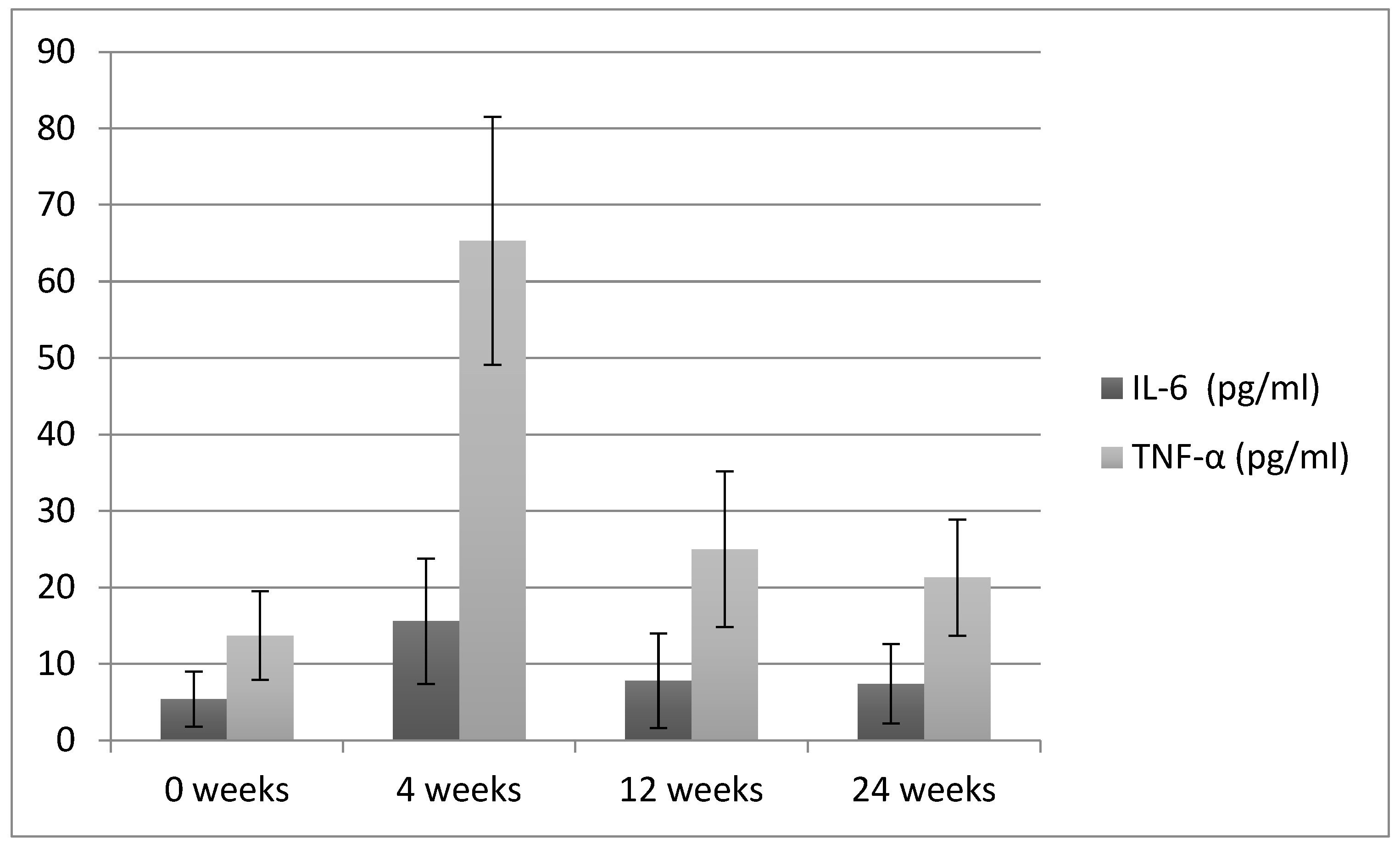
3.3. Volume and Cytokine Profile of the GCF

Mean GCF volume and cytokine profile were presented in Table 2. Mean GCF volume at week 4 (1.3 ± 0.6 μL) was significantly higher from the baseline (0.6 ± 0.2 μL) (p < 0.05). Mean volume at week 12 (0.8 ± 0.3 μL) and 24 (0.8 ± 0.5 μL) was found to be comparable to control (p > 0.05). GCF levels of IL-6 were increased at week 4 (15.6 ± 8.2 pg/mL) from the baseline (5.4 ± 3.6 pg/mL) (p < 0.05). However, there was no significant difference in GCF IL-6 level at week 12 (7.8 ± 6.2 pg/mL) and 24 (7.4 ± 5.2 pg/mL) from the baseline (p > 0.05). Moreover, level of TNF-α in GCF at week 4 (65.3 ± 16.2 pg/mL), 12 (25 ± 10.2 pg/mL), and 24 (21.3 ± 7.6 pg/m) was found to be higher than the baseline (13.7 ± 5.8 pg/mL) (p < 0.05) (Figure 4).

4. Discussion

The present study was based on the null hypothesis that periodontal parameters, GCF volume and GCF cytokine (IL-6 and TNF-α) levels in patients treated with lumineers would be comparable before and after treatment. It was observed that ceramic lumineer treatment showed similar clinical periodontal parameters and GCF cytokine profile (IL-6 and TNF-α) levels from baseline to 24-week follow-up. Therefore, the postulated null hypothesis was accepted. Multiple explanations for these outcomes include ceramic biocompatibility, oral hygiene improvements, socio-economic status of patients, and expertise of treatment providers.
Primary use of lumineers were emphasized to the advantage of patients who did not want their teeth to be prepared [5]. Lumineers, due to their higher esthetics, biocompatibility, and clinical longevity, are considered as an alternative to complete-coverage restorations [24]. Studies suggest that researches are focused mostly on esthetic and technical aspects of ceramic veneers [25,26]. However, investigations pertaining to gingival and periodontal outcomes of ceramic lumineer restorations are limited and showed heterogeneous results. In addition, an appreciation of restoration and periodontal interface is critical in determining the successful clinical outcomes and to ensure satisfactory function, form and esthetics of the dentition [27].
GCF levels of inflammatory cytokines (IL-6 and TNF-α) have found to be increased in different inflammatory conditions. Cytokine balance plays an important part in stability and early progression of inflammation, however their detrimental role in progression of periodontal disease is gaining strength [15,17,28]. Macrophages and T-cells secrete TNF-α, which play important role in early induction of periodontal inflammation. Furthermore, it may synergistically stimulate human gingival fibroblasts to enhance IL-6 secretion. Elevation of both IL-6 and TNF-α causes periodontal and gingival breakdown via disturbances in lipid metabolism [29]. It is also suggested that IL-6 exerts its influence by myeloid cell differentiation, up-regulation of B lymphocytes and T cell populations [17]. IL-6 therefore amplifies the host response and regulates periodontal tissue homeostasis through stimulation of osteoclastic activity and growth of periodontopathogenic bacteria [30]. Moreover, increased IL-6 levels are reported in periodontitis and a significant decrease in its level following periodontal therapy indicates its role as an early disease onset biomarker for periodontal inflammation [17,31].
The results of the present study demonstrated that PI and BOP scores at week 4 were significantly higher from the baseline. There are multiple reasons accredited to this outcome. Considering the esthetic requirement, most of the lumineers’ margins in the present study were placed subgingivally or equigingivally, this might have caused reversible damage to the junctional epithelium and connective tissue [32]. Furthermore, subgingival margin tends to accumulate more plaque and exerts potentially undesirable effects on the health of surrounding tissues. Moreover, the role of soft tissue manipulation during margin preparation, gingival retraction, and impression making cannot be neglected. In addition, a direct relationship exists between over contouring and soft tissue inflammation, as it causes plaque accumulation and soft tissue disturbance due to change in emergence profile. Furthermore, undetected excess cement in the sulcus area might also have contributed to this outcome [33].
Interestingly, PI and BOP findings become comparable to the baseline at week 12 and 24. In addition, PPD and CAL remains unaffected after lumineer placement and appeared comparable to baseline at 4, 12, and 24 weeks follow-up. These outcomes are in accordance with the findings of the study conducted by Arif et al., [34]. According to which, gingival response after lumineer placement was satisfactory with GI and PPD scores remaining within normal range. A possible explanation for this finding is the biocompatibility of ceramics and its ability to minimize toxic and inflammatory cellular effects with minimal bacterial growth around the cervical margin [35]. Hahn et al., in their study reported that ceramic restorations accumulated less plaque around the restoration margins along with the reduced bacterial viability. In addition, oral hygiene instructions and plaque control are pivotal for a healthy perio-restorative interface [36]. As oral hygiene instructions were reinforced at regular review appointments, it is the author’s opinion that plaque control and therefore BOP considerably improved with consequent follow-up examinations at 12 and 24 weeks in patients [37]. Hussain, in her study, found that regular oral hygiene instructions improved the periodontal parameters and plaque control in patients with dental veneers [38]. It is also suggested that education level and socioeconomic status have a positive indirect influence on oral health and plaque control of individuals [39]. As a majority of participants (87.4%) enrolled in the present study were well educated and belonged to good socio-economic background, this might have contributed to the results of the study. Furthermore, it is reported in previous studies that females exhibit better behavior and attitude towards oral hygiene practices than males [40,41]. Incidentally, the majority (59.25%) of patients in the present study were females, therefore gender distribution and their behavior towards oral hygiene practices positively influenced the study findings.
GCF flow rate is an indicator of increased tissue permeability which represents early stages of inflammation. This outflow removes the non-adherent microbes and their toxins from dento-gingival space. The findings of the present study showed that GCF volume and GCF levels of IL-6 and TNF-α at week 4 was significantly higher compared to baseline. IL-6 and TNF-α cytokines in GCF are pro-inflammatory biomarkers, suggestive of deterioration and clinical inflammatory response in periodontium [18,42]. Subgingival margin, biological width violation, overzealous cord placement before lumineers placement margin adaptation, lumineer gingival contour, and cementation might be the reasons for the greater volume of GCF and cytokine level derangement [29,43]. This result is in line with the outcomes of the study conducted by Kowashi et al., according to which GCF flow rate increases to 5.5-fold during 0–21 days in gingivitis experiment study [44]. However, GCF flow rate and proinflammatory cytokines levels returned to baseline at week 12 and 24. This result is in accordance with the findings of clinical studies which suggested that highly glazed porcelain retains less plaque than tooth enamel [35].
It is well known, that individuals with periodontal infections and ongoing or previous periodontal disease may respond poorly to dental restorative treatments relative to healthy people. In addition, habitual tobacco smokers, diabetics, pregnant women and elderly patients are susceptible to periodontal disease [45,46,47]. In the present study, patients with chronic periodontitis, systemic disease, and smokers were excluded that affects the periodontal response in such patients as a result of Lumineer treatment. Therefore, further studies assessing the clinical periodontal parameters and GCF cytokine profile of diabetics and smokers treated with esthetic lumineer restorations are recommended.

5. Conclusions

GCF levels of pro-inflammatory biomarkers (IL-6 and TNF-α) are indicative of periodontal disease initiation and progression. Adhesive ceramic lumineer treatment showed a transient increase in GCF IL-6 and TNF-α levels at four weeks of treatment indicating initiation of gingival inflammation. However, at twenty-four weeks, the IL-6 normalized to baseline levels, however TNF-α levels remained increased suggesting a sub-clinical inflammatory response. The clinical periodontal parameters among patients treated with ceramic lumineers at baseline and twenty-four week follow-up were comparable.

6. Clinical Significance

Lumineers are conservative esthetic indirect restorations, however they are over contoured as little or no tooth preparation is performed which may contribute to plaque accumulation and compromised periodontal health. The study observed that clinical periodontal parameters and GCF cytokine levels of IL-6 and TNF-α among patients treated with adhesive esthetic lumineer restorations at baseline and twenty-four week follow-up were mostly comparable. Suggesting that the clinical application of ceramic lumineers for esthetic rehabilitation is a viable option with minimal risks of compromising periodontal health.

Author Contributions

A.A. (Ali Alrahlah) and M.A.: Data collection, study design, data assessment, and manuscript writing, data analysis, final manuscript approval. A.A. (Abadulaziz Alshuwaier) and M.E.: Data inference, experiments performance, study design, manuscript drafting, data analysis, and manuscript approval. K.M.A. and E.A.: Conceptualization, Data collection, funding, resources, data interpretation, writing, revise, and editing and final manuscript approval. F.V. and T.A.: Data collection, data interpretation, funding, resources, software, writing, revise, and editing, funding acquisition and final manuscript approval. E.A.A. and A.A. (Abdullah Alshahrani): Conceptualization. Review and revise manuscript, software, resources, visualization. All authors have read and agreed to the published version of the manuscript.

Funding

This research was funded by Vice Deanship of Scientific Research Chairs, Engr. Abdullah Bugshan research chair for Dental and Oral Rehabilitation at King Saud University.

Institutional Review Board Statement

The present study was performed following guidelines recognized by the Declaration of Helsinki as revised in 2013 for experimentation involving human patients. The ethic review committee and institutional review board of specialist practice and research center (UDPRC) in Riyadh, Saudi Arabia, approved the study protocol with reference number-E-19-4329.

Informed Consent Statement

All participants completed and signed an informed consent to voluntarily participate in the study. All participants were informed that they could withdraw their participation at any stage of the investigation without consequences.

Data Availability Statement

The data is available on personal need basis on contacting the corresponding author of the published article.

Acknowledgments

The authors are grateful to the Deanship of Scientific Research, King Saud University for funding through Vice Deanship of Scientific Research Chairs, Engr. Abdullah Bugshan research chair for Dental and Oral Rehabilitation.

Conflicts of Interest

The authors declare that they have no conflict of interest and all authors have read and approved the final draft.

References

  1. Binshabaib, M.; Alharthi, S.S.; Akram, Z.; Khan, J.; Rahman, I.; Romanos, G.E.; Javed, F. Clinical periodontal status and gingival crevicular fluid cytokine profile among cigarette-smokers, electronic-cigarette users and never-smokers. Arch. Oral Biol. 2019, 102, 212–217. [Google Scholar] [CrossRef]
  2. Alhekeir, D.F.; Al-Sarhan, R.A.; Al Mashaan, A.F. Porcelain laminate veneers: Clinical survey for evaluation of failure. Saudi Dent. J. 2014, 26, 63–67. [Google Scholar] [CrossRef] [Green Version]
  3. Walls, A.W. The use of adhesively retained all-porcelain veneers during the management of fractured and worn anterior teeth: Part 2. Clinical results after 5 years of follow-up. Br. Dent. J. 1995, 178, 337–340. [Google Scholar] [CrossRef]
  4. Abduljabbar, T.; Akram, Z.; Vohra, F.; Warnakulasuriya, S.; Javed, F. Assessment of interleukin-1β, interleukin-6, and tumor necrosis factor-Α levels in the peri-implant sulcular fluid among waterpipe (narghile) smokers and never-smokers with peri-implantitis. Clin. Implant Dent. Relat. Res. 2018, 20, 144–150. [Google Scholar] [CrossRef] [PubMed]
  5. Zlatanovska, K.; Dimova, C.; Zarkova-Atanasova, J. Minimally Invasive Aesthetic Solutions—Porcelain Veneers and Lumineers. Defect Diffus. Forum 2017, 376, 111–120. [Google Scholar] [CrossRef]
  6. Mitthra, S.; Anuradha, B.; Pia, J.C.; Subbiya, A. Veneers−Diagnostic and Clinical Considerations: A Review. Indian J. Public Health Res. Dev. 2019, 10, 2143. [Google Scholar] [CrossRef]
  7. Beier, U.S.; Kapferer, I.; Burtscher, D.; Dumfahrt, H. Clinical performance of porcelain laminate veneers for up to 20 years. Int. J. Prosthodont. 2012, 25, 79–85. [Google Scholar] [PubMed]
  8. Jang, J.-H.; Lee, S.-H.; Paek, J.; Kim, S.-Y. Splinted Porcelain Laminate Veneers with a Natural Tooth Pontic: A Provisional Approach for Conservative and Esthetic Treatment of a Challenging Case. Oper. Dent. 2015, 40, E257–E265. [Google Scholar] [CrossRef]
  9. Pneumans, M. The Clinical Performance of Veneer Restorations and their Influence on the Periodontium. Influ. Periodontium 1997, 1, 34–41. [Google Scholar]
  10. Kihn, P.W.; Barnes, D.M. The clinical longevity of porcelain veneers: A 48-month clinical evaluation. J. Am. Dent. Assoc. 1998, 129, 747–752. [Google Scholar] [CrossRef]
  11. Shah, H.H.; Shah, J.H.; Patel, S.A.; Shah, H.P. Esthetic management of fluoresced teeth with ceramic veneers and direct composite bonding—An overview and a case presentation. J. Clin. Diagn. Res. 2015, 9, ZD28–ZD30. [Google Scholar]
  12. Zhyvotovskyi, I.V.; Sylenko, Y.I.; Khrebor, M.V.; Shlykova, O.A.; Izmailova, O.V. Dynamics of the level of pro- and anti-inflammatory cytokines in the crevicular fluid after direct and indirect restoration. Ukr. Dent. Alm. 2020, 46–50. [Google Scholar] [CrossRef]
  13. Chang, K.-C.J.; Wheater, M.A.; Jacobs, L.C.; Litonjua, L.A. Interleukins in gingival crevicular fluid in patients with definitive full-coverage restorations. Compend. Contin. Educ. Dent. 2014, 35, e18–e24. [Google Scholar] [PubMed]
  14. Kourkouta, S.; Walsh, T.T.; Davis, L.G. The effect of porcelain laminate veneers on gingival health and bacterial plaque characteristics. J. Clin. Periodontol. 1994, 21, 638–640. [Google Scholar] [CrossRef]
  15. Isola, G.; Polizzi, A.; Patini, R.; Ferlito, S.; Alibrandi, A.; Palazzo, G. Association among serum and salivary A. actinomycetemcomitans specific immunoglobulin antibodies and periodontitis. BMC Oral Health 2020, 20, 1–3. [Google Scholar] [CrossRef]
  16. Matarese, G.; Isola, G.; Anastasi, G.P.; Favaloro, A.; Milardi, D.; Vermiglio, G.; Vita, G.; Cordasco, G.; Cutroneo, G. Immunohistochemical analysis of TGF-β1 and VEGF in gingival and periodontal tissues: A role of these biomarkers in the pathogenesis of scleroderma and periodontal disease. Int. J. Mol. Med. 2012, 30, 502–508. [Google Scholar] [CrossRef] [PubMed] [Green Version]
  17. Isola, G.; Giudice, A.L.; Polizzi, A.; Alibrandi, A.; Murabito, P.; Indelicato, F. Identification of the different salivary Interleukin-6 profiles in patients with periodontitis: A cross-sectional study. Arch. Oral Biol. 2021, 122, 104997. [Google Scholar] [CrossRef]
  18. Akram, Z.; Abduljabbar, T.; Abu Hassan, M.I.; Javed, F.; Vohra, F. Cytokine Profile in Chronic Periodontitis Patients with and without Obesity: A Systematic Review and Meta-Analysis. Dis. Markers 2016, 2016, 1–12. [Google Scholar] [CrossRef] [Green Version]
  19. Reid, J.S.; Kinane, D.F.; Adonogianaki, E. Gingival health associated with porcelain veneers on maxillary incisors. Int. J. Paediatr. Dent. 1991, 1, 137–141. [Google Scholar] [CrossRef]
  20. Ainamo, J.; Bay, I. Problems and proposals for recording gingivitis and plaque. Int. Dent. J. 1975, 25, 229–235. [Google Scholar]
  21. Silness, J.; Löe, H. Periodontal Disease in Pregnancy II. Correlation Between Oral Hygiene and Periodontal Condition. Acta Odontol. Scand. 1964, 22, 121–135. [Google Scholar] [CrossRef]
  22. Al-Hamoudi, N.; Alsahhaf, A.; Al Deeb, M.; Alrabiah, M.; Vohra, F.; Abduljabbar, T. Effect of scaling and root planing on the expression of anti-inflammatory cytokines (IL-4, IL-9, IL-10, and IL-13) in the gingival crevicular fluid of electronic cigarette users and non-smokers with moderate chronic periodontitis. J. Periodontal Implant Sci. 2020, 50, 74–82. [Google Scholar] [CrossRef]
  23. Vohra, F.; Akram, Z.; Bukhari, I.; Sheikh, S.; Riny, A.; Javed, F. Comparison of Periodontal Inflammatory Parameters and Whole Salivary Cytokine Profile Among Saudi Patients with Different Obesity Levels. Int. J. Periodontics Restor. Dent. 2018, 38, e119–e126. [Google Scholar] [CrossRef]
  24. Öztürk, E.; Bolay, Ş.; Hickel, R.; Ilie, N. Shear bond strength of porcelain laminate veneers to enamel, dentine and enamel-dentine complex bonded with different adhesive luting systems. J. Dent. 2013, 41, 97–105. [Google Scholar] [CrossRef]
  25. Turgut, S.; Bagis, B. Colour stability of laminate veneers: An in vitro study. J. Dent. 2011, 39 (Suppl. 3), e57–e64. [Google Scholar] [CrossRef]
  26. Turgut, S.; Bagis, B. Effect of resin cement and ceramic thickness on final color of laminate veneers: An in vitro study. J. Prosthet. Dent. 2013, 109, 179–186. [Google Scholar] [CrossRef]
  27. Bennani, V.; Ibrahim, H.; Al-Harthi, L.; Lyons, K.M. The periodontal restorative interface: Esthetic considerations. Periodontology 2000 2017, 74, 74–101. [Google Scholar] [CrossRef]
  28. Souza, F.I. Laminados Cerâmicos Cimentados Sobre Dentes Não Preparados. Estudo Clínico, Prospectivo e Longitudinal Sobre a Adaptação Marginal e Avaliação do Comportamento Periodontal Pelo Uso de Biomarcadores do Fluido Gengival Crevicular. Available online: https://repositorio.unesp.br/handle/11449/180305 (accessed on 8 January 2021).
  29. Celik, N.; Askın, S.; Gul, M.A.; Seven, N. The effect of restorative materials on cytokines in gingival crevicular fluid. Arch. Oral Biol. 2017, 84, 139–144. [Google Scholar] [CrossRef]
  30. Bennermo, M.; Held, C.; Stemme, S.; Ericsson, C.-G.; Silveira, A.; Green, F.; Tornvall, P. Genetic Predisposition of the Interleukin-6 Response to Inflammation: Implications for a Variety of Major Diseases? Clin. Chem. 2004, 50, 2136–2140. [Google Scholar] [CrossRef] [Green Version]
  31. Tymkiw, K.D.; Thunell, D.H.; Johnson, G.K.; Joly, S.; Burnell, K.K.; Cavanaugh, J.E.; Brogden, K.A.; Guthmiller, J.M. Influence of smoking on gingival crevicular fluid cytokines in severe chronic periodontitis. J. Clin. Periodontol. 2010, 38, 219–228. [Google Scholar] [CrossRef]
  32. Savadi, A.; Rangarajan, V.; Savadi, R.C.; Satheesh, P. Biologic Perspectives in Restorative Treatment. J. Indian Prosthodont. Soc. 2011, 11, 143–148. [Google Scholar] [CrossRef]
  33. Block, P. Restorative margins and periodontal health: A new look at an old perspective. J. Prosthet. Dent. 1987, 57, 683–689. [Google Scholar] [CrossRef]
  34. Arif, R.; Dennison, J.B.; Garcia, D.; Yaman, P. Gingival Health of Porcelain Laminate Veneered Teeth: A Retrospective Assessment. Oper. Dent. 2019, 44, 452–458. [Google Scholar] [CrossRef] [PubMed]
  35. Guilardi, L.F.; Soares, P.; Werner, A.; de Jager, N.; Pereira, G.K.; Kleverlaan, C.J.; Rippe, M.P.; Valandro, L.F. Fatigue performance of distinct CAD/CAM dental ceramics. J. Mech. Behav. Biomed. Mater. 2020, 103, 103540. [Google Scholar]
  36. Peumans, M.; Van Meerbeek, B.; Lambrechts, P.; Vanherle, G. Porcelain veneers: A review of the literature. J. Dent. 2000, 28, 163–177. [Google Scholar] [CrossRef]
  37. Daood, U.; Abduljabbar, T.; Al-Hamoudi, N.; Akram, Z. Clinical and radiographic periodontal parameters and release of collagen degradation biomarkers in naswar dippers. J. Periodontal Res. 2018, 53, 123–130. [Google Scholar] [CrossRef] [PubMed]
  38. Sabir Hussain, A. Oral Health and Dental Veneers: Clinical Tips. Am. J. Biomed. Sci. Res. 2019, 2. [Google Scholar] [CrossRef]
  39. Ghosh-Dastidar, R.; Gillam, D.G.; Islam, S.S. Socio-Demographic and Oral Health Related Risk Factors for Periodontal Disease in Inner North East London (INEL) Adults: A Secondary Analysis of the INEL Data. Int. J. Dent. Oral Health 2016, 2. [Google Scholar] [CrossRef]
  40. Östberg, A.-L.; Halling, A.; Lindblad, U. Gender differences in knowledge, attitude, behavior and perceived oral health among adolescents. Acta Odontol. Scand. 1999, 57, 231–236. [Google Scholar] [CrossRef]
  41. Al-Omari, Q.D.; Hamasha, A.A.-H. Gender-Specific Oral Health Attitudes and Behavior among Dental Students in Jordan. J. Contemp. Dent. Pract. 2005, 6, 107–114. [Google Scholar] [CrossRef]
  42. Alwan, A.H. Determination of Interleukin-1β (IL-1 β) and Interleukin-6(IL6) in Gingival Crevicular Fluid in Patients with Chronic Periodontitis. IOSR J. Dent. Med. Sci. 2015, 14, 81–90. [Google Scholar]
  43. Brunton, P.A.; Wilson, N.H. Preparations for porcelain laminate veneers in general dental practice. Br. Dent. J. 1998, 184, 553–556. [Google Scholar] [CrossRef] [PubMed]
  44. Kowashi, Y.; Jaccard, F.; Cimasoni, G. Sulcular polymorphonuclear leucocytes and gingival exudate during experimental gingivitis in man. J. Periodontal Res. 1980, 15, 151–158. [Google Scholar] [CrossRef] [PubMed]
  45. Tomar, S.L. Smoking-attributable periodontitis in the United States: Findings from NHANES III Tobacco control View project Global Adult Tobacco Survey View project. J. Periodontol. 2000, 71, 743–751. [Google Scholar] [CrossRef]
  46. Laine, M.A. Effect of pregnancy on periodontal and dental health. Acta Odontol. Scand. 2002, 60, 257–264. [Google Scholar] [CrossRef] [PubMed]
  47. Campus, G.; Salem, A.; Uzzau, S.; Baldoni, E.; Tonolo, G. Diabetes and Periodontal Disease: A Case-Control Study. J. Periodontol. 2005, 76, 418–425. [Google Scholar] [CrossRef] [PubMed]
Figure 1. Flow diagram for study protocol.
Applsci 11 02829 g001
Figure 2. Plaque index (PI) and bleeding on probing (BOP) percentage (%) observations from baseline to 24-week follow-up among patients treated with lumineers.
Applsci 11 02829 g002
Figure 3. Comparison of periodontal pocket depth (PPD) and clinical attachment loss (CAL) in millimeters (mm) as observed at baseline and different follow-up durations in patients treated with lumineers.
Applsci 11 02829 g003
Figure 4. Comparison of IL-6 and TNF-α pg/mL as observed at baseline and different follow-up durations in patients treated with lumineers.
Applsci 11 02829 g004
Table 1. General characteristics of the study population.
Study Characteristics
Number of Patients27
Age range (Years)23.5–40.6
Gender (F/M)16/11
Number of Lumineers288
Maxilla/Mandible196/92
Table 2. Gingiva crevicular fluid (GCF) flow rate and periodontal parameters observed in the study.
ParametersBaseline (Control)4 Weeks12 Weeks24 Weeksp-Value *
Mean PI (%)15.2 ± 4.3 A25.4 ± 6.7 B17.8 ± 7.3 A17.1 ± 4.5 A<0.05
Mean BOP (%)12.8 ± 5.6 A28.6 ± 7.8 B16.1 ± 8.3 A16.5 ± 5.5 A<0.05
Mean PPD (mm)2.4 ± 0.5 A2.6 ± 0.7 A2.8 ± 0.9 A2.7 ± 0.6 A>0.05
Mean CAL (mm)1.1 ± 0.5 A1.1 ± 0.8 A1.3 ± 0.5 A1.3 ± 0.7 A>0.05
Mean GCF volume (μL)0.6 ± 0.2 A1.3 ± 0.6 B0.8 ± 0.3 A0.8 ± 0.5 A<0.05
IL-6 (pg/mL)5.4 ± 3.6 A15.6 ± 8.2 B7.8 ± 6.2 A7.4 ± 5.2 A<0.05
TNF-α (pg/mL)13.7 ± 5.8 A65.3 ± 16.2 B25 ± 10.2 C21.3 ± 7.6 C<0.05
* ANOVA, Dissimilar capital superscript alphabets denote statistically significant difference in same row (Tukey–Kramer Multiple comparisons test).
Publisher’s Note: MDPI stays neutral with regard to jurisdictional claims in published maps and institutional affiliations.

