Article

Platelet-Rich Plasma in Anterior Cruciate Ligament Quadriceps Tendon Bone Reconstruction—Impact of PRP Administration on Pain, Range of Motion Restoration, Knee Stability, Tibial Tunnel Widening and Functional Results

1
Artromedical Orthopaedic Clinic, Antracytowa 1, 97-400 Belchatow, Poland
2
Twin Cities Orthopedics, Edina, MN 55121, USA
3
Orthopedic and Trauma Department, Veteran’s Memorial Teaching Hospital in Lodz, Medical University of Lodz, 90-549 Lodz, Poland
*
Author to whom correspondence should be addressed.
Appl. Sci. 2021, 11(9), 3993; https://doi.org/10.3390/app11093993
Submission received: 30 March 2021 / Revised: 14 April 2021 / Accepted: 27 April 2021 / Published: 28 April 2021
(This article belongs to the Special Issue Multicriterial Assessment of Ligament Healing in the Knee Joint)

Abstract

:
Background: Using Platetet-Rich Plasma (PRP) in anterior cruciate ligament reconstruction (ACLR) has been suggested to improve patient outcomes. The aim of this study was to assess the impact of PRP administration on pain, range of motion (ROM) restoration and the functional results of ACLR performed with quadriceps tendon bone (QTB) autografts. Methods: A total of 106 patients were included in this multicenter study. Fifty-two patients underwent single-bundle QTB ACLR and 54 patients underwent the same procedure with additional PRP administration. Results: Mean time of need for on-demand analgesia was 8 days in the PRP group and 11 days in no-PRP group. Symmetric full extension was restored in a mean of 40 days in the PRP group and 53 days in the no-PRP group. Ninety degrees of flexion was restored at a mean of 21 days in the PRP group and 25 days in the no-PRP group. At 18 months postoperatively, the mean side-to-side difference in anterior tibial translation with the use of an arthrometer (Rolimeter, Aircast Europa) was 1.3 mm in the PRP group vs. 2.7 mm in the no-PRP group. Mean tibial tunnel widening was 1.4 mm in the PRP group vs. 2.1 mm in the no-PRP group. The mean score in the pain section of the KOOS scale was 93 in the PRP group vs. 89 in the no-PRP group. For the IKDC scale, 53 patients in the PRP group graded A or B and 1 patient graded C. In the no-PRP group, 48 patients graded A or B and 4 patients graded C or D. Conclusions: The use of PRP in QTB ACLR may decrease the need for on-demand analgesia and accelerate ROM restoration as well as improve knee stability, lessen the extent of tibial tunnel widening and potentially diminish pain at 18 months postoperatively. Further studies will be needed to confirm all authors’ conclusions.

1. Introduction

The use of a quadriceps tendon with an attached bone block (QTB) as a source of a graft for anterior cruciate ligament (ACL) reconstruction (ACLR) has gained popularity in recent years. Its advocates point out its greater cross-sectional area and higher failure load than a patellar tendon graft, making it a feasible option for an ACL graft choice [1,2]. Additionally, it enables the bone-to-bone healing, which is faster and results in stronger incorporation than bone-to-tendon healing mode for all soft tissue grafts [3,4]. These assumptions are being verified by recent systematic reviews that report that quadriceps tendon is equal to other graft options for ACLR in terms of stability, functional outcomes and complications [5,6].
The outcome of an ACLR is strongly influenced by the progress of graft remodeling. To enhance the progress of ligamentization, various modalities have been proposed, with the use of platelet rich plasma (PRP) being one of the most extensively used in a variety of clinical settings [7,8,9,10,11,12]. PRP is a first-generation autologous platelet concentrate used to promote wound healing and tissue regeneration through the release of growth factors [13]. However, despite its clinical popularity, the utility of PRP in ACL reconstruction is still controversial [8]. Thus, the purpose of this study was to assess the impact of PRP on pain, range of motion (ROM) restoration, knee stability, tibial tunnel widening and functional results.

2. Materials and Methods

2.1. Patient Selection
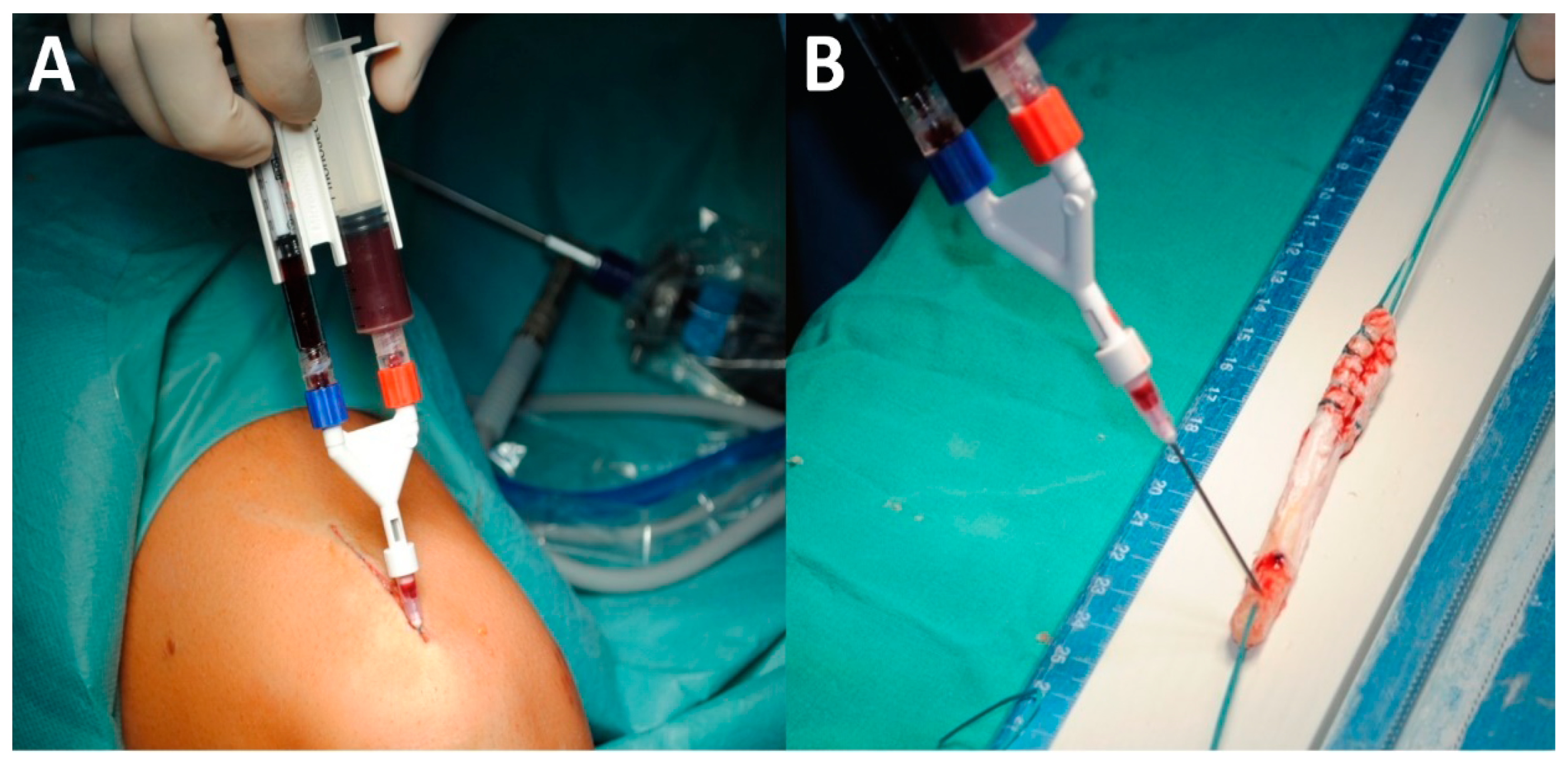
This was a retrospective multicenter clinical study. All patients undergoing ACL reconstruction with a QTB graft between 2008 to 2010 in the Maków Mazowiecki District Hospital, Bełchatów MegaMed Hospital and Radom “Ortopedia” Hospital that fulfilled the study criteria were invited to participate. We included consecutive patients who were scheduled to undergo ACL reconstruction. Qualification criteria for this procedure were: symptomatic anterior instability of the knee with full preoperative ROM. Qualification of all patients to ACLR was performed by the same single surgeon. Excluded were patients with more than 3 mm of additional instabilities (medial, lateral or posterior instability), any rotational instability of the knee, more than 1/3 of width lateral or 2/3 medial meniscectomy, more than 2° ICRS cartilage damage, more than 10° limb malalignment in the frontal plane and hip or ankle diseases. From 106 patients included in the study, 52 were randomly assigned to the group that underwent single-bundle QTB ACLR and 54 to the group that underwent the same procedure with an additional PRP administration. Neither the researchers nor patients were blinded.

2.2. Procedure

Single bundle ACL reconstructions were performed in an anatomic, transportal manner with anteromedial femoral tunnel using a quadriceps tendon-bone full thickness autograft (9 mm × 95−100 mm) (Figure 1). The graft was harvested as described in a recent technical article [14]. Grafts were approximately 9 mm in diameter, both on the soft tissue portion and on the bone plug. They were harvested with the use of straight 8-mm-wide osteotome used as the guide. The soft tissue portion was prepared by incising quadriceps tendon with surgical knife and straight scissors up to about 8 cm proximal to the patella. The bone plug was about 20 mm long and it was harvested with use of a power saw with a 7-mm depth limiter and an osteotome. Grafts were stabilized with titanium screws in the femoral and tibial tunnels and with sutures tied over the bone bridge as additional stabilization on the tibia. For the PRP group, it was obtained from a sample of the patient’s blood and processed using the Zimmer Biomet GPS® Platelet Concentration System. Minimal infrapatellar fat pad was resected. Saline passage was stopped at the initiation of injecting PRP (before placing the graft in the joint) in order to avoid rinsing off the PRP. In the PRP group, it was administered to the graft harvest site, onto the intra-tunnel portions of the graft (Figure 2) and into the reconstruction tunnels (Figure 3). Tunnels were injected with PRP both before and after graft insertion (through the cannulated interference screw, Figure 3). Any remaining plasma and thrombin were administered onto the skin wounds. All cases were operated and qualified by a single surgeon. ACLR was a second stage surgery, all additional procedures (i.e., suturing the meniscus) were performed during the first stage surgeries at least 6 to 8 weeks before the ACLR. All patients underwent the same postoperative rehabilitation protocol. They spent the first 3 days after the surgery in the hospital with on-demand analgesia. A postoperative brace was set at 15 to 90 degrees of flexion and administered for use while ambulating for the first three weeks. Patients were allowed to remove the brace during activities such as sitting or lying in bed. After the third week, the brace was maintained with no ROM limitation to 12 weeks postoperatively. Crutches were obligatory for 14 days for ambulation and up to 6 weeks for outside locomotion. Patients were recommended to perform weightbearing as tolerated. Home exercises for ROM restoration started at the first postoperative day; however, patients were warned to perform them in the pain-free zone. Physical therapy with a specialist started at 7 days after surgery and was performed twice a week up to 6 months postoperatively.
Quadriceps tendon-bone full-thickness autograft visible after harvesting and preparation. The graft was harvested as described in a recent technical article [14]. Grafts were approximately 9 mm in diameter, both on the soft tissue portion and on the bone plug. They were harvested with the use of straight 8-mm-wide osteotome used as the guide. The soft tissue portion was prepared by incising quadriceps tendon with surgical knife and straight scissors up to about 8cm proximal to the patella. The bone plug was about 20 mm long and it was harvested with use of a power saw with a 7-mm depth limiter and an osteotome.

2.3. Assessment of Outcomes

Patients were evaluated postoperatively by three independent non-blinded surgeons at 2 weeks, 6 weeks, 3 months, 6 months and at a final follow-up of 18 months. In the first days after surgery, the following early postoperative outcomes were assessed: body temperature, need for on-demand analgesia, presence of palpable hematoma/swelling, time to walking without crutches, and the time of return to full knee extension and to 90 degrees of knee flexion.
The following middle-term outcomes were evaluated at the final follow-up. Harvest site pain was assessed in scale of 0—no pain at all; 1—slight pain; 2—moderate pain; 3—severe pain. Simple dynamic testing was performed, consisting of jumping and twisting on one knee. The possible scores were: 2 points—3 jumps, 40 cm each, performed on a flexed knee, with 90 degrees of body twist in every jump and with 5 s of balance after the jumps; 1 point—as above, but without twisting the body in the jumps; 0 points—inability to complete this test. Knee injury and Osteoarthritis Outcome Score (KOOS) and International Knee Documentation Committee (IKDC) functional scales were collected. Stability was evaluated by the means of an arthrometer (Rolimeter, Aircast Europe) used to assess side-to-side differences in anterior tibial translation. Tibial tunnel widening was also measured manually at the 18 months postoperatively on AP and lateral radiographs (Figure 4).

2.4. Statistical Analysis

The data were collected in the years 2008—2010 and the results of this study were presented at 8th Biennial ISAKOS Congress in 2011, Rio de Janeiro, Brazil and at 15th Congress of ESSKA in 2012, Geneva, Switzerland. Unfortunately, the original individual patients sheets with recorded individual outcomes were unavailable. Therefore, for the sake of preparation of this article, data were retrieved from the outcomes sheets, making it impossible to perform a statistical analysis of continuous variables. For nominal variables, data were analyzed using a Chi square test, Chi square test with Yates correction or a double-tailed exact Fisher test, accordingly to the number of patients included in the analysis of the given variable. All statistical analyses were performed in the Statistica version 13.3 software (StatSoft Poland, Kraków, Poland).

3. Results

3.1. Early Postoperative Outcomes

The mean time of the presence of an elevated body temperature and mean body temperature were respectively 3.5 days, 37.7 Celsius degrees in the PRP group vs. 3 days, 37.4 Celsius degrees in the no-PRP group. Palpable hematoma/swelling was present in the PRP group for an average of 22 days vs. 21 days in the no-PRP group. The on-demand analgesia in the PRP group was administered for mean of 8 days, whereas patients in the no-PRP group required it for a mean time of 11 days. The mean time to walking without crutches was 21 days in the PRP group vs. 25 days in the no-PRP group. Full knee extension was restored in a mean time of 40 days in the PRP group vs. 53 days in no-PRP group. Ninety degrees of flexion was restored in a mean time of 21 days in the PRP group vs. 25 days in the no-PRP group (Table 1.)

3.2. Mid-Term Outcomes

At 18 months after surgery, harvest site pain was present in three patients in each group. On the scale of 0 to 3, the mean pain intensity was 1.3 in the PRP group vs. 2.3 in the no-PRP group. Results of dynamic testing with maximum score of 2 points were 1.98 points in the PRP group vs. 1.8 points in the no-PRP group. For the KOOS scale, the results were as following: for the PRP group vs. the no-PRP group, respectively: in the Symptoms section 92 vs. 90; in the Pain section 93 vs. 89; in the Function, daily living section 94 vs. 92; in the Function, sport activities section 90 vs. 88; in the Quality of life section 83 vs. 85. As for the IKDC scale, 53 patients in the PRP group graded A or B and 1 patient graded C. In the no-PRP group, 48 patients graded A or B and 4 patients graded C or D. This difference was not significant (p = 0.201). The mean side-to-side difference in anterior tibial translation during the stability test performed with the use of the arthrometer (Rolimeter, Aircast Europa) was 1.3 mm in PRP group vs. 2.7 mm in no-PRP group. Tibial tunnel widening at 18 months after the surgery was present in 23 out of 54 patients in the PRP group (42.6%), compared with 28 out of 52 patients in the no-PRP group (53.8%), p = 0.246. Mean widening in the PRP group was 1.4 mm vs. 2.1 mm in the no-PRP group (Table 2).

4. Discussion

The most important findings of the study were that the use of PRP in QTB ACLR may decrease the need for on-demand analgesia and accelerate ROM restoration as well as improve knee stability, lessen the extent of tibial tunnel widening and potentially diminish pain at 18 months postoperatively.
A small elongation of the time of elevated body temperature and a small elevation in the mean body temperature were observed in the PRP group compared to the no-PRP group. Mean body temperature did not exceed 38 Celsius degrees, in agreement with the study of Zhang et al., who reported no case of fever in the series of 30 patients who underwent PRP injection in the knee [15]. Regarding the presence of hematoma/swelling, there was only a slight difference in time of their presence between groups in our study. Such data supports the results of Seijas et al., who reported no significant difference in postoperative swelling between PRP and control groups [16]. Patients in the PRP group needed on-demand analgesia for an average 3 days less than patients in the no-PRP group. This corresponds with the results of de Almeida et al., who reported a lower Visual Analog Score (VAS) in the PRP group on the first postoperative day [17].
Patients who had PRP administration were able to walk without crutches for an average of 4 days faster than patients from the no-PRP group. To our knowledge, no data regarding the impact of PRP on the necessity of crutches support were reported in the literature. What is more, both restoration of full knee extension and 90 degrees knee flexion occurred earlier in the PRP group. This stands in contrast to the study of Azcárate et al., who showed no significant difference in the range of knee motion when adding PRP to the ACLR [18]. Such outcomes of our study could result from earlier walking without crutches.
A recent systematic review reported that there is no current evidence that using PRP in ACLR improves functional outcomes [8]. This stays in accordance with results of our study, in which only a slight superiority was noted in most of subsections of KOOS scale. What is more, in the Quality of life section there was an advantage of the no-PRP group. The same applied to the IKDC scale, where the superior results of patients from PRP group did not reach statistical significance.
At 18 months postoperatively, harvest site pain was rare in our patients, being present in less than 6% of patients in both groups. However, the pain intensity was greater in the no-PRP group than in PRP group, reflected also by the lower mean score in Pain section of KOOS scale in the no-PRP group. This is in agreement with the study of Cervellin et al., who reported improved outcomes for the Victorian Institute Sport Assessment (VISA) pain scale in the PRP group at 12 months of follow-up [19]. The same authors reported no significant differences in the VAS at 12 months of follow-up [19]. What is more, in the recent double-blind randomized controlled trial by Walters et al., there was no difference in the VAS between the PRP and no-PRP groups at 12 weeks, 6 months, 1 year and 2 years after surgery [20]. However, VISA pain scale seems more comparable to Pain section of KOOS used in our study, because both these scales consisted of the set of questions assessing pain during different activities [19].
There was a slight tendency towards superiority of the PRP group in dynamic test performance; however, it was not supported by the functional scales. The potential mechanism of faster maturation of the graft and better proprioception in the PRP group could explain such differences [9,21]. However, it may be too slight to gain clinical significance. Using PRP resulted in 1.4 mm lower side-to-side difference in anterior tibial translation during the arthrometer (Rolimeter, Aircast Europa) stability test. This is comparable with the study of Mirzatolooei et al., who reported mean side to side difference in the PRP group to be 1.1 mm, compared with 2.2 mm in the control group [22].
Tibial tunnel widening was observed in a slightly lower percentage of patients in the PRP group; however, this difference did not reach statistical significance. However, mean widening was lower in the PRP group. This parameter is of crucial importance, because massive widening of the tunnel makes it impossible for the graft to properly incorporate into bone and remodel. Therefore, lower degrees of tibial tunnel widening can be considered as an indicator of improved graft incorporation into bone [23,24]. The results of our study are in agreement with the literature reporting improved incorporation of graft into bone when ACLR is enhanced with PRP administration [16,25,26]. Rupreht et al. assessed the formation of the tunnel wall cortical bone encircling the tibial tunnel of an ACLR, which was recognized as an indicator of graft incorporation. They found significantly higher percentages of cortical bone created at two and a half months and six months after surgery in the PRP group compared with controls [25]. Seijas et al. reported better remodeling of ACL grafts in the PRP group than in controls at 4 months, 6 months and 12 months postoperatively [16]. In the study of Ventura et al., patients who underwent an ACLR with PRP administration achieved higher density of the graft at 6 months, comparable to the density of the posterior cruciate ligament (PCL) [26].
The main limitation of this study was that it was not possible to perform statistical analysis for continuous outcomes. The second limitation was that this study was retrospective with all inherent limitations of such study design. On the other hand, groups in this study were larger than in any other study included in the recent systematic review by Davey et al. and we also analyzed some outcomes that were not reported before in the literature [8]. An advantage of our study was that it concerned the impact of PRP administration on QTB ACLR, which due to our knowledge was not reported in the literature before.

5. Conclusions

The use of PRP in QTB ACLR may decrease the need for on-demand analgesia and accelerate ROM restoration as well as improve knee stability, lessen the extent of tibial tunnel widening and potentially diminish pain at 18 months postoperatively. Further studies will be needed to confirm all authors’ conclusions.

Author Contributions

Conceptualization, K.M., M.E., M.M., R.F.L.; methodology, K.M.; formal analysis, M.M.; investigation, K.M., M.E., M.M.; resources, K.M., M.E., M.M.; data curation, K.M., M.E., M.M.; writing—original draft preparation, M.E., M.M.; writing—review and editing, K.M., R.F.L.; supervision, K.M., R.F.L.; project administration, K.M. All authors have read and agreed to the published version of the manuscript.

Funding

This research received no external funding.

Institutional Review Board Statement

Ethical review and approval were waived for this study, due to the fact that PRP administration was a standard procedure covered and approved by the national healthcare system.

Informed Consent Statement

Informed consent was obtained from all subjects involved in the study.

Data Availability Statement

The available datasets reported in this study are available upon the reasonable request from the corresponding author.

Conflicts of Interest

The authors declare no conflict of interest.

Abbreviations

PRP—platelet-rich plasma; ACL—anterior cruciate ligament; ACLR—anterior cruciate ligament reconstruction; ROM—range of motion; QTB—quadriceps tendon bone; KOOS—Knee injury and Osteoarthritis Outcome Score; IKDC—and International Knee Documentation Committee; SSD—side to side difference; NA—not available; A—available.

References

  1. Lin, K.M.; Boyle, C.; Marom, N.; Marx, R.G. Graft Selection in Anterior Cruciate Ligament Reconstruction. Sports Med. Arthrosc. 2020, 28, 41–48. [Google Scholar] [CrossRef]
  2. Harris, N.L.; Smith, D.A.B.; Lamoreaux, L.; Purnell, M. Central Quadriceps Tendon for Anterior Cruciate Ligament Reconstruction. Am. J. Sports Med. 1997, 25, 23–28. [Google Scholar] [CrossRef]
  3. Suzuki, T.; Shino, K.; Nakagawa, S.; Nakata, K.; Iwahashi, T.; Kinugasa, K.; Otsubo, H.; Yamashita, T. Early integration of a bone plug in the femoral tunnel in rectangular tunnel ACL reconstruction with a bone-patellar tendon-bone graft: A prospective computed tomography analysis. Knee Surg. Sport. Traumatol. Arthrosc. 2011, 19, S29–S35. [Google Scholar] [CrossRef] [PubMed]
  4. Hofbauer, M.; Soldati, F.; Szomolanyi, P.; Trattnig, S.; Bartolucci, F.; Fu, F.; Denti, M. Hamstring tendon autografts do not show complete graft maturity 6 months postoperatively after anterior cruciate ligament reconstruction. Knee Surg. Sport. Traumatol. Arthrosc. 2019, 27, 130–136. [Google Scholar] [CrossRef] [PubMed] [Green Version]
  5. Mouarbes, D.; Menetrey, J.; Marot, V.; Courtot, L.; Berard, E.; Cavaignac, E. Anterior Cruciate Ligament Reconstruction: A Systematic Review and Meta-analysis of Outcomes for Quadriceps Tendon Autograft Versus Bone—Patellar Tendon—Bone and Hamstring-Tendon Autografts. Am. J. Sports Med. 2019, 47, 3531–3540. [Google Scholar] [CrossRef] [PubMed]
  6. Slone, H.S.; Romine, S.E.; Premkumar, A.; Xerogeanes, J.W. Quadriceps tendon autograft for anterior cruciate ligament reconstruction: A comprehensive review of current literature and systematic review of clinical results. Arthrosc. J. Arthrosc. Relat. Surg. 2015, 31, 541–554. [Google Scholar] [CrossRef] [PubMed]
  7. Chahla, J.; Kennedy, M.I.; Aman, Z.S.; LaPrade, R.F. Ortho-Biologics for Ligament Repair and Reconstruction. Clin. Sports Med. 2019, 38, 97–107. [Google Scholar] [CrossRef] [PubMed]
  8. Davey, M.S.; Hurley, E.T.; Withers, D.; Moran, R.; Moran, C.J. Anterior Cruciate Ligament Reconstruction with Platelet-Rich Plasma: A Systematic Review of Randomized Control Trials. Arthrosc. J. Arthrosc. Relat. Surg. 2020, 36, 1204–1210. [Google Scholar] [CrossRef] [PubMed]
  9. Dallo, I.; Chahla, J.; Mitchell, J.J.; Pascual-Garrido, C.; Feagin, J.A.; LaPrade, R.F. Biologic Approaches for the Treatment of Partial Tears of the Anterior Cruciate Ligament: A Current Concepts Review. Orthop. J. Sport. Med. 2017, 5. [Google Scholar] [CrossRef] [Green Version]
  10. Giudice, A.; Barone, S.; Giudice, C.; Bennardo, F.; Fortunato, L. Can platelet-rich fibrin improve healing after surgical treatment of medication-related osteonecrosis of the jaw? A pilot study. Oral Surg. Oral Med. Oral Pathol. Oral Radiol. 2018, 126, 390–403. [Google Scholar] [CrossRef]
  11. Fortunato, L.; Bennardo, F.; Buffone, C.; Giudice, A. Is the application of platelet concentrates effective in the prevention and treatment of medication-related osteonecrosis of the jaw? A systematic review. J. Cranio-Maxillo-Facial Surg. Off. Publ. Eur. Assoc. Cranio-Maxillo-Facial Surg. 2020, 48, 268–285. [Google Scholar] [CrossRef] [PubMed]
  12. Kwapisz, A.; Prabhakar, S.; Compagnoni, R.; Sibilska, A.; Randelli, P. Platelet-Rich Plasma for Elbow Pathologies: A Descriptive Review of Current Literature. Curr. Rev. Musculoskelet. Med. 2018, 11, 598–606. [Google Scholar] [CrossRef] [Green Version]
  13. Bennardo, F.; Liborio, F.; Barone, S.; Antonelli, A.; Buffone, C.; Fortunato, L.; Giudice, A. Efficacy of platelet-rich fibrin compared with triamcinolone acetonide as injective therapy in the treatment of symptomatic oral lichen planus: A pilot study. Clin. Oral Investig. 2021. [Google Scholar] [CrossRef] [PubMed]
  14. Malinowski, K.; Paszkowski, J.; Mostowy, M.; Góralczyk, A.; LaPrade, R.F.; Hermanowicz, K. Quadriceps Tendon-Bone Full-Thickness Autograft: Reproducible and Easy Harvesting Technique Using Simple Surgical Tools. Arthrosc. Tech. 2021. [Google Scholar] [CrossRef]
  15. Zhang, H.; Bai, Y.; Liu, C.; Jin, S.; Su, K.; Liu, Y.; Lü, Z. Effect of intra-articular injection of platelet-rich plasma on interleukin-17 expression in synovial fluid and venous plasma of knee osteoarthritis patients. Zhongguo Xiufu Chongjian Waike Zazhi = Chin. J. Reparative Reconstr. Surg. 2017, 31, 918–921. [Google Scholar] [CrossRef]
  16. Seijas, R.; Ares, O.; Catala, J.; Alvarez-Diaz, P.; Cusco, X.; Cugat, R. Magnetic resonance imaging evaluation of patellar tendon graft remodelling after anterior cruciate ligament reconstruction with or without platelet-rich plasma. J. Orthop. Surg. (Hong Kong) 2013, 21, 10–14. [Google Scholar] [CrossRef] [PubMed]
  17. De Almeida, A.M.; Demange, M.K.; Sobrado, M.F.; Rodrigues, M.B.; Pedrinelli, A.; Hernandez, A.J. Patellar tendon healing with platelet-rich plasma: A prospective randomized controlled trial. Am. J. Sports Med. 2012, 40, 1282–1288. [Google Scholar] [CrossRef] [PubMed]
  18. Azcárate, A.V.; Lamo-Espinosa, J.; Beola, J.D.A.; Gonzalez, M.H.; Gasque, G.M.; Nin, J.R.V. Comparison between two different platelet-rich plasma preparations and control applied during anterior cruciate ligament reconstruction. Is there any evidence to support their use? Injury 2014, 45, S36–S41. [Google Scholar] [CrossRef]
  19. Cervellin, M.; de Girolamo, L.; Bait, C.; Denti, M.; Volpi, P. Autologous platelet-rich plasma gel to reduce donor-site morbidity after patellar tendon graft harvesting for anterior cruciate ligament reconstruction: A randomized, controlled clinical study. Knee Surg. Sport. Traumatol. Arthrosc. 2012, 20, 114–120. [Google Scholar] [CrossRef]
  20. Walters, B.L.; Porter, D.A.; Hobart, S.J.; Bedford, B.B.; Hogan, D.E.; McHugh, M.M.; Klein, D.A.; Harousseau, K.; Nicholas, S.J. Effect of Intraoperative Platelet-Rich Plasma Treatment on Postoperative Donor Site Knee Pain in Patellar Tendon Autograft Anterior Cruciate Ligament Reconstruction: A Double-Blind Randomized Controlled Trial. Am. J. Sports Med. 2018, 46, 1827–1835. [Google Scholar] [CrossRef]
  21. Di Matteo, B.; Andriolo, L.; Filardo, G.; Loibl, M.; Zellner, J.; Koch, M.; Angele, P. Biologic agents for anterior cruciate ligament healing: A systematic review. World J. Orthop. 2016, 7, 592–603. [Google Scholar] [CrossRef]
  22. Mirzatolooei, F.; Alamdari, M.T.; Khalkhali, H.R. The impact of platelet-rich plasma on the prevention of tunnel widening in anterior cruciate ligament reconstruction using quadrupled autologous hamstring tendon: A randomised clinical trial. Bone Jt. 2013, 95B, 65–69. [Google Scholar] [CrossRef]
  23. Colombet, P.; Graveleau, N.; Jambou, S. Incorporation of Hamstring Grafts Within the Tibial Tunnel After Anterior Cruciate Ligament Reconstruction: Magnetic Resonance Imaging of Suspensory Fixation Versus Interference Screws. Am. J. Sports Med. 2016, 44, 2838–2845. [Google Scholar] [CrossRef] [PubMed]
  24. Lui, P.P.Y.; Ho, G.; Shum, W.T.; Lee, Y.W.; Ho, P.Y.; Lo, W.N.; Lo, C.K. Inferior tendon graft to bone tunnel healing at the tibia compared to that at the femur after anterior cruciate ligament reconstruction. J. Orthop. Sci. 2010, 15, 389–401. [Google Scholar] [CrossRef] [PubMed]
  25. Rupreht, M.; Vogrin, M.; Hussein, M. MRI evaluation of tibial tunnel wall cortical bone formation after platelet-rich plasma applied during anterior cruciate ligament reconstruction. Radiol. Oncol. 2013, 47, 119–124. [Google Scholar] [CrossRef] [PubMed] [Green Version]
  26. Ventura, A.; Terzaghi, C.; Borgo, E.; Verdoia, C.; Gallazzi, M.; Failoni, S. Use of growth factors in ACL surgery: Preliminary study. J. Orthop. Traumatol. 2005, 6, 76–79. [Google Scholar] [CrossRef]
Figure 1. Quadriceps tendon bone full-thickness autograft.
Applsci 11 03993 g001
Figure 2. Administration of the PRP onto the harvest site and intra-tunnel portions of the graft: (A) administration of the PRP onto the harvest site of the QTB full thickness autograft; (B) administration of the PRP onto the intra-tunnel portions of the QTB full thickness autograft. PRP, platelet-rich plasma; QTB, quadriceps tendon bone.
Applsci 11 03993 g002
Figure 3. Administration of PRP into the reconstruction tunnels before and after quadriceps tendon bone graft insertion. Administration of the PRP in the tunnels: (A) tibial tunnel before the graft insertion, view from the anterolateral arthroscopic portal; (B) tibial tunnel after the graft insertion, through the interference screw; (C) femoral tunnel before the graft insertion, view from the anterolateral arthroscopic portal; (D) femoral tunnel after the graft insertion, through the interference screw, view from the anterolateral arthroscopic portal. PRP, platelet-rich plasma.
Applsci 11 03993 g003
Figure 4. Assessment of tibial tunnel widening on an AP radiograph 18 months postoperatively. Radiograph of the left knee performed in the anteroposterior projection with straight knee. The tibial tunnel width was measured manually. A line was drawn at the widest point of the tunnel, perpendicular to its long axis.
Applsci 11 03993 g004
Table 1. Early postoperative outcomes.
OutcomePRP Group, MeanNo-PRP Group, Mean
Time of presence of fever3.5 days3 days
Body temperature37.7 °C37.4 °C
Time of presence of palpable
hematoma/swelling
22 days21 days
Time of need for on-demand analgesia8 days11 days
Time to walking without crutches21 days25 days
Time of restoring full knee extension40 days53 days
Time of restoring 90° of knee flexion21 days25 days
PRP, platelet-rich protein; °C, Celsius degrees.
Table 2. Functional outcomes at 18 months postoperatively.
OutcomePRP GroupNo-PRP Groupp Value Availability
Harvest site pain, number of patients3/54 patients3/52 patientsNA
Harvest site pain, intensity scale 0—3mean 1.3mean 2.3NA
Dynamic test score scale 0—2mean 1.98mean 1.8NA
KOOS Symptomsmean 92mean 90NA
KOOS Painmean 93mean 89NA
KOOS Function daily livingmean 94mean 92NA
KOOS Function sport activitiesmean 90mean 88NA
KOOS Quality of livemean 83mean 85NA
IKDC grade A17 patients16 patientsA
IKDC grade B36 patients32 patientsA
IKDC grade C1 patients2 patientsA
IKDC grade D0 patients2 patientsA
Anterior tibial translation SSD1.3 mm2.7 mmNA
Presence of tibial widening23/54 patients28/52 patientsA
Mean tibial widening1.4 mm2.1 mmNA
PRP, platelet-rich plasma; KOOS, Knee injury and Osteoarthritis Outcome Score; IKDC, International Knee Documentation Committee; SSD, side to side difference; NA, not available; A, available.
Publisher’s Note: MDPI stays neutral with regard to jurisdictional claims in published maps and institutional affiliations.

