Article

The Impact of Radiation to Epicardial Adipose Tissue on Prognosis of Esophageal Squamous Cell Carcinoma Receiving Neoadjuvant Chemoradiotherapy and Esophagectomy

1
Department of Radiation Oncology, MacKay Memorial Hospital, Taipei 10449, Taiwan
2
Department of Biomedical Imaging and Radiological Sciences, National Yang Ming Chiao Tung University, Taipei 11221, Taiwan
3
Department of Medicine, MacKay Medical College, New Taipei City 25245, Taiwan
4
Division of Thoracic Surgery, Department of Surgery, MacKay Memorial Hospital, Taipei 10449, Taiwan
5
Department of Medical Research, China Medical University Hospital, Taichung 404332, Taiwan
6
School of Chinese Medicine, College of Chinese Medicine, China Medical University, Taichung 406040, Taiwan
7
Department of Radiology, MacKay Memorial Hospital, Taipei 10449, Taiwan
8
Department of Nursing, MacKay Junior College of Medicine, Nursing and Management, Taipei 11260, Taiwan
*
Authors to whom correspondence should be addressed.
Appl. Sci. 2021, 11(9), 4023; https://doi.org/10.3390/app11094023
Submission received: 15 March 2021 / Revised: 24 April 2021 / Accepted: 26 April 2021 / Published: 28 April 2021
(This article belongs to the Special Issue Applications of Medical Physics)

Abstract

:
The epicardial adipose tissue (EAT), mainly composed of brown adipose tissue, is a metabolically active tissue releasing various bioactive factors with a critical role in metabolic diseases. The EAT is often irradiated during radiotherapy in patients with esophageal cancer due to its proximity to the target region. We aimed to evaluate the effect of radiation to the EAT on survival outcomes in patients with esophageal cancer receiving neoadjuvant chemoradiotherapy followed by esophagectomy. We analyzed data on 36 patients with esophageal cancer treated with trimodal therapy between 2012 and 2017. The median follow-up period was 22.0 months. The 3-year overall survival and progression-free survival rates were 39.7% and 32.5%, respectively. Multivariate analysis revealed that higher EAT-REI was independently associated with worse overall survival (hazard ratio: 1.002, p = 0.028) and progression-free survival (hazard ratio: 1.002, p = 0.03). The cutoff value with the highest accuracy for avoiding mortality was EAT-REI = 68.8 cGy/mL (area under the curve, 0.78, p = 0.006). The 3-year overall survival rate in patients with EAT-REI ≥68.8 and <68.8 was 21.7% and 71.9%, respectively (p = 0.003). The EAT should be considered an organ at risk during radiotherapy in patients with esophageal cancer. EAT-REI might serve as a biomarker of survival outcomes in these patients.

1. Introduction

Esophageal cancer ranks as the seventh most common type of cancer and the sixth leading cause of cancer-related mortality worldwide [1]. Neoadjuvant chemoradiation therapy (NACRT) followed by surgery has become the standard of treatment for advanced esophageal cancer, with an improvement in survival compared with surgery alone [2,3,4,5].
The modern radiotherapy technique can deliver a focused dose to targets while minimizing the doses to normal organs. Many thoracic organs are located near the esophagus, and these normal organs could also be irradiated during esophageal cancer radiotherapy. Previous studies have reported that the radiation dose-volume to the lung is associated with worse survival outcomes in these patients; hence, minimizing the lung radiation dose-volume was one of the treatment goals during the radiotherapy planning process. In order to optimize the lung radiation dose-volume, the heart, trachea/bronchus, great vessels, spinal cord, muscles, adipose tissue, and other soft tissues may receive a higher radiation dose. A higher RT dose delivered to the lung and heart has been reported to impair the survival of lung and breast cancer patients [6,7,8]. However, the impact of adipose tissues in the thorax, such as the left main coronary artery fat tissue, peri-thoracic adipose tissue (TAT), subcutaneous adipose tissue (SAT), and visceral adipose tissue (VAT), still needs to be determined.
Epicardial adipose tissue (EAT) is a unique thoracic adipose tissue located between the myocardium and the visceral layer of the pericardium. EAT is a metabolically active tissue releasing various bioactive factors that can affect the prognosis of patients with metabolic diseases [9,10]. It is characterized by a highly active fatty acid metabolism and high expression of thermogenic genes. EAT is considered to function in a manner similar to brown adipose tissue with the expression of uncoupling protein-1 (UCP-1), brown adipocyte differentiation transcription factor PR-domain-missing 16 (PRDM16), and peroxisome-proliferator-activated receptor γ co-activator-1α (PGC-1α) [11]. Hence, the radiation dose on EAT may have an impact on the survival outcomes of patients with esophageal cancer undergoing NACRT. As EAT is commonly irradiated during esophageal cancer radiotherapy, the associations between radiation on EAT and survival need to be determined.
We hypothesized that the EAT dose-volume could impact the survival outcomes of patients with esophageal squamous cell carcinoma (ESCC) undergoing NACRT. This study aimed to evaluate the EAT dose-volume and their associations with survival outcomes in patients with ESCC undergoing NACRT.

2. Materials and Methods

2.1. Patients

For analysis of thoracic adipose tissues including EAT, the main criteria for enrolment of patients were ESCC located at the middle to lower-third esophagus, in stage IIA to stage IIIC, and with radiation fields covering these tissues. A total of 36 patients who had been treated with NACRT between July 2012 and December 2017 in a single institute were included in this study. The exclusion criteria included distant metastasis and incomplete CCRT course. This study is retrospective research.

2.2. Neoadjuvant Chemoradiotherapy and Surgery

The NACRT comprised concurrent RT and chemotherapy. The prescribed radiation dose delivered to gross tumors and enlarged lymph nodes was 40–48 Gy, while that delivered to elective regional lymphatics was 36.0–43.2 Gy, delivered in 20–24 fractions with simultaneously integrated boost planning technique using intensity-modulated radiation therapy (Figure 1A). IMRT was performed once daily, 5 days a week. The target volume included the primary tumor (2 Gy per fraction) and lymphadenopathy plus a 1 cm circumferential margin and a 3 to 4 cm longitudinal margin. Elective nodal (1.8 Gy per fraction) irradiation was also included in the target volume as per the physician’s discretion [12]. The normal tissue constraints were as follows: a maximal dose of 45 Gy to the spinal cord, the lung volume received 20 Gy or a radiation dose (V20) of ≤30%, and a mean heart dose of ≤30 Gy. A dose-volume histogram (DVH) parameter of Vx was defined as the percentage of the total organ volume receiving a radiation dose of x (Gy) or more. All patients underwent concurrent chemotherapy during the RT course with weekly cisplatin (30 mg/m2).

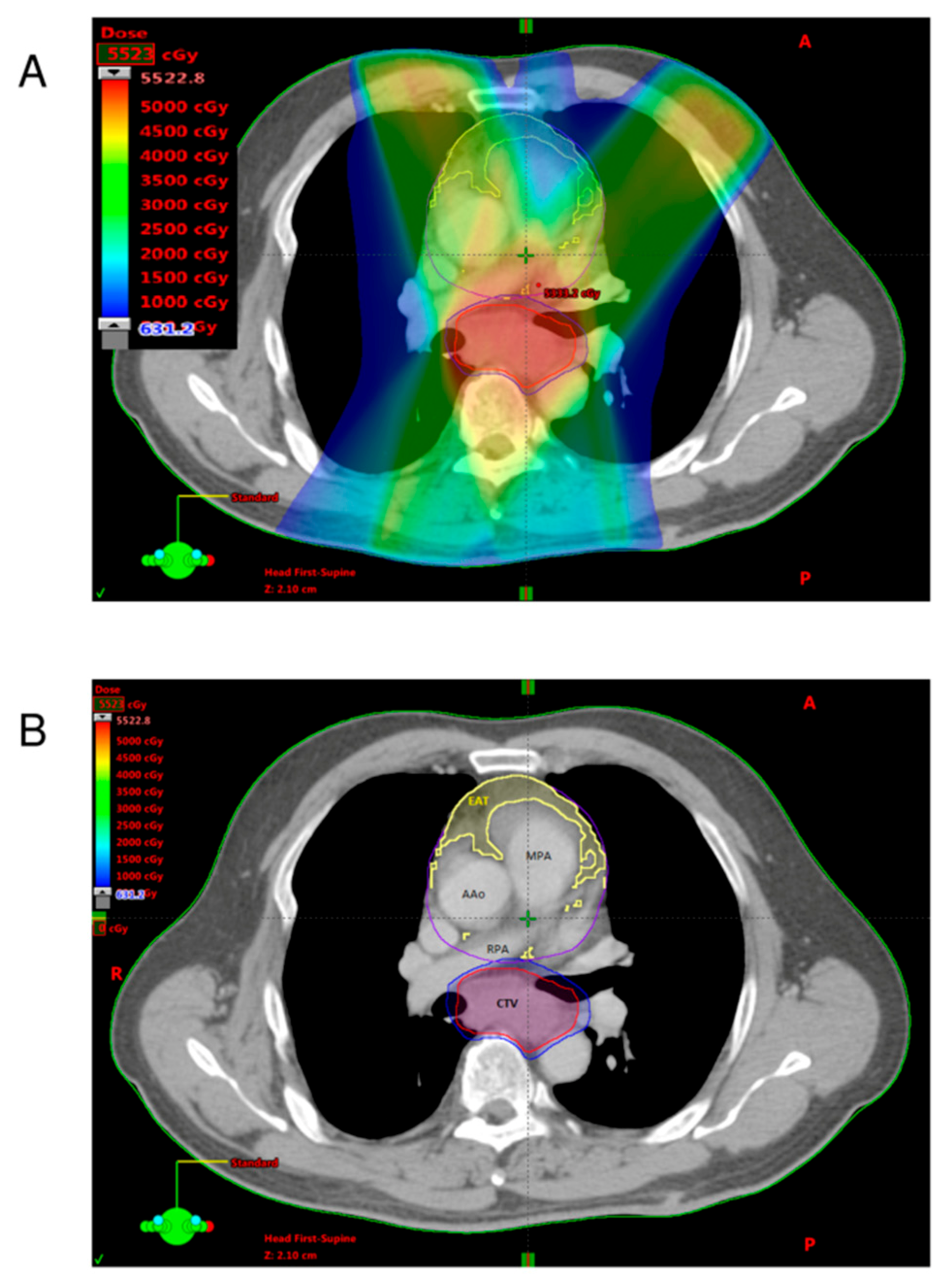
2.3. Quantification of Adipose Tissues

Computed tomography (CT) is a standard imaging modality for simulation and is used in the RT planning system prior to NACRT. Using anatomy to delineate the region of interest (ROI) and the Hounsfield unit (HU) to measure the radiodensity of structures such as tumor and adipose tissue, data on both context and structure volume could be obtained. Images were obtained using a CT scanner (Big-Bore CT simulator, Philips, Amsterdam, Netherlands) equipped for simulation with the following specifications: scanning with 16 × 0.75 mm collimation, rotation time of 420 msec, and tube voltage of 120 kV. The EAT was contoured based on the anatomical boundary between the outer wall of the myocardium and the visceral layer of pericardium from the level of the left main coronary artery to the cardiac base, ranging from −195 to −45 HU (Figure 1B) [13,14,15]. The left main coronary artery for fat thickness, peri-thoracic adipose tissue (TAT) for peri-thoracic aortic fat volume, and subcutaneous adipose tissue (SAT) and visceral adipose tissue (VAT) of the thorax for fat areas were delineated according to the methods shown in our previous publications [13,16,17,18]. For example, the TAT tissue was defined as the adipose tissue surrounding the thoracic aorta, extending 67.5 mm caudally from the level of the bifurcation of pulmonary arteries.

2.4. Definition of EAT-REI

The radiation dosimetric parameters included radiation exposure intensity (REI). EAT-REI was defined as the mean radiation dose divided by volume of EAT (cGy/mL). The calculation equation for EAT-REI was listed below
EAT-REI = mean dose of EAT/volume of EAT (cGy/mL)
Quantification of the adipose tissues was performed by reconstruction of delineated images, and calculation was performed using Varian Eclipse 11.3 (Varian Medical Systems, Palo Alto, CA, USA).

2.5. Nutrition Status Evaluation

The patient characteristics with nutrition status, metabolic disease, and cardiac diseases of this cohort and their association with the outcomes were analyzed. As demonstrated in Table 1, the metabolic and cardiac diseases, as well as nutrition status, in terms of pre-treatment BMI, albumin level, lymphocyte count, triglyceride, and total cholesterol were listed.

2.6. Surveillance and Recurrence Evaluation

The patients were followed up monthly for 3–6 months, then every 3 months for the first year, and then every 6 months thereafter. The follow-up evaluation included clinical examination, blood tests, chest/abdominal CT, and upper gastrointestinal panendoscopy with biopsies. Further imaging studies were performed if there was clinical suspicion of recurrence. Recurrence was diagnosed on the basis of the results of physical or radiographic examinations or pathological confirmation [19].

2.7. Statistical Analysis

Quantitative data were expressed as means ± standard deviation (SD) and categorical data as frequencies and proportions. Comparisons between the participants were performed using the independent t-test and chi-square test, as appropriate. Cox proportional hazards regression analysis was performed to assess the prognostic factors for overall survival (OS) and progression-free survival (PFS). A logistic regression model was used to determine the significance of covariate-adjusted associations between variables and OS and PFS. All statistical analyses were carried out using Statistical Package for the Social Sciences for Windows, SPSS® software V. 22.0 (IBM Corp., New York, NY, USA; formerly SPSS Inc., Chicago, IL, USA), and p < 0.05 was considered significant.

3. Results

3.1. Patients and Clinical Outcome

All 36 patients underwent esophagectomy after NACRT (Table 1), with a median follow-up of 22.0 (interquartile range: 11.3–36.3) months, and showed a pCR rate of 33.3%. The 3-year PFS and OS values were 32.5% and 39.7%, respectively.

3.2. Analysis of ROC Curve

ROC analysis provides tools to select possibly optimal models and to independently discard suboptimal ones. ROC analysis is related in a direct and natural way to cost/benefit analysis of diagnostic decision making. In our study, the area under the curve (AUC) for PFS and OS for patient was 0.715 and 0.779 (Figure 2), indicating that REI improved the discrimination ability for OS.

3.3. Analysis of Radiotherapy Dosimetric Parameters

For OS, the significant univariate prognostic factors were TAT, EAT (V10), and EAT-REI. The results of multivariate Cox regression for OS showed that only EAT-REI (HR: 1.002; 95% CI: 1.000–1.004; p = 0.028) (Table 2) was significant. Patients with a smaller EAT-REI had better OS than those with a higher EAT-REI (p < 0.05). Univariate analysis for PFS revealed that TAT, SAT, EAT (V5, V10, and V20), and EAT-REI were significant, but only EAT-REI was significant, as shown in the multivariate analysis (HR: 1.002; 95% CI: 1.000–1.004; p = 0.03) (Table 3). The receiver-operating characteristics analysis showed that the area under curve (AUC) values of EAT-REI for OS/PFS were 0.779 (95% CI: 0.623–0.936, p = 0.006) / 0.715 (95% CI: 0.534–0.897, p = 0.048) and the cut-off value of REI was 68.8 cGy/mL, while the sensitivity and 1-specificity (false positive) was 0.826 and 0.308, respectively. Indicating an informative predictor for Kaplan–Meier curves for 36-month survival according to EAT-REI cut-off value (68.8 cGy/mL) were significant for PFS Kaplan–Meier curves (65.9% and 13.0%, p = 0.001) and OS Kaplan–Meier curves (71.9% and 21.7%, p = 0.003) (Figure 3). With regard to the radiotherapy parameters evaluated by DVH, the quantities of high-dose and low-dose planning target volumes had no significant impact on clinical outcome.
Further analysis of EAT-REI was performed to clarify its clinical significance. The EAT-REI was defined as the mean dose divided by the volume of EAT. The mean doses of EAT had no significant impact on OS and PFS (p > 0.05). However, the patients with a larger volume of EAT had a trend of less progression without statistical significance (p > 0.05). Thus, the lower EAT-REI mainly, but not completely, resulted from the larger volume of EAT.

3.4. Analysis of EAT-REI Ratio High and Low Group

The comparison of high and low EAT-REI groups shows no significant difference among the age, sex, BMI, CRP level, albumin level, lymphocyte count, triglyceride, and total cholesterol. In clinical T stage (p = 0.69), clinical N stage (p = 0.92), and cTNM stage (p = 0.72) also showed no significant difference (Table 4).

3.5. Analysis of Nutrition Parameters

The distribution of metabolic disease and cardiac diseases has no significant correlation to OS and PFS. Among the nutrition parameters, no significant survival impact was noted by BMI, albumin level, lymphocyte count, triglyceride, and total cholesterol. As for correlation between nutrition parameters and EAT or EAT-REI, only BMI has a significant correlation to EAT volume.

4. Discussion

EAT is mainly composed of brown adipose tissue [11] and has an impact on the clinical outcomes of patients with metabolic diseases such as coronary artery disease [20] and diabetes mellitus [21]. EAT is a metabolically active tissue releasing various bioactive factors that can affect the prognosis of metabolic diseases [9]. Local expression of chemokine (monocyte chemotactic protein (MCP)-1) and inflammatory cytokines (interleukin (IL)-1β, IL-6, and tumor necrosis factor (TNF)-α) was observed in CAD patients. Significant changes in IL-1β, IL-6, MCP-1, and TNF-α mRNA and protein were observed in the epicardial adipose stores [9,22]. Ionizing radiation is known to induce inflammatory reactions and lipid remodeling of adipose tissues [23,24,25], but its role in brown adipose tissue or EAT remains unclear. Our investigations indicate that lower radiation exposure intensity may have a correlation with better survival. Whether this correlation resulted from radiation-induced inflammatory reactions or radiation-modulated metabolic changes still needs to be clarified.
The EAT-REI defined as mean radiation dose divided by volume of EAT is a unique biomarker for radiobiological effect derived from conventional physic dosimetric parameters. The radiation effects from exposure dose and irradiated volume may simultaneously contribute to this radiobiological marker. Growing evidence demonstrated that local radiation may have a systemic effect due to the release of soluble mediators within the radiation field. To determine whether EAT-REI has a role in this effect, which is compatible with the simultaneously irradiated dose and volume concerned, further in vivo studies using experimental animals are warranted.
In the era of dose painting RT, the use of CT scan images for simulation and RT planning software is a routine process. Analysis of the volume of EAT and the radiation dose distribution to EAT using these imaging data, therefore, is feasible and applicable. After analyzing the correlation between radiation dosimetric parameters and clinical outcomes, we found that EAT-REI might be a novel imaging biomarker from the radiobiological aspect. This implies that EAT-REI may have the potential to be adopted as a constraint when planning for RT. To the best of our knowledge, this is the first study to report EAT-REI as a constraint to RT planning.
The limitations of this study were mainly due to the retrospective analysis of both clinical factors and radiotherapy parameters. To ensure the estimated radiotherapy parameters were retrieved from the same RT planning algorithm and system, and the sample size of eligible patients was relatively small (N = 36). Another limitation of this retrospective analysis was the lack of measurement of additional biochemical parameters related to EAT-REI. These study limitations could be overcome by further prospective investigations.

5. Conclusions

The radiation exposure intensity of epicardial adipose tissue might be an important factor, with an impact on overall survival of ESCC patients receiving NACRT and esophagectomy. However, the biological meaning of EAT-REI still needs to be clarified.

Author Contributions

Y.-J.C., S.-M.H. and C.-H.Y. designed the study. H.-C.T. performed the study and drafted the manuscript with the help of J.L. and C.-H.Y. provided assistance in CT measurement. W.-C.H., H.-C.L. and C.-H.C. contributed to the collection and analysis of surgical patients’ data. Y.-C.H. analyzed the data. C.-J.L. helped in the collection of patient and measurement data. Y.-J.C. and S.-M.H. critically reviewed the data and content and provided the final approval of the version to be submitted. All authors have read and agreed to the published version of the manuscript.

Funding

This research was supported by MacKay Memorial Hospital (Grant number: MMH-E-109-13 and MMH-E-110-13).

Institutional Review Board Statement

The study protocol was granted institutional review board approval (serial number: 18MMHIS194e, MacKay Memorial Hospital, Taipei, Taiwan).

Informed Consent Statement

Patient consent was waived due to retrospective analysis and it was approved by IRB in MacKay Memorial Hospital.

Data Availability Statement

The datasets presented in this study are available from corresponding author on reasonable request.

Conflicts of Interest

The authors declare no conflict of interest in this work.

References

  1. Bray, F.; Ferlay, J.; Soerjomataram, I.; Siegel, R.L.; Torre, L.A.; Jemal, A. Global cancer statistics 2018: GLOBOCAN estimates of incidence and mortality worldwide for 36 cancers in 185 countries. CA Cancer J. Clin. 2018, 68, 394–424. [Google Scholar] [CrossRef] [PubMed] [Green Version]
  2. Shapiro, J.; van Lanschot, J.J.B.; Hulshof, M.; van Hagen, P.; van Berge Henegouwen, M.I.; Wijnhoven, B.P.L.; van Laarhoven, H.W.M.; Nieuwenhuijzen, G.A.P.; Hospers, G.A.P.; Bonenkamp, J.J.; et al. Neoadjuvant chemoradiotherapy plus surgery versus surgery alone for oesophageal or junctional cancer (CROSS): Long-term results of a randomised controlled trial. Lancet Oncol. 2015, 16, 1090–1098. [Google Scholar] [CrossRef]
  3. Sjoquist, K.M.; Burmeister, B.H.; Smithers, B.M.; Zalcberg, J.R.; Simes, R.J.; Barbour, A.; Gebski, V. Survival after neoadjuvant chemotherapy or chemoradiotherapy for resectable oesophageal carcinoma: An updated meta-analysis. Lancet Oncol. 2011, 12, 681–692. [Google Scholar] [CrossRef]
  4. Tepper, J.; Krasna, M.J.; Niedzwiecki, D.; Hollis, D.; Reed, C.E.; Goldberg, R.; Kiel, K.; Willett, C.; Sugarbaker, D.; Mayer, R. Phase III trial of trimodality therapy with cisplatin, fluorouracil, radiotherapy, and surgery compared with surgery alone for esophageal cancer: CALGB 9781. J. Clin. Oncol. Off. J. Am. Soc. Clin. Oncol. 2008, 26, 1086–1092. [Google Scholar] [CrossRef] [PubMed] [Green Version]
  5. Muro, K.; Lordick, F.; Tsushima, T.; Pentheroudakis, G.; Baba, E.; Lu, Z.; Cho, B.C.; Nor, I.M.; Ng, M.; Chen, L.T.; et al. Pan-Asian adapted ESMO Clinical Practice Guidelines for the management of patients with metastatic oesophageal cancer: A JSMO-ESMO initiative endorsed by CSCO, KSMO, MOS, SSO and TOS. Ann. Oncol. Off. J. Eur. Soc. Med. Oncol. 2019, 30, 34–43. [Google Scholar] [CrossRef] [PubMed]
  6. Bradley, J.D.; Paulus, R.; Komaki, R.; Masters, G.; Blumenschein, G.; Schild, S.; Bogart, J.; Hu, C.; Forster, K.; Magliocco, A.; et al. Standard-dose versus high-dose conformal radiotherapy with concurrent and consolidation carboplatin plus paclitaxel with or without cetuximab for patients with stage IIIA or IIIB non-small-cell lung cancer (RTOG 0617): A randomised, two-by-two factorial phase 3 study. Lancet Oncol. 2015, 16, 187–199. [Google Scholar] [CrossRef] [PubMed] [Green Version]
  7. Darby, S.C.; Ewertz, M.; McGale, P.; Bennet, A.M.; Blom-Goldman, U.; Bronnum, D.; Correa, C.; Cutter, D.; Gagliardi, G.; Gigante, B.; et al. Risk of ischemic heart disease in women after radiotherapy for breast cancer. N. Engl. J. Med. 2013, 368, 987–998. [Google Scholar] [CrossRef] [Green Version]
  8. Lee, J.; Lin, J.B.; Sun, F.J.; Lu, K.W.; Lee, C.H.; Chen, Y.J.; Huang, W.C.; Liu, H.C.; Wu, M.H. Dosimetric predictors of acute haematological toxicity in oesophageal cancer patients treated with neoadjuvant chemoradiotherapy. Br. J. Radiol. 2016, 89, 20160350. [Google Scholar] [CrossRef] [PubMed] [Green Version]
  9. Mazurek, T.; Zhang, L.; Zalewski, A.; Mannion, J.D.; Diehl, J.T.; Arafat, H.; Sarov-Blat, L.; O’Brien, S.; Keiper, E.A.; Johnson, A.G.; et al. Human epicardial adipose tissue is a source of inflammatory mediators. Circulation 2003, 108, 2460–2466. [Google Scholar] [CrossRef] [Green Version]
  10. Demir, E.; Harmankaya, N.O.; Kirac Utku, I.; Aciksari, G.; Uygun, T.; Ozkan, H.; Demir, B. The Relationship between Epicardial Adipose Tissue Thickness and Serum Interleukin-17a Level in Patients with Isolated Metabolic Syndrome. Biomolecules 2019, 9, 97. [Google Scholar] [CrossRef] [PubMed] [Green Version]
  11. Sacks, H.S.; Fain, J.N.; Holman, B.; Cheema, P.; Chary, A.; Parks, F.; Karas, J.; Optican, R.; Bahouth, S.W.; Garrett, E.; et al. Uncoupling protein-1 and related messenger ribonucleic acids in human epicardial and other adipose tissues: Epicardial fat functioning as brown fat. J. Clin. Endocrinol. Metab. 2009, 94, 3611–3615. [Google Scholar] [CrossRef] [PubMed]
  12. Hou, T.C.; Dai, K.Y.; Wu, M.C.; Hua, K.L.; Tai, H.C.; Huang, W.C.; Chen, Y.J. Bio-physic constraint model using spatial registration of delta 18F-fluorodeoxyglucose positron emission tomography/computed tomography images for predicting radiation pneumonitis in esophageal squamous cell carcinoma patients receiving neoadjuvant chemoradiation. OncoTargets Ther. 2019, 12, 6439–6451. [Google Scholar] [CrossRef] [Green Version]
  13. Yun, C.H.; Lin, T.Y.; Wu, Y.J.; Liu, C.C.; Kuo, J.Y.; Yeh, H.I.; Yang, F.S.; Chen, S.C.; Hou, C.J.; Bezerra, H.G.; et al. Pericardial and thoracic peri-aortic adipose tissues contribute to systemic inflammation and calcified coronary atherosclerosis independent of body fat composition, anthropometric measures and traditional cardiovascular risks. Eur. J. Radiol. 2012, 81, 749–756. [Google Scholar] [CrossRef] [PubMed]
  14. Mahabadi, A.A.; Massaro, J.M.; Rosito, G.A.; Levy, D.; Murabito, J.M.; Wolf, P.A.; O’Donnell, C.J.; Fox, C.S.; Hoffmann, U. Association of pericardial fat, intrathoracic fat, and visceral abdominal fat with cardiovascular disease burden: The Framingham Heart Study. Eur. Heart J. 2009, 30, 850–856. [Google Scholar] [CrossRef] [Green Version]
  15. Lehman, S.J.; Massaro, J.M.; Schlett, C.L.; O’Donnell, C.J.; Hoffmann, U.; Fox, C.S. Peri-aortic fat, cardiovascular disease risk factors, and aortic calcification: The Framingham Heart Study. Atherosclerosis 2010, 210, 656–661. [Google Scholar] [CrossRef] [PubMed] [Green Version]
  16. Yun, C.H.; Bezerra, H.G.; Wu, T.H.; Yang, F.S.; Liu, C.C.; Wu, Y.J.; Kuo, J.Y.; Hung, C.L.; Lee, J.J.; Hou, C.J.; et al. The normal limits, subclinical significance, related metabolic derangements and distinct biological effects of body site-specific adiposity in relatively healthy population. PLoS ONE 2013, 8, e61997. [Google Scholar] [CrossRef] [PubMed]
  17. Lai, Y.H.; Yun, C.H.; Yang, F.S.; Liu, C.C.; Wu, Y.J.; Kuo, J.Y.; Yeh, H.I.; Lin, T.Y.; Bezerra, H.G.; Shih, S.C.; et al. Epicardial adipose tissue relating to anthropometrics, metabolic derangements and fatty liver disease independently contributes to serum high-sensitivity C-reactive protein beyond body fat composition: A study validated with computed tomography. J. Am. Soc. Echocardiogr. Off. Publ. Am. Soc. Echocardiogr. 2012, 25, 234–241. [Google Scholar] [CrossRef] [PubMed]
  18. Lai, Y.H.; Hou, C.J.; Yun, C.H.; Sung, K.T.; Su, C.H.; Wu, T.H.; Yang, F.S.; Hung, T.C.; Hung, C.L.; Bezerra, H.G.; et al. The association among MDCT-derived three-dimensional visceral adiposities on cardiac diastology and dyssynchrony in asymptomatic population. BMC Cardiovasc. Disord. 2015, 15, 142. [Google Scholar] [CrossRef] [Green Version]
  19. Lin, J.B.; Hung, L.C.; Cheng, C.Y.; Chien, Y.A.; Lee, C.H.; Huang, C.C.; Chou, T.W.; Ko, M.H.; Lai, Y.C.; Liu, M.T.; et al. Prognostic significance of lung radiation dose in patients with esophageal cancer treated with neoadjuvant chemoradiotherapy. Radiat. Oncol. 2019, 14, 85. [Google Scholar] [CrossRef] [Green Version]
  20. Wu, F.Z.; Chou, K.J.; Huang, Y.L.; Wu, M.T. The relation of location-specific epicardial adipose tissue thickness and obstructive coronary artery disease: Systemic review and meta-analysis of observational studies. BMC Cardiovasc. Disord. 2014, 14, 62. [Google Scholar] [CrossRef]
  21. Wang, C.P.; Hsu, H.L.; Hung, W.C.; Yu, T.H.; Chen, Y.H.; Chiu, C.A.; Lu, L.F.; Chung, F.M.; Shin, S.J.; Lee, Y.J. Increased epicardial adipose tissue (EAT) volume in type 2 diabetes mellitus and association with metabolic syndrome and severity of coronary atherosclerosis. Clin. Endocrinol. 2009, 70, 876–882. [Google Scholar] [CrossRef] [PubMed]
  22. Parisi, V.; Rengo, G.; Pagano, G.; D’Esposito, V.; Passaretti, F.; Caruso, A.; Grimaldi, M.G.; Lonobile, T.; Baldascino, F.; De Bellis, A.; et al. Epicardial adipose tissue has an increased thickness and is a source of inflammatory mediators in patients with calcific aortic stenosis. Int. J. Cardiol. 2015, 186, 167–169. [Google Scholar] [CrossRef] [PubMed]
  23. Poglio, S.; Galvani, S.; Bour, S.; André, M.; Prunet-Marcassus, B.; Pénicaud, L.; Casteilla, L.; Cousin, B. Adipose tissue sensitivity to radiation exposure. Am. J. Pathol. 2009, 174, 44–53. [Google Scholar] [CrossRef] [PubMed] [Green Version]
  24. Xiao, Y.; Mo, W.; Jia, H.; Yu, D.; Qiu, Y.; Jiao, Y.; Zhu, W.; Koide, H.; Cao, J.; Zhang, S. Ionizing radiation induces cutaneous lipid remolding and skin adipocytes confer protection against radiation-induced skin injury. J. Dermatol. Sci. 2020, 97, 152–160. [Google Scholar] [CrossRef]
  25. Lee, J.; Lin, J.B.; Wu, M.H.; Jan, Y.T.; Chang, C.L.; Huang, C.Y.; Sun, F.J.; Chen, Y.J. Muscle radiodensity loss during cancer therapy is predictive for poor survival in advanced endometrial cancer. J. Cachexia Sarcopenia Muscle 2019, 10, 814–826. [Google Scholar] [CrossRef] [Green Version]
Figure 1. (A) The dose distribution was in color wash with IMRT treatment planning; (B) the epicardial adipose tissue (yellow) was contoured based on the anatomical boundary of heart, ranging from −195 to −45 HU. Abbreviations: EAT, epicardial adipose tissue; MPA, main pulmonary artery; AAo, ascending aorta; RPA, right pulmonary artery; CTV, clinical target volume.
Applsci 11 04023 g001
Figure 2. Receiver-operating characteristic curves for (A) progression-free survival AUC = 0.715 and (B) overall survival AUC = 0.779. AUC, area under the curve; CI, confidence interval.
Applsci 11 04023 g002
Figure 3. Kaplan–Meier estimates of the 3-year (A) progression-free survival p = 0.001 and (B) overall survival p = 0.003 according to EAT-REI cut-off value (68.8 cGy/mL).
Applsci 11 04023 g003
Table 1. Patient and tumor characteristics.
CharacteristicsOverall (n = 36)
Age (years)58.8 ± 8.6
Sex
 Man34 (94.4%)
 Woman2 (5.6%)
BMI (kg/m2)22.1 ± 4.3
Albumin (g/dL)4.0 ± 0.5
Hb (g/dL)12.0 ± 1.4
Lymphocyte (%)21.8 ± 9.0
Cholesterol (mg/dL)181.5 ± 40.0
Triglycerides (mg/dL)116.2 ± 57.2
Metabolic diseases6 (16.7%)
Cardiac diseases9 (25.0%)
Clinical T stage
 cT1-29 (25.0%)
 cT3-427 (75.0%)
Clinical N stage
 cN0-119 (52.8%)
 cN2-317 (47.2%)
cTNM stage
 II10 (27.8%)
 III26 (72.2%)
Target volume (mL)688.8 ± 271.2
Heart volume (mL)621.4 ± 117.1
Heart mean dose (Gy)25.9 ± 6.9
Dose-volume of EAT
 Volume (mL)19.2 (8.4–29.4)
 Mean dose (Gy)21.5 (16.2–26.4)
 V5 *16.5 (8.2–26.6)
 V1014.0 (6.8–21.9)
 V2010.2 (3.5–16.5)
 V304.6 (1.5–11.4)
 V401.1 (0.5–3.5)
SAT volume (mL)1239.8 (682.8–1795.5)
SAT mean dose (Gy)6.7 (5.4–7.8)
VAT volume (mL)124.2 (76.3–251.3)
VAT mean dose (Gy)22.5 (17.7–26.6)
* Vx = volume (mL) of EAT receiving X Gy or more; data are mean ± standard deviation, median (interquartile range), or n (%). Abbreviations: EAT, epicardial adipose tissue; SAT, subcutaneous adipose tissue; VAT, visceral adipose tissue.
Table 2. Univariate and multivariate Cox proportional hazards model for overall survival.
Univariate Multivariate
CharacteristicsHR (95% CI)p ValueHR (95% CI)p Value
Age1.005 (0.953–1.060)0.859
BMI (kg/m2)0.999 (0.907–1.099)0.979
Albumin (g/dL)0.772 (0.351–1.702)0.522
Hb (g/dL)0.986 (0.733–1.326)0.926
Lymphocyte (%)0.976 (0.929–1.025)0.325
Cholesterol (mg/dL)1.002 (0.987–1.017)0.785
Triglycerides (mg/dL)1.000 (0.991–1.008)0.931
Metabolic diseases1.392 (0.470–4.123)0.550
Cardiac diseases0.558 (0.188–1.655)0.293
Clinical T (T1–2 vs. T3–4)0.658 (0.303–1.432)0.292
Clinical N (N0–1 vs. N2–3)1.235 (0.534–2.854)0.621
cTNM stage (II vs. III)0.790 (0.321–1.943)0.608
Pathological response (non-pCR vs. pCR)0.561 (0.218–1.445)0.231
Target volume1.001 (1.000–1.003)0.125
Heart mean dose1.000 (1.000–1.001)0.639
Dose-volume of EAT *
Volume (mL)0.981 (0.957–1.007)0.149
Mean dose (Gy)1.000 (1.000–1.001)0.702
 V50.971 (0.943–1.000)0.051
 V100.967 (0.935–1.000)0.0490.988 (0.952–1.026)0.529
 V200.960 (0.917–1.005)0.081
V300.964 (0.904–1.029)0.269
V400.887 (0.746–1.055)0.177
REI of EAT (EAT-REI)1.002 (1.001–1.004)0.0021.002 (1.000–1.004)0.028
TAT volume (mL)0.999 (0.999–1.003)0.0491.000 (0.999–1.000)0.322
TAT mean dose (Gy)1.001 (0.999–1.000)0.294
SAT volume (mL)0.999 (0.999–1.000)0.062
SAT mean dose (Gy)1.001 (0.998–1.003)0.501
VAT volume (mL)0.995 (0.991–1.000)0.054
VAT mean dose (Gy)1.000 (1.000–1.001)0.472
* Vx = volume (mL) of EAT receiving X Gy or more; REI(EAT-REI) was defined as the EAT mean dose divided by volume (cGy/mL). Abbreviations: CI, confidence interval; DVH, dose-volume histogram; REI, radiation exposure intensity; HR, hazard ratio; TAT, total adipose tissue.
Table 3. Univariate and multivariate Cox proportional hazards model for progression-free survival.
Univariate Multivariate
CharacteristicsHR (95% CI)p ValueHR (95% CI)p Value
Age1.014 (0.965–1.066)0.572
BMI (kg/m2)0.974 (0.873–1.087)0.637
Albumin (g/dL)0.694 (0.277–1.738)0.435
Hb (g/dL)0.998 (0.719–1.385)0.989
Lymphocyte (%)0.958 (0.904–1.015)0.149
Cholesterol (mg/dL)0.992 (0.973–1.011)0.429
Triglycerides (mg/dL)0.996 (0.986–1.007)0.510
Metabolic diseases0.040 (0.000–20.334)0.311
Cardiac diseases0.344 (0.078–1.522)0.160
Clinical T (T1–2 vs. T3–4)0.904 (0.444–1.839)0.780
Clinical N (N0–1 vs. N2–3)1.524 (0.689–3.375)0.299
cTNM stage (II vs. III)0.828 (0.342–2.004)0.675
Pathological response (non-pCR vs. pCR)0.694 (0.288–1.669)0.414
Target Volume1.001 (1.000–1.002)0.164
Heart mean dose1.000 (0.999–1.000)0.663
Dose-volume of EAT *
Volume (mL)0.982 (0.959–1.005)0.114
Mean dose (Gy)1.000 (0.999–1.000)0.751
 V50.969 (0.943–0.996)0.0261.131 (0.933–1.369)0.209
 V100.965 (0.935–0.996)0.0490.796 (0.608–1.041)0.095
 V200.958 (0.918–1.000)0.0491.116 (0.943–1.321)0.200
V300.959 (0.902–1.029)0.182
V400.871 (0.735–1.032)0.111
REI of EAT (EAT-REI)1.003 (1.001–1.004)0.0021.002 (1.000–1.004)0.030
TAT volume (mL)0.999 (0.999–1.000)0.0140.970 (0.998–1.000)0.060
TAT mean dose (Gy)1.000 (0.998–1.002)0.896
SAT volume (mL)0.999 (0.999–1.000)0.018
SAT mean dose (Gy)1.000 (0.998–1.002)0.855
VAT volume (mL)0.995 (0.991–1.000)0.0351.003 (0.995–1.010)0.458
VAT mean dose (Gy)1.000 (0.999–1.001)0.739
* Vx = volume (mL) of EAT receiving X Gy or more. Abbreviations: CI, confidence interval; DVH, dose-volume histogram; REI, radiation exposure intensity; HR, hazard ratio; TAT, total adipose tissue.
Table 4. Compare EAT-REI ratio with high and low patient groups.
CharacteristicsEAT-REI ≥ 68.8 (n = 23)EAT-REI < 68.8 (n = 13)p Value
Age (years),57.3 ± 7.961.6 ± 9.40.15
Sex 0.60
 Man22 (95.7%)12 (92.3%)
 Woman1 (4.3%)1 (7.7%)
BMI (kg/m2)21.2 ± 4.423.6 ± 3.70.11
CRP (mg/dL)7.0 ± 6.26.2 ± 5.50.66
Albumin (g/dL)3.9 ± 0.54.0 ± 0.60.71
Hb (g/dL)11.9 ± 1.412.3 ± 1.50.38
Lymphocyte (number/uL)1414.4 ± 770.51744.2 ± 631.40.20
Cholesterol (mg/dL)178.6 ± 48.6186.4 ± 18.80.60
Triglycerides (mg/dL)115.8 ± 52.9116.8 ± 66.10.77
Metabolic diseases4 (17.4%)2 (15.4%)1.00
Cardiac diseases4 (17.4%)5 (38.5%)0.24
Clinical T stage 0.69
 cT1–25 (21.7%)4 (30.8%)
 cT3–418 (78.3%)9 (69.2%)
Clinical N stage 0.92
 cN0–112 (52.2%)7 (53.8%)
 cN2–311 (47.8%)6 (46.2%)
cTNM stage 0.72
 II7 (30.4%)3 (23.1%)
 III16 (69.6%)10 (76.9%)
Publisher’s Note: MDPI stays neutral with regard to jurisdictional claims in published maps and institutional affiliations.

