Article

Fermented Antler Recovers Stamina, Muscle Strength and Muscle Mass in Middle-Aged Mice

1
R&BD Center, hy Co., Ltd., 22, Giheungdanji-ro 24beon-gil, Giheung-gu, Yongin-si 17086, Gyeonggi-do, Korea
2
Department of Oriental Medicine Biotechnology, College of Life Sciences, Kyung Hee University, Yongin-si 17104, Gyeonggi-do, Korea
3
Mbiometherapeutics Co., Ltd., Yongin-si 16942, Gyeonggi-do, Korea
*
Authors to whom correspondence should be addressed.
These authors contributed equally to this work.
Appl. Sci. 2022, 12(1), 106; https://doi.org/10.3390/app12010106
Submission received: 12 November 2021 / Revised: 14 December 2021 / Accepted: 20 December 2021 / Published: 23 December 2021
(This article belongs to the Special Issue Functional Food and Nutrition)

Abstract

:
In a previous study, we found that Lactobacillus curvatus HY7602-fermented antler (FA) improved exercise endurance by increasement of muscle mass and strength in a young mouse model. In this study, we investigated the effect of FA on recovery of muscle mass and strength in aging-induced muscle loss. We have used a middle-aged model in which muscle decline begins in many mammalian species. All mice performed treadmill exercise and forced swimming, and measured muscle grip strength. Then, calf muscle weight and histological analysis, blood biomarker and gene expression in soleus muscle tissue were measured. Muscle strength and forced swimming time were significantly increased in the FA-intake groups compared to controls. The levels of muscle and liver damage-related indicators (ATL, ALP, LDH and CK) and muscle endurance, fatigue and exercise performance-related indicators (lactate and creatinine) were significantly improved by FA supplementation. In addition, FA regulates genes related to muscle protein degradation (Atrogin-1 and MuRF1) and muscle fiber synthesis (MyoD and Myf5), resulting in increased muscle mass, and fiber diameter and area values. The Bax/Bcl-2 ratio, related to apoptosis in skeletal muscle was significantly decreased. These results demonstrate that FA improves exercise performance with ameliorating blood biomarkers and also increases muscle mass and muscle strength by inhibiting muscle proteolysis and promoting muscle synthesis in a middle-aged mouse.

1. Introduction

Deer antlers, a rare example of an organ that is regenerated annually, have been traditionally used for health promotion purposes in Korea, Japan and China. It is known that deer antlers contain various pharmacologically active substances include gangliosides, collagens, phospholipids, chondroitin, glucosamines, hyaluronic acid and pantocrin (70% ethanol extract) [1,2], and it has been reported to have a positive effect on physical strength and immunity [3], bone growth and development [4], an anti-fatigue effect [5] and endurance during exercise [2].
Fermentation of deer antler has been carried out by several researchers and has shown that the functionality of antler is improved; improvement of stimulation of differentiation and mineralization of osteoblasts [6], hematopoiesis of murine marrow cells [7] and enhancement of viability and interleukin-12 production in spleen cells [8]. Then we selected lactic acid bacteria that can grow from antlers extract and presented the research results related to the improvement of muscular endurance by fermented antlers with these Lactobacilli [2].
It is known that muscle strength and mass generally decrease with age [9]. Sarcopenia, the progressive loss of muscle mass due to aging, causes various negative health phenomena (reduction in exercise performance, weight gain, falls and osteoporosis, quality of life, etc.). In particular, a decrease in muscle mass and its function leads to reduced physical activity, leading to a progressive decline of energy consumption, weight gain and obesity. This phenomenon, in turn, promotes the secretion of inflammatory cytokines that negatively affect muscle cells, resulting in aggravation of sarcopenia [10].
Because, as aging progresses, the synthesis rates of muscle filament and mitochondrial protein decreases [11], and middle age is a transition period from youth to senior, it is the starting time when physical strength and muscle strength diminish [12,13]. Since middle-aged people are at their most active period in terms of their social activities, it is important for them to retain their physiological strength. In addition, as muscle mass and strength have a large effect on health, researches about these factors in middle-aged people are important [14].
There are many trials (pharmaceutical and food supplement, etc.) to treat sarcopenia. Unfortunately, they have ineffective or limited efficacy. According to Jones et al., a safe and highly effective way in aged adults is resistance exercise for increasing muscle mass and strength [15].
The present study aimed to investigate whether the fermented antler improves skeletal muscular tissue and function, weakened by aging, in middle-aged mice. To this end, the mice were fed with fermented antlers and creatine that is a nitrogenous organic acid that elevates supplying energy to muscle cells and enhances the performance of exercise for 4 weeks. Then we observed that changes in periodical muscle strength, muscle mass, blood parameters related to muscle damage, muscle fiber cross-sectional area, and genes associated with muscle protein synthesis and degradation and apoptosis in skeletal muscle.

2. Materials and Methods

2.1. Antler

The antlers used in this experiment were derived from raw materials that met the standards of herbal medicine standard (The Korean Herbal Pharmacopoeia, Ministry of Food and Drug Safety, Korea) collected from Cervus elaphus Linné. The middle/lower sections of the hot air-dried antlers were crushed with a grinder and then stored at −80 °C.

2.2. Lactic Acid Bacteria and Growth Conditions

Lactobacillus curvatus strain HY7602 was cultured in De Man, Rogosa and Sharpe (MRS) media (BD Difco, Sparks, MD, USA) broth at 37 °C for 24 h, collected and freeze-dried for use in fermentation.

2.3. Preparation of Fermented Antler

The fermented antler was prepared as described previously [2]. First, to make the antler extract, the crushed antler was mixed with distilled water (1:30, w/v), refluxed at 95 °C for 3 h, and filtered. After that, 1% lactic acid bacteria (L. curvatus HY7602, v/v) was added to the antler extract and fermented at 37 °C for 24 h, and then sterilized. The final fermented antler (FA) sample was stored at 4 °C.

2.4. Animals, Diet and Experimental Design

Male C57BL/6J mice at 4-week-old (young) and 40-week-old (middle-aged) were purchased from DooYeol Biotech (Seoul, Korea). Under a 12 h light–dark cycle, the mice were housed individually in separate cages maintained at constant temperature and humidity (22 ± 1 °C, 55 ± 10%). During the one-week acclimation period, mice were fed the AIN-93G diet. After this period, the mice were divided into four groups of five based on body weight, grip strength and lactate levels (Table 1). Mice in young and middle-age were fed AIN-93G diet, and those in the experimental groups were fed AIN-93G diets containing FA (250 mg/kg/day) or creatine (75 mg/kg/day; positive control). The food intake and body weights of mice were measured weekly. All animal studies were conducted according to the hy Co., Ltd. (Yongin-si, Korea) and approved by Institutional Animal Care and Use Committee of hy Co., Ltd. (IACUC approval number, AEC-20081229-0003).

2.5. Treadmill Exercise Performance Test and Grip Strength Measurement

The treadmill exercise was performed once a week as described previously [2]. One week before the treatment period, all mice were accustomed to running on a variable speed-belt treadmill (JD-A-09, Jeong Do B&P, Seoul, Korea). Exercise performance was started at 14 m/min for the first 2–3 min, on a 11° incline, and then continued at 21 m/min until exhaustion. The grip strength of mice forelimbs was measured using a rip strength meter equipped with a pull bar. To measure grip strength, gently pull the tail until the mouse released the bar (Bioseb, Virtolles CEDEX, France). Each mouse was tested twice to obtain the peak value.

2.6. Swimming Endurance

The swimming endurance of all mice was tested immediately before sacrifice, as described previously [2]. The test was conducted at 25 ± 1 °C, with water to a depth of 25 cm water in an acrylic plastic pool (24 cm × 14 cm × 14 cm) until exhaustion (defined as not rising to the surface within 5 s to breathe.). After the test, mice were sacrificed immediately under deep CO2 anesthesia.

2.7. Tissue Collection and Serum Biochemistry

Mice skeletal muscles were collected and weighed. The soleus was separated and stored at −80 °C for RT-PCR. Blood was collected by cardiac puncture into a serum tube and centrifuged at 1000× g for 15 min at 4 °C, and the serum was stored at −80 °C. Alanine aminotransferase (ALT), alkaline phosphatase (ALP), lactate dehydrogenase (LDH), creatine kinase (CK), creatinine, glucose and lactate were measured using an automated analyzer (Hitachi 7020, Hitachi, Japan).

2.8. Histological Analysis

The gastrocnemius muscle was fixed in 10% formalin, paraffin-embedded and stained with hematoxylin and eosin (H&E) for histological analysis. Three animals per group were chosen for muscle fiber size morphometry and three representative pictures were taken from muscle section. Images were captured at 10× magnification, and fiber area diameters were measured using a Motic digital microscope image analysis system (Motic Optical Instruments Co., Ltd., Xiamen, China).

2.9. Real-Time RT-PCR

RNA extraction from the tissue and RT-PCR analysis were performed, as previously described [2]. Total RNA was extracted using a Qiagen RNA Prep kit (Qiagen, Valencia, CA, USA) from soleus muscle tissue and amplified. The amplified products were analyzed using the QuantStudio 6 real-time polymerase chain reaction program (Thermo Fisher 150 Scientific, Waltham, MA, USA). The resulting cDNA is used as a template in RT-PCR reactions to detect the expression of mRNA encoding muscle atrophy F-box (Atrogin-1), muscle RING-finger protein-1 (MuRF1), myoblast determination protein 1 (MyoD), myogenic factor 5 (Myf5), B-cell lymphoma protein 2 (Bcl-2) and apoptosis regulation Bcl-2-associated X (Bax).

2.10. Statistical Analysis

All data are expressed as mean ± SD. The values obtained from the experimental results were evaluated using unpaired Student’s t-tests in SPSS software (SPSS; version 26.0 software, IBM, Somers, NY, USA), and those with p-values < 0.05 (*) as compared with the young control or p-values < 0.05 (#) as compared with the middle-aged control mice were considered significant.

3. Results

3.1. FA Supplementation Improves Muscle Strength

We observed the effect of FA intake on muscle strength in middle-aged mice. After 4 weeks of dietary intake and exercise, grip strength at weeks 0 and 4 were compared. The young mouse group increased muscle strength by about 0.34 N for 4 weeks, whereas the middle-aged mouse group showed little increase. However, the middle-aged mouse group fed a diet containing creatine or FA increased muscle strength significantly (Figure 1a,b). Changes in dietary intake and body weight were almost similar for each experimental group (Figure 1c,d).

3.2. FA Supplementation Ameliorated Exercise Performance and Related Blood Indices

The intake of FA also showed an effect in the forced swimming test (Figure 2). The younger group of mice continued swimming for 6.24 min, while the group of middle-aged mice only lasted about 2.58 min. Groups of middle-aged mice fed a diet containing creatine or FA revealed significant differences by continuing to swim for 5.97 and 6.81 min, respectively.
As a final stage of the experiment, after the forced swimming test, mice were sacrificed and their blood samples were collected to observe the biochemical indicators. In the case of ALT, ALP, LDH and CK, which are indicators that increase when muscle and liver tissue are damaged by heavy exercise, it was found that middle-aged mice significantly increased compared to young mice. In addition, it was recognized that the intake of FA significantly reduced these increased blood markers (Table 2). Lactate, creatinine and glucose related to muscular endurance, fatigue and exercise performance were significantly increased in middle-aged mice compared to younger mice, and FA alleviated all these tendencies. In particular, lactate and creatinine were decreased to a significant level. We could confirm that FA can protect muscle tissue during exercise and improve exercise performance through all the blood indicators presented.

3.3. FA Supplementation Increased Muscle Mass and Histological Section Area of Muscle Fiber

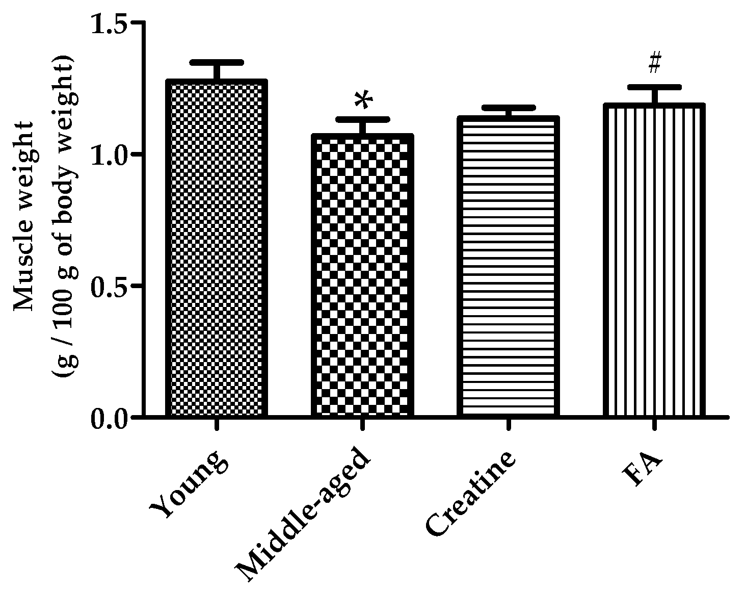
In order to verify the increase in muscle mass according to FA intake, the gastrocnemius and soleus were recovered after sacrifice, and their weight was measured, and the ratio of muscle to total body weight was compared. As shown in Figure 3, middle-aged mice showed a significant decrease in the percentage of muscle mass compared to young mice. On the other hand, the percentage of muscle mass in middle-aged mice fed FA was significantly increased, supporting that ingestion of FA had a positive effect on the increase in muscle mass.
To examine the increase in muscle mass according to FA intake from a histological point of view, tissue sections of gastrocnemius were analyzed by staining with hematoxylin and eosin. It can be revealed that the muscle fibers of middle-aged mice were thinner than those of young mice, and the muscle fibers of middle-aged mice fed creatine or FA were thicker (Figure 4a). The cross-sectional length and area of the muscle fibers were measured, and it was shown that the results of the histological evidence showed a statistically significant difference (Figure 4b,c).

3.4. FA Supplementation Altered mRNA Expression Related in Muscle Hypertrophy

In soleus muscle tissue, the expression of genes related to muscle protein degradation (Atrogin-1 and MuRF1) and muscle synthesis (MyoD and Myf5) were examined. Atrogin-1 was significantly increased in the middle-aged mouse control group compared to young mice, and the creatine intake group and FA intake group were significantly decreased compared to the middle-aged mice. This trend was even greater in MuRF1, which increased twice in the middle-aged mouse experimental group compared to the young mouse, and the FA intake group significantly decreased to a level of 55% compared to the middle-aged mouse (Figure 5a). On the other hand, the expression of MyoD and Myf5 related to muscle synthesis by ingestion of FA showed a tendency to decrease significantly in middle-aged mice compared to young mice, and it was confirmed that FA significantly improved both factors (Figure 5b).
Then, the gene expression of Bax, Bcl-2 and the expression ratio of these genes related to muscle cell apoptosis were compared. In mouse soleus muscle tissue, the expression of the Bax, which caused mitochondria damage and apoptosis in muscle cells, was decreased in the FA-treated group. On the other hand, the expression of the Bcl-2 gene, which is also involved in apoptosis but has pro-survival activity depending on the ratio with Bax, was meaningfully increased in the FA-treated group. As a result, the ratio of Bax/Bcl-2 was significantly decreased in the FA treatment group (Figure 5c).

4. Discussion

As interest in aging and quality of life increases, research and interest in sarcopenia are expanding [16,17,18]. Sarcopenia is closely related to an increase in age, but it is also highly correlated with exercise and nutritional status [19,20,21,22]. Appropriate exercise promotes muscle production and prevents loss [23,24,25]. In the elderly, maintaining muscle strength through resistance exercise is one of the effective methods [15,26,27], and proper nutrition also plays an important role [28,29]. However, since the intestinal microbiota changes with age [30], and its function may be lowered [31], ingestion of nutrients through fermented food may be a more effective method for middle-aged or elderly adults [32,33]. We studied fermented antler, which is a functional ingredient that has been traditionally widely used in Korea and China, and has reported that exercise performance of young mice improved through the antler fermentation process [2].
There have been reports of a decrease in muscle function and changes in mitochondria before complete sarcopenia, even in middle-aged mice [34]. The purpose of this investigation is to examine whether the effects of improving muscular endurance and increasing muscle mass by ingestion of fermented antlers are found in middle-aged mice. Based on a report on the correlation between mouse age and human age, mice aged 10 months were selected to examine the effects of fermented antlers [35].
As the balance of synthesis and decomposition of muscle gradually weakened by aging, muscle mass decreases. In order to maintain muscle mass despite aging, it is necessary to promote the synthesis of muscle protein or suppress aging-induced muscle protein degradation along with appropriate exercise stimulation. As in a previous study, using young mice, middle-aged mice that consumed FA for 4 weeks (11-month-aged finally) and mice exercised with a treadmill showed significant increases in grip strength, ratio of muscle mass and exercise performance during forced swimming, unlike middle-aged mice that did not consume FA.
We compared the expression of genes involved in the degradation and synthesis of muscle proteins in the muscle tissue of middle-aged mice to check the effect of FA on middle-aged mice at the molecular level. As a result, the intake of FA significantly improved the expression of Atrogin-1 and MuRF1, which are involved in muscle protein degradation, in middle-aged mice. It also increased the expression of MyoD and Myf5, which are involved in muscle protein synthesis. The expression patterns of the above four genes in middle-aged mice compared to young mice were all significantly different, and FA intake improved them all, and it was concluded that they contributed to the improvement of muscle mass and muscle strength.
In addition, changes in the expression of Bax (BCL2 associated X, apoptosis regulator) and Bcl-2 (B-cell lymphoma 2) genes related to muscle cell apoptosis were also correlated with the improvement of muscle mass and strength. Bax, together with Bak (Bcl-2 antagonist/killer 1), is a kind of nuclear-encoded protein present in higher eukaryotes and can induce apoptotic cell death by penetrating the outer membrane of mitochondria [36]. In addition, Bcl-2 family proteins are involved in apoptosis, and although the specific mechanism is controversial, in some cases, they exhibit pro-survival activity and prevent apoptosis [37]. There is a report that the expression of Bax protein decreases and Bcl-2 level increases through exercise, which can alleviate the Bax/Bcl-2 ratio that increases with age in the aging heart and consequent apoptosis [38]. Our experimental results could be inferred that the decrease in Bax expression and the increase in Bcl-2 expression through exercise in middle-aged mice were further increased through FA intake.
In general, the thickness of muscle fibers is correlated with muscle strength, and the thickness can be improved through exercise [39,40]. The molecular level results were consistent with the results of comparing muscle fiber thickness through histological analysis of our gastrocnemius muscle. The H&E staining test result was observed to be significantly increased in the middle-aged mice fed FA, which was significantly increased compared to the muscle fiber thickness of the young mouse.
Furthermore, an increase in exercise time and improvement in blood parameters related to fatigue were also confirmed in mice fed FA. With FA ingestion, the levels of ALT, ALP, LDH and CK in the blood, which are indicators related to muscle tissue damage caused by strenuous exercise, were significantly controlled [41,42]. In addition, blood lactate and creatinine levels, which are indicators of muscle endurance and fatigue following exercise, were also significantly lowered, showing that they contributed to the improvement of exercise performance in mice fed FA [43,44].
According to recent reports on the relationship between probiotics and aging, some live probiotics may help improve weakness of muscle, bone and cognitive function by regulating the gut microbiome in aged mice [45,46]. Although there is a difference in that the probiotics HY7602 in the FA used in our study are sterilized, it is possible that HY7602 improved muscle mass and exercise performance by affecting the gut microbiome of middle-aged mice. These reports suggest that further investigation is needed to determine whether the muscle mass improving effect of FA is due to antler fermentation, the probiotics HY7602 contained in the FA, or both.

5. Conclusions

Through this study, it could be seen that antler fermented with lactic acid bacteria contributes to the recovery of muscle mass and strength in middle-aged mice, which lose muscle strength with age. Furthermore, in the case of old, various pro-inflammatory factors increase and physiological activity is lowered, so it is to be studied whether fermented antlers can exhibit the effect in elderly mice in the future. In addition, it is necessary to search for specific functional substances in fermented antlers and/or probiotics that regulate muscular health.
Our findings show that antler fermented with HY7602 can be used as an excellent dietary supplement to help improve muscle strength and muscle mass in young as well as middle-aged mice. Therefore, it is necessary to confirm whether the same effect will appear in the human body in the future, and through this, its utility as a raw material for natural health supplements that will improve the quality of life due to sarcopenia, which increases with age, may increase.

Author Contributions

Conceptualization, Y.-T.K., H.J., S.-H.K., K.H., J.-J.S., J.-L.L., D.-C.Y. and S.C.K.; methodology, Y.-T.K. and H.J.; software, Y.-T.K. and H.J.; validation, Y.-T.K. and H.J.; formal analysis, Y.-T.K. and H.J.; investigation, S.-H.K., H.J. and Y.-T.K.; writing—original draft preparation, Y.-T.K. and H.J.; writing—review and editing, K.H., D.-C.Y. and S.C.K.; visualization, H.J. All authors have read and agreed to the published version of the manuscript.

Funding

This research received no external funding.

Institutional Review Board Statement

Animal experiments were reviewed and approved by the Institutional Animal Care and Use Committee of hy Co., Ltd. (IACUC approval number AEC-2021-00004-Y).

Informed Consent Statement

Not applicable.

Data Availability Statement

The data presented in this study are available in the article.

Conflicts of Interest

The authors declare no conflict of interest.

References

  1. Ivankina, N.F.; Isay, S.V.; Busarova, N.G.; Mischenko, T. Prostaglandin-like activity, fatty acid and phospholipid composition of sika deer (Cervus nippon) antlers at different growth stages. Comp. Biochem. Physiol. B Comp. Biochem. 1993, 106, 159–162. [Google Scholar] [CrossRef]
  2. Jung, S.; Kim, S.-H.; Jeung, W.; Ra, J.; Heo, K.; Shim, J.-J.; Lee, J.-L. Fermented Antler Improves Endurance during Exercise Performance by Increasing Mitochondrial Biogenesis and Muscle Strength in Mice. Appl. Sci. 2021, 11, 5386. [Google Scholar] [CrossRef]
  3. Jang, D.W.; Ameer, K.; Oh, J.H.; Park, M.K. Optimization and Pretreatment for Hot Water Extraction of Korean Deer (Cervus canadensis Erxleben) Velvet Antlers. J. Microbiol. Biotechnol. 2020, 30, 1116–1123. [Google Scholar] [CrossRef]
  4. Earnest, C.P.; Quindry, J.; Panton, L.; Broeder, C. Effect of Deer Antler Velvet on Aerobic, Anaerobic and Strength Performance. Cent. Eur. J. Sport Sci. Med. 2015, 9, 17–26. [Google Scholar]
  5. Jo, K.; Jang, W.Y.; Yun, B.S.; Kim, J.S.; Lee, H.S.; Chang, Y.B.; Suh, H.J. Effect of Deer Antler Extract on Muscle Differentiation and 5-Aminoimidazole-4-Carboxamide Ribonucleoside (AICAR)-Induced Muscle Atrophy in C2C12 Cells. Food Sci. Anim. Resour. 2021, 41, 623–635. [Google Scholar] [CrossRef]
  6. Lee, H.S.; Kim, M.K.; Kim, Y.K.; Jung, E.Y.; Park, C.S.; Woo, M.J.; Lee, S.H.; Kim, J.S.; Suh, H.J. Stimulation of osteoblastic differentiation and mineralization in MC3T3-E1 cells by antler and fermented antler using Cordyceps militaris. J. Ethnopharmacol. 2011, 133, 710–717. [Google Scholar] [CrossRef]
  7. Park, Y.; Choi, H.-S.; Lee, H.-S.; Suh, H.J. Hematopoietic effect of deer antler extract fermented by Bacillus subtilis on murine marrow cells. Nutr. Res. Pract. 2015, 9, 451–458. [Google Scholar] [CrossRef] [Green Version]
  8. Yang, H.-Y.; Kim, Y.; Joo, H.-G. Fermented antler extract enhances the viability and interleukin-12 production of spleen cells. Korean J. Vet. Res. 2016, 56, 183–187. [Google Scholar] [CrossRef] [Green Version]
  9. Critchley, M. Neurological disabilities in extreme old age. Pa Med. J. 1963, 66, 35–37. [Google Scholar]
  10. De Beas-Jiménez, J.D.; López-Lluch, G.; Sánchez-Martínez, I.; Muro-Jiménez, A.; Rodríguez-Bies, E.; Navas, P. Sarcopenia, implications of physical exercise in its pathophysiology. prevention and treatment. Rev. Andal. De Med. Del Deporte 2011, 4, 158–166. [Google Scholar]
  11. Nair, K.S. Age-Related Changes in Muscle. In Mayo Clinic Proceedings; Elsevier: Amsterdam, The Netherlands, 2000; Volume 75, pp. S14–S18. [Google Scholar]
  12. Cruz-Jentoft, A.J.; Hughes, B.D.; Scott, D.; Sanders, K.M.; Rizzoli, R. Nutritional strategies for maintaining muscle mass and strength from middle age to later life: A narrative review. Maturitas 2020, 132, 57–64. [Google Scholar] [CrossRef]
  13. Tanimoto, Y.; Watanabe, M.; Kono, R.; Hirota, C.; Takasaki, K.; Kono, K. Aging changes in muscle mass of Japanese. Nihon Ronen Igakkai Zasshi. Jpn. J. Geriatr. 2010, 47, 52–57. [Google Scholar] [CrossRef] [Green Version]
  14. Mijnarends, D.M.; Luiking, Y.C.; Halfens, R.J.G.; Evers, S.; Lenaerts, E.L.A.; Verlaan, S.; Wallace, M.; Schols, J.M.G.A.; Meijers, J.M.M. Muscle, health and costs: A glance at their relationship. J. Nutr. Health Aging 2018, 22, 766–773. [Google Scholar] [CrossRef] [Green Version]
  15. Jones, T.E.; Stephenson, K.W.; King, J.G.; Knight, K.R.; Marshall, T.L.; Scott, W.B. Sarcopenia—Mechanisms and treatments. J. Geriatr. Phys. 2009, 32, 83–89. [Google Scholar] [CrossRef]
  16. Rizzoli, R.; Reginster, J.-Y.; Arnal, J.-F.; Bautmans, I.; Beaudart, C.; Bischoff-Ferrari, H.; Biver, E.; Boonen, S.; Brandi, M.-L.; Chines, A.; et al. Quality of life in sarcopenia and frailty. Calcif. Tissue Int. 2013, 93, 101–120. [Google Scholar] [CrossRef]
  17. Beaudart, C.; Locquet, M.; Reginster, J.-Y.; Delandsheere, L.; Petermans, J.; Bruyère, O. Quality of life in sarcopenia measured with the SarQoL®: Impact of the use of different diagnosis definitions. Aging Clin. Exp. Res. 2018, 30, 307–313. [Google Scholar] [CrossRef] [Green Version]
  18. Woo, T.; Yu, S.; Adams, R.; Visvanathan, R. The Association Between Sarcopenia and Quality of Life is Different in Community Dwelling Older Australian Men and Women. Geriatr. Med. Care 2018, 2, 31. [Google Scholar] [CrossRef]
  19. Santilli, V.; Bernetti, A.; Mangone, M.; Paoloni, M. Clinical definition of sarcopenia. Clin. Cases Min. Bone Metab. 2014, 11, 177–180. [Google Scholar] [CrossRef]
  20. Cruz-Jentoft, A.J.; Baeyens, J.P.; Bauer, J.M.; Boirie, Y.; Cederholm, T.; Landi, F.; Martin, F.C.; Michel, J.P.; Rolland, Y.; Schneider, S.M.; et al. Sarcopenia: European consensus on definition and diagnosis: Report of the European Working Group on Sarcopenia in Older People. Age Ageing 2010, 39, 412–423. [Google Scholar] [CrossRef] [Green Version]
  21. Cruz-Jentoft, A.J.; Bahat, G.; Bauer, J.; Boirie, Y.; Bruyère, O.; Cederholm, T.; Cooper, C.; Landi, F.; Rolland, Y.; Sayer, A.A.; et al. Sarcopenia: Revised European consensus on definition and diagnosis. Age Ageing 2019, 48, 16–31. [Google Scholar] [CrossRef] [Green Version]
  22. Bloom, I.; Shand, C.; Cooper, C.; Robinson, S.; Baird, J. Diet Quality and Sarcopenia in Older Adults: A Systematic Review. Nutrients 2018, 10, 308. [Google Scholar] [CrossRef] [Green Version]
  23. Sartori, R.; Romanello, V.; Sandri, M. Mechanisms of muscle atrophy and hypertrophy: Implications in health and disease. Nat. Commun. 2021, 12, 330. [Google Scholar] [CrossRef]
  24. Shen, L.; Meng, X.; Zhang, Z.; Wang, T. Physical Exercise for Muscle Atrophy. Adv. Exp. Med. Biol. 2018, 1088, 529–545. [Google Scholar] [CrossRef]
  25. Stene, G.B.; Helbostad, J.L.; Balstad, T.R.; Riphagen, I.I.; Kaasa, S.; Oldervoll, L.M. Effect of physical exercise on muscle mass and strength in cancer patients during treatment—a systematic review. Crit. Rev. Oncol. Hematol. 2013, 88, 573–593. [Google Scholar] [CrossRef] [Green Version]
  26. Kalyani, R.R.; Corriere, M.; Ferrucci, L. Age-related and disease-related muscle loss: The effect of diabetes, obesity, and other diseases. Lancet. Diabetes Endocrinol. 2014, 2, 819–829. [Google Scholar] [CrossRef] [Green Version]
  27. Moore, D.R.; Kelly, R.P.; Devries, M.C.; Churchward-Venne, T.A.; Phillips, S.M.; Parise, G.; Johnston, A.P. Low-load resistance exercise during inactivity is associated with greater fibre area and satellite cell expression in older skeletal muscle. J. Cachexia Sarcopenia Muscle 2018, 9, 747–754. [Google Scholar] [CrossRef]
  28. Morley, J.E.; Argiles, J.M.; Evans, W.J.; Bhasin, S.; Cella, D.; Deutz, N.E.P.; Doehner, W.; Fearon, K.C.H.; Ferrucci, L.; Hellerstein, M.K. Nutritional recommendations for the management of sarcopenia. J. Am. Med. Dir. Assoc. 2010, 11, 391–396. [Google Scholar] [CrossRef] [Green Version]
  29. Malafarina, V.; Uriz-Otano, F.; Iniesta, R.; Gil-Guerrero, L. Effectiveness of nutritional supplementation on muscle mass in treatment of sarcopenia in old age: A systematic review. J. Am. Med. Dir. Assoc. 2013, 14, 10–17. [Google Scholar] [CrossRef]
  30. Saraswati, S.; Sitaraman, R. Aging and the human gut microbiota—From correlation to causality. Front. Microbiol. 2015, 5, 764. [Google Scholar] [CrossRef]
  31. Kim, S.; Jazwinski, S.M. The gut microbiota and healthy aging: A mini-review. Gerontology 2018, 64, 513–520. [Google Scholar] [CrossRef]
  32. Dimidi, E.; Cox, S.R.; Rossi, M.; Whelan, K. Fermented foods: Definitions and characteristics, impact on the gut microbiota and effects on gastrointestinal health and disease. Nutrients 2019, 11, 1806. [Google Scholar] [CrossRef] [Green Version]
  33. Taylor, B.C.; Lejzerowicz, F.; Poirel, M.; Shaffer, J.P.; Jiang, L.; Aksenov, A.; Litwin, N.; Humphrey, G.; Martino, C.; Miller-Montgomery, S. Consumption of fermented foods is associated with systematic differences in the gut microbiome and metabolome. Msystems 2020, 5, e00901–e00919. [Google Scholar]
  34. Del Campo, A.; Contreras-Hernández, I.; Castro-Sepúlveda, M.; Campos, C.A.; Figueroa, R.; Tevy, M.F.; Eisner, V.; Casas, M.; Jaimovich, E. Muscle function decline and mitochondria changes in middle age precede sarcopenia in mice. Aging 2018, 10, 34. [Google Scholar] [CrossRef] [Green Version]
  35. Flurkey, K.; Currer, J.M.; Harrison, D.E. Chapter 20—Mouse Models in Aging Research. In The Mouse in Biomedical Research, 2nd ed.; Fox, J.G., Davisson, M.T., Quimby, F.W., Barthold, S.W., Newcomer, C.E., Smith, A.L., Eds.; Academic Press: Burlington, MA, USA, 2007; pp. 637–672. [Google Scholar] [CrossRef]
  36. Kim, H.; Rafiuddin-Shah, M.; Tu, H.C.; Jeffers, J.R.; Zambetti, G.P.; Hsieh, J.J.; Cheng, E.H. Hierarchical regulation of mitochondrion-dependent apoptosis by BCL-2 subfamilies. Nat. Cell Biol. 2006, 8, 1348–1358. [Google Scholar] [CrossRef]
  37. Daniel, P.T.; Schulze-Osthoff, K.; Belka, C.; Güner, D. Guardians of cell death: The Bcl-2 family proteins. Essays Biochem. 2003, 39, 73–88. [Google Scholar] [CrossRef] [Green Version]
  38. Kwak, H.B.; Song, W.; Lawler, J.M. Exercise training attenuates age-induced elevation in Bax/Bcl-2 ratio, apoptosis, and remodeling in the rat heart. Faseb J. 2006, 20, 791–793. [Google Scholar] [CrossRef]
  39. Starkey, D.B.; Pollock, M.L.; Ishida, Y.; Welsch, M.A.; Brechue, W.F.; Graves, J.E.; Feigenbaum, M.S. Effect of resistance training volume on strength and muscle thickness. Med. Sci. Sports Exerc. 1996, 28, 10. [Google Scholar] [CrossRef]
  40. Hughes, S.M.; Schiaffino, S. Control of muscle fibre size: A crucial factor in ageing. Acta Physiol. Scand. 1999, 167, 307–312. [Google Scholar] [CrossRef] [PubMed]
  41. Salahshoor, T.; Farzanegi, P.; Habibian, M. Synergistic effects of omega 3 supplementation and exercise on markers of liver (ALP, AST, and ALT) and muscle (LDH and CK) damage in male karate athletes. J. Appl. Sci. Agric. 2014, 9, 245–249. [Google Scholar]
  42. Wu, R.-E.; Huang, W.-C.; Liao, C.-C.; Chang, Y.-K.; Kan, N.-W.; Huang, C.-C. Resveratrol protects against physical fatigue and improves exercise performance in mice. Molecules 2013, 18, 4689. [Google Scholar] [CrossRef]
  43. Kan, N.-W.; Ho, C.-S.; Chiu, Y.-S.; Huang, W.-C.; Chen, P.-Y.; Tung, Y.-T.; Huang, C.-C. Effects of resveratrol supplementation and exercise training on exercise performance in middle-aged mice. Molecules 2016, 21, 661. [Google Scholar] [CrossRef] [PubMed] [Green Version]
  44. Wei, L.; Wen, Y.-T.; Lee, M.-C.; Ho, H.-M.; Huang, C.-C.; Hsu, Y.-J. Effects of isolated soy protein and strength exercise training on exercise performance and biochemical profile in postpartum mice. Metabolism 2019, 94, 18–27. [Google Scholar] [CrossRef] [PubMed]
  45. Lee, C.-C.; Liao, Y.-C.; Lee, M.-C.; Lin, K.-J.; Hsu, H.-Y.; Chiou, S.-Y.; Young, S.-L.; Lin, J.-S.; Huang, C.-C.; Watanabe, K. Lactobacillus plantarum TWK10 attenuates aging-associated muscle weakness, bone loss, and cognitive impariment by modulating the gut microbiome in mice. Front. Nutr. 2021, 8, 708096. [Google Scholar] [CrossRef] [PubMed]
  46. Ni, Y.; Yang, X.; Zheng, L.; Wang, Z.; Wu, L.; Jiang, J.; Yang, T.; Ma, L.; Fu, Z. Lactobacillus and Bifidobacterium improves physiological function and cognitive ability in aged mice by the regulation of gut microbiota. Mol. Nutr. Food Res. 2019, 63, e1900603. [Google Scholar] [CrossRef]
Figure 1. Effect of FA on grip strength. The forelimb grip strength of each mouse was measured at weeks 0 and 4 (a). Data are expressed as the mean ± SD (n = 5 mice per group), p-values < 0.05 (*) as compared with week 0. Muscle strength improvement from week 0 to week 4 (b), daily food intake (c), and weekly body weight (d). Data are expressed as the mean ± SD (n = 5 mice per group), and p-values < 0.05 (*) as compared with the young control or p-values < 0.05 (#) as compared with the middle-aged control mice.
Applsci 12 00106 g001
Figure 2. Effect of FA on swimming capability of mice. Data are expressed as the mean ± SD (n = 5 mice per group), and p-values < 0.05 (*) as compared with the young control or p-values < 0.05 (#) as compared with the middle-aged control mice.
Applsci 12 00106 g002
Figure 3. Relative weight of muscle (gastrocnemius + soleus) to body weight. Data are expressed as the mean ± SD (n = 5 mice per group), and p-values < 0.05 (*) as compared with the young control or p-values < 0.05 (#) as compared with the middle-aged control mice.
Applsci 12 00106 g003
Figure 4. Skeletal muscle fiber stained-images, diameter and area. (a) Representative gastrocnemius muscle histological images, (b) quantification of skeletal muscle fiber diameter, (c) and fiber area. Data are expressed as the mean ± SD (n = 3 mice per group), and p-values < 0.05 (*) as compared with the young control or p-values < 0.05 (#) as compared with the middle-aged control mice.
Applsci 12 00106 g004
Figure 5. Gene expression in soleus muscle tissue related to muscle protein-degradation (Atrogin-1 and MuRF1) (a) and protein-synthesis (MyoD and Myf5) (b), and apoptosis (Bax and Bcl-2) with the ratio of Bax/Bcl-2 (c). FA, fermented antler. Data are expressed as the mean ± SD (n = 5 mice per group), and p-values < 0.05 (*) as compared with the young control or p-values < 0.05 (#) as compared with the middle-aged control mice.
Applsci 12 00106 g005aApplsci 12 00106 g005b
Table 1. Initial body weight, grip strength and lactate level in experimental groups.
Young (One Group)Middle-Aged (Three Groups)
TreatmentsDiet OnlyDiet Only+Creatine+FA
Body weight (g)25.20 ± 1.14
(range, 23.43–27.18)
31.69 ± 2.15
(range, 28.37–36.75)
Grip strength (N)1.04 ± 0.22
(range, 0.65–1.25)
0.94 ± 0.20
(range, 0.55–1.35)
Lactate (mmol/L)3.56 ± 0.78
(range, 2.9–4.9)
3.26 ± 0.62
(range, 2.5–4.9)
The data are expressed as the mean ± SD (n = 5 mice per group).
Table 2. Biochemical analysis of serum of different groups of mice after the exhaustive swimming test.
YoungMiddle-AgedCreatineFA
ALT (U/L)16.3 ± 1.421.2 ± 2.3 *19.1 ± 2.816.8 ± 3.0 #
ALP (U/L)169 ± 26222 ± 19 *177 ± 24 #159 ± 14 #
LDH (U/L)135 ± 9226 ± 54 *194 ± 11141 ± 24 #
CK (U/L)29 ± 360 ± 14 *46 ± 1032 ± 6 #
Lactate (mg/dL)106.1 ± 13.4163.4 ± 30.1 *118.2 ± 8.6 #10.6 ± 17.5 #
Creatinine (mg/dL)0.45 ± 0.040.49 ± 0.02 *0.48 ± 0.020.45 ± 0.02 #
Glucose (mg/dL)257 ± 28357 ± 60 *329 ± 61284 ± 63
The data are expressed as the mean ± SD (n = 5 mice per group), and p-values < 0.05 (*) as compared with the young control or p-values < 0.05 (#) as compared with the middle-aged control mice. ALT, alanine aminotransferase; ALP, alkaline phosphatase; LDH, lactate dehydrogenase; CK, creatine kinase.
Publisher’s Note: MDPI stays neutral with regard to jurisdictional claims in published maps and institutional affiliations.