Article

Lack of Adverse Effects of Cold Physical Plasma-Treated Blood from Leukemia Patients: A Proof-of-Concept Study

1
Molecular and Cellular Biology Research Center, Faculty of Medicine, Mazandaran University of Medical Sciences, Sari 4847191971, Iran
2
Department of Immunology, School of Medicine, Shahid Beheshti University of Medical Sciences, Tehran 1985717443, Iran
3
Department of the Laboratory of Sciences, School of Allied Medical Sciences, Mazandaran University of Medical Sciences, Sari 4847191971, Iran
4
Department of Internal Medicine, School of Medicine, Mazandaran University of Medical Sciences, Sari 4847191971, Iran
5
Department of Atomic and Molecular Physics, Faculty of Basic Sciences, University of Mazandaran, Babolsar 4741613534, Iran
6
ZIK Plasmatis, Leibniz Institute for Plasma Science and Technology (INP), Felix-Hausdorff-Str. 2, 17489 Greifswald, Germany
7
Department of Immunology, School of Medicine, Mazandaran University of Medical Sciences, Sari 4847191971, Iran
*
Authors to whom correspondence should be addressed.
These authors contributed equally to this work.
Appl. Sci. 2022, 12(1), 128; https://doi.org/10.3390/app12010128
Submission received: 19 November 2021 / Revised: 17 December 2021 / Accepted: 17 December 2021 / Published: 23 December 2021

Abstract

:
Chronic lymphocytic leukemia (CLL) is the most common blood malignancy with multiple therapeutic challenges. Cold physical plasma has been considered a promising approach in cancer therapy in recent years. In this study, we aimed to evaluate the cytotoxic effect of cold plasma or plasma-treated solutions (PTS) on hematologic parameters in the whole blood of CLL patients. The mean red blood cell count, white blood cell (WBC) count, platelet and hemoglobin counts, and peripheral blood smear images did not significantly differ between treated and untreated samples in either CLL or healthy individuals. However, both direct plasma and indirect PTS treatment increased lipid peroxidation and RNS deposition in the whole blood of CLL patients and in healthy subjects. In addition, the metabolic activity of WBCs was decreased with 120 s of cold plasma or PTS treatment after 24 h and 48 h. However, cold plasma and PTS treatment did not affect the prothrombin time, partial thromboplastin time, nor hemolysis in either CLL patients or in healthy individuals. The present study identifies the components of cold plasma to reach the blood without disturbing the basic parameters important in hematology, confirming the idea that the effect of cold plasma may not be limited to solid tumors and possibly extends to hematological disorders. Further cellular and molecular studies are needed to determine which cells in CLL patients are targeted by cold plasma or PTS.

1. Introduction

Chronic lymphocytic leukemia (CLL) is the most common hematologic malignancy worldwide [1,2], which is characterized by the accumulation of monoclonal CD5+ mature B cells in the bone marrow, peripheral blood, and lymphoid organs, such as lymph nodes, and spleen [2,3]. Increasing the number of mature B cells in the blood reduces the ability of the bone marrow to produce red blood cells (RBCs) and platelets (PT) in CLL patients [4]. According to reported evidence, there is a relationship between induction of oxidative stress and antileukemia chemotherapeutic agents [5,6,7]. Leukemic cells have increased basal oxidative stress, making them vulnerable to chemotherapeutic drugs that further elevate ROS levels [8,9,10]. There is no doubt that chemotherapy drugs cause inevitable damage to normal human cells. Therefore, the development of new avenues, such as gas plasma technology, may be promising for CLL treatment.
In recent decades, scientists have proposed the ROS-generating action as the primary treatment modality of gas plasma technology, as this partially ionized gas generates a plethora of reactive oxygen and nitrogen species (ROS/RNS) simultaneously [11]. Besides the treatment of chronic wounds, which has entered clinical practice [12], the opportunity of cancer treatment came into researchers’ focus in the past years [13]. It has been demonstrated that ROS/RNS produced by cold plasma can stimulate cellular signaling pathways, including JNK, p38 [14], and p53 [15], and can promote mitochondrial dysfunction and caspase activation, ultimately leading to apoptosis or non-apoptotic cell death [16,17]. Another mode of action increasingly investigated is the generation of plasma-treated solutions (PTS) [18]. These plasma-oxidized liquids are dominated by the long-lived oxidation products of the initially short-lived reactive species. Among the long-lived oxidants are hydrogen peroxide (H2O2), and nitrite (NO2) and nitrate (NO3), which have been attributed synergistic effects in anticancer activity [19].
Preclinical reports are promising that cold plasma or PTS can effectively induce cell death in various types of solid tumors, including melanoma [20,21,22], glioblastoma [23], breast [24], lung [25], gastric [26], prostate [27], and colon cancers [28], partially in a selective manner. However, successful clinical application of cold-plasma-treated tumors showing an evident decline of tumor mass is still limited to case reports of head and neck cancer palliation [29], and an immunological dimension of this treatment has been suggested [30]. Apart from in vitro reports, the role of plasma medicine in hematological malignancies has not been reported.
To this end, we exposed the whole blood of healthy donors and of CLL patients to cold plasma or to PTS ex vivo. While we did not investigate leukemia cells directly, we identified cold plasma and PTS components to reach the blood without disturbing main hematological parameters, being the first proof-of-concept studies on the adverse effects of cold plasma and PTS in this matrix, demonstrating the general feasibility of this approach.

2. Materials and Methods

2.1. Patients and Controls

Nine untreated CLL patients (6 males and 3 females; mean age of 63.4 ± 12.9 years) and nine sex- and age-matched healthy control subjects referred to the Hematology and Oncology Clinic of Imam Khomeini Hospital in Sari affiliated with Mazandaran University of Medical Sciences between January 2020 to January 2021 were included in this study. Written consent letters were collected from all participants, and this research was accepted by the ethics committee of the Mazandaran University of Medical Sciences (ethical approval code: IR.MAZUMS.REC.1398.6889). The patients’ ages ranged from 48 to 72 years, and most patients were male (66.6%). The CLL patients were diagnosed and selected by a hematology–oncology specialist based on white blood cell count, cell morphology, immunophenotyping analysis, clinical symptoms, and microscopic observations of peripheral blood smear according to the standards outlined by the WHO [31]. All patients and healthy participants included in this study had no history of receiving chemotherapy or immunosuppressive drugs, or of having autoimmune diseases or other blood malignancies. In addition, they had not been infected with any chronic viral diseases, such as the Epstein–Barr virus (EBV), human betaherpesvirus 5/cytomegalovirus (CMV), human immunodeficiency virus (HIV), hepatitis B virus (HBV), hepatitis C virus (HCV), or any congenital or acquired immunodeficiency identifiable upon anamnesis (Table 1). Venous blood was drawn. Samples were divided into two tubes under sterile conditions. One was used for complete blood counts with ethylenediaminetetraacetic acid (EDTA as potassium salt spray-coated on the blood tubes) as an anticoagulant. The other was collected and mixed at a 1:9 ratio with 3.2% trisodium citrate for decalcification. The samples were then centrifuged at 1000× g to obtain supernatants (blood plasma) not containing blood cells. Finally, prothrombin time (PT) and partial thromboplastin time (PTT) were measured as per standard hematology guidelines to assess the ability of blood plasma factors to perform coagulation.

2.2. Cold Plasma Jet Device and Sample Treatment

As previously reported, the adopted plasma jet device consisted of a copper cylindrical tube as a fed electrode, a Pyrex tube as a dielectric, and a ground-based copper ring electrode [21]. The interspace of two electrodes is ~7 mm. The plasma is discharged between two electrodes, using high voltage and igniting argon gas (2.5 L/min). The peak voltage (PV) and pulse repetition frequency (PRF) was held steady at 0–20 kV and 9 kHz during treatment. For treatment, 1 mL of citrated blood and 1 mL of blood containing EDTA were cultured separately from each voFlunteer in 24-well plates in triplicates. Then, the cells were divided into two groups for treatment: one was exposed to the cold plasma directly at a distance of 3 cm from the blood surface at two separate treatment times (60 s and 120 s), and the other group was mixed with PBS (phosphate-buffered saline) that had been pretreated with cold plasma for 120 s (plasma-treated solution— PTS). Plasma treatment times were established based on preliminary tests on RNS production. The dilution factor of PBS or PTS added to the blood was 25% (1:4 dilution of PBS or PTS with blood). After exposure, the blood was incubated at 37 °C for morphological, molecular, and biochemical investigations.

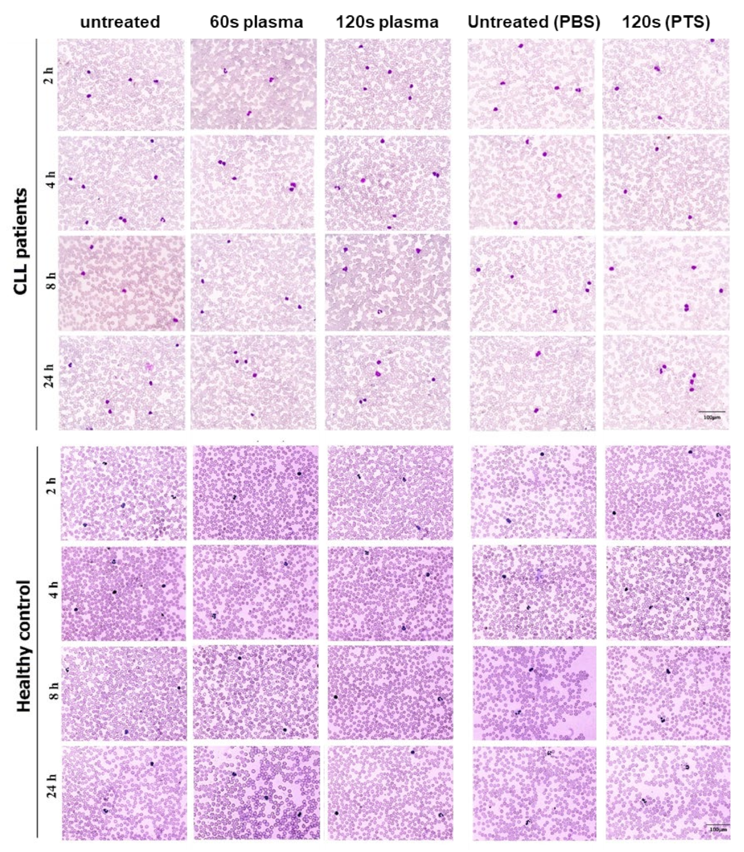
2.3. Complete Blood Cell Count (CBC) and Imaging of Peripheral Blood Smear

Complete blood cell counts (CBC) were performed by an automatic hematology analyzer (KX-21N; Sysmex, Norderstedt, Germany) at 2 h, 4 h, 8 h, and 24 h after cold plasma or PTS exposure. Red blood cells (RBC), white blood cells (WBC), platelets (PLT), hemoglobin (Hb), and red cell distribution width (RDW) were measured. Specifically, for the hemogram determination, three hydraulic subsystems were used for analyzing the four parameters, being the RBC channel, the WBC channel, the PLT channel, and a separate Hb channel. For the preparation and imaging of peripheral blood smears, blood smears were prepared and stained with May-Grünwald-Giemsa at 2 h, 4 h, 8 h, and 24 h after cold plasma or PTS treatment. A hematologist then examined smears to investigate the morphological changes of the WBCs, including hypersegmentation, toxic granulation, smudge cells, and morphological changes of the RBCs, including anisocytosis and microcytosis, hypochromia, and echinocytes. Microscopy of the smears was performed manually.

2.4. RNS and Lipid Peroxidation in Blood Plasma Treated with Cold Plasma or PTS

The nitrite (NO2)/nitrate (NO3) colorimetric Griess assay [33] was used according to the manufacturer’s protocol to measure the concentrations of RNS introduced by cold plasma and PTS treatment in isolated EDTA blood plasma at 2 h, 4 h, 8 h, and 24 h after treatment. The Griess reagents include 0.1% N-naphthyl-ethylenediamine dissolved in ionized water and 2% sulphanilamide in 5% HCl. This reagent reacts with nitrite in blood plasma and was incubated for 30 min at 37 °C to form a purple product. Then, the absorbance of each reaction mixture was measured at 540 nm using a BioTek microplate reader (BioTek, Winooski, Vermont, USA). Lipid peroxidation results from oxidative damage, and malondialdehyde (MDA) is a valuable marker for oxidative stress, which was analyzed according to the lipid peroxidation assay instruction at 2 h, 4 h, 8 h, and 24 h after plasma exposure with cold plasma or PTS. Briefly, 20% trichloroacetic acid (TCA) was added to the blood plasma to precipitate proteins. Then, 0.6% thiobarbituric acid (TBA) was added to the mixture. MDA in the blood plasma reacts with TBA to produce an MDA–TBA compound. This compound was detected colorimetrically at 535 nm (BioTek, Winooski, Vermont, USA). MDA leveled up to 1 nmol/well.

2.5. MTT Assay for Cytotoxicity of Cold Plasma and PTS Treatment

After 12 h, 24 h, and 48 h of treatment of the whole blood of 4 CLL patients and 4 sex- and age-matched healthy control subjects with cold plasma or PTS, the metabolic activity of WBCs was measured using the MTT (3-(4,5-dimethylthiazol-2-yl)-2,5-diphenyltetrazolium bromide; Sigma, St. Louis, Missouri, USA) assay according to the manufacturer’s protocol. Briefly, the samples were centrifuged and the blood plasma was isolated. Next, RBCs were removed using lysis buffer added to the pellet, and WBCs were isolated and cultured in isolated blood plasma. MTT solution (at a final concentration of 5 mg/mL in PBS) was added to each well. Following incubation for 3 h at standard culture conditions, the supernatant was removed, and 150 μL of DMSO was added to each well. The absorbance was measured using a BioTek microplate reader (BioTek, Winooski, Vermont, USA) at 570 nm.

2.6. Prothrombin Time (PT) and Partial Thromboplastin Time (PTT) Measurements

The PT and PTT from isolated citrated blood plasma were measured after 2 h, 4 h, 8 h, and 24 h of cold plasma or PTS exposure, using a Thermo Fisher kit according to the manufacturer’s protocol. A control sample (blood plasma from clinically healthy individuals) was included in each run. For the PT test, 100 µL of citrated blood plasma was incubated for 1 min at 37 °C with 200 µL of PT reagent. The time from mixing the reagent with blood plasma to the initiation of clot formation (detected visually) was determined as PT in seconds (s). For the PTT test, 100 µL of citrated blood plasma was added to 100 µL of PTT reagent and then incubated for 3 min at 37 °C. Subsequently, 100 µL of heated calcium chloride (37 °C) was added for activating the intrinsic (plasmatic) clotting cascade. Finally, the time (s) for clot formation (visually detected) was determined as PTT.

2.7. Quantification of Hemolysis

The RBCs were isolated by centrifugation (3000 rpm at 10 min) from 2 mL of whole blood and resuspended in 500 μL of PBS. From this initial erythrocyte stock, 50 μL was added to 1 mL of PBS and, following exposure to cold plasma (60 s and 120 s) or PTS (ratio of PTS to whole blood: 1:5), after 6 h of incubation, samples were centrifuged at 500× g for 5 min. Then, the supernatant was removed, and the hemolytic activity was quantified using spectrophotometry at 414 nm. In this experiment, deionized water (dH2O) and 10% SDS (sodium dodecyl sulfate) were used as positive controls. As an additional readout, blood agar medium was prepared and poured into 60 mm dishes using isolated RBCs that were washed in PBS 3-times and incubated for 24 h at 37 °C. After preparing the plates, the samples were exposed directly to cold plasma (120 s) or PTS. After an additional 24 h of incubation, the hemolytic activity was assessed from the diameter of the hemolytic zone. In this experiment, deionized water (dH2O) and 10% SDS were dropped onto blood agar plates as positive controls.

2.8. Statistical Analysis

Results are presented as the mean ± SEM of at least four independent experiments. The t-test for paired data or the 2-way ANOVA analysis for repeated measures was used. To perform pairwise comparisons between groups, Tukey’s post hoc test was used. All graphs were performed using Prism 7.0 (GraphPad Software, San Diego, California, USA), with p < 0.05 considered statistically significant.

3. Results

Neither cold plasma treatment nor PTS exposure affected hematological parameters or blood cell morphology. Analysis of CBC parameters in the blood of CLL patients after cold plasma treatment (60 s or 120 s) and PTS exposure (120 s) regarding mean counts of RBCs, WBCs, platelets, and hemoglobin identified no significant changes between treated and untreated samples in either CLL or healthy subjects (Figure 1). Blood smear images of the whole blood of CLL patients and healthy counterparts that had received cold plasma treatment (60 s or 120 s) or PTS exposure (120 s) were investigated after 2 h, 4 h, 8 h, and 24 h post-exposure by a hematologist. The images contain information on the morphological changes, and the results were compared with the untreated group. Peripheral blood smear images showed no significant morphological changes in WBCs, including hypersegmentation, toxic granulation, smudge cells, as well as morphological changes in RBCs, including anisocytosis, microcytosis, hypochromia, and echinocytes among either the treatment or control groups in both CLL and healthy subjects (Figure 2).

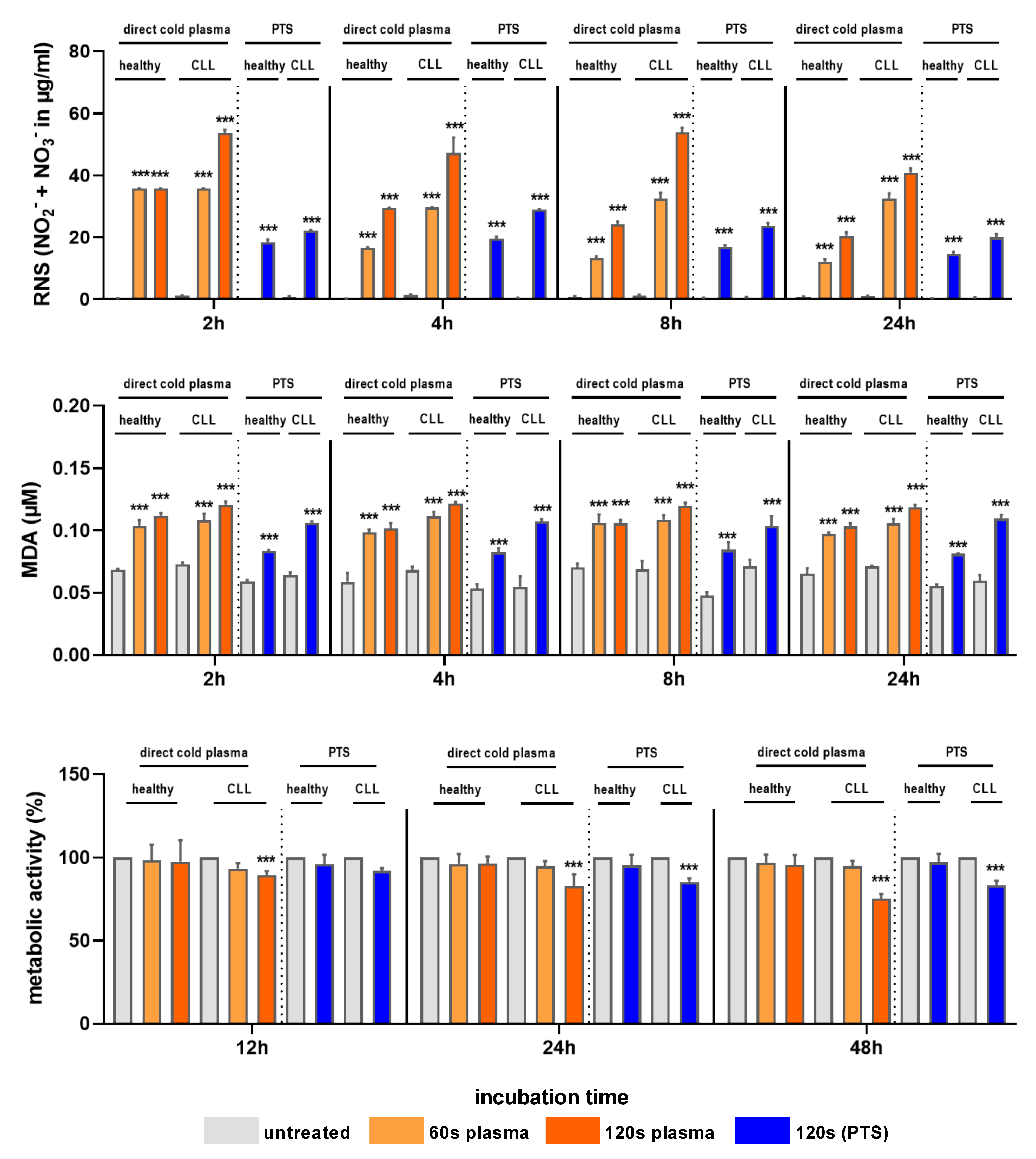
3.1. Cold Plasma and PTS Increased RNS Levels, Lipid Peroxidation, and Cytotoxicity

ROS/RNS are known cold plasma effectors in cells. We, thus, examined the ability of cold plasma to deposit RNS in whole blood by running subsequent analyses in blood plasma. Both cold plasma treatment and PTS exposure significantly increased RNS production in the blood plasma of CLL patients and healthy controls compared to untreated controls. However, the RNS concentration produced by cold plasma was considerably higher than the concentration introduced by PTS (Figure 3). Both cold plasma and PTS treatments showed increased lipid peroxidation levels, as determined using MDA (Figure 3). MDA levels were equally high in both treatment regimens and independent of the cohort (CLL vs. healthy). Next, we investigated the cytotoxicity of cold plasma treatment and PTS exposure in WBCs using the MTT assay (Figure 3). The treatment decreased the metabolic activity of leukocytes in CLL patients in a dose-dependent manner. The metabolic activity of WBCs was reduced with 120 s of cold plasma or PTS treatment after 24 h and 48 h (p < 0.001), while cold plasma was not cytotoxic in cells of healthy patients.

3.2. Cold Plasma and PTS Treatment Did Not Affect the PT, PTT, or Hemolysis

The median prothrombin time (PT) and partial thromboplastin time (PTT) from the citrated blood plasma of CLL patients and healthy individuals were analyzed 2 h, 4 h, 8 h, and 24 h after cold plasma or PTS exposure. Significant changes were not observed (Figure 4). Next, hemolysis was investigated macroscopically (Figure 5a) and quantitatively (Figure 5b). Compared to the positive controls SDS and dH2O, no hemolytic activity was observed with either cold plasma or PTS treatment in either CLL or healthy volunteer whole blood 6 h post-treatment for the specific plasma source used in this study.

4. Discussion

Plasma jet technology has attracted widespread attention in biomedical applications. It has been reported that cold plasma and PTS can selectively induce cell death in various tumor cells, typically by producing a wide range of ROS/RNS [34,35]. Enhanced basal RNS, especially nitric oxide (NO), might play a role in the prevention and progression of cancer. The selective nature of cold plasma is hypothesized to rely on ill-fated metabolism and baseline ROS/RNS in cancer cells compared to non-malignant counterparts, suggesting the former to be more sensitive to external ROS/RNS stressors and eventually leading to cell death [36,37]. PTS also contains high levels of H2O2 and NO2− capable of inducing programmed cell death at higher concentrations in malignant cells [38,39]. Using a plasma device that can operate in RNS production mode, this study provides information about the effects of cold-plasma-produced RNS on the blood of leukemia patients, as we reported decreased cell viability in the whole blood of patients with hematologic malignancies, namely CLL. In our ex vivo study protocol of patient and healthy donor blood, we aimed to identify the effect of cold plasma or PTS-mediated effects on hematological parameters such as CBC, WBC, RBC morphology, PT, PTT, cytotoxicity in WBC, RNS, lipid peroxidation, and hemolytic activity. Our findings showed that cold plasma exposure and PTS treatment increased blood plasma RNS and MDA levels in both CLL patients and healthy individuals without significantly altering complete blood count, WBC, RBC morphology, or PT and PTT time.
Our data suggest a selective effect on reduced metabolic activity in cells of CLL blood compared to healthy donor blood. Therefore, considering that the plasma therapy does not affect non-malignant leukocyte cells to a significant extent, the cold plasma treatment and the PTS exposure using the current plasma setup seem to be cytotoxic to mature malignant B cells known to be accumulated in the blood of CLL patients [40]. A range of previous in vitro studies on leukemia cells of different phenotypes, such as acute myeloid leukemia (AML) and acute T-cell leukemia, have provided evidence of cold-plasma-induced toxicity. This includes THP-1 and Jurkat cells [41,42,43], TK.6 cells [44], MOLM13 [45,46], U937 [41,42,47,48,49,50], and Molt-4 [41,50], which are among the previously in vitro studied cell lines. However, a study directly comparing the sensitivity of Jurkat and THP-1 leukemia cells with their non-malignant counterparts, T-helper cells and monocytes, revealed that the leukemia cells were much more resistant to cold-plasma-induced toxicity compared to the healthy donor cells [51]. Although the cells in that study were cultured in a cell culture medium and not derived from whole donor or patient blood, further research is needed to assess the selectivity of the plasma approach based on single-cell data. To the best of our knowledge, our report is the first study to show that both cold plasma treatment and PTS exposure reduced the metabolic activity of whole blood cells in CLL patients. However, further cellular and molecular studies are needed to determine which cells in CLL patients are targeted by cold plasma and PTS.
The direct plasma treatment induced greater MDA levels than PTS (indirect) exposure. This might be due to the delivery of relatively short-lived reactive species, such as peroxynitrite, shown to be generated by plasma jets [52] that can induce lipid peroxidation and MDA formation. The half-life of peroxynitrite is about two seconds; hence, it is likely absent in the PTS, leading to less MDA formation. At the same time, PTS induced a more considerable decline in metabolic activity than the direct plasma treatment. This might be due to the reactive species being degraded by the blood’s antioxidant during the direct treatment, ultimately leading to a lower overall concentration of plasma-derived oxidants, such as hydrogen peroxide that was shown to be toxic to white blood cells [53]. In the case of PTS, a higher concentration accumulates in the plasma-treated solution, which is then added to the blood cells at once, exposing a fraction of blood cells to that higher concentration during the mixing, and subsequently inducing more significant toxicity. The fact that not directly plasma-treated healthy leukocytes but CLL-derived leukocytes showed a decline in metabolic activity nevertheless suggests a particular specificity of the exposure-induced toxicity. This was also the case with PTS. Nevertheless, it should be mentioned that the PTS is radically different from the direct treatment in terms of reactive species generation and treatment application. In the PTS approach, a higher concentration of the long-lived oxidant hydrogen peroxide is added, but then quickly diluted in the matrix, leading to potentially different results than the direct treatment where more short-lived species are being deposited to the sample [54].
This study had several limitations. First, a preliminary plasma treatment time dose-escalation based on toxicity was not performed, ultimately leading to a modest decline in the blood cells’ metabolic activity. However, at 120 s, the direct plasma treatment was already relatively long. Hence, the current plasma-jet-based treatment scheme might be unsuitable for therapeutic action in the blood due to practical treatment time constraints. However, the current study served as a proof-of-concept trial, rather than as a therapeutic attempt. Moreover, the current study lacks single-cell analysis data done via, e.g., flow cytometry with fluorescently conjugated antibodies staining individual cell populations to demonstrate that the decline in metabolic activity and, therefore, likely cell death induction was present in the leukemia cell population specifically. In addition, measuring reactive species concentrations immediately after exposure to plasma or PTS, as well as testing liquids with equivalent ROS/RNS concentrations, would have been desirable, but was not possible in our study. Moreover, the possibility of endogenous ROS/RNS production, as known for phagocytes generating, for instance, superoxide and/or nitric oxide after activation after direct gas plasma or PTS exposure was not explored.

5. Conclusions

This proof-of-concept study provides evidence of non-hemolytic cold plasma operation modes for treating whole blood. At the same time, a decline in cellular activity was observed. These data suggest the principal feasibility of cold plasma exposure as an adjuvant option for leukemia treatment. While the exact integration of cold plasma into realistic clinical treatment schemes awaits further studies, the current research supports the idea that the effectiveness of cold plasma might not be limited to solid tumors but may be extended to hematological disorders, as well. Adapting cold plasma technology systems to the specific needs of such a future therapy within a clinically applicable approach is awaited. A potential modus operandi would be to perform extracorporeal blood circulation (leukapheresis unit) in conjunction with a plasma discharge integrated within a flow system and a liquid discharge without external gas. Such an apparatus would be conceivable as a medical device. However, the approach using plasma-treated solutions (PTS) might fall within the approval procedure of drugs, with much more effort needed to translate this concept into clinics.

Author Contributions

Conceptualization, A.R.; methodology, M.G., M.A., A.M., E.Z. and F.S.; software, M.G. and M.A.; validation, M.G., S.B. and A.R.; formal analysis, M.G.; investigation, M.G., M.A., A.M., E.Z. and F.S.; resources, A.R.; data curation, S.B. and A.R.; writing—original draft preparation, M.G. and S.B.; writing—review and editing, M.G. and S.B.; visualization, M.G. and S.B.; supervision, A.R.; project administration, A.R.; funding acquisition, A.R. All authors have read and agreed to the published version of the manuscript.

Funding

This study was financially supported by a grant (grant no: 6889) from the Research and Technology Council of Mazandaran University of Medical Sciences.

Institutional Review Board Statement

The study was conducted according to the guidelines of the Declaration of Helsinki, and approved by the Ethics Committee of the Mazandaran University of Medical Sciences (ethical approval code: IR.MAZUMS.REC.1398.6889).

Informed Consent Statement

Informed consent was obtained from all subjects involved in the study.

Data Availability Statement

Data are available from the corresponding author upon reasonable request.

Acknowledgments

The authors thank the patients and their families for their support, cooperation, and patience. We would like to thank the staff of the departments associated with the care and management of the patients.

Conflicts of Interest

The authors declare no conflict of interest.

References

  1. Amar, A. Chronic lymphocytic leukemia and second primary malignancies: A relationship revisited. Indian J. Med. Paediatr. Oncol. 2020, 41, 787–788. [Google Scholar] [CrossRef]
  2. Nabhan, C.; Rosen, S.T. Chronic lymphocytic leukemia: A clinical review. JAMA 2014, 312, 2265–2276. [Google Scholar] [CrossRef] [Green Version]
  3. Rezazadeh, H.; Astaneh, M.; Tehrani, M.; Hossein-Nataj, H.; Zaboli, E.; Shekarriz, R.; Asgarian-Omran, H. Blockade of PD-1 and TIM-3 immune checkpoints fails to restore the function of exhausted CD8+ T cells in early clinical stages of chronic lymphocytic leukemia. Immunol. Res. 2020, 68, 269–279. [Google Scholar] [CrossRef] [PubMed]
  4. Hatton, M.P.; Rubin, P.A. Chronic lymphocytic leukemia of the orbit. Arch. Ophthalmol. 2002, 120, 990–991. [Google Scholar] [PubMed]
  5. Weniger, M.A.; Rizzatti, E.G.; Pérez-Galán, P.; Liu, D.; Wang, Q.; Munson, P.J.; Raghavachari, N.; White, T.; Tweito, M.M.; Dunleavy, K. Treatment-induced oxidative stress and cellular antioxidant capacity determine response to bortezomib in mantle cell lymphoma. Clin. Cancer Res. 2011, 17, 5101–5112. [Google Scholar] [CrossRef] [PubMed] [Green Version]
  6. Goel, A.; Spitz, D.R.; Weiner, G.J. Manipulation of cellular redox parameters for improving therapeutic responses in B-cell lymphoma and multiple myeloma. J. Cell. Biochem. 2012, 113, 419–425. [Google Scholar] [CrossRef] [Green Version]
  7. Yosifov, D.Y.; Idler, I.; Bhattacharya, N.; Reichenzeller, M.; Close, V.; Ezerina, D.; Scheffold, A.; Jebaraj, B.M.C.; Kugler, S.; Bloehdorn, J. Oxidative stress as candidate therapeutic target to overcome microenvironmental protection of CLL. Leukemia 2020, 34, 115–127. [Google Scholar] [CrossRef]
  8. Ahmad, R.; Tripathi, A.K.; Tripathi, P.; Singh, R.; Singh, S.; Singh, R.K. Oxidative stress and antioxidant status in patients with chronic myeloid leukemia. Indian J. Clin. Biochem. 2008, 23, 328–333. [Google Scholar] [CrossRef] [PubMed] [Green Version]
  9. Kay, N.E. ROS: Double-edged sword for leukemic cells. Blood 2006, 107, 2212–2213. [Google Scholar] [CrossRef]
  10. Samimi, A.; Kalantari, H.; Lorestani, M.Z.; Shirzad, R.; Saki, N. Oxidative stress in normal hematopoietic stem cells and leukemia. Apmis 2018, 126, 284–294. [Google Scholar] [CrossRef] [PubMed]
  11. Privat-Maldonado, A.; Schmidt, A.; Lin, A.; Weltmann, K.D.; Wende, K.; Bogaerts, A.; Bekeschus, S. ROS from Physical Plasmas: Redox Chemistry for Biomedical Therapy. Oxid. Med. Cell. Longev. 2019, 2019, 9062098. [Google Scholar] [CrossRef] [Green Version]
  12. Bekeschus, S.; von Woedtke, T.; Emmert, S.; Schmidt, A. Medical gas plasma-stimulated wound healing: Evidence and mechanisms. Redox Biol. 2021, 46, 102116. [Google Scholar] [CrossRef]
  13. Dai, X.; Bazaka, K.; Richard, D.J.; Thompson, E.R.W.; Ostrikov, K.K. The Emerging Role of Gas Plasma in Oncotherapy. Trends Biotechnol. 2018, 36, 1183–1198. [Google Scholar] [CrossRef] [PubMed]
  14. Ahn, H.J.; Kim, K.I.; Hoan, N.N.; Kim, C.H.; Moon, E.; Choi, K.S.; Yang, S.S.; Lee, J.-S. Targeting cancer cells with reactive oxygen and nitrogen species generated by atmospheric-pressure air plasma. PLoS ONE 2014, 9, e86173. [Google Scholar]
  15. Ma, Y.; Ha, C.S.; Hwang, S.W.; Lee, H.J.; Kim, G.C.; Lee, K.-W.; Song, K. Non-thermal atmospheric pressure plasma preferentially induces apoptosis in p53-mutated cancer cells by activating ROS stress-response pathways. PLoS ONE 2014, 9, e91947. [Google Scholar] [CrossRef] [PubMed]
  16. Chung, W.-H. Mechanisms of a novel anticancer therapeutic strategy involving atmospheric pressure plasma-mediated apoptosis and DNA strand break formation. Arch. Pharmacal Res. 2016, 39, 1–9. [Google Scholar] [CrossRef]
  17. Conway, G.E.; He, Z.; Hutanu, A.L.; Cribaro, G.P.; Manaloto, E.; Casey, A.; Traynor, D.; Milosavljevic, V.; Howe, O.; Barcia, C.; et al. Cold Atmospheric Plasma induces accumulation of lysosomes and caspase-independent cell death in U373MG glioblastoma multiforme cells. Sci. Rep. 2019, 9, 12891. [Google Scholar] [CrossRef] [PubMed] [Green Version]
  18. Tanaka, H.; Bekeschus, S.; Yan, D.; Hori, M.; Keidar, M.; Laroussi, M. Plasma-Treated Solutions (PTS) in Cancer Therapy. Cancers 2021, 13, 1737. [Google Scholar] [CrossRef]
  19. Bauer, G. The synergistic effect between hydrogen peroxide and nitrite, two long-lived molecular species from cold atmospheric plasma, triggers tumor cells to induce their own cell death. Redox Biol. 2019, 26, 101291. [Google Scholar] [CrossRef] [PubMed]
  20. Alimohammadi, M.; Golpour, M.; Sohbatzadeh, F.; Hadavi, S.; Bekeschus, S.; Niaki, H.A.; Valadan, R.; Rafiei, A. Cold atmospheric plasma is a potent tool to improve chemotherapy in melanoma in vitro and in vivo. Biomolecules 2020, 10, 1011. [Google Scholar] [CrossRef]
  21. Rafiei, A.; Sohbatzadeh, F.; Hadavi, S.; Bekeschus, S.; Alimohammadi, M.; Valadan, R. Inhibition of murine melanoma tumor growth in vitro and in vivo using an argon-based plasma jet. Clin. Plasma Med. 2020, 19, 100102. [Google Scholar] [CrossRef]
  22. Yazdani, Z.; Mehrabanjoubani, P.; Rafiei, A.; Biparva, P.; Kardan, M. Combined Effect of Cold Atmospheric Plasma and Curcumin in Melanoma Cancer. BioMed Res. Int. 2021, 2021, 1969863. [Google Scholar] [CrossRef]
  23. Gjika, E.; Pal-Ghosh, S.; Kirschner, M.E.; Lin, L.; Sherman, J.H.; Stepp, M.A.; Keidar, M. Combination therapy of cold atmospheric plasma (CAP) with temozolomide in the treatment of U87MG glioblastoma cells. Sci. Rep. 2020, 10, 1969863. [Google Scholar] [CrossRef] [PubMed]
  24. Mahdikia, H.; Saadati, F.; Freund, E.; Gaipl, U.S.; Majidzadeh-a, K.; Shokri, B.; Bekeschus, S. Gas plasma irradiation of breast cancers promotes immunogenicity, tumor reduction, and an abscopal effect in vivo. Oncoimmunology 2021, 10, 1859731. [Google Scholar] [CrossRef] [PubMed]
  25. Yazdani, Z.; Mehrabanjoubani, P.; Biparva, P.; Rafiei, A. Cytotoxicity Effect of Cold Atmospheric Plasma on Melanoma (B16-F10), Breast (MCF-7) and Lung (A549) Cancer Cell Lines Compared with Normal Cells. J. Maz. Univ. Med. Sci. 2020, 30, 38–48. [Google Scholar]
  26. Takeda, S.; Yamada, S.; Hattori, N.; Nakamura, K.; Tanaka, H.; Kajiyama, H.; Kanda, M.; Kobayashi, D.; Tanaka, C.; Fujii, T. Intraperitoneal administration of plasma-activated medium: Proposal of a novel treatment option for peritoneal metastasis from gastric cancer. Ann. Surg. Oncol. 2017, 24, 1188–1194. [Google Scholar] [CrossRef] [PubMed]
  27. Gümbel, D.; Daeschlein, G.; Ekkernkamp, A.; Kramer, A.; Stope, M.B. Cold atmospheric plasma in orthopaedic and urologic tumor therapy. GMS Hyg. Infect. Control. 2017, 12, 1–10. [Google Scholar] [CrossRef]
  28. Canady, J.; Gordon, S.; Zhuang, T.; Wigh, S.; Rowe, W.; Shashurin, A.; Chiu, D.; Jones, S.; Wiley, K.; Cohen, E. Cold Atmospheric Plasma (CAP) Combined with Chemo-Radiation and Cytoreductive Surgery: The First Clinical Experience for Stage IV Metastatic Colon Cancer. In Comprehensive Clinical Plasma Medicine; Springer: Berlin/Heidelberg, Germany, 2018; pp. 163–183. [Google Scholar]
  29. Metelmann, H.-R.; Seebauer, C.; Miller, V.; Fridman, A.; Bauer, G.; Graves, D.B.; Pouvesle, J.-M.; Rutkowski, R.; Schuster, M.; Bekeschus, S.; et al. Clinical experience with cold plasma in the treatment of locally advanced head and neck cancer. Clin. Plas. Med. 2018, 9, 6–13. [Google Scholar] [CrossRef]
  30. Witzke, K.; Seebauer, C.; Jesse, K.; Kwiatek, E.; Berner, J.; Semmler, M.L.; Boeckmann, L.; Emmert, S.; Weltmann, K.D.; Metelmann, H.R.; et al. Plasma medical oncology: Immunological interpretation of head and neck squamous cell carcinoma. Plasma Process. Polym. 2020, 17, e1900258. [Google Scholar] [CrossRef]
  31. Jaffe, E.S. The 2008 WHO classification of lymphomas: Implications for clinical practice and translational research. ASH Educ. Program Book 2009, 2009, 523–531. [Google Scholar]
  32. Rostami, S.; Fathollahpour, A.; Abdi, M.; Naderi, K. Alteration in Prooxidant-antioxidant Balance Associated with Selenium Concentration in Patients with Congenital Hypothyroidism. J. Med. Biochem. 2018, 37, 355–363. [Google Scholar] [CrossRef] [PubMed] [Green Version]
  33. Sun, J.; Zhang, X.; Broderick, M.; Fein, H. Measurement of nitric oxide production in biological systems by using Griess reaction assay. Sensors 2003, 3, 276–284. [Google Scholar] [CrossRef] [Green Version]
  34. Ratovitski, E.A.; Cheng, X.; Yan, D.; Sherman, J.H.; Canady, J.; Trink, B.; Keidar, M. Anti-cancer therapies of 21st century: Novel approach to treat human cancers using cold atmospheric plasma. Plasma Process. Polym. 2014, 11, 1128–1137. [Google Scholar] [CrossRef]
  35. Yan, D.; Talbot, A.; Nourmohammadi, N.; Cheng, X.; Canady, J.; Sherman, J.; Keidar, M. Principles of using cold atmospheric plasma stimulated media for cancer treatment. Sci. Rep. 2015, 5, 18339. [Google Scholar] [CrossRef] [PubMed] [Green Version]
  36. Salunkhe, S.; Mishra, S.V.; Ghorai, A.; Hole, A.; Chandrani, P.; Dutt, A.; Chilakapati, M.; Dutt, S. Metabolic rewiring in drug resistant cells exhibit higher OXPHOS and fatty acids as preferred major source to cellular energetics. Biochim. Biophys. Acta (BBA)-Bioenerg. 2020, 1861, 148300. [Google Scholar] [CrossRef] [PubMed]
  37. Perillo, B.; Di Donato, M.; Pezone, A.; Di Zazzo, E.; Giovannelli, P.; Galasso, G.; Castoria, G.; Migliaccio, A. ROS in cancer therapy: The bright side of the moon. Exp. Mol. Med. 2020, 52, 192–203. [Google Scholar] [CrossRef]
  38. Turrini, E.; Laurita, R.; Simoncelli, E.; Stancampiano, A.; Catanzaro, E.; Calcabrini, C.; Carulli, G.; Rousseau, M.; Gherardi, M.; Maffei, F. Plasma-activated medium as an innovative anticancer strategy: Insight into its cellular and molecular impact on in vitro leukemia cells. Plasma Process. Polym. 2020, 17, 2000007. [Google Scholar] [CrossRef]
  39. Jezeh, M.A.; Tayebi, T.; Khani, M.R.; Niknejad, H.; Shokri, B. Direct cold atmospheric plasma and plasma-activated medium effects on breast and cervix cancer cells. Plasma Process. Polym. 2020, 17, 1900241. [Google Scholar] [CrossRef]
  40. Rozman, C.; Montserrat, E. Chronic lymphocytic leukemia. N. Engl. J. Med. 1995, 333, 1052–1057. [Google Scholar] [CrossRef] [Green Version]
  41. Jawaid, P.; Rehman, M.U.; Zhao, Q.L.; Takeda, K.; Ishikawa, K.; Hori, M.; Shimizu, T.; Kondo, T. Helium-based cold atmospheric plasma-induced reactive oxygen species-mediated apoptotic pathway attenuated by platinum nanoparticles. J. Cell. Mol. Med. 2016, 20, 1737–1748. [Google Scholar] [CrossRef]
  42. Wolff, C.M.; Kolb, J.F.; Weltmann, K.-D.; von Woedtke, T.; Bekeschus, S. Combination treatment with cold physical plasma and pulsed electric fields augments ROS production and cytotoxicity in lymphoma. Cancers 2020, 12, 845. [Google Scholar] [CrossRef] [PubMed] [Green Version]
  43. Turrini, E.; Laurita, R.; Stancampiano, A.; Catanzaro, E.; Calcabrini, C.; Maffei, F.; Gherardi, M.; Colombo, V.; Fimognari, C. Cold Atmospheric Plasma Induces Apoptosis and Oxidative Stress Pathway Regulation in T-Lymphoblastoid Leukemia Cells. Oxidative Med. Cell. Longev. 2017, 2017, 4271065. [Google Scholar] [CrossRef] [PubMed]
  44. Bekeschus, S.; Schmidt, A.; Kramer, A.; Metelmann, H.R.; Adler, F.; von Woedtke, T.; Niessner, F.; Weltmann, K.D.; Wende, K. High throughput image cytometry micronucleus assay to investigate the presence or absence of mutagenic effects of cold physical plasma. Environ. Mol. Mutagen. 2018, 59, 268–277. [Google Scholar] [CrossRef] [PubMed]
  45. Xu, D.; Ning, N.; Xu, Y.; Wang, B.; Cui, Q.; Liu, Z.; Wang, X.; Liu, D.; Chen, H.; Kong, M.G. Effect of cold atmospheric plasma treatment on the metabolites of human leukemia cells. Cancer Cell Int. 2019, 19, 135. [Google Scholar] [CrossRef]
  46. Xu, D.; Ning, N.; Xu, Y.; Xia, W.; Liu, D.; Chen, H.; Kong, M.G. Effect of He Plasma Jet Versus Surface Plasma on the Metabolites of Acute Myeloid Leukemia Cells. Front. Oncol. 2021, 11, 552480. [Google Scholar] [CrossRef]
  47. Maikho, T.; Patwardhan, R.S.; Das, T.N.; Sharma, D.; Sandur, S.K. Cold atmospheric plasma-modulated phorbol 12-myristate 13-acetate-induced differentiation of U937 cells to macrophage-like cells. Free Radic. Res. 2018, 52, 212–222. [Google Scholar] [CrossRef]
  48. Tabuchi, Y.; Uchiyama, H.; Zhao, Q.-L.; Yunoki, T.; Andocs, G.; Nojima, N.; Takeda, K.; Ishikawa, K.; Hori, M.; Kondo, T. Effects of nitrogen on the apoptosis of and changes in gene expression in human lymphoma U937 cells exposed to argon-based cold atmospheric pressure plasma. Int. J. Mol. Med. 2016, 37, 1706–1714. [Google Scholar] [CrossRef] [Green Version]
  49. Ji, H.W.; Kim, H.; Kim, H.W.; Yun, S.H.; Park, J.E.; Choi, E.H.; Kim, S.J. Genome-wide comparison of the target genes of the reactive oxygen species and non-reactive oxygen species constituents of cold atmospheric plasma in cancer cells. Cancers 2020, 12, 2640. [Google Scholar] [CrossRef]
  50. Moniruzzaman, R.; Rehman, M.U.; Zhao, Q.-L.; Jawaid, P.; Takeda, K.; Ishikawa, K.; Hori, M.; Tomihara, K.; Noguchi, K.; Kondo, T. Cold atmospheric helium plasma causes synergistic enhancement in cell death with hyperthermia and an additive enhancement with radiation. Sci. Rep. 2017, 7, 11659. [Google Scholar] [CrossRef] [PubMed]
  51. Bundscherer, L.; Bekeschus, S.; Tresp, H.; Hasse, S.; Reuter, S.; Weltmann, K.-D.; Lindequist, U.; Masur, K. Viability of Human Blood Leukocytes Compared with Their Respective Cell Lines after Plasma Treatment. Plasma Med. 2013, 3, 71–80. [Google Scholar] [CrossRef]
  52. Clemen, R.; Freund, E.; Mrochen, D.; Miebach, L.; Schmidt, A.; Rauch, B.H.; Lackmann, J.W.; Martens, U.; Wende, K.; Lalk, M.; et al. Gas Plasma Technology Augments Ovalbumin Immunogenicity and OT-II T Cell Activation Conferring Tumor Protection in Mice. Adv. Sci. 2021, 8, 2003395. [Google Scholar] [CrossRef] [PubMed]
  53. Bekeschus, S.; Kolata, J.; Winterbourn, C.; Kramer, A.; Turner, R.; Weltmann, K.D.; Broker, B.; Masur, K. Hydrogen peroxide: A central player in physical plasma-induced oxidative stress in human blood cells. Free Radic. Res. 2014, 48, 542–549. [Google Scholar] [CrossRef] [PubMed]
  54. Freund, E.; Bekeschus, S. Gas Plasma-Oxidized Liquids for Cancer Treatment: Preclinical Relevance, Immuno-Oncology, and Clinical Obstacles. IEEE Trans. Radiat. Plasma Med. Sci. 2021, 5, 761–774. [Google Scholar] [CrossRef]
Figure 1. CBC parameters in CLL patient- and healthy volunteer-derived blood receiving either cold plasma (60 s or 120 s) or PTS (120 s) treatment, with subsequent analyses at 2 h, 4 h, 8 h, and 24 h. No significant difference was observed for any of the parameters between the control groups against either cold plasma treatment or PTS exposure in both healthy and CLL blood. Data are represented as mean + SEM of four donors each.
Applsci 12 00128 g001
Figure 2. Morphological analysis of CLL patient- and healthy volunteer-derived blood receiving either cold plasma (60 s or 120 s) or PTS (120 s), with subsequent analyses at 2 h, 4 h, 8 h, and 24 h. No macroscopic difference was observed for the May-Grünwald staining between the control groups and either cold plasma treatment or PTS exposure in either healthy or CLL blood, as shown for one representative donor.
Applsci 12 00128 g002
Figure 3. RNS (nitrite + nitrate) and MDA concentrations in CLL patient- and healthy volunteer-derived blood receiving either cold plasma (60 s or 120 s) or PTS (120 s), with subsequent analyses at 2 h, 4 h, 8 h, and 24 h, as well as metabolic activity of cultured leukocytes assessed at 12 h, 24 h, and 48 h after exposure. Data are represented as mean + SEM of three donors each; *** denotes p < 0.001.
Applsci 12 00128 g003
Figure 4. Hemostasis parameters. The median prothrombin time (PT) and partial thromboplastin time (PTT) in citrated blood plasma of patients with CLL and healthy volunteers were analyzed at 2 h, 4 h, 8 h, and 24 h after either cold plasmas (60 s or 120 s) or PTS (120 s) exposure. The results showed no significantly different results when comparing untreated to plasma groups. Data are represented as mean + SEM of four donors each.
Applsci 12 00128 g004
Figure 5. Investigation of hemolysis of RBCs isolated from CLL patients and healthy individuals after exposure to either cold plasma (60 s or 120 s) or PTS (120 s) after 24 h culture on agar plates (a). The quantitative spectrophotometric experiment was performed at 6 h (b). Deionized water (dH2O) and SDS 10% served as positive controls. Data are represented as mean + SEM of four donors each; *** denotes p < 0.001.
Applsci 12 00128 g005
Table 1. Main hematological characteristics of CLL patients. Reference data are added from a healthy Iranian population (n = 60, [32]), CD19+ count in a reference population is the median from public databases. M: male; F: female; WBC: white blood cell count; RBC: red blood cell count; PLT: platelet count; Hb: hemoglobin; HCT: hematocrit; MCHC: mean corpuscular hemoglobin concentration; RDW: red cell distribution width; NA: not analyzed.
Sex Age WBC (103/mm3) RBC (106/mm3) Hb (g/DL) HCT (%) MCHC (g/DL) RDW (FL) PLT
(103/mm3)
CD19 (%)
1M636.65.114.242.233.612.3126NA
2M5510.14.08.839.334.813.713965
3M7117.85.112.140.333.213.211171
4M9041.83.013.132.333.412.615791
5M4816.04.313.237.735.012.825480
6F7251.95.310.635.629.817.528768
7F5619.44.713.439.933.613.322765
8F52107.23.112.337.834.913.093NA
9M6470.73.611.031.135.413.812778
Ref--8.34.612.337.432.513.3334(14)
Publisher’s Note: MDPI stays neutral with regard to jurisdictional claims in published maps and institutional affiliations.

