Article

A Herbal Concoction of Cinnamomum cassia and Artemisa annua Extracts Ameliorates Allergic Rhinitis in OVA-Induced Balb/C Mice by Inhibiting Th2 Signaling

R&BD Center, Hy Co., Ltd., 22, Giheungdanji-ro 24beon-gil, Giheung-gu, Yongin-si 17086, Korea
*
Author to whom correspondence should be addressed.
Appl. Sci. 2022, 12(1), 340; https://doi.org/10.3390/app12010340
Submission received: 20 October 2021 / Revised: 22 December 2021 / Accepted: 25 December 2021 / Published: 30 December 2021
(This article belongs to the Topic Applied Sciences in Functional Foods)

Abstract

:
Allergic rhinitis (AR) is an inflammatory airway disease (IAD) that is characterized by itching, nasal obstruction, and sneezing. AR is induced by Th-2 inflammatory responses such as those mediated by IgE and IL-4. This study aims to investigate the therapeutic effects of an herbal concoction, which is a combination of Cinnamomum cassia and Artemisa annua extracts (CIAR) against ovalbumin (OVA)-induced allergic rhinitis in a Balb/C mouse model. The effect of CIAR on the Th-2 mediated inflammatory response in the AR mouse model was studied by analyzing blood or nasal fluid samples. Experimental results revealed that OVA inhalation increased IgE, IL-4, IL-33, and TSLP levels, leading to Th2-type cytokine response. CIAR was found to significantly reduce the Th-2 response and levels of cytokines, including IL-4, IL-33, and thymic stromal lymphopoietin (TSLP). CIAR also down-regulated eosinophil (EOS) and basophil (BASO) levels in the blood. Histological analyses demonstrated decreased OVA-induced thickness of the respiratory epithelium in the CIAR-treated group. Collectively, our results suggest that the herbal concoction CIAR can effectively ameliorate the development of allergic rhinitis through the inhibition of Th-2 mediated responses.

1. Introduction

AR affects nearly 30% of the world’s population and is becoming a worldwide health problem with an increasing incidence every year [1,2]. It is a hypersensitivity reaction that is characterized by sneezing, itching of the nose, and rhinorrhea subsequent to exposure to certain allergens [3]. These symptoms develop due to the secretion of inflammatory mediators from mast cells, eosinophils (EOS), and basophils (BASO) [4]. Moreover, Th-2-related signals, including immunoglobulin E (IgE) and interleukin 4 (IL-4), trigger inflammatory reactions and are recognized as critical mediators of allergic rhinitis [5]. On exposure to allergens, immune cells such as eosinophils, basophils, and mast cells (MCs) secrete secondary signaling molecules including IgE, IL-4, and tumor necrosis factor (TNF), which cause differentiation of naïve CD4+ T cells into Th2 cells that in turn trigger acute allergic responses like allergic rhinitis [6,7].
Recently, Park et al. reported that Th-2 cells are stimulated by the epithelial-derived cytokines IL-25, IL-33, and thymic stromal lymphopoietin (TSLP), the levels of which are regulated by the activity of protein arginine methyltransferase 1(PRMT1). This causes the activation of B cells or EOS and the subsequent development of allergic rhinitis. They further demonstrated that the elevated secretion of epithelial cytokines, including IL-25, IL-33, and TSLP, is essential for Th-2 cell differentiation. Thus, previous studies have established an association between the epithelium mediators IL-25, IL-33, and TSL, as well as IgE and IL-4 Th-2 signaling molecules with allergic rhinitis [8].
Cinnamomum cassia has traditionally been used in Asian cultures for its beneficiary effect on dyspepsia, peptic ulcers, and ischemic brain injury [9]. Artemisa annua has also been reported to possess anti-cancer, anti-adipogenic, anti-osteoporotic, and anti-malarial properties [10,11]. Several studies have reported that Cinnamomum cassia and Artemisa annua have a preventive effect on allergic rhinitis in human clinical trials via their anti-inflammatory effects [12,13]. However, there are no reports on the ameliorative effect of the combination of these herbs, CIAR (Cinnamomum cassia and Artemisa annua), on Th-2 signaling mediated inflammatory responses.
Therefore, this study aims to investigate the anti-allergic effect of CIAR in the OVA-induced rhinitis mouse model by analyzing the improvement in the levels of Th-2 signal molecules in serum and nasal fluid.

2. Materials and Methods

2.1. Reagents and Equipment

Fine chemicals and assay kits used in the study include ovalbumin, dexamethasone and A23187 (Sigma Aldrich, St. Louis, MO, USA), mouse IL-4, IL-5 and TNF ELISA kit (R&D systems, Minneapolis, MN, USA), mouse IgE ELISA kit (Invitrogen, Carisbad, CA, USA), mouse IL-25, IL-33 and TSLP ELISA kit (Abcam, Cambridge, UK), and mouse ECP ELISA kit (MyBiosource, San Diego, CA, USA). The standards used in the chemical analysis, organic acids (malic acid, acetic acid, maleic acid, succinic acid, propionic acid, tartaric acid, and butyric acid) and flavonoids (quercetin, kaempferol, and isorhamnetin), were obtained from Sigma Aldrich. Amino acids standard complex was purchased from Hitachi. CIAR was prepared as previously published by Kim et al. [14]. Briefly, dried Cinnamomum cassia (CI) and Artemisa annua (AR) were purchased from Humanherb Co., Ltd. (Daegu, Korea). Grounded samples (100 g) were mixed and stirred for 24 h at 50–70 °C in 1 L of 50% ethanol. The obtained extract was concentrated in a rotary vacuum evaporator for 24 h. The concentrated material was re-suspended in water for experiments.

2.2. Animals

Balb/c mice (male, 6 weeks old, 22 ± 3 g) were purchased from DooYeol Biotech (Seoul, Korea) and housed in the animal laboratory (22 ± 2 °C, relative humidity of 50 ± 20%, 12-h light/dark cycle, non-specific pathogen free, fed a standard diet with ultraviolet sterilizer. All experimental procedures were approved by the Ethics Review Committee of the Hy Company Limited R&D Center, Yongin-si, Korea (AEC-2021-0006).

2.3. Mouse Model of Ovalbumin-Induced Allergic Rhinitis

The allergic rhinitis mice model was generated according to the modified method published by Kim et al. [15]. Briefly, male Balb/c mice were randomly divided into six groups of 7–8 mice each: (1) control group (vehicle), (2) OVA-induced group (OVA), (3) dexamethasone-treated group (Dex), (4) CIAR low-dose-administered group (CIAR-L), (5) CIAR middle-dose-administered group (CIAR-M), and (6) CIAR high-dose-administered group (CIAR-H). Mice were intraperitoneally (i.p.) sensitized with 50 μg of OVA emulsified with 0.15 mL of 1% aluminum hydroxide (w/v) in phosphate-buffered saline (PBS, pH 7.4) on days 1, 8, and 15. A similar volume of saline was administered to the control group. Challenges were continued with repeated exposures to an aerosol of 1% OVA by nebulization (NE-C28, Omron, Seoul, Korea). The challenge procedure was conducted daily for 30 min from days 25 to 29. The dexamethasone, CIAR (mixed CI and AR powder with 1:1 ratio), low-, middle-, and high-dose groups were orally gavaged from days 16 to 29 using PBS as a vehicle. CIAR low-dose, middle-dose, and high-dose groups were administered CIAR at 150 mg/kg (CIAR-L), 300 mg/kg (CIAR-M), and 450 mg/kg (CIAR-H) once a day, respectively. Dexamethasone was administered at 500 μg/kg. On day 30, mice were sacrificed by CO2 inhalation, followed by extraction of the nasal cavity and collection of nasal fluid and blood. All organs and tissues were stored at −70 °C for subsequent cytokine and histopathological analysis.

2.4. Cell Cultures

The mouse macrophage cells (RAW264.7) and rat basophilic leukemia cells (RBL-2H3) were cultured in Dulbecco’s modified Eagle’s medium (DMEM) containing 1% antibiotic-antimycotic solution (Gibco) and 10% heat-inactivated fetal bovine serum in 5% CO2 at 37 °C. For in vitro assay, cells (3 × 10⁵ cells/mL) were incubated with lipopolysaccharide (LPS, 1 μg/mL) or A23187 (1 μM) in the presence or absence of CIAR (50 μg/mL of final concentration, mixed with 1:1 ratio with CI and AR), CI (50 μg/mL) or AR (50 μg/mL) for 24 h. The culture supernatants were recovered for analysis by ELISA.

2.5. Analysis of Cytokines in Blood and Nasal Fluid Samples

Blood and nasal fluid samples were prepared as previously published by Bae et al. with slight modifications [16]. To separate serum, blood was kept at 25 °C for 30 min and then centrifuged at 2000× g for 10 min. Serum was analyzed by mouse ELISA kits for cytokines and ECP levels. For the estimation of immunocyte counts, whole blood was collected in EDTA treated tubes and was analyzed using Mindray, BC-5000Vet. Cell counts were indicated as multiples of 1 × 10³ cells/μL.

2.6. Histological Examination of Nasal Epithelial Thickness

Nasal tissue was fixed with 10% formalin solution and processed at KPC (Gwangju-si, Korea). The thickness of the epithelium was measured using an Olympus CK2 microscope (Tokyo, Japan) under 4× or 20× magnification. The epithelial thickness was analyzed using Image J software (National Institutes of Health; NIH, Bethesda, MD, USA).

2.7. HPLC Analysis of Organic Acids and Flavonoids

The organic acids and flavonoids of Cinnamomun cassia and Artemisa annua samples were determined using an Agilent 1280 HPLC system with a UV detector (Agilent Techonologies, St. Louis, MO, USA). The chromatographic separation of amino acids was achieved using a Prevail OA column (4.6 × 250 mm, 5 µm; Alltech) or Aminex HPX-87H column (4 × 250 mm, 0.45 µm; Biorad), and flavonoids were analyzed to use a CAPCELL PAC C18 UG120 (4.6 × 250 mm, 5 µm; SHISEIDO). The Alltech prevail OA column was used to analyze malic acid, acetic acid, maleic acid, succinic acid, and propionic acid at a wavelength of 540 nm, and the Bio-rad Aminex HPx-87H column was used to analyze butyric acid and tartaric acid at a wavelength of 210 nm. The flavonoids were detected at 370 nm wavelength. The 25 mM phosphate buffer was used as mobile phase with Alltech Prevail OA column, and 0.01 N sulfuric acid solution was used with Bo-rad Aminex HPX-87H column. The flavonoid analysis mobile phase consisted of 0.1% phosphoric acid in acetonitrile (solvent A) and 0.1% phosphoric acid in water (solvent B). All mobile flow rate was 1 mL/min.

2.8. The Condition of the Automated Amino Acid Analyzer

To analyze amino acids, an automated high-speed amino acid analyzer L-8900 (Hitachi, Tokyo, Japan) was used. The analytical column was a Hitachi HPLC Packed column (Ion-exchange resin, 4.6 × 60 mm, 3 µm; Hitachi). The visible detector (Hitachi) was used as analytical detector and was set to measure at 570 nm wavelength (phosphoserine, aspartic acid, thereonine, glutamic acid, glyccine, alanine, valine, cystine, isoleucine, leucine, phenylalanine, and g-amino-n-butyric acid). Four different Ph buffer solutions (Mitsubishi) were prepared as mobile phase; ph values were 3.3, 3.2, 4.0, and 4.9, respectively. The flow rate of the amino acid analyzer was 1 mL/min, and the injecting volume was 20 µL. For post-column reaction analysis, glacial acetic acid contained lithium acetate dihydrate, and propylene glycol monomethyl ether was prepared (Wako). During the running of the samples, the separate delivery pump for the ninhydrin reagent automatically mixed these 2 solutions, which were kept under nitrogen in the amino acid analyzer.

2.9. Statistical Analysis

All data are indicated as mean ± standard error. Significant differences were statistically analyzed using a one-way analysis of variance, followed by Duncan’s multiple range test (p < 0.05 or 0.01). The obtained data were analyzed using GraphPad Prism 5 program (GraphPad Software, Inc., San Diego, CA, USA.).

3. Results

3.1. Reduced Immunocyte Counts and Eosinophil Cationic Protein (ECP) Levels in the Mouse Model of OVA-Induced Allergic Rhinitis after CIAR Administration

The availability of CIAR to alleviate OVA-induced AR in mice was tested by measuring changes in immune cell composition and serum ECP levels (Figure 1). This effect was further investigated in a dose-dependent manner by the administration of three incremental concentrations of CIAR. OVA-induced AR resulted in an increase in the eosinophil and basophil cell populations in comparison to the control group. Oral administration of CIAR at doses of 150 mg/kg, 300 mg/kg, and 450 mg/kg caused the count eosinophil counts to drop to 6.94 ± 0.56, 6.24 ± 1.25, and 5.12 ± 0.44 × 10³ cells/µL, respectively (p < 0.05). Similarly, the basophil counts were reduced to 1.18 ± 0.25, 1.02 ± 0.29, and 0.9 ± 0.2 × 10³ cells/µL, respectively, in response to the incremental doses of CIAR (p < 0.05). Thus, eosinophil and basophil cell populations showed a significant decrease as a consequence of CIAR administration in a dose-dependent manner. ECP levels were closely related to eosinophil immune responses and the subsequent activation of allergic rhinitis. Serum ECP was found to be elevated in the OVA-induced mice group (p < 0.05), Moreover, ECP concentrations were significantly decreased to 4.74 ± 1.13, 4.13 ± 0.94, and 3.96 ± 0.68 ng/mL post oral administration of CIAR at 150 mg/kg, 300 mg/kg, and 450 mg/kg, respectively (p < 0.05).

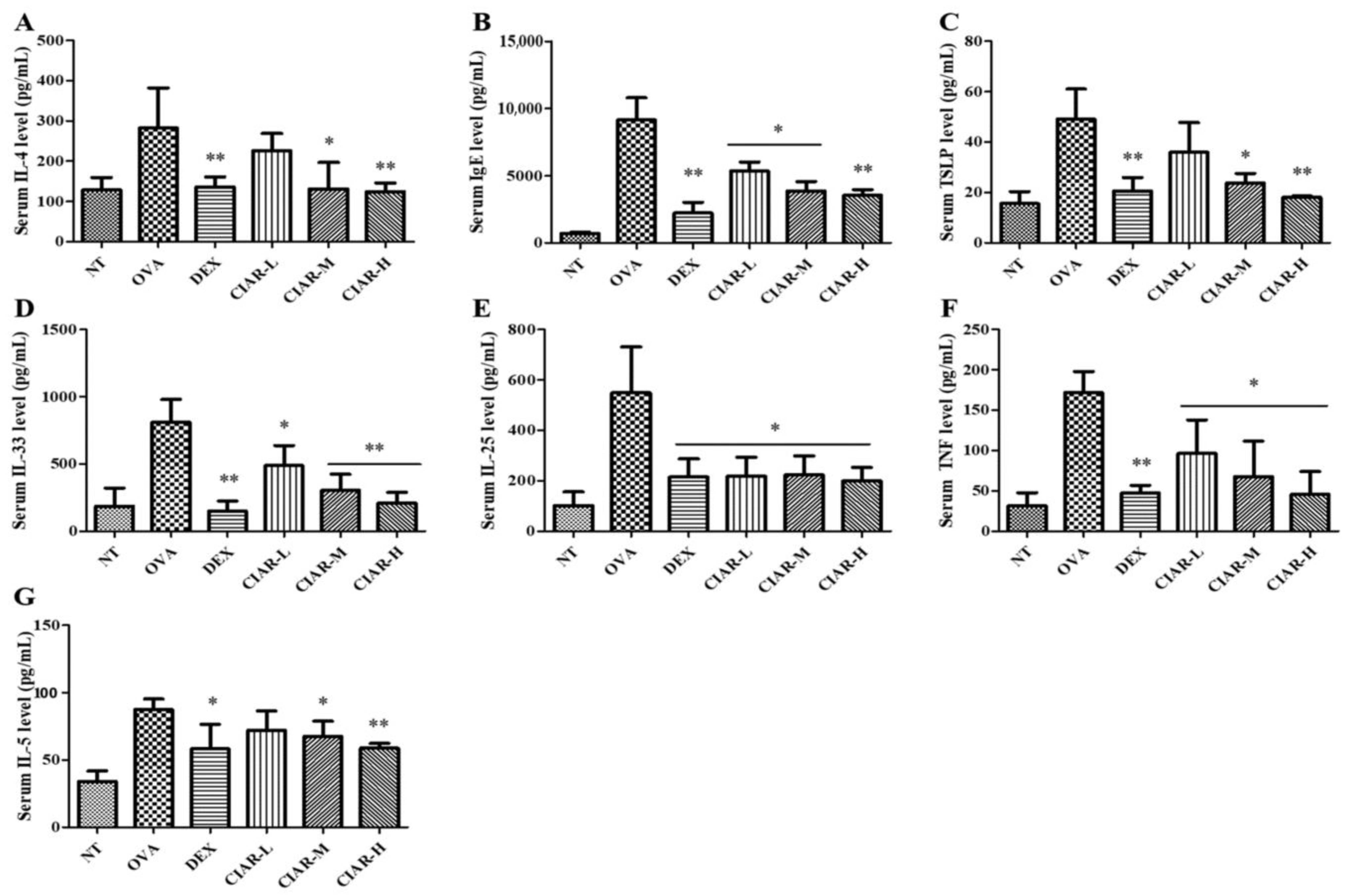
3.2. Inhibition of Th-2 Cytokine Production in Serum of Mice with OVA Induced Allergic Rhinitis

The inhibitory effect of CIAR on the secretion of Th-2 mediated inflammatory cytokines was examined (Figure 2). In comparison to the control group, serum levels of IgE, IL-4, IL-5, TNF, IL-25, IL-33, and TSLP were elevated post inhalation of OVA (p < 0.05). As shown in Figure 2, administration of the three different doses of CIAR inhibited the secretion of Th-2 cytokines, including IL-4, IL-5, and TNF. Furthermore, Th-2 mediated signal transduction induced by OVA was significantly suppressed by CIAR administration, as seen by the decrease in IgE, IL-25, IL-33, and TSLP levels as compared to the control group. This was especially apparent in the CIAR-M and CIAR-H groups, which showed significant inhibition of the Th-2 mediated response in AR.

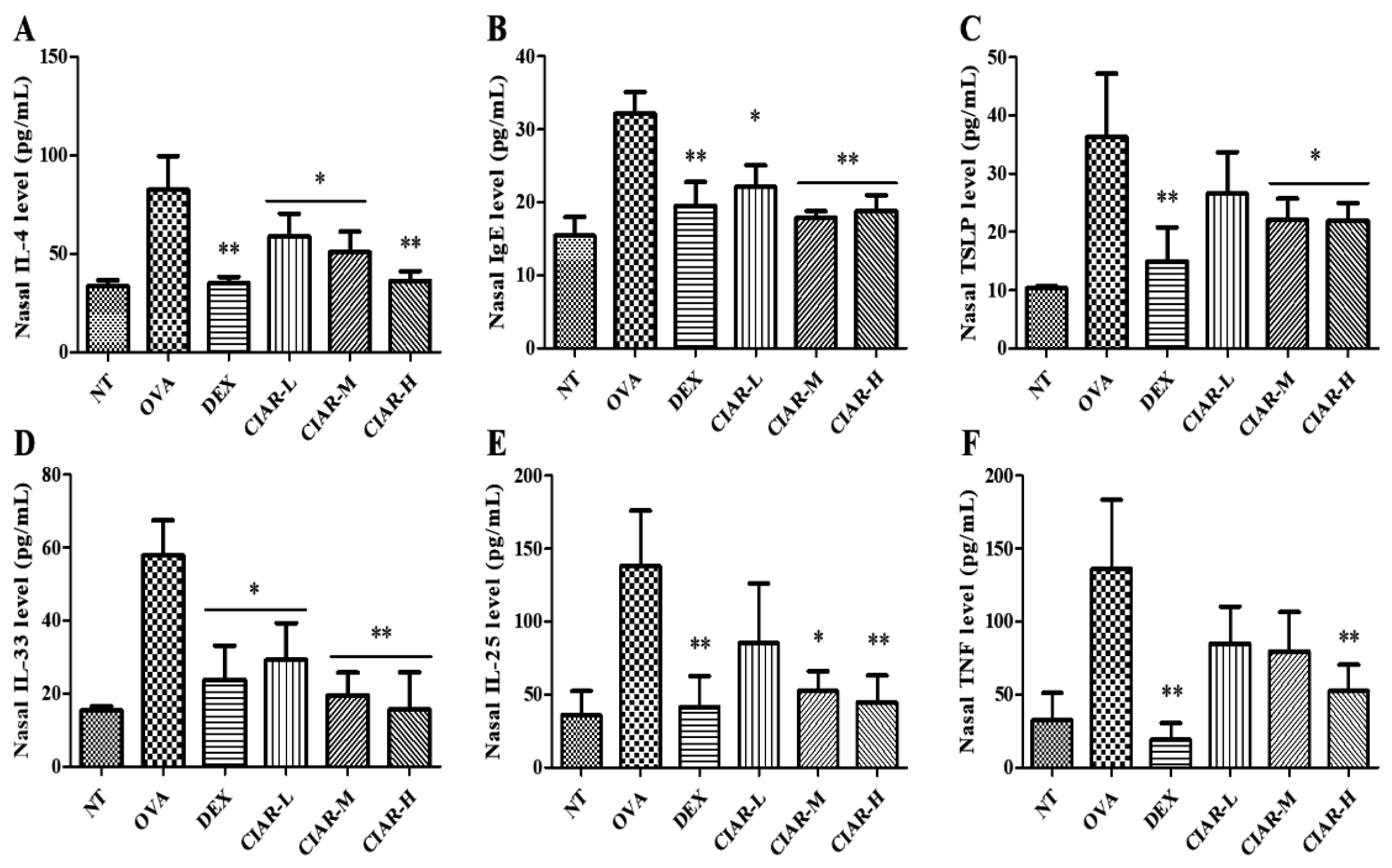
3.3. Effects of CIAR on Th2 Cytokine Production in OVA Induced Mouse Nasal Fluid

Inhibition of Th-2 cytokine levels was further investigated in mice nasal fluid (Figure 3). Exposure to OVA caused an elevation in the levels of nasal Th-2 signaling molecules. However, the administration of CIAR in incremental doses significantly reduced the production of nasal Th-2 cytokine-related signaling molecules. In concordance with the results obtained in serum analysis, the CIAR-M and CIAR-H groups demonstrated a higher degree of suppression of the inflammatory effects induced by OVA (p < 0.05).

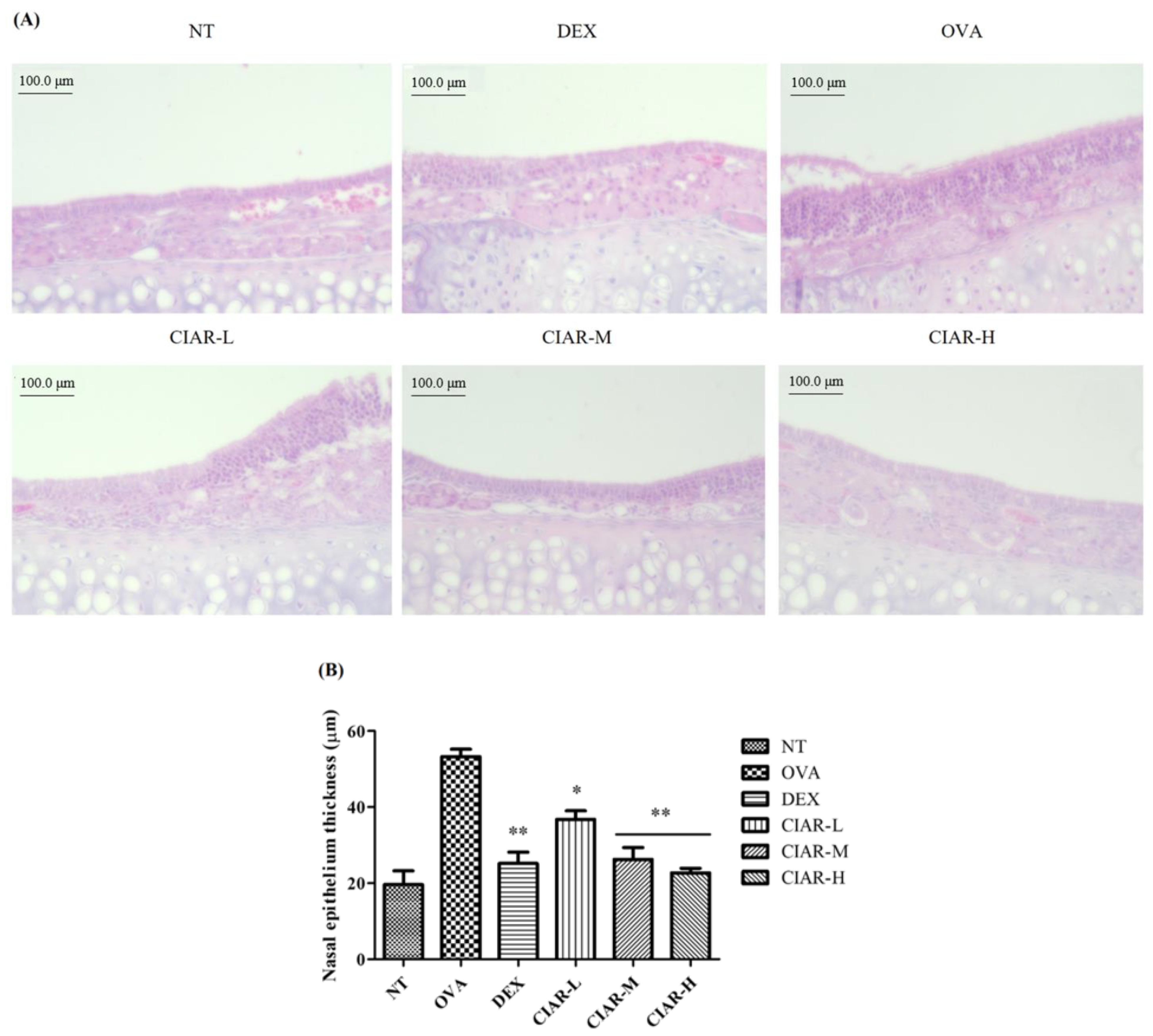
3.4. CIAR Ameliorates Nasal Respiratory Epithelial Thickness Damaged by OVA Inhalation in Mice with Allergic Rhinitis

Nasal epithelial tissue thickness was assessed by histological examination. Representative immunohistochemical images of mouse nasal respiratory epithelium are shown in Figure 4 and Table 1. The increase in respiratory epithelium thickness from 19.65 ± 3.65 to 53.25 ± 1.96 µm was observed post exposure to inhaled OVA (p < 0.05). Administration of CIAR at doses of 150 mg/kg, 300 mg/kg, and 450 mg/kg resulted in a reduction of the respiratory epithelial thickness to 36.77 ± 2.25, 26.28 ± 3.09, and 22.72 ± 1.23 µm, respectively (p < 0.05).

3.5. Synergistic Effect of CIAR in In Vitro Assays on Levels of Inflammatory Cytokines

We investigated the inhibitory action of CIAR, CI (Cinnamomum cassia), and AR (Artemisa annua) on the levels of IL-4, IL-33, TNF, and TSLP in RAW264.7 and RBL-2H3 cells (Figure 5). CI and AR treatment demonstrated inhibition of IL-4 production in RBL-2H3 cells on stimulation with A23187 (p < 0.05). CI and AR treatment also lowered COX2, TSLP, IL-33, and TNF in RAW264.7 cells induced by LPS (p < 0.05). Furthermore, a synergistic effect on the reduction of IL-4, IL-33, TNF, and TSLP was observed in the presence of CIAR.

3.6. The Analyzed Chemical Components in CIAR, Organic Acids, Amino Acids, and Flavonoids

The chemical components, organic acids, flavonoids, and amino acids, contained in CI, AR were analyzed with HPLC or an automatic amino acid analyzer. The measured contents are tabulated in Table 2, Table 3 and Table 4 about organic acids, amino acids, and flavonoids, respectively. Organic acids were detected in HPLC, and CI showed higher amounts than AR organic acids contents, generally. Especially at succinic acid, butyric acid of CI resulted in 49.22 ± 3.4 and 314.09 ± 10.62 mg/g, respectively, and AR resulted in non-detection (Table 2). At amino acids analysis, AR was generally measured to have higher amounts than CI. Thereonine, glycine, isoleucine, leucine, and phenylalanine contents of AR resulted to 405.3 ± 3.4, 131.9 ± 3.4, 136.7 ± 3.4, 417.3 ± 6.8, and 412.5 ± 6.8 μg/g, respectively (Table 3). Flavonoids’ analysis results are shown in Table 4.

4. Discussion

Allergic rhinitis, a hypersensitivity immune disorder, is a common inflammatory disease that affects all age groups [17]. Approximately 10% of the world population suffers from AR, and its incidence has been observed to rise rapidly every year [2,18]. Patients of AR present with symptoms including itching, runny nose, and sneezing, which can significantly affect functionality [19]. Many studies have demonstrated that Th-2 mediated cytokines play a critical role in allergic inflammatory rhinitis and the importance of the relationship between Th-2 cells and allergic rhinitis is well established [20]. Activated Th-2 cell secrete signal molecules including TNF, IL-4, and IL-13 that activate B cells and mast cells. Post this induction, activated B cells produce IgE that stimulates mast cells and basophils, leading to an allergic reaction. Furthermore, Th-2 cells activate eosinophils via IL-3 or IL-5, resulting in cell migration into the vascular endothelium. Therefore, inhibition of an upstream pathway such as the Th-2 signaling pathway in allergic response may result in a better clinical outcome as compared to inhibition of downstream steps in the inter-related signal transduction pathways [21].
Previous studies show that TSLP, IL-25, and IL-33, which are produced in epithelial cells, are critical Th-2 differentiators [22]. Allergens or chemicals can promote protein arginine methyltransferase 1(PRMT1) expression via mitogen-activated protein kinase (MAPK) phosphorylation in epithelial cells, which in turn can stimulate Th-2 cells, Mast cells, and type 2 innate lymphoid (ILC2) cells via secreted TSLP, IL-25, and IL-33. During this process, elevated IL-4, IL-5, and IL-13 levels lead to eosinophil and mast cell degranulation, IgE synthesis from B cells, and the subsequent development of a characteristic type 2 immune environment [23,24].
In this study, we used an allergic rhinitis mouse model that, on induction with OVA-inhalation, showed pathological changes associated with AR including nasal itching and sneezing, as reported previously [25]. We observed sensitization and challenge with OVA-induced allergic rhinitis and a concomitant significant increase in immunocytes populations, including eosinophils and basophils in serum. Furthermore, the oral administration of CIAR at three incremental doses resulted in the alleviation of immunocyte levels in a dose-dependent manner. ECP is a cytotoxic protein granule produced by eosinophils and may play a role in the pathophysiology of allergic rhinitis, such as epithelial cell damage and non-specific hyperreactivity [26]. We were able to demonstrate an elevation in serum ECP post OVA inhalation, which was reversed by the administration of CIAR in a dose-dependent manner.
The induced mice showed an up-regulation of inflammatory Th-2 mediated signal molecules in serum and nasal fluid as previously reported [4,27]. We used the Balb/C mouse to induce allergic rhinitis because, according to a previous study, inflammation reactions are better expressed in Balb/C mice than in other mice models. Moreover, Hiroyuki et al. reported that the Balb/C mouse model is recommended to use to study related to Th-2 response inflammation or allergic study [28,29]. Dexamethasone, used as a positive control, showed alleviated effect against inflammation induced by OVA. According to Kim et al. [4], dexamethasone suppressed the cytokine level such as IL-4 and immunocytes such as eosinophils, and we also used it as a positive agent [4]. Th-2 cytokines induce inflammatory reactions, such as immunocyte activation and production of IgE as well as epithelial-derived cytokines [30,31]. In our analysis, serum IgE, IL-4, TNF, and IL-5 were decreased significantly in CIAR administrated group. Further, TSLP, IL-24, and IL-33 levels were also reduced post-CIAR administration in OVA-induced mice. Induced allergic rhinitis with OVA is resulted in high levels of Th-2 signal expression in nasal fluid, as reported previously [32]. Similarly, CIAR administration effectively inhibited Th-2 inflammatory cytokines and related signal molecules in nasal fluid in a dose manner. But unlike serum, IL-5 cytokine level change was not observed in nasal fluid. This may be due to immunocytes change, which is observed in serum but not in nasal fluid, and according to Benson et al., it has been reported that high IL-5 increases in allergic rhinitis patients with increased immune cells such as eosinophils and neutrophils [33]. Additionally, according to Kim et al., immunocytes were not observed in mouse nasal fluid [4]. Therefore, IL-5 was not observed in mouse nasal fluid where immune cells were not counted.
Previous studies reported increased thickness of the nasal mucosal tissue of mice induced by OVA [34,35]. Th-2 cells activated with OVA secrete IL-4 or IL-5 cytokine, which stimulates B cells, eosinophils, basophils, and mast cells. The subsequent production of IgE, IL-33, IL-25, and TSLP mediators induce degranulation of effector cells results in nasal epithelial damage and thickening [36]. As depicted in Figure 3, the thickened nasal epithelium was alleviated to a thickness of 36.78 ± 2.25, 26.28 ± 3.09, and 22.72 ± 1.23 from 53.26 ± 1.96 µm when administered with CIAR at doses of 150 mg/kg, 300 mg/kg, and 450 mg/kg, respectively.
In addition, we confirmed the inhibitory effects of CIAR, CI, and AR on Th-2 signaling pathways in-vitro on Raw264.7 and RBL-2H3 cells. The lRaw264.7 macrophage cells and RBL-2H3 basophil cells upregulated production of IL-4, IL-33, TNF, and TSLP on stimulation with LPS or A23187, respectively, as previously reported [16,37,38,39]. Treatment of these cells by CI and AR extracts suppressed the stimulation of these signal molecules. Moreover, the inhibitory effect of these extracts on the TH-2 signaling pathway was synergistic when used together (CIAR).
To guess the reason for the synergic effect of mixed CI and AR showed, we analyzed the phytochemicals or bioactive components with HPLC or amino acids analyzer. As previously reported, organic acids, flavonoids, or amino acids have many functional effects, especially anti-inflammation [40,41]. As we tabulated in Table 2, AR contains more amino acid contents than CI, such as Ile and Leu, which have already been reported to have anti-inflammatory effects [42,43]. Interestingly, the analysis results of organic acids showed different patterns. As shown in Table 3, CI contains more organic acids contents than AR, such as succinic acid, tartaric acid, and butyric acid, which are also reported to have anti-inflammatory functions [44,45]. Therefore, we carefully considered this point as one of the reasons for synergic effects. By mixing together, CIAR could show anti-inflammatory effects from both organic acids and amino acids, resulting in a synergic effect. However, further study about the mechanism or interaction of bioactive chemicals is needed.
Collectively, our findings suggest CIAR, an herbal concoction of CI (Cinnamomum cassia) and AR (Artemisa annua) extracts, may possibly suppress Th-2 cytokines and the related signal mediated allergic rhinitis response, thereby decreasing the nasal respiratory epithelial thickness and alleviating allergic rhinitis inflammation in mouse models.

5. Conclusions

Our study suggests that herbal concoction, which is a combination of Cinnamomum cassia and Artemisa annua (CIAR) extracts, has ameliorating effects against induced allergic rhinitis mouse model. In blood or nasal fluid, herbal concoction shows that significantly reduce the Th-2 response and levels of cytokines, including IL-4, IL-33, and TSLP, which related Th-2 related signals. CIAR also down-regulated immune cell composition in blood. In addition, histological analyses demonstrated decreased OVA-induced thickness of the respiratory epithelium in the CIAR-treated group. These results suggest that the herbal concoction CIAR can effectively ameliorate the development of allergic rhinitis through the inhibition of Th-2 mediated responses. Further studies are required on the identification of mechanisms with other immune signals.

Author Contributions

Conceptualization, C.B., J.K., S.P., J.S., and J.L.; methodology, C.B.; software, C.B.; validation, C.B.; formal analysis, C.B.; investigation, C.B. and J.K.; writing—original draft preparation, C.B.; writing—review and editing, S.P.; visualization, C.B. All authors have read and agreed to the published version of the manuscript.

Funding

This research received no external funding.

Institutional Review Board Statement

Animal experiments were reviewed and approved by the Institutional Animal Care and Use Committee of hy Co., Ltd.

Informed Consent Statement

Not applicable.

Data Availability Statement

The data presented in this study are available in the article.

Conflicts of Interest

The authors declare no conflict of interest.

References

  1. Eifan, A.O.; Durham, S.R. Pathogenesis of rhinitis. Clin. Exp. Allergy 2007, 46, 1139–1151. [Google Scholar] [CrossRef]
  2. Skoner, D.P. Allergic thinitis: Definition, epidemiology, pathophysiology, decetion, and diagnosis. J. Allergy Clin. Immunol. 2001, 108, S2–S8. [Google Scholar] [CrossRef]
  3. Zhang, Y.L.; Shin, H.J.; Lee, J.H.; Lee, J. Antiallergic effect of hizikia fusiformis in an ovalbumin-induced allergic rhinitis mouse model. Clin. Exp. Otorhinolaryngol. 2019, 12, 196–205. [Google Scholar] [CrossRef] [Green Version]
  4. Kim, H.I.; Kim, J.K.; Kim, J.Y.; Ham, M.J.; Kim, D.H. Fermented red ginseng and ginsenoside Rd alleviate ovalbumin-induced allergic rhinitis in mice by suppressing IgE, interleukin-4, and interleukin-5 expression. J. Ginseng Res. 2019, 43, 635–644. [Google Scholar] [CrossRef]
  5. Galli, S.J.; Tsai, M.; Piliponsky, A.M. The development of allergic inflammation. Nature 2008, 454, 445–454. [Google Scholar] [CrossRef] [PubMed] [Green Version]
  6. Kulkarni, R.; Behboudi, S.; Sharif, S. Insights into hrole of Toll-like receptors in modulation of T cell responses. Cell Tissue Res. 2011, 343, 141–152. [Google Scholar] [CrossRef] [PubMed]
  7. Anand, P.; Singh, B.; Jaggi, A.S.; Singh, N. Mast cells: An expanding pathophysiological role from allergy to other disorders. Naunyn-Schmiedeberg’s Arch. Pharmacol. 2012, 385, 657–670. [Google Scholar] [CrossRef] [PubMed]
  8. Park, J.Y.; Choi, J.H.; Lee, S.N.; Cho, H.J.; Ahn, J.S.; Kim, Y.B.; Park, D.Y.; Park, S.C.; Kim, S.I.; Kang, M.J. Protein arginine methyltransferase 1 contributes to the development of allergic rhinitis by promoting the production of epithelial-derived cytokines. J. Allergy Clin. Immunol. 2021, 147, 1720–1731. [Google Scholar] [CrossRef] [PubMed]
  9. Zaidi, F.Z.; Aziz, M.; Muhammad, J.S.; Kadowaki, M. Review: Diverse pharmacological properties of Cinnamomum cassiaL A review. Pak. J. Pham. Sci. 2015, 28, 1433–1438. [Google Scholar]
  10. Alesaeidi, S.; Miraj, S. A syatematic review of anti-malarial properties, immunosuppressive properties, anti-inflammatory properties, and anti-cancer properties of Artemisia annua. Electron. Physician 2016, 25, 3150–3155. [Google Scholar] [CrossRef] [PubMed] [Green Version]
  11. Feng, X.; Cao, S.; Qiu, F.; Zhang, B. Traditional application and modern pharmacological research of Artemisia annua L. Pharm. Ther. 2020, 216, 107650. [Google Scholar] [CrossRef]
  12. Steels, E.; Deshpande, P.; Thakurdesai, P.; Dighe, S.; Collet, T. A randomized, double-blind placebo-controlled study of intranasal standardized cinnamon bark extract for seasonal allergic rhinitis. Complement. Med. 2019, 47, 102198. [Google Scholar] [CrossRef]
  13. Lou, H.; Wang, X.; Wei, Q.; Zhao, C.; Xing, Z.; Zhang, Q.; Meng, J.; Zhang, S.; Zhou, H.; Ma, R. Artemisia Annua sublingual immunotherapy for seasonal allergic rhinitis: A multicenter, randomized trial. World Allergy Organ J. 2020, 13, 100458. [Google Scholar] [CrossRef]
  14. Kim, J.; Kim, J.Y.; Jeong, J.W.; Choi, I.D.; Park, S.D.; Lee, J.L.; Sim, J.H. Potential of Lilium lancifolium in alleviating pain and inflammation in a rat model of monosodium iodoacetate-induced osteoarthritis. Korean J. Plant Res. 2020, 33, 638–644. [Google Scholar]
  15. Kim, W.G.; Kang, G.D.; Kim, H.I.; Han, M.J.; Kim, D.H. Bifidobacterium longum IM55 and Lactobacillus plantarum IM76 alleviate allergic rhinitis in mice by restoring Th2/Treg imbalance and gut microbiota disturbance. Benef. Microbes 2019, 10, 55–67. [Google Scholar] [CrossRef] [PubMed]
  16. Bae, C.H.; Kim, J.S.; Nam, W.; Kim, H.; Kim, J.; Nam, B.; Park, S.; Lee, J.; Sim, J. Fermented red ginseng alleviates ovalbumin induced inflammation in mice by suppressing interleukin-4 and immunoglobulin E expression. J. Med. Food 2021, 24, 569–576. [Google Scholar] [CrossRef] [PubMed]
  17. Wikstén, J.; Toppila-Salmi, S.; Mäkelä, M. Primary prevention of airway allergy. Curr. Treat. Options Allergy 2018, 5, 347–355. [Google Scholar] [CrossRef] [Green Version]
  18. Shin, J.H.; Roh, D.; Lee, D.H.; Kim, S.W.; Kim, S.W.; Cho, J.H.; Kim, B.H.; Kim, B.Y. Allergic rhinitis and rhinosinusitis synergistically compromise the mental health and health-related quality of life of Korean adults: A nationwide population-based survey. PLoS ONE 2018, 13, e0191115. [Google Scholar] [CrossRef] [PubMed] [Green Version]
  19. Zhang, X.; Lan, F.; Zhang, Y.; Zhang, L. Chinese herbal medicine to treat allergic rhinitis: Evidence from a meta-analysis. Allergy Asthma Immunol. Res 2018, 10, 34–42. [Google Scholar] [CrossRef] [PubMed] [Green Version]
  20. Degirmenci, P.B.; Aksun, S.; Altin, Z.; Bilgir, F.; Arslan, I.B.; Colak, H.; Ural, B.; Kahraman, D.S.; Diniz, G.; Ozdemir, B.; et al. Allergic rhinigit and its relationship with IL-10, IL-17, TGF-β, IFN-γ, IL 22, and IL-35. Dis. Mark. 2018, 6, 9131432. [Google Scholar]
  21. Holgate, S.T.; Broide, D. New targets for allergic rhinitis-a disease of civilization. Nat. Rev. Drug Discov. 2003, 2, 902–914. [Google Scholar] [CrossRef]
  22. Hong, H.; Liao, S.; Chen, F.; Yang, Q.; Wang, D.Y. Role of IL-25, IL-33, and TSLP in triggering united airway diseases toward type 2 inflammation. Allergy 2020, 75, 2794–2804. [Google Scholar] [CrossRef]
  23. Yao, X.J.; Liu, X.F.; Wang, X.D. Potential role of interleukin-25/interleukin-33/thymic stromal lymphopoietin-fibrocyte axis in the pathogenesis of allergic airway diseases. Chin. Med. J. 2018, 131, 1983–1989. [Google Scholar] [CrossRef]
  24. Patel, N.N.; Kohanski, M.A.; Maina, I.W.; Workman, A.D.; Herbert, D.R.; Cohen, N.A. Sentinels at the wall: Epithelial-derived cytokines serve as triggers of upper airway type 2 inflammation. Int. Forum Allergy Rhinol. 2019, 9, 93–99. [Google Scholar] [CrossRef] [Green Version]
  25. Kim, S.R.; Choi, H.S.; Seo, H.S.; Choi, Y.K.; Shin, Y.C.; Ko, S.G. Topical application of herbal mixture extract inhibits ovalbumin-or 2,4-dinitrochlorobenzene-induced atopic dermatitis. Evid. Based Complement. Altern. Med. 2012, 2012, 545497. [Google Scholar] [CrossRef]
  26. Jung, H.W.; Jung, J.K.; Kim, Y.H.; Kang, J.S.; Park, Y.K. Effect of KOBO3, a polyherbal medicine, on ovalbumin-induced allergic rhinitis in guinea pigs. Chin. Med. 2012, 7, 27. [Google Scholar] [CrossRef] [PubMed] [Green Version]
  27. Lee, E.J.; Song, M.J.; Kwon, H.S.; Ji, G.E.; Sung, M.K. Oral administration of fermented red ginseng suppressed ovalbumin-induced allergic responses in female BALB/c mice. Phytomedicine 2012, 19, 896–903. [Google Scholar] [CrossRef] [PubMed]
  28. Okano, M.; Nishizaki, K.; Abe, M.; Wang, M.M.; Yoshino, T.; Satoskar, A.R.; Masuda, Y.; Han, D.A. Strain-dependent induction of allergic rhinitis without adjuvant in mice. Allergy 1999, 6, 593–601. [Google Scholar] [CrossRef] [PubMed]
  29. Watanabe, H.; Numata, K.; Ito, T.; Takagi, K.; Matsukawa, A. Innate immune response in th1- and th2-dominant mouse strain. Shock 2004, 5, 460–466. [Google Scholar] [CrossRef]
  30. Yang, E.J.; Lee, J.S.; Song, B.B.; Yun, C.Y.; Kim, D.H.; Kim, I.S. Anti-inflammatory effects of ethanolic extract from Lagerstroemia indica on airway inflammation in mice. J. Ethnopharmacol. 2011, 136, 422–427. [Google Scholar] [CrossRef]
  31. Sun, Q.; Yang, X.; Zhong, B.; Jiao, F.; Li, C.; Li, D.; Li, X.; Sun, J.; Lu, S. Upregulated protein arginine methyltransferase 1 by IL-4 increases eotaxi-1 expression I airway epithelial cells and participates in antigen-induced pulmonary inflammation in rats. J. Immunol. 2012, 188, 3506–3512. [Google Scholar] [CrossRef] [Green Version]
  32. Kim, K.A.; Jung, J.H.; Choi, Y.S.; Kang, G.; Kim, S.T. Anti-inflammatory effect of wogonin on allergic responses in ovalbumin-induced allergic rhinitis in the mouse. Allergy Rhonol. 2018, 9, 2152656718764143. [Google Scholar] [CrossRef] [Green Version]
  33. Benson, M.; Strannegard, I.L.; Wennergren, G.; Strannegard, O. Interleukin-5 and interleukin-8 in relation to eosinophils and neutrophils in nasal fluids from school children with seasonal allergic rhinitis. Pediatr. Allergy Immunol. 1999, 10, 178–185. [Google Scholar] [CrossRef] [PubMed]
  34. Dong, F.; Tan, J.; Zheng, Y. Chlorogenic acid alleviated allergic inflammatory responses through regulation th1/th2 balance in ovalbumin-induced allergic rhinitis mice. Med. Sci. Monit. 2020, 26, e923358. [Google Scholar] [CrossRef] [PubMed]
  35. Cho, S.H.; Oh, S.Y.; Zhu, Z.; Lee, J.; Lane, A.P. Spontaneous eosinophilic nasal inflammation in a genetically-mutant mouse: Comparative study with an allefgic inflammation model. PLoS ONE 2012, 7, e35114. [Google Scholar] [CrossRef] [PubMed] [Green Version]
  36. Soyka, M.B.; Veen, W.; Holzmann, D.; Akdis, M.; Akdis, C.A. Scientific foundations of allergen-specific immunotherapy for allergic disease. Transl. Basic Res. Clin. Pract. 2014, 146, 1347–1357. [Google Scholar] [CrossRef]
  37. Han, N.R.; Moon, P.D.; Kim, H.M.; Jeong, H.J. TSLP exacerbates septic inflammation via murine double minute 2(MDM2) signaling pathway. J. Clin. Med. 2019, 8, 1350. [Google Scholar] [CrossRef] [Green Version]
  38. Hong, J.; Bae, S.; Jhun, H.; Lee, S.; Choi, J.; Kang, T.; Kwak, A.; Hong, K.; Kim, E.; Jo, S.; et al. Identification of constitutively active interleukin 33 (IL-33) splice variant. J. Biol. Chem. 2011, 286, 20078–20086. [Google Scholar] [CrossRef] [Green Version]
  39. Eliopoulos, A.G.; Dumitru, C.D.; Wang, C.C.; Cho, J.; Tsichlis, P.N. Induction of cox-2 by lps in macrophages is regulated by tpl2-dependent creb activation signals. EMBO J. 2002, 21, 4831–4840. [Google Scholar] [CrossRef] [Green Version]
  40. He, F.; Wu, C.; Li, P.; Li, N.; Zhang, D.; Zhu, Q.; Ren, W.; Peng, Y. Functions, and signaling pathways of amino acids in intestinal inflammation. BioMed Res. Int. 2018, 2018, 9171905. [Google Scholar] [CrossRef]
  41. Grilli, E.; Tugnoli, B.; Passey, J.L.; Stahl, C.H.; Piva, A.; Moeser, A.J. Impact of dietary organic acids and botanicals on intestinal integrity and inflammation in weaned pigs. BMC Vet. Res. 2015, 11, 96. [Google Scholar] [CrossRef] [PubMed] [Green Version]
  42. Nicastro, H.; Carvalho, M.; Barquilha, G.; Ferreira, L.S. Leucine supplementation: A possible anti-inflammatory strategy evidences from a pilot study. SL Nutr. Metab. 2017, 1, 114. [Google Scholar]
  43. Gu, C.; Mao, X.; Chen, D.; Yu, B.; Yang, Q. Isoleucine plays an important role for maintaining immune function. Curr. Protein Pept. Sci. 2019, 7, 644–651. [Google Scholar] [CrossRef] [PubMed]
  44. Sturm, E.M.; Knuplez, E.; Marsche, G. Role of short chain fatty acids and apolipoproteins in the regulation of eosinophilia-associated diseases. Int. J. Mol. Sci. 2021, 22, 4377. [Google Scholar] [CrossRef]
  45. Liu, P.; Wang, L.; Li, H.; Tan, L.; Ying, X.; Ju, B. Two new organic acids from Portulaca oleracea L. and their anti-inflammatory and anticholinesterase activities. Nat. Prod. Res. 2021, 8, 1–9. [Google Scholar] [CrossRef] [PubMed]
Figure 1. Effects of CIAR on immune cell composition, (A) eosinophil and (B) basophil population (×103 cells/μL), and (C) ECP (eosinophil cation protein) level in blood of mice with ovalbumin-induced allergic rhinitis. Data are shown as the mean ± SE. Significant differences are indicated as * p < 0.05 and ** p < 0.01, compared to the OVA group. NT—normal mice; OVA—OVA-exposed mice; DEX—dexamethasone-treated OVA group (1 mg/kg/day); CIAR-L—low-dose-administered OVA group (150 mg/kg/day); CIAR-M—middle-dose-administered OVA group (300 mg/kg/day); CIAR-H—high-dose-administered OVA group (450 mg/kg/day).
Applsci 12 00340 g001
Figure 2. Effects of Th-2 related signal level of CIAR on blood of mice with ovalbumin-induced allergic rhinitis. Effect on (A) IL-4, (B) IgE, (C) TSLP, (D) IL-33, (E) IL-25, (F) TNF, and (G) IL-5 levels in serum samples. Data are shown as the mean ± SE. Significant differences are indicated as * p < 0.05 and ** p < 0.01, compared to the OVA group. NT—normal mice; OVA—OVA-exposed mice; DEX—dexamethasone-treated OVA group (1 mg/kg/day); CIAR-L—low-dose-administered OVA group (150 mg/kg/day); CIAR-M—middle-dose-administered OVA group (300 mg/kg/day); CIAR-H—high-dose-administered OVA group (450 mg/kg/day).
Applsci 12 00340 g002
Figure 3. Effects of Th-2 related signal level of CIAR on nasal fluid of mice with ovalbumin-induced allergic rhinitis. Effect on (A) IL-4, (B) IgE, (C) TSLP, (D) IL-33, (E) IL-25, and (F) TNF levels in nasal fluid samples. Data are shown as the mean ± SE. Significant differences are indicated as * p < 0.05 and ** p < 0.01, compared to the OVA group. NT—normal mice; OVA—OVA-exposed mice; DEX—dexamethasone-treated OVA group (1 mg/kg/day); CIAR-L—low-dose-administered OVA group (150 mg/kg/day); CIAR-M—middle-dose-administered OVA group (300 mg/kg/day); CIAR-H—high-dose-administered OVA group (450 mg/kg/day).
Applsci 12 00340 g003
Figure 4. Histopathological changes in the nasal epithelial tissues and thickness. (A) Representative images of hematoxylin and eosin-stained sections of the respiratory epithelium. (B) Thickness of nasal epithelium. Data are shown as the mean ± SE. Significant differences are indicated as * p < 0.05 and ** p <0.01, compared to the OVA group. NT—normal mice; OVA—OVA-exposed mice; DEX—dexamethasone-treated OVA group (1 mg/kg/day); CIAR-L—low-dose-administered OVA group (150 mg/kg/day); CIAR-M—middle-dose-administered OVA group (300 mg/kg/day); CIAR-H—high-dose-administered OVA group (450 mg/kg/day). The scale bar length is 100.0 µm.
Applsci 12 00340 g004
Figure 5. Effects of CI, AR, and CIAR in LPS-induced RAW264.7 cells or A23187-induced RBL-2H3 cells. (A) Reduced cytokine IL-4 levels in RBL-2H3 cells. (BD) show reduced cytokine levels in LPS-induced RAW264.7 cells. Data are shown as the mean ± SE. Significant differences are indicated as ** p < 0.01 and * p < 0.05, compared to the LPS group. NT—normal cell; LPS—LPS-treated group (1 μg/mL); A23187—A23187-treated group (1 μM); DEX—dexamethasone-treated group (100 mg/mL); CI, AR, and CIAR—CI-, AR-, and CIAR-treated group (50 μg/mL).
Applsci 12 00340 g005
Table 1. The values for the thickness of the respiratory epithelium in OVA-induced mice.
GroupNTOVADexCIAL-LCIAL-MCIAR-H
Epithelium thickness19.64 ± 3.6553.26 ± 1.9625.17 ± 2.99 **36.77 ± 2.25 *26.28 ± 3.09 **22.72 ± 1.23 **
The data are presented as the mean ± SEM. * p < 0.05, ** p < 0.01 versus OVA group.
Table 2. Contents of organic acids in CI and AR.
CIAR
Malic acid6.54 ± 1.2112.77 ± 2.1
Acetic acid6.64 ± 1.241.27 ± 0.78
Maleic acid0.37 ± 0.090.32 ± 0.94
Succinic acid49.22 ± 3.4N.D.
Propionic acid5.43 ± 1.236.12 ± 2.01
Tartaric acid3.55 ± 0.722.36 ± 0.65
Butyric acid314.09 ± 10.62N.D.
N.D.—non-detected.
Table 3. Contents of amino acids in CI and AR.
CIAR
P-Ser133.5 ± 6.7340.5 ± 13.6
Asp81.56 ± 10.5894.5 ± 3.4
ThrN.D.405.3 ± 3.4
GluN.D.1079.1 ± 13.6
GlyN.D.131.9 ± 3.4
AlaN.D.1899.3 ± 6.8
Val227.4 ± 71052.8 ± 3.4
CysN.D.64.8 ± 3.4
IleN.D.136.7 ± 3.4
LeuN.D.417.3 ± 6.8
PheN.D.4125.5 ± 6.78
g-ABA180.4 ± 31148.7 ± 3.4
P-ser—phosphoserine; Asp—aspartic acid; Thr—thereonine; Glu—glutamic acid; Gly—glycine; Ala—alanine; Val—valine; Cys—cystine; Ile—isoleucine; Leu—leucine; Phe—phenylalanine; g-ABA—g-amino-n-butyric acid; N.D.—non-detected.
Table 4. Contents of flavonoids in CI and AR.
CIAR
Quercetin4.2 ± 0.677.3 ± 0.71
Kaemferol2.5 ± 0.422.1 ± 0.78
IsorhamneticN.D.N.D.
N.D.—non-detected.
Publisher’s Note: MDPI stays neutral with regard to jurisdictional claims in published maps and institutional affiliations.

