Article

Immunoenhancement Effects of the Herbal Formula Hemomine on Cyclophosphamide-Induced Immunosuppression in Mice

1
Department of Food Science and Nutrition, Pusan National University, Busan 46241, Korea
2
Department of Companion Animal Health, Daegu Haany University, Gyeongsan-si 38610, Korea
3
Food Safety and Processing Research Division, National Institute of Fisheries Science, Busan 46083, Korea
4
Department of Anatomy and Histology, College of Korean Medicine, Daegu Haany University, Gyeongsan-si 38610, Korea
5
Department of Food and Nutrition, Gachon University, Seongnam-si 13120, Korea
*
Authors to whom correspondence should be addressed.
These authors contributed equally to this work.
Appl. Sci. 2022, 12(10), 4935; https://doi.org/10.3390/app12104935
Submission received: 25 March 2022 / Revised: 10 May 2022 / Accepted: 11 May 2022 / Published: 13 May 2022
(This article belongs to the Special Issue Functional Food and Chronic Disease II)

Abstract

:
Hemomine is an herbal blend comprising Angelicae Gigantis Radix and other herbs known to have immunomodulatory effects. We examined the immunopotentiating effect of this herbal blend on cyclophosphamide (CPA)-induced immunosuppression. Male mice were assigned to one of six groups: the intact control and five CPA treatment groups (one control, one reference (β-glucan), and three with the application of hemomine at different concentrations; 4, 2, or 1 mL/kg; n = 10 per group). Mice were injected with CPA to induce myelosuppression and immunosuppression, after which they received one of the experimental treatments. In immunosuppressed mice, hemomine treatment alleviated the noticeable reductions in body, spleen, and submandibular lymph node weights caused by CPA; caused changes in hematological markers; induced the reduced levels of serum IFN-γ and spleen TNF-α, IL-1β, and IL-10 by CPA; improved natural killer cell activities in the spleen and peritoneal cavity; and also improved lymphoid organ atrophy in a dose-dependent manner. We demonstrate that hemomine, a mixture of six immunomodulatory herbs, is an effective immunomodulatory agent, with the potential to enhance immunity.

1. Introduction

The side effects associated with the administration of chemotherapeutic medication are currently regarded as an issue of major concern in chemotherapy [1]. Both cytotoxic and immunomodulatory chemotherapy cause myelosuppression and immunosuppression, which degrade body functions and reduce life quality [2]. Long-term chemotherapy can also contribute to the development of immunodeficiency, which can enhance the susceptibility to infections and reduce cancer immunosurveillance [3]. Although certain immunomodulatory drugs can be used to prevent these side effects, these too are known to have associated adverse effects, including fever, flu-like symptoms, and allergic reactions [4]. In contrast, natural herbal preparations are considered potentially effective immunomodulators that have little or no adverse effects [5].
Hemomine (HM; ~60% solids, LJG305) is a modified version of Samul-Tang, also known as Siwu-Tang, a well-known herbal blend used for treating hematological disease that consists of the same amount of four medicinal herbs containing root extracts from Angelicae Gigantis Radix, Cnidium officinale Makino, Paeonia lactiflora Pall, and Rehmannia glutinosa Liboschitz ex Steudel [6]. Hemomine additionally contains Astragalus membranaceus and Glycyrrhiza uralensis [7]. Among these constituents, the predominant component, Angelicae Gigantis Radix (3.9%), contains a range of bioactive chemicals, including nodakenin, decursin, decursinol angelate, and decursinol [8]. Previous studies have examined the immunomodulatory effects of Angelicae Gigantis Radix on cyclophosphamide (CPA)-induced immunosuppression [9]. Furthermore, the aforementioned bioactive components of Angelicae Gigantis Radix have been established to have anti-inflammatory activities [10,11,12,13]. Similar anti-inflammatory and immunomodulatory effects have been demonstrated for other herbs in the HM preparation: A. membranaceus [14], P. lactiflora [15,16], C. officinale Makino [17], and G. uralensis [18,19]. However, despite the fact that herbal formulations are assumed to be more synergistic than individual immunomodulatory herbal substances, there is a lack of research on the effects of these immunomodulatory herbal mixes on immunological modulation. Therefore, it is anticipated that the combination of these herbs in HM would have a more pronounced immunomodulatory effect.
Cyclophosphamide is a well-known alkylating chemical that inhibits cell division by promoting cross-linking in DNA and inhibiting the proliferation of neoplastic cells [20]. This cytotoxic drug is used to treat a range of autoimmune diseases and cancers, including lymphoma, myeloma, and chronic lymphocytic leukemia [21]. However, CPA therapy has been reported to have adverse effects on hematological parameters and lymphoid organs, thereby resulting in myelosuppression and immunosuppression [22,23]. Consequently, CPA is also utilized in myelosuppressive and immunosuppressive animal models to investigate the immunomodulatory effects of natural compounds [24,25].
In this study, we investigated the immune-enhancing effects of HM on CPA-induced myelosuppression and immunosuppression in mice, which have been employed as a useful animal model for evaluating the immunomodulatory effects of natural substances. Given that each of the constituent herbs comprising HM has potent anti-inflammatory properties, we hypothesized that this herbal blend would have excellent immunomodulatory properties. To verify this assumption, we used model mice to determine changes in body weight, immune organ indices, hematological parameters, serum and splenic cytokines levels, and natural killer (NK) cell activities, and also examined lymphoid organ histopathology.

2. Materials and Methods

2.1. Chemicals

Hemomine (HM, LJG305) was supplied by Aribio Inc. Ltd. (Jechon, Korea). This product comprises a 60% herbal blend (Angelicae Gigantis Radix, Astragalus membranaceus, Cnidium officinale, Glycyrrhiza uralensis, Paeonia lactiflora, and Rehmannia glutinosa [7]). Other constituents include l-arginine, l-isoleucine, l-leucine, l-valine, and B vitamins. The lowest dose of HM used in this study was 1 mL/kg, which was calculated by multiplying the company’s recommended dosage for human by the conversion rate of animal doses to human-equivalent doses (12-fold) [26], and we also assessed the effects of 2 and 4 mL/kg, representing sequential two-fold increases in the administered dosage. In our previous study, there was no toxic response up to 2 mL/kg when various dosages were treated [7], and Samul-Tang, the base of hemomine, has been reported to have no toxicity up to 5000 mg/kg [27]. Furthermore, even at a somewhat higher concentration, it was deemed to be effective. Depending on the results, we treated hemomine at concentrations ranging from 1 mL/kg to 4 mL/kg in this study. As a reference drug for comparison, we used β-1,3/1,6-glucan isolated from Aureobasidium pullulans SM2001 (Glucan Corp., Busan, Korea), a well-established immunomodulatory polysaccharide, which was administered at a dosage of 250 mg/kg based on previously described usage [28].

2.2. Study Design

The study was approved by the Institutional Animal Care and Use Committee of Daegu Haany University (DHU2014-070, Gyeongsan, Korea). A total of 60 male ICR mice (6 weeks old) were purchased from OrientBio (Seongnam, Korea) and maintained at 20–25 °C and 50–55% humidity under a 12-h light/dark cycle. Having allowed the mice to acclimatize for 7 days, the animals were allocated to one of six treatment groups (n = 10 per group, Figure 1). On days 3 and 1 preceding oral treatment, CPA (10 mL/kg; Sigma-Aldrich, St. Louise, MO, USA) was intraperitoneally injected at 150 and 110 mg/kg of body weight, respectively. Intact control animals were injected with an identical volume of saline. HM (4, 2, or 1 mL/kg) and β-glucan (250 mg/kg) were orally administrated four times at 12-h intervals, starting on the day following the second CPA injection, according to our previous techniques [28,29]. Thereafter, the mice were sacrificed by CO2 inhalation, and blood and organ samples were collected, including the spleen and the left-side submandibular lymph node (LN).

2.3. Hematology

All assessed hematological parameters were determined using an automated hematology cell counter (Cell-DYN3700; Abbott Lab, Chicago, IL, USA) at the Veterinary Teaching Hospital, Kyungpook National University (Daegu, Korea). We counted total leukocytes (lymphocytes, neutrophils, monocytes, eosinophils, and basophils) and obtained values for the number of erythrocytes, hemoglobin, hematocrit, mean corpuscular volume, mean corpuscular hemoglobin, mean corpuscular hemoglobin concentration, and platelets [29].

2.4. NK Cell Activities

The activities of NK cells in the spleen and peritoneum were evaluated using the 51Cr release assay [30,31]. Peritoneal cells were recovered by intraperitoneal washes, whereas spleens were homogenized, and the homogenate supernatants were collected by centrifugation. Red blood cells were lysed in cold 1% ammonium oxalate for 10 min. Cells obtained from the spleen and peritoneum were washed with Hanks Solution (Gibco BRL, Grand Island, NY, USA) and cultured overnight. HTLA-230 neuroblastoma target cells were labeled with Na2O4 for 2 h (100 μCi/L × 106 cells) (ICN Biomedicals, Asse, Belgium), and were thereafter treated with splenocytes or peritoneal cells, as effector cells, for 6 h at 37 °C, which were applied at 100:1 and 10:1 effector:target cell ratios, respectively. The amounts of radioactivity released into the supernatants were determined using a Cobra 5002 gamma counter (Canberra Packard, Meriden, CT, USA). Percentage target cell lysis was calculated using the following formula:
[(Exp − S)/(M − S) × 100]%,
where Exp is the released 51Cr value, S is the spontaneously released 51Cr value, and M is the highest 51Cr value [29].

2.5. Serum and Splenic Cytokine Measurements

Serum IFN-γ was measured by ELISA (BD Biosciences, San Jose, CA, USA). Splenic cytokine (TNF-α, IL-1β, and IL-10) levels were also measured by ELISA (TNF-α: BD Biosciences; IL-1β and IL-10: Genzyme, Westborough, MA, USA) according to the manufacturers’ procedures. Spleens (10 mg) were homogenized in 1 mL lysis buffer containing PBS, 2 mM PMSF, and 1 mg/mL aprotinin, leupeptin, and pepstatin A [32]. Protein concentrations were quantified by Bradford assay, and the same amounts of protein samples and standards were loaded onto the ELISA plate. The results were expressed as pg/mg of protein.

2.6. Histopathology

Spleens and submandibular LNs were fixed in 10% formalin for 24 h, and slides were prepared for hematoxylin and eosin staining. Histological assessments were performed under blinded conditions. The total and cortical thickness of spleens (from the apex of the anterior border to the center of the posterior border; mm/spleen), white pulp cell counts (pulps/mm2 of spleen), total and cortical thickness of the submandibular LNs (μm/central regions), and the number of follicles (number/mm2 of cortex) were calculated using an i-Solution FL 9.1 image analyzer (IMT i-Solution, Vancouver, BC, Canada) [28,29].

2.7. Statistical Analysis

All values are expressed as the means ± standard deviations (SDs) of values obtained for 10 mice. Variance of homogeneity was initially assessed using the Levene test [33]. Having established homogeneity, data were analyzed using a one-way analysis of variance (ANOVA) followed by a least-significant differences test (LSD) or the non-parametric Mann–Whitney test. Statistical analyses were conducted using SPSS 14K (IBM SPSS, Armonk, NY, USA), and p-values were considered significant at the 0.05 level.

3. Results

3.1. Changes in Body and Organ Weights

To investigate the immunomodulatory effects of HM on CPA-induced immunosuppressive mice, mice were injected with CPA and given HM extract four times. CPA injection greatly reduced representative immunity markers, such as body weight, spleen, and LN weight, whereas HM extracts considerably reversed these reductions in a dose-dependent manner.
During the 5-day experiment, all CPA-treated mice underwent a significant initial weight loss compared with the intact controls (p < 0.05). However, on receiving the experimental treatments (β-glucan and 4, 2, and 1 mL/kg HM), the treated mice showed significant body-weight gains of 114.3%, 132.3%, 118.45%, and 89.92%, respectively, compared with CPA control mice (p < 0.05) (Table 1).
Compared with the intact control mice, all CPA-treated mice were found to have significantly lower initial spleen weights (p < 0.05), particularly the CPA controls, which showed a −58.17% reduction. However, compared with these CPA control mice, all animals receiving each of the four subsequent experimental treatments showed significant increases in relative spleen weights of 48.34%, 61.82%, 49.82%, and 34.73% for the β-glucan and 4, 2, and 1 mL/kg HM treatments, respectively (p < 0.05) (Table 1).
Compared with the intact control mice, all CPA-treated animals had significantly lower submandibular LN relative weights at the end of the treatment period (p < 0.05), particularly the CPA controls, which showed a change of −74.6%. However, compared with the CPA control mice, those receiving the β-glucan and 4 and 2 mL/kg HM treatments showed significant increases in the submandibular LN relative weights of 70.03%, 155.79%, and 90.20%, respectively (p < 0.05), although no significant difference was detected at the lowest dose of 1 mL/kg HM (Table 1).

3.2. Hematological Changes

To evaluate immune and hemopoietic function, we investigated the effect of HM on immune-related hematological parameters in myelosuppressed mice. The results obtained for hematological parameters are shown in Table 2.
Among the 13 parameters assessed, only the levels of white blood cells (WBCs), red blood cells (RBCs), hemoglobin (HGB), hematocrit (HCT), and platelets (PLTs) in CPA-treated mice showed significant declines compared with those in the intact controls (p < 0.05), with the exceptions being the levels of HGB and HCT in the 4 mL/kg HM-treated mice (Table 2).
Compared with the intact control mice, the levels of WBCs, RBCs, HGB, HCT, and PLTs in the CPA control animals were reduced by −96.55%, −49.17%, −28.88%, −12.34%, and −47.05%, respectively, whereas compared with the CPA controls, we recorded 154.34%, 174.01%, 163.34%, and 81.35% (WBCs); 45.06%, 67.68%, 46.00%, and 32.66% (RBCs); 32.09%, 38.00%, 32.25%, and 23.54% (HGB); 9.39%, 14.54%, 8.67%, and 6.16% (HCT); and 36.63%, 59.64%, 37.17%, and 22.43% (PLTs) increases in the in β-glucan- and 4, 2, and 1 mL/kg HM-treated mice, respectively (Table 2).

3.3. Changes in Serum and Splenic Cytokine Levels

To evaluate the effects of HM on immune-associated cytokines, the levels of IFN-γ in serum and TNF-α, IL-1β, and IL-10 in spleens were measured by ELISA. Compared with the intact control mice, the levels of IFN-γ in serum and TNF-α, IL-1β, and IL-10 in spleens were significantly lower in the CPA-treated mice (p < 0.05). However, compared with the CPA control mice, all animals receiving experimental treatments (β-glucan and 4, 2, and 1 mL/kg HM) showed significant increases (p < 0.05). In addition, mice administered the different HM dosages showed dose-dependent increases, and effects comparable to those observed in the 2 mL/kg HM-administered mice were recorded in those receiving 250 mg/kg β-glucan (Table 3).
Compared with the intact controls, the levels of IFN-γ in serum and TNF-α, IL-1β, and IL-10 in spleens in CPA control mice were reduced by −72.78%, −70.75%, −70.03%, and −67.41%, respectively, whereas compared with the CPA controls, levels were increased by 96.06%, 144.67%, 97.42%, and 39.25% (serum IFN-γ); 96.90%, 141.52%, 97.31%, and 64.82% (splenic TNF-α); 97.11%, 144.38%, 97.23%, and 69.67% (splenic IL-1β); and 91.70%, 140.80%, 94.27%, and 47.18% (splenic IL-10) in the in β-glucan- and 4, 2, and 1 mL/kg HM-treated mice, respectively (Table 3).

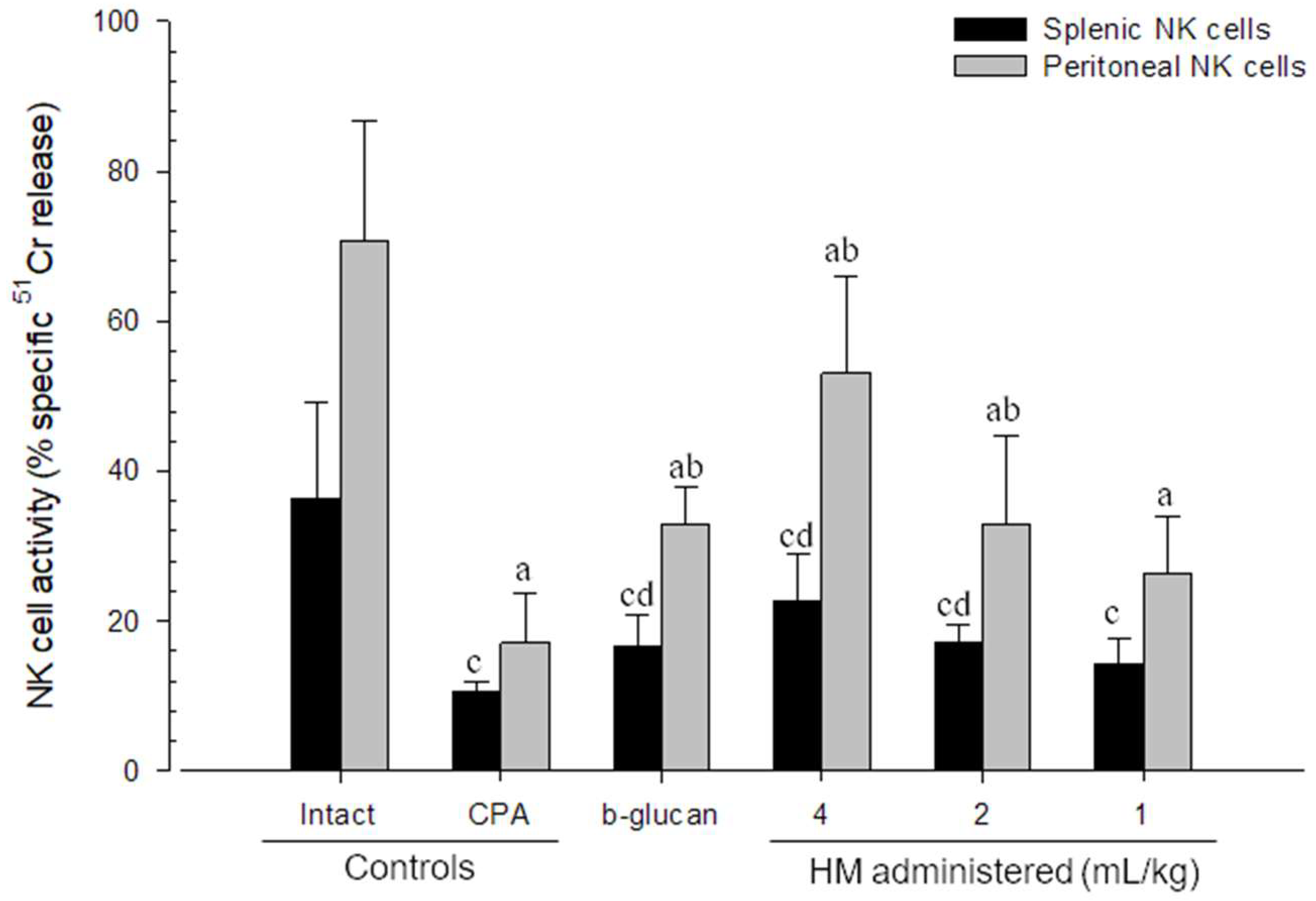
3.4. Changes in NK Cell Activities

The activity of NK cells is considered to be a marker of intrinsic functional immunity. We isolated NK cells from the spleen and peritoneal cavity of the mice and assessed their activity to determine the effects of HM on NK cell activities. Compared with the intact control mice, all CPA-treated mice were found to have significantly lower spleen and peritoneal cavity NK cell activities (p < 0.05), whereas compared with the CPA controls, those mice receiving β-glucan and 4 and 2 mL/kg HM showed significant increases in activity. Despite the fact that mice treated with the lowest dose of HM (1 mL/kg) showed no significant differences compared with the CPA control mice, we detected dose-dependent effects on CPA-induced reductions in NK cell activities in all HM-treated mice. In addition, effects comparable to those observed in 2 mL/kg HM-administered mice were recorded in those receiving 250 mg/kg β-glucan (Figure 2).
Compared with the intact control mice, there were reductions of −71.10% and −75.83% in NK cell activities in the spleen and peritoneal cavity of CPA control mice, whereas compared with the CPA control mice, we detected increases of 57.45%, 114.75%, 62.39%, and 37.18% (splenic NK cell activities) and 92.19%, 211.10%, 92.59%, and 54.70% (peritoneal NK cell activities) in the β-glucan- and 4, 2, and 1 mL/kg HM-treated mice, respectively (Figure 2).

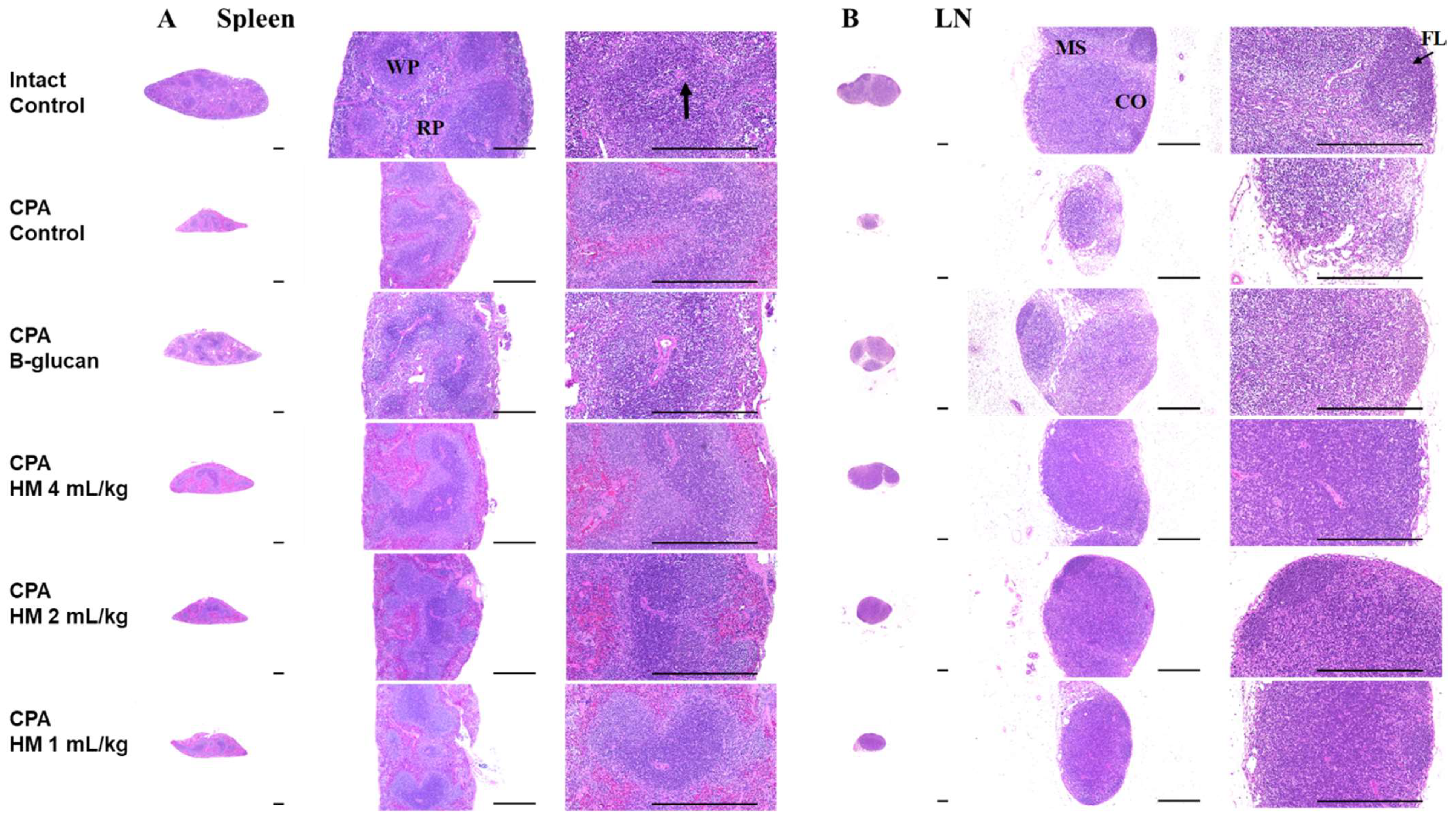
3.5. Effects on Splenic and Submandibular Lymph Node Histopathology

In this study, the effects of HM on the histomorphology of immunological organs (spleen and submandibular LN) were investigated.
Compared with the intact control mice, those treated with CPA showed atrophic changes and reductions in the numbers of white pulp lymphoid cells in the spleen, and we recorded significantly lower (p < 0.05) total and cortical thicknesses and white pulp counts, with the exception being white pulp cell counts in the 4 mL/kg HM-treated mice. However, compared with the intact controls, the experimental treatments significantly reduced these splenic atrophic alterations, with total and cortical spleen thickness and splenic white pulp cell counts in the CPA control mice being reduced by −52.53%, −52.29%, and −70.42%, respectively. Furthermore, compared with the CPA controls, we detected increases of 67.09%, 73.58%, 64.46%, and 35.69% (total spleen thickness); 48.96%, 80.46%, 51.89%, and 28.51% (cortical spleen thickness); and 33.33%, 200.00%, 142.86%, and 78.57% (white pulp cell counts) in β-glucan and 4, 2, and 1 mL/kg HM-treated mice, respectively (Table 4 and Figure 3).
Compared with the intact control mice, we detected significant reductions (p < 0.05) in total and cortical thickness and follicle numbers in the submandibular LN of CPA-treated mice, with the exception of the cortical thickness in the 4 mL/kg HM-treated mice. The experimental treatments were found to promote significant reductions in the atrophic alterations observed in the submandibular LN. Compared with the intact control mice, the total and cortical submandibular LN thickness and follicle counts in CPA control mice were reduced by −60.91%, −66.41%, and −84.44%, respectively. Furthermore, compared with the CPA control mice, we detected increases of 104.46%, 129.13%, 108.22%, and 63.41% (total submandibular LN thickness); 114.08%, 176.67%, 118.99%, and 72.72% (cortical submandibular LN thickness); and 190.48%, 342.86%, 195.24%, and 80.95% (follicle numbers) in the β-glucan- and 4, 2, and 1 mL/kg HM-treated mice, respectively (Table 4 and Figure 3).

4. Discussion

In this study, we demonstrated that treatment with the mixed herbal formula hemomine reversed CPA-induced myelosuppression and immunosuppression in a dose-dependent manner. CPA therapy was found to promote reductions in body weight, immune organ indices, hematological parameters, serum and splenic cytokine levels, and NK cell activities, thereby indicating toxicity and myelosuppressive properties [34]. However, in the CPA-treated mice subsequently administered HM, we detected dose-dependent increases in the markers reduced by CPA, which were comparable to those obtained with the positive control β-glucan, thereby indicating that these substances can attenuate the toxicity and myelosuppressive effects of CPA. Previously, polysaccharides, particularly β-glucan, have been shown to enhance immune function and are considered to have potential utility as effective agents that could be administered to ameliorate the immunosuppressive effects associated with anti-cancer, sepsis, and high-dose chemotherapy, with few side effects [28,35]. Similar to β-glucan, we established that HM has potential as a promising hematopoietic agent and immunomodulator.
Immunomodulators are substances that aid the immune system in restoring homeostasis in a beneficial way and are being developed to enhance immune responses to infection and cancer, as well as to reduce the immunological response to transplanted organs and to treat autoimmune diseases [36]. Furthermore, immunomodulators are utilized in anti-cancer chemotherapy to prevent myelosuppression and immunosuppression. Many of the currently used chemotherapeutic drugs, including CPA, can cause severe myelosuppression and immunodeficiency, which to varying extents limits the optimization of drug treatments [2]. Although CPA, which contributes to retarding tumor growth by reducing the levels of immune response effectors, is acknowledged to be particularly beneficial as a part of immunosuppressive therapy, [37], its use has been linked to a range of adverse side effects, including nausea, vomiting, panleukopenia, nonregenerative anemia, and thrombocytopenia [29,38]. Consequently, immunomodulators should ideally be co-administered to reduce or eliminate these undesirable effects. However, given that the use of currently administered immunomodulators also tends to be associated with certain side effects, including fatigue, diarrhea, and low blood cell counts, there have been concerted efforts to identify natural compounds, such polysaccharides, that can reduce myelosuppression and enhance immunological responses [39].
Among the numerous clinical hematological indicators, WBCs, RBCs, and platelets are routinely employed to assess immunological activity and infection. WBCs, which include granulocytes, monocytes, and lymphocytes, serve as biomarkers of immunological activation and infection, whereas RBCs are involved in tissue oxygenation and cell communication with platelets and macrophages, both of which play essential roles in the immune response, with the former serving as an indicator of the response to foreign chemicals [40].
Myelosuppression, also referred to as bone marrow suppression, is a reduction in bone marrow activity, which leads to a reduction in blood cell synthesis. CPA has the effect of suppressing the activity of antioxidant enzymes in the bone marrow, thereby promoting oxidative stress, and increases myelosuppression and anemia by lowering RBC, WBC, and PLT levels [41]. In the present study, we found that mice administered CPA were characterized by marked reductions in WBCs, RBCs, hemoglobin, hematocrit, and platelets. Moreover, we also detected reductions in the numbers of immune cells, such as lymphocytes and neutrophils, although the percentage of WBCs remained relatively constant. Collectively, these observations thus indicate that CPA therapy can be a cause of myelosuppressive nonregenerative anemia, panleukopenia, and thrombocytopenia. These myelosuppressive changes were, nevertheless, relieved by the subsequent administration of HM, which has previously been proven to be effective against anemia [7].
Immune cells (T cells, B cells, and macrophages) release cytokines, immunomodulating compounds that contribute to the regulation of immune responses and play key roles in immune cell development and tissue healing [42]. Among these, IFN-γ is a pro-inflammatory cytokine produced by NK and Th1 cells that causes disease-related tissue damage. However, it is also one of the cytokines that plays a role in tissue homeostasis and protection in homeostatic circumstances by inducing host defense and immunological responses and minimizing tissue damage [43]. TNF-α, produced by macrophages and Th1 cells, and IL-1β, produced by macrophages, play roles in the regulation of normal inflammatory reactions and can be utilized as biomarkers to assess inflammatory conditions [44,45]. T regulatory cell-derived IL-10 is an important regulatory cytokine that suppresses excessive T cell responses and contributes to regulating the immune response [46]. In the present study, we established that CPA treatment causes not only myelosuppression and quantitative reductions in immune-related cells but also impairs the qualitative functioning of immune systems, including a reduction in cytokine levels. Notably, however, we found that co-administration of HM had the effect of restoring these reduced cytokine levels.
We also established that CPA treatment has certain immunosuppressive effects, among which were losses in body and lymphoid organ weights, atrophic changes in lymphoid organs, and reductions in NK cell activities. The spleen and submandibular LN are essential immune organs, in the former of which the white pulp area, in particular, is involved in innate and adaptive immunity via mediation of the differentiation, development, and maturation of T and B cells, along with macrophages. Lymph nodes in the submandibular region also produce lymphocytes and filter out foreign particles that can potentially cause infections. Losses in body, spleen, and LN weights attributable to a reduction in lymphocytes and atrophy of the lymphoid organs are also used as markers of immunosuppression [44,45]. Among the different types of lymphocytes, NK cells are considered the most potent effectors in the innate immune system, identifying and killing diseased or damaged cells to control the development of malignancies and infections. However, many of the currently used immunosuppressive drugs have the effect of reducing both the numbers and activities of NK cells [47]. We nonetheless demonstrated that the administration HM had dose-dependent innate and adaptive immunological-enhancing effects with respect to gains in body and lymphoid organ weights, reducing the atrophy of immune organs, restoring NK cell activity, and ameliorating the immunosuppression attributable to CPA.
A limitation of this study is the lack of investigation into the underlying mechanisms of the effect of the herbal mixture on immunological regulation. Other polyphenols have been studied as potential immunomodulatory medicines by activating the MAPK/NF-κB signaling pathway [48]. To determine the underlying mechanism, more research is needed to determine the effect of HM on the MAPK/NF-κB signaling pathway.
Previously, it has been established that extracts of Angelicae Gigantis Radix [9], Astragalus membranaceus [14], Paeonia lactiflora [15,16], Cnidium officinale [17], and Glycyrrhiza uralensis Fischer [18,19] have potent immunomodulatory effects. Furthermore, nodakenin, the main constituent of Angelicae Gigantis Radix, has been reported to have anti-inflammatory activities [10,49]. However, in recent years, researchers have been interested in herbal mixtures incorporating numerous herbal extracts because the formulations have synergistic benefits, such as boosting the bioavailability of active compounds, improving therapeutic effects, and reducing adverse effects [50]. The benefits of Samul-Tang, a mixture of immunomodulatory herbs, have only been studied using anemic models; no research on the synergistic effect of herbal mixtures on immune modulation has been done. We discovered that HM, a mixture of immunomodulatory herbs, will have considerable promise as a potent natural immunomodulatory agent, with therapeutic applications in the treatment of a range of immune disorders, with comparatively little or no toxicity. The immunomodulatory benefits of HM may be attributable to the immunomodulatory actions of the individual herbal extracts comprising this preparation. However, this herbal mixture is thought to be synergistic, and our findings could serve as a starting point for further research into the synergistic effect of the immunomodulatory herbal mixture.

5. Conclusions

The findings of this study revealed that administration of the herbal preparation hemomine, a mixture of six immunomodulatory herbs, at three different dosages (4, 2, and 1 mL/kg) significantly alleviated cyclophosphamide-induced immunosuppression in mice. The immunomodulatory effects of 2 mL/kg hemomine were similar to those of 250 mg/kg β-glucan, while a higher dose of hemomine therapy (4 mL/kg) exhibited greater immunostimulatory effects. We accordingly believe hemomine to be a promising immunomodulator with the potential to limit the adverse effects of chemotherapeutic medications.

Author Contributions

Conceptualization and methodology: J.W.K., S.K.K. and H.-J.L.; Formal analysis: H.K., J.W.K. and S.K.K.; Writing—original draft preparation: H.K.; Writing—review and editing: H.K., Y.-K.K. and H.-J.L.; Supervision: S.K.K. and H.-J.L. All authors have read and agreed to the published version of the manuscript.

Funding

This study was supported by a grant from the National Institute of Fisheries Science in Korea (R2022066) and partly supported by the Gachon University Research Fund of 2018 (GCU-2018-0369).

Institutional Review Board Statement

The animal study protocol was approved by the Institutional Animal Care and Use Committee of Daegu Haany University (DHU2014-070, Gyeongsan, Korea).

Informed Consent Statement

Not applicable.

Conflicts of Interest

The authors declare no conflict of interest.

References

  1. Carr, C.; Ng, J.; Wigmore, T. The side effects of chemotherapeutic agents. Curr. Anaesth. Crit. Care 2008, 19, 70–79. [Google Scholar] [CrossRef]
  2. Daniel, D.; Crawford, J. Myelotoxicity from Chemotherapy. In Seminars in Oncology; Elsevier: Amsterdam, The Netherlands, 2006; pp. 4–85. [Google Scholar]
  3. López, M.M.; Valenzuela, J.E.; Álvarez, F.C.; López-Álvarez, M.R.; Cecilia, G.S.; Paricio, P.P. Long-term problems related to immunosuppression. Transpl. Immunol. 2006, 17, 31–35. [Google Scholar] [CrossRef] [PubMed]
  4. Lee, S.J.; Chinen, J.; Kavanaugh, A. Immunomodulator therapy: Monoclonal antibodies, fusion proteins, cytokines, and immunoglobulins. J. Allergy Clin. Immunol. 2010, 125, S314–S323. [Google Scholar] [CrossRef] [PubMed]
  5. Jantan, I.; Ahmad, W.; Bukhari, S.N.A. Plant-derived immunomodulators: An insight on their preclinical evaluation and clinical trials. Front. Plant Sci. 2015, 6, 655. [Google Scholar] [CrossRef]
  6. Kim, J.; You, S. Samul-tang ameliorates oocyte damage due to cyclophosphamide-induced chronic ovarian dysfunction in mice. Sci. Rep. 2020, 10, 21925. [Google Scholar] [CrossRef] [PubMed]
  7. Ku, S.K.; Kim, H.; Kim, J.W.; Kang, K.S.; Lee, H.-J. Ameliorating effects of herbal formula hemomine on experimental subacute hemorrhagic anemia in rats. J. Ethnopharmacol. 2017, 198, 205–213. [Google Scholar] [CrossRef]
  8. Kim, S.-J.; Ko, S.-M.; Choi, E.-J.; Ham, S.-H.; Kwon, Y.-D.; Lee, Y.-B.; Cho, H.-Y. Simultaneous determination of decursin, decursinol angelate, nodakenin, and decursinol of angelica gigas nakai in human plasma by uhplc-ms/ms: Application to pharmacokinetic study. Molecules 2018, 23, 1019. [Google Scholar] [CrossRef] [Green Version]
  9. Park, K.J.; Lee, B.C.; Lee, J.S.; Cho, M.H. Angelica gigas nakai extract ameliorates the effects of cyclophosphamide on immunological and hematopoietic dysfunction in mice. J. Med. Plant Res. 2014, 8, 657–663. [Google Scholar]
  10. Rim, H.-K.; Cho, W.; Sung, S.H.; Lee, K.-T. Nodakenin suppresses lipopolysaccharide-induced inflammatory responses in macrophage cells by inhibiting tumor necrosis factor receptor-associated factor 6 and nuclear factor-κb pathways and protects mice from lethal endotoxin shock. J. Pharmacol. Exp. Ther. 2012, 342, 654–664. [Google Scholar] [CrossRef]
  11. Kim, J.-H.; Jeong, J.-H.; Jeon, S.-T.; Kim, H.; Ock, J.; Suk, K.; Kim, S.-I.; Song, K.-S.; Lee, W.-H. Decursin inhibits induction of inflammatory mediators by blocking nuclear factor-κb activation in macrophages. Mol. Pharmacol. 2006, 69, 1783–1790. [Google Scholar] [CrossRef]
  12. Shehzad, A.; Parveen, S.; Qureshi, M.; Subhan, F.; Lee, Y.S. Decursin and decursinol angelate: Molecular mechanism and therapeutic potential in inflammatory diseases. Inflamm. Res. 2018, 67, 209–218. [Google Scholar] [CrossRef] [PubMed]
  13. Kim, W.-J.; Lee, M.-Y.; Kim, J.-H.; Suk, K.; Lee, W.-H. Decursinol angelate blocks transmigration and inflammatory activation of cancer cells through inhibition of pi3k, erk and nf-κb activation. Cancer Lett. 2010, 296, 35–42. [Google Scholar] [CrossRef] [PubMed]
  14. Auyeung, K.K.; Han, Q.-B.; Ko, J.K. Astragalus membranaceus: A review of its protection against inflammation and gastrointestinal cancers. Am. J. Chin. Med. 2016, 44, 1–22. [Google Scholar] [CrossRef] [PubMed]
  15. He, D.-Y.; Dai, S.-M. Anti-inflammatory and immunomodulatory effects of Paeonia lactiflora pall., a traditional chinese herbal medicine. Front. Pharmacol. 2011, 2, 10. [Google Scholar] [CrossRef] [Green Version]
  16. Wang, Q.-S.; Gao, T.; Cui, Y.-L.; Gao, L.-N.; Jiang, H.-L. Comparative studies of paeoniflorin and albiflorin from Paeonia lactiflora on anti-inflammatory activities. Pharm. Biol. 2014, 52, 1189–1195. [Google Scholar] [CrossRef]
  17. Jeong, J.B.; Hong, S.C.; Jeong, H.J.; Koo, J.S. Anti-inflammatory effects of ethyl acetate fraction from cnidium officinale makino on lps-stimulated raw 264.7 and thp-1 cells. Korean J. Plant Res. 2012, 25, 299–307. [Google Scholar] [CrossRef] [Green Version]
  18. Han, M.H.; Lee, M.H.; Hong, S.H.; Choi, Y.H.; Moon, J.S.; Song, M.K.; Kim, M.J.; Shin, S.J.; Hwang, H.J. Comparison of anti-inflammatory activities among ethanol extracts of sophora flavescens, glycyrrhiza uralensis and dictamnus dasycarpus, and their mixtures in raw 246.7 murine macrophages. J. Life Sci. 2014, 24, 329–335. [Google Scholar] [CrossRef]
  19. Park, E.; Kum, S.; Wang, C.; Park, S.Y.; Kim, B.S.; Schuller-Levis, G. Anti-inflammatory activity of herbal medicines: Inhibition of nitric oxide production and tumor necrosis factor-α secretion in an activated macrophage-like cell line. Am. J. Chin. Med. 2005, 33, 415–424. [Google Scholar] [CrossRef]
  20. Crook, T.R.; Souhami, R.L.; Mclean, A.E. Cytotoxicity, DNA cross-linking, and single strand breaks induced by activated cyclophosphamide and acrolein in human leukemia cells. Cancer Res. 1986, 46, 5029–5034. [Google Scholar]
  21. Emadi, A.; Jones, R.J.; Brodsky, R.A. Cyclophosphamide and cancer: Golden anniversary. Nat. Rev. Clin. Oncol. 2009, 6, 638. [Google Scholar] [CrossRef]
  22. Angulo, I.; de las Heras, F.G.; Garcıa-Bustos, J.F.; Gargallo, D.; Munoz-Fernández, M.A.; Fresno, M. Nitric oxide-producing cd11b+ ly-6g (gr-1)+ cd31 (er-mp12)+ cells in the spleen of cyclophosphamide–treated mice: Implications for t-cell responses in immunosuppressed mice. Blood 2000, 95, 212–220. [Google Scholar] [CrossRef] [PubMed]
  23. Xun, C.; Thompson, J.; Jennings, C.; Brown, S.; Widmer, M. Effect of total body irradiation, busulfan-cyclophosphamide, or cyclophosphamide conditioning on inflammatory cytokine release and development of acute and chronic graft-versus-host disease in h-2-incompatible transplanted scid mice. Blood 1994, 83, 2360–2367. [Google Scholar] [CrossRef] [PubMed] [Green Version]
  24. Wang, H.; Wang, M.; Chen, J.; Tang, Y.; Dou, J.; Yu, J.; Xi, T.; Zhou, C. A polysaccharide from strongylocentrotus nudus eggs protects against myelosuppression and immunosuppression in cyclophosphamide-treated mice. Int. Immunopharmacol. 2011, 11, 1946–1953. [Google Scholar] [CrossRef] [PubMed]
  25. Shruthi, S.; Vijayalaxmi, K.; Shenoy, K.B. Immunomodulatory effects of gallic acid against cyclophosphamide-and cisplatin-induced immunosuppression in swiss albino mice. Indian J. Pharm. Sci. 2018, 80, 150–160. [Google Scholar] [CrossRef] [Green Version]
  26. Nair, A.B.; Jacob, S. A simple practice guide for dose conversion between animals and human. J. Basic Clin. Pharm. 2016, 7, 27–31. [Google Scholar] [CrossRef] [Green Version]
  27. Yoo, S.-R.; Jeong, S.-J.; Shin, H.-K. Single oral dose toxicity evaluation of samul-tang, a traditional herbal formula, in crl: Cd (sd) rats. J. Korean Med. Sci. 2014, 35, 28–33. [Google Scholar] [CrossRef]
  28. Yoon, H.-S.; Kim, J.-W.; Cho, H.-R.; Moon, S.-B.; Shin, H.-D.; Yang, K.-J.; Lee, H.-S.; Kwon, Y.-S.; Ku, S.-K. Immunomodulatory effects of aureobasidium pullulans sm-2001 exopolymers on cyclophosphamide-treated mice. J. Microbiol. Biotechnol. 2010, 20, 438–445. [Google Scholar] [CrossRef]
  29. Kim, J.W.; Choi, J.-S.; Seol, D.J.; Choung, J.J.; Ku, S.K. Immunomodulatory effects of kuseonwangdogo-based mixed herbal formula extracts on a cyclophosphamide-induced immunosuppression mouse model. Evid. Based Complement. Altern. Med. 2018, 2018, 1–14. [Google Scholar] [CrossRef] [Green Version]
  30. Hotchkiss, R.; Osborne, D.; Lappas, G.; Karl, I. Calcium antagonists decrease plasma and tissue concentrations of tumor necrosis factor-alpha, interleukin-1 beta, and interleukin-1 alpha in a mouse model of endotoxin. Shock 1995, 3, 337–342. [Google Scholar]
  31. Hubbell, H.R.; Kvalnes-Krick, K.; Carter, W.A.; Strayer, D.R. Antiproliferative and immunomodulatory actions of β-interferon and double-stranded rna, individually and in combination, on human bladder tumor xenografts in nude mice. Cancer Res. 1985, 45, 2481–2486. [Google Scholar]
  32. Clark, B.D.; Bedrosian, I.; Schindler, R.; Cominelli, F.; Cannon, J.G.; Shaw, A.R.; Dinarello, C.A. Detection of interleukin 1 alpha and 1 beta in rabbit tissues during endotoxemia using sensitive radioimmunoassays. J. Appl. Physiol. 1991, 71, 2412–2418. [Google Scholar] [CrossRef] [PubMed]
  33. Levene, A. Pathological factors influencing excision of tumours in the head and neck. Part I. Clin. Otolaryngol. Allied Sci. 1981, 6, 145–151. [Google Scholar] [CrossRef] [PubMed]
  34. Kanno, T.Y.N.; Sensiate, L.A.; Paula, N.A.d.; Salles, M.J.S. Toxic effects of different doses of cyclophosphamide on the reproductive parameters of male mice. Braz. J. Pharm. Sci. 2009, 45, 313–319. [Google Scholar] [CrossRef] [Green Version]
  35. Harada, T.; Miura, N.; Adachi, Y.; Nakajima, M.; Yadomae, T.; Ohno, N. Effect of scg, 1, 3-β-d-glucan from sparassis crispa on the hematopoietic response in cyclophosphamide induced leukopenic mice. Biol. Pharm. Bull. 2002, 25, 931–939. [Google Scholar] [CrossRef] [Green Version]
  36. Bascones-Martinez, A.; Mattila, R.; Gomez-Font, R.; Meurman, J.H. Immunomodulatory drugs: Oral and systemic adverse effects. Med. Oral Patol. Oral Cir. Bucal. 2014, 19, e24–e31. [Google Scholar] [CrossRef]
  37. Peng, S.; Lyford-Pike, S.; Akpeng, B.; Wu, A.; Hung, C.-F.; Hannaman, D.; Saunders, J.R.; Wu, T.-C.; Pai, S.I. Low-dose cyclophosphamide administered as daily or single dose enhances the antitumor effects of a therapeutic hpv vaccine. Cancer Immunol. Immunother. 2013, 62, 171–182. [Google Scholar] [CrossRef]
  38. Eid, R.A.; Razavi, G.S.E.; Mkrtichyan, M.; Janik, J.; Khleif, S.N. Old-school chemotherapy in immunotherapeutic combination in cancer, a low-cost drug repurposed. Cancer Immunol. Res. 2016, 4, 377–382. [Google Scholar] [CrossRef] [Green Version]
  39. Tzianabos, A.O. Polysaccharide immunomodulators as therapeutic agents: Structural aspects and biologic function. Clin. Microbiol. Rev. 2000, 13, 523–533. [Google Scholar] [CrossRef]
  40. Zhang, J.; Zhou, H.-C.; He, S.-B.; Zhang, X.-F.; Ling, Y.-H.; Li, X.-Y.; Zhang, H.; Hou, D.-D. The immunoenhancement effects of sea buckthorn pulp oil in cyclophosphamide-induced immunosuppressed mice. Food Funct. 2021, 12, 7954–7963. [Google Scholar] [CrossRef]
  41. Botnick, L.E.; Hannon, E.C.; Vigneulle, R.; Hellman, S. Differential effects of cytotoxic agents on hematopoietic progenitors. Cancer Res. 1981, 41, 2338–2342. [Google Scholar]
  42. Zhou, Y.; Chen, X.; Yi, R.; Li, G.; Sun, P.; Qian, Y.; Zhao, X. Immunomodulatory effect of tremella polysaccharides against cyclophosphamide-induced immunosuppression in mice. Molecules 2018, 23, 239. [Google Scholar] [CrossRef] [PubMed] [Green Version]
  43. Ivashkiv, L.B. Ifnγ: Signalling, epigenetics and roles in immunity, metabolism, disease and cancer immunotherapy. Nat. Rev. Immunol. 2018, 18, 545–558. [Google Scholar] [CrossRef] [PubMed]
  44. Yu, F.; Zhang, Z.; Ye, S.; Hong, X.; Jin, H.; Huang, F.; Yang, Z.; Tang, Y.; Chen, Y.; Ding, G. Immunoenhancement effects of pentadecapeptide derived from cyclina sinensis on immune-deficient mice induced by cyclophosphamide. J. Funct. Foods 2019, 60, 103408. [Google Scholar] [CrossRef]
  45. Kaneko, N.; Kurata, M.; Yamamoto, T.; Morikawa, S.; Masumoto, J. The role of interleukin-1 in general pathology. Inflamm. Regen. 2019, 39, 12. [Google Scholar] [CrossRef] [Green Version]
  46. Couper, K.N.; Blount, D.G.; Riley, E.M. Il-10: The master regulator of immunity to infection. J. Immunol. 2008, 180, 5771–5777. [Google Scholar] [CrossRef]
  47. Pontrelli, P.; Rascio, F.; Castellano, G.; Grandaliano, G.; Gesualdo, L.; Stallone, G. The role of natural killer cells in the immune response in kidney transplantation. Front. Immunol. 2020, 11, 1454. [Google Scholar] [CrossRef]
  48. Liu, Y.; Wu, X.; Jin, W.; Guo, Y. Immunomodulatory effects of a low-molecular weight polysaccharide from enteromorpha prolifera on raw 264.7 macrophages and cyclophosphamide-induced immunosuppression mouse models. Mar. Drugs 2020, 18, 340. [Google Scholar] [CrossRef]
  49. Lim, J.-Y.; Lee, J.-H.; Yun, D.-H.; Lee, Y.-M.; Kim, D.-K. Inhibitory effects of nodakenin on inflammation and cell death in lipopolysaccharide-induced liver injury mice. Phytomedicine 2020, 81, 153411. [Google Scholar] [CrossRef]
  50. Zhou, X.; Seto, S.W.; Chang, D.; Kiat, H.; Razmovski-Naumovski, V.; Chan, K.; Bensoussan, A. Synergistic effects of chinese herbal medicine: A comprehensive review of methodology and current research. Front. Pharmacol. 2016, 7, 201. [Google Scholar] [CrossRef] [Green Version]
Figure 1. Study experimental design. In this study, we examined the effects of treatments on six groups comprising 10 mice each. In the five groups initially treated with CPA, mice were intraperitoneally administered 150 and 110 mg/kg of body weight CPA on days 3 and 1 day prior to oral treatment. The experimental compounds were orally administered four times at 12-h intervals, commencing on the day following the second CPA administration. Control mice without initial CPA treatment were intraperitoneally administered an identical volume of sterile saline. CPA: Cyclophosphamide, HM: Hemomine (60% LJG305).
Applsci 12 04935 g001
Figure 2. NK cell activities in response to hemomine treatment in intact or CPA-induced immunosuppressed mice. The results are expressed as the means ± SDs (n = 10). NK: Natural killer, CPA: Cyclophosphamide, HM: Hemomine (60% LJG305). Different letters above bars indicate a significant difference at the p < 0.05 level compared with the a intact control and b CPA-treated control mice, as determined using the LSD test, and with the c intact control and d CPA-treated control mice using the Mann–Whitney test.
Applsci 12 04935 g002
Figure 3. Representative histopathological images of the spleen and left submandibular LN from intact or CPA-induced immunosuppressed mice. All hematoxylin–eosin stained. CPA: Cyclophosphamide, HM: Hemomine (60% LJG305), LN: Lymph node. Scale bar: 400 µm. (A) Splenic histopathology. WP: white pulp, RP: red pulp, Arrow: central arteriole. (B) LN histopathology. CO: cortex; MS: medullary sinus, FL: follicle.
Applsci 12 04935 g003
Table 1. Changes in weight gain and relative organ weights following hemomine treatment in intact or CPA-induced immunosuppressed mice.
GroupWeight Gain 1 (g)Relative Organ Weight 2 (% of Body Weights)
SpleenLN
Intact Control3.35 ± 0.620.297 ± 0.0500.057 ± 0.016
CPA-Control−2.19 ± 0.89 a0.124 ± 0.030 a0.014 ± 0.007 a
CPA-β-glucan0.48 ± 0.83 ab0.184 ± 0.038 ab0.024 ± 0.007 ab
CPA-HM 4 mL/kg1.13 ± 0.86 ab0.201 ± 0.032 ab0.037 ± 0.009 ab
CPA-HM 2 mL/kg0.63 ± 1.22 ab0.186 ± 0.032 ab0.027 ± 0.008 ab
CPA-HM 1 mL/kg−0.40 ± 0.66 ab0.167 ± 0.020 ab0.022 ± 0.006 a
Values are expressed as the means ± SDs (n = 10). 1 Weight gain (g) = Body weight at sacrifice–Body weight on the initial CPA treatment. 2 Relative organ weight (%) = Organ weight/Body weight at sacrifice × 100. CPA: Cyclophosphamide, HM: Hemomine (60% LJG305), LN: Submandibular lymph node, left side. Different superscript letters indicate a significant difference at p < 0.05 compared with the a intact control and b CPA-treated control mice, as determined using the LSD test.
Table 2. Hematological values following hemomine treatment in intact or CPA-induced immunosuppressed mice.
Groups ItemsControlsReferenceHM
IntactCPAβ-Glucan4 mL/kg2 mL/kg1 mL/kg
WBC (K/μL)9.02 ± 3.330.31 ± 0.11 c0.79 ± 0.12 cd1.24 ± 0.26 cd0.82 ± 0.19 cd0.56 ± 0.13 cd
LYM (%)77.49 ± 7.3176.10 ± 5.2177.15 ± 8.4976.38 ± 5.6876.79 ± 7.2775.74 ± 6.48
NEU (%)15.92 ± 5.5617.69 ± 4.9416.47 ± 8.4217.07 ± 5.1917.00 ± 5.3018.01 ± 6.48
MON (%)3.94 ± 1.273.86 ± 1.273.85 ± 1.843.94 ± 1.573.85 ± 2.033.89± 1.86
EOS (%)0.93 ± 0.900.96 ± 0.840.97 ± 0.450.99 ± 0.660.97 ± 0.590.99 ± 0.47
BAS (%)0.81 ± 0.700.82 ± 0.500.81 ± 0.600.78 ± 0.460.81 ± 0.580.83 ± 0.36
RBC (M/μL)9.68 ± 0.794.92 ± 0.73 a7.14 ± 0.64 ab8.25 ± 1.19 ab7.18 ± 0.51 ab6.53 ± 0.59 ab
HGB (g/dL)18.10 ± 1.1912.87 ± 1.91 a17.00 ± 0.89 ab17.76 ± 1.16 b17.02 ± 0.61 ab15.90 ± 0.95 ab
HCT (%)44.08 ± 2.1538.64 ± 2.60 a42.27 ± 0.99 ab44.26 ± 2.65 b41.99 ± 1.33 ab41.02 ± 0.88 ab
MCV (fl)52.04 ± 2.9951.62 ± 4.0651.23 ± 3.2351.33 ± 2.4452.08 ± 1.9751.77 ± 2.37
MCH (pg)18.20 ± 0.9117.81 ± 1.4017.75 ± 1.0717.77 ± 1.0317.92 ± 1.3417.80 ± 1.34
MCHC (g/dL)19.61 ± 1.7319.56 ± 1.5019.48 ± 1.5119.64 ± 1.3619.73 ± 1.2319.41 ± 1.16
PLT (M/μL)1665.4 ± 217.6881.8 ± 155.3 a1204.8 ± 97.6 ab1407.7 ± 111.6 ab1209.6 ± 174.7 ab1079.6 ± 120.6 ab
Values are expressed as the means ± SDs (n = 10). CPA: Cyclophosphamide, HM: Hemomine (60% LJG305), WBC: white blood cells, LYM: lymphocytes, NEU: neutrophils, MON: monocytes, EOS: eosinophils, BAS: basophils, RBC: red blood cells, HGB: hemoglobin, HCT: hematocrit, MCV: mean corpuscular volume, MCH: mean corpuscular hemoglobin, MCHC: mean corpuscular hemoglobin concentration, PLT: platelets. Different letters indicate significant differences at p < 0.05 level compared with the a intact control and b CPA-treated control mice, as determined using the LSD test, and with the c intact control and d CPA-treated control mice using the Mann–Whitney test.
Table 3. Changes in the serum and splenic cytokine contents following hemomine treatment in intact or CPA-induced immunosuppressed mice.
SerumSpleen
GroupsIFN-γ (pg/mL)TNF-α (pg/mg Protein)IL-1β (pg/mg Protein)IL-10 (pg/mg Protein)
Intact Control276.08 ± 38.51109.91 ± 28.3066.45 ± 10.0499.46 ± 22.61
CPA Control75.15 ± 21.29 a32.15 ± 10.06 c19.92 ± 10.54 a32.42 ± 10.20 a
CPA-β-glucan147.35 ± 32.12 ab63.30 ± 12.65 cd39.26 ± 10.35 ab62.14 ± 11.26 ab
CPA-HM 4 mL/kg183.88 ± 33.80 ab77.64 ± 17.22 cd48.67 ± 10.23 ab78.06 ± 16.76 ab
CPA-HM 2 mL/kg148.37 ± 14.65 ab63.43 ± 13.12 cd39.28 ± 14.32 ab62.97 ± 19.12 ab
CPA-HM 1 mL/kg104.65 ± 22.03 ab52.98 ± 11.37 cd33.79 ± 10.46 ab47.71 ± 11.01 ab
Values are expressed as the means ± SDs (n = 10). CPA: Cyclophosphamide, HM: Hemomine (60% LJG305). Different letters indicate significant differences at the p < 0.05 level compared with the a intact control and b CPA-treated control mice, as determined using the LSD test, and with the c intact control and d CPA-treated control mice using the Mann–Whitney test.
Table 4. Histomorphometrical analysis of response to hemomine treatment in intact or CPA-induced immunosuppressed mice.
Groups ItemsControlsReferenceHM
IntactCPAβ-Glucan4 mL/kg2 mL/kg1 mL/kg
Spleen thickness
Total (μm)1762.17 ± 161.91836.47 ± 178.99 a1397.67 ± 194.24 ab1451.95 ± 193.96 ab1375.67 ± 160.47 ab1134.98 ± 250.88 ab
Cortex (μm)414.55 ± 52.99197.79 ± 29.12 a294.63 ± 36.89 ab356.94 ± 41.23 ab300.42 ± 41.84 ab254.18 ± 44.89 ab
WP number (/mm2)14.20 ± 2.154.20 ± 0.92 c9.80 ± 1.32 cd12.60 ± 1.43 d10.20 ± 1.40 cd7.50 ± 0.71 cd
LN thickness
Total (μm)1148.41 ± 110.11448.88 ± 115.39 a917.77 ± 104.45 ab1028.52 ± 127.04 ab934.67 ± 105.72 ab733.50 ± 182.62 ab
Cortex (μm)833.29 ± 118.01279.90 ± 101.44 a599.21 ± 119.30 ab774.40 ± 132.92 b612.95 ± 137.40 ab483.44 ± 128.17 ab
Follicle number (/mm2)13.50 ± 2.802.10 ± 1.20 c6.10 ± 1.20 cd9.30 ± 2.45 cd6.20 ± 1.69 cd3.80 ± 1.03 cd
Values are expressed as the mean ± SD (n = 10). CPA: Cyclophosphamide, HM: Hemomine (60% LJG305), WP: White pulp, LN: Submandibular lymph node, left side. Different letters indicate a significant difference at the p < 0.05 level compared with the a intact control and b CPA-treated control mice, as determined using the LSD test, and with the c intact control and d CPA-treated control mice using the Mann–Whitney test.
Publisher’s Note: MDPI stays neutral with regard to jurisdictional claims in published maps and institutional affiliations.

