Article

Dependence of Electric Pulse Mediated Growth Factor Release on the Platelet Rich Plasma Separation Method

1
GE Research, Niskayuna, NY 12309, USA
2
School of Nuclear Engineering, Purdue University, West Lafayette, IN 47906, USA
3
Elmore Family School of Electrical and Computer Engineering, Purdue University, West Lafayette, IN 47907, USA
4
Department of Agricultural and Biological Engineering, Purdue University, West Lafayette, IN 47907, USA
*
Authors to whom correspondence should be addressed.
Appl. Sci. 2022, 12(10), 4965; https://doi.org/10.3390/app12104965
Submission received: 21 April 2022 / Revised: 9 May 2022 / Accepted: 12 May 2022 / Published: 14 May 2022
(This article belongs to the Special Issue Biomedical Applications of Pulsed Power and Plasmas)

Abstract

:
Platelet rich plasma (PRP) has been explored for multiple clinical applications, including dentistry, orthopedics, sports medicine, diabetic foot ulcers, and cosmetic treatments. Topical applications of PRP typically use thrombin to induce platelet activation, which is accompanied by growth factor release and clotting of the PRP, prior to treatment. Injectable PRP treatments typically use non-activated PRP under the assumption that collagen at the site of the injury mediates platelet activation to ensure growth factor release in vivo. Ex-vivo electrical stimulation of platelets is emerging as a robust, easy to use, instrument-based PRP activation technique to facilitate growth factor release with or without clotting, while providing tunability of growth factor release, clot mechanical properties (when desired), and serotonin release from the dense granules. This paper briefly reviews the key results of the electrical activation of platelets and demonstrates successful growth factor release by electrical ex-vivo stimulation without clotting for three types of PRP separated from whole blood using available commercial kits: Harvest, EmCyte and Eclipse. While these three types of PRP feature a wide range of platelet and red blood cell content compared to whole blood, we demonstrate that pulsed electric fields enable growth factor release for all these biological matrices generated using whole blood from four human donors. These experiments open opportunities for using electrically stimulated PRP with released growth factors without clotting for injectable platelet treatments in relevant clinical applications.

1. Introduction

Platelet rich plasma (PRP) is an autologous biological matrix containing growth factors and proteins that are important for the wound healing cascade and tissue regeneration [1,2,3,4,5,6]. Clinical workflows leveraging PRP for wound healing use either the topical path or the injectable path. Both workflows entail drawing whole blood from the patient and separating the PRP, typically using a commercially available kit. The topical path involves activation with a mix of thrombin and calcium chloride. While bovine thrombin (BT) is most commonly utilized in the clinic, other alternatives exist, such as recombinant thrombin, human thrombin from donor plasma, and autologous thrombin. Thrombin triggers growth factor release and clotting. Injectable treatments typically use non-activated PRP.
To date, clinical exploration using PRP includes diabetic foot ulcers [3], cardiac surgery [4], orthopedics and sports medicine [7], and, increasingly, cosmetic applications [8,9,10,11,12]. For diabetic foot ulcers, activated and clotted PRP (“platelet gel”) is typically applied on the wound, while most orthopedic, sports medicine, and cosmetic applications use injectable, non-activated PRP. It is hypothesized that in injectable workflows, platelets are activated by the collagen at the site of the injury.
Various challenges with bovine thrombin activation [13,14,15,16,17], including lack of standardization, expense, potential side effects, and workflow complexity, have motivated exploration into alternative ex vivo platelet activation methods. Pulsed electric fields (PEFs), which have been applied for multiple applications in cancer treatment [18] and microorganism inactivation [19], have also been proposed for platelet activation [18] with promising results [19,20,21,22,23,24,25,26,27,28,29,30,31,32]. PEFs can facilitate growth factor release from PRP with or without clotting [25,28,29,32]—a unique capability that cannot be matched by thrombin. Another exciting discovery has been the ability to tune the level of growth factor release [28,29,32,33] and the mechanical properties of the clot when clotting with growth factor release was desired [28]. PEFs also provide the capability to tune the serotonin release from platelet dense granules [32] and induce differential growth factor release during PEF activation [28]. PEFs facilitate physiological activation of platelets and not just mechanical fragmentation of platelets [28]. Since there is no current standardization for bovine thrombin activation, an instrument-based PEF platelet activator [22,23,24,27] would provide the first standardized PRP activation method, which could facilitate clinical protocols for the numerous topical and injectable applications described above.
PEF platelet activation was successfully tested for in vitro cell proliferation assays [23,24,26] and in animal wound healing studies [34,35,36]. It should be mentioned that electrical ex-vivo activation of platelets is currently explored not only for wound healing and tissue regeneration, but also for cell culture [37].
Most previous work in PEF platelet activation used Harvest PRP [21,22,23,24,25,26,27,28,29,30,32] or whole blood [31], which was tested since some clinicians have started to inject whole blood instead of non-activated PRP to achieve comparable clinical benefits [38,39,40,41]. Since there are many PRP separation devices available commercially, it is of great interest to test the activation of other types of biological matrices besides the Harvest PRP or whole blood. In this paper, we report the first experimental demonstration of PEF activation of three different types of PRP, separated from the whole blood of four human donors by three commercial kits (Harvest, Eclipse and EmCyte).
Some of the most interesting clinical application of platelets use injectable non-activated PRP for orthopedic or cosmetic applications. In the study presented here, we treated the three PRPs with a PEF activation protocol designed to trigger growth factor release without clotting. Essentially, we exposed PRP separated from whole blood to PEFs with no calcium chloride added. With electrical activation, we propose the injection of a biological matrix with already released growth factors at a known level, as opposed to injecting non-activated PRP, under the hypothesis that the collagen at the injury site would activate the injected platelets.
We emphasize that we have demonstrated several methods for using PEFs to induce growth factor release without clotting. Applying specific PEFs to PRP separated from whole blood [25] or using specific combinations of PEF parameters with different concentrations of CaCl2 in the PRP [28,29,32] can induce growth factor release without clotting. These experimental protocols facilitate obtaining PRP based biological matrices with tunable growth factor release that could be tested clinically in applications requiring injectable platelet based therapeutic mixes.
We will further show that reducing the pulse duration from 5 μs to 2 μs has a negligible effect on growth factor release. While initial experiments assumed the need for very short electric pulses for platelet activation (pulse durations of tens to hundreds of nanoseconds) [20], subsequent studies have demonstrated that a wide range of PEFs durations may be used with excellent results [24,28,29,30,31,32]. Microsecond PEF generators provide a practical path toward deploying simple, low-cost instruments for PEF based platelet activation to the clinic because they are typically easier and less expensive to build and mitigate potential safety concerns by using lower voltages. In fact, recent work showed that extending PEF duration by more than two orders of magnitude [30] compared to the initial research [20] can still facilitate platelet activation [30]. The same study [30] highlighted the opportunity of using appropriate PEFs to obtain growth factor release profiles similar to, and even greater than, what may be achieved using bovine thrombin.
Section 2 summarizes the materials and methods used in these experiments. Section 3 summarizes the results. We discuss the results in Section 4 and provide concluding remarks in Section 5.

2. Materials and Methods

2.1. PRP Systems and Protocols

We used three commercially available PRP systems to prepare PRP. The first PRP system was the Harvest SmartPrep2 System (Harvest Technologies, Plymouth, MA, USA), using either one or two 60 mL process disposables. Briefly, 8 mL of anticoagulant citrate dextrose A (ACD-A) was drawn into the blood draw syringe, after which 2 mL was transferred to the sterile closed SmartPreP2 process disposable through the white resealable injection port into the plasma chamber of the disposable. We next added 54 mL of the subject’s blood to the remaining 6 mL of ACD-A in the blood draw syringe. The blood was then transferred to the SmartPreP2 process disposable through the red resealable injection port into the SmartPreP2 process disposable, and the cartridge was placed in the automated Harvest centrifuge. During spinning, a floating shelf in the cartridge compartment with the blood rises to just below the buffy coat/red blood cell interface, allowing automated decanting of platelets, plasma and white blood cells into the plasma compartment of the cartridge for subsequent recovery. Approximately 10 mL of PRP was withdrawn from each process disposable (i.e., the yellow spacer was not removed), and the resultant PRP was tested for platelet concentration and pooled prior to further treatment when more than one disposable was used.
The second PRP system was the Eclipse PRP (Eclipse Medical, The Colony, TX, USA), which used up to four of the 22 mL platelet separation tubes per donor, while following the manufacturer’s protocol (including removing ~75% of the platelet-poor plasma). Briefly, 20 mL of the subject’s blood was drawn directly into a 22 mL platelet separation tube containing 2 mL of MNC7 (Eclipse Medical, The Colony, TX, USA), a proprietary anticoagulant, and a thixotropic separator gel. The tube was then spun in a centrifuge with a relative centrifugal force (RCF) of 1500 for 10 min and a braking time of 30–45 s. Upon completing centrifugation, we removed the platelet poor plasma (PPP) fraction by withdrawing ~75% of the plasma fraction. The platelets were then resuspended by gently inverting/shaking the tube between one and seven times. Approximately 3–6 mL of PRP was withdrawn from each tube, tested for platelet concentration, and pooled prior to further treatment.
The third PRP system was the EmCyte PurePRP II (EmCyte Corporation, Fort Myers, FL, USA), using the 60 mL concentrating device and following GS60 Pure II Protocol A. Briefly, 10 mL of sodium citrate anticoagulant was first drawn into a 60 mL syringe, followed by 50 mL of subject blood. The anticoagulated whole blood was then transferred into the concentrating device and placed into the centrifuge for an initial spin of 1.5 min at 3800 RPM. Using the 60 mL syringe, the platelet plasma suspension was withdrawn from the concentrating device and transferred to the concentrating accessory (a second device). The platelet plasma suspension was then spun in the centrifuge a second time for 5 min at 3800 RPM. After centrifugation, the platelet poor plasma was withdrawn from the concentrating accessory, leaving approximately 7 mL of PRP, which we then withdrew from the concentrating accessory, tested for platelet concentration, and pooled prior to further treatment.

2.2. Donors, Blood Collection and Preparation of PRP

This study was conducted at the Pennsylvania Biotechnology Center and all subjects provided informed consent. Blood was collected from four healthy volunteers aged ≥18 years with platelet counts in the normal range (150,000 to 450,000 µL−1). For each donor, three separate draws were conducted by a nurse phlebotomist, one for each of the three PRP systems. Obtaining at least 10 mL of testable PRP for each PRP system typically required more than one collection device. The Harvest, EmCyte, and Eclipse each required two, two, and four collection devices, respectively. In addition, a 3 mL whole blood sample was collected from each volunteer for each PRP preparation for complete blood cell analysis, and a 12 mL whole blood sample (4 × 3 mL) was collected for baseline growth factor analysis (none of which were anticoagulated). Complete blood cell counts were performed on the whole blood and the concentrated PRP using a Horiba ABX Micros 60 hematology analyzer.

2.3. PRP Study Design

PRP activation by PEF (conditions described below) and bovine thrombin (100 NIH units/µL final concentration in a 0.1% bovine serum albumin (BSA) solution, Sigma-Aldrich Co., St. Louis, MO, USA) were evaluated. Whole blood was activated only with bovine thrombin. Activation by bovine thrombin was achieved by adding 10% by volume to the unactivated PRP or whole blood (e.g., 130 µL bovine thrombin was added to 1.3 mL unactivated PRP or whole blood in a microcentrifuge tube), yielding a final bovine thrombin concentration of 10 NIH units/µL. Unactivated PRP and whole blood was also used as a negative control. Endpoints included growth factors released from platelets into the supernatant (EGF, PDGF-AA, VEGF, and soluble P-selectin). All endpoints were measured in supernatant samples taken 15 ± 2 min after activation, which were created by transferring the PRP and whole blood samples to microcentrifuge tubes and spinning them in an Eppendorf microcentrifuge at 2000 G for 10 min at room temperature. The supernatant samples were then stored in a −80 °C freezer until analysis.

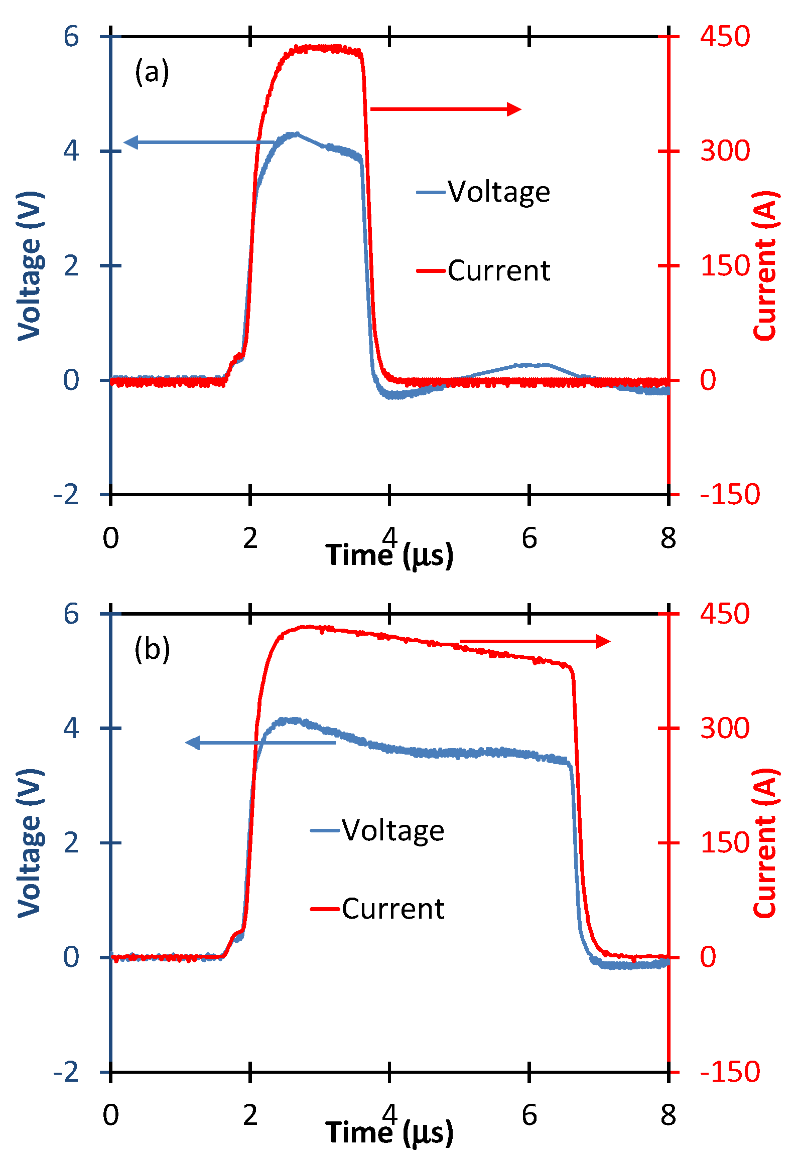
2.4. PEF Stimulation of PRP

PEF stimulation of PRP was performed using a specially designed instrument prototype (GE Research, Niskayuna, NY, USA) [27]. The instrument accounts for the specific electrical properties of the PRP, which is typically more conductive than the standard buffers used in electroporation. Concentrated PRP (400 µL) was placed in a 2 mm electroporation cuvette (Molecular BioProducts, San Diego, CA, USA) and exposed to two conditions: PEF A and PEF F (Figure 1). During exposure to PEFs, it is critical to fill the area between the electrodes in the cuvettes with liquid sample to mitigate arcing or flashover [24,27] while avoiding overfilling to minimize the amount of sample not between the electrodes that will be untreated. We applied a single electric pulse to the cuvette for PRP stimulation. We selected the 5 μs pulse duration as the baseline based on prior results [28] and the 2 μs pulse duration to assess changes due to reducing pulse duration. We used a Tektronix DPO 4140B-L oscilloscope to measure the voltage pulses applied to the cuvettes and included a Pearson 110 current monitor to simultaneously measure the current across the cuvette.

2.5. Growth Factor Release

Levels of EGF, PDGF-AA, VEGF and soluble P-selectin [see the soluble P-selectin results in Appendix A] in the supernatants of the treated PRP were measured in duplicate using commercially available ELISA kits (EGF, VEGF and P-selectin from R&D Systems, Minneapolis, MN, USA; PDGF-AA, RayBiotech, Norcross, GA, USA).

2.6. Statistical Analysis
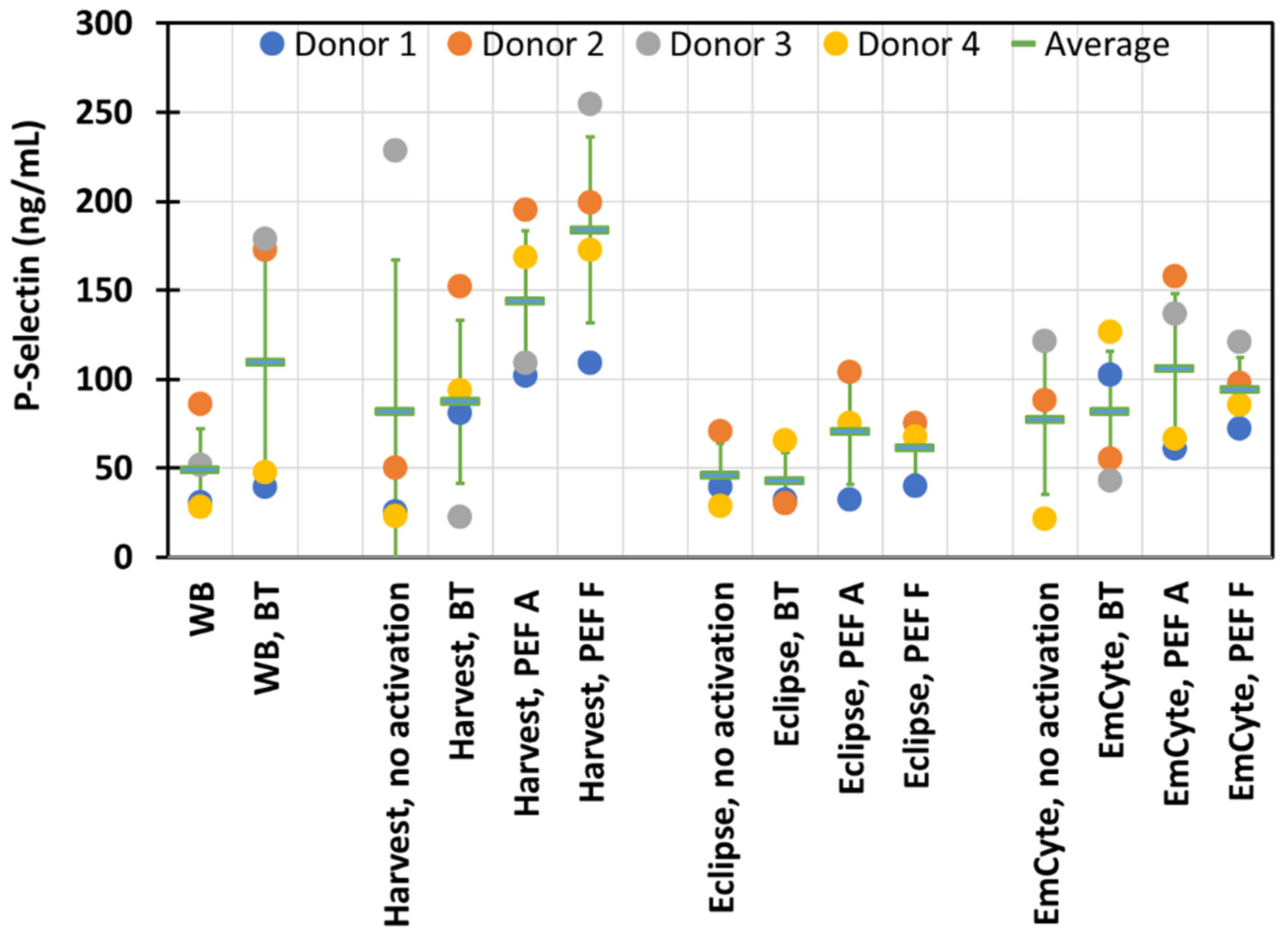
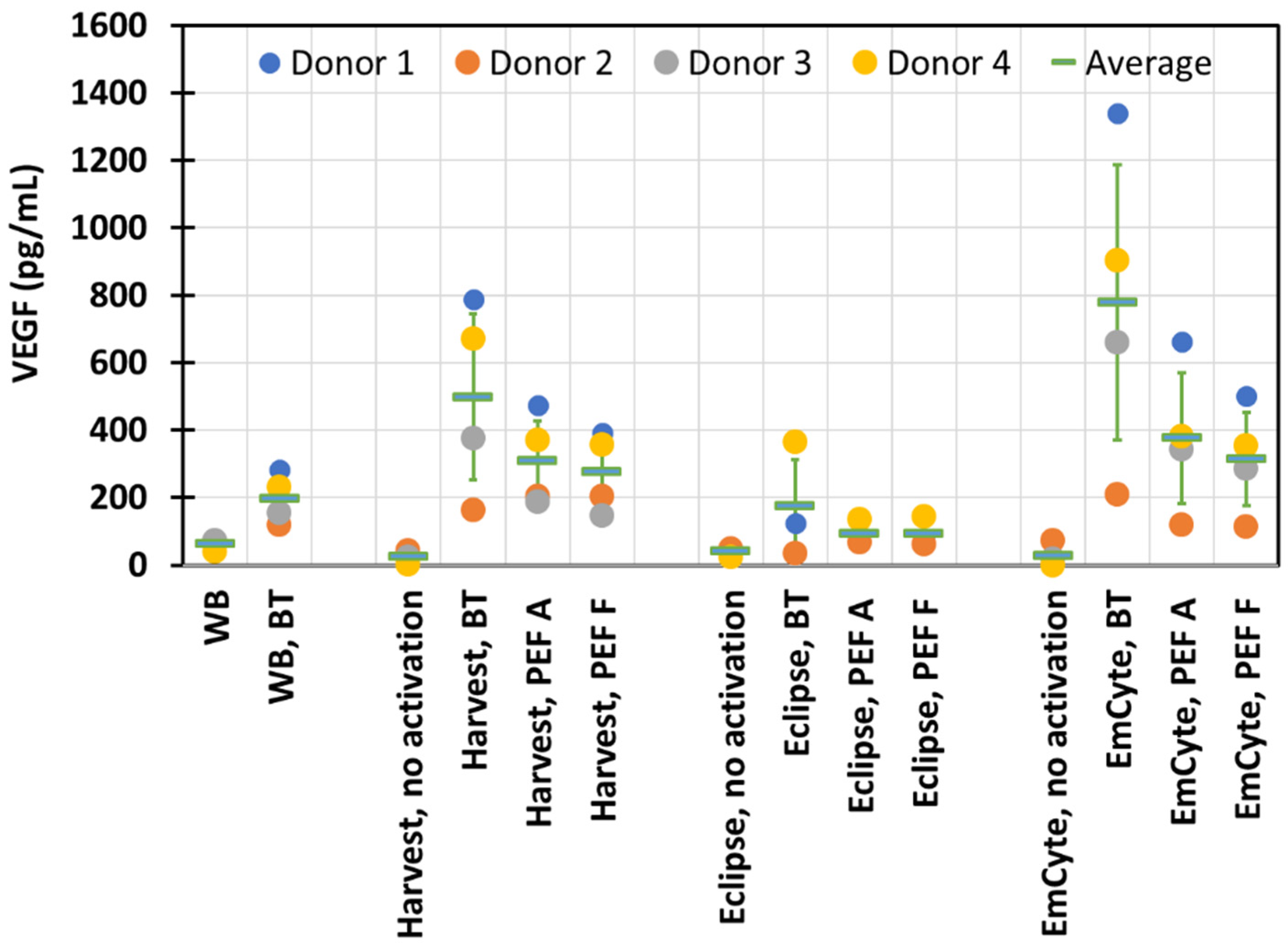
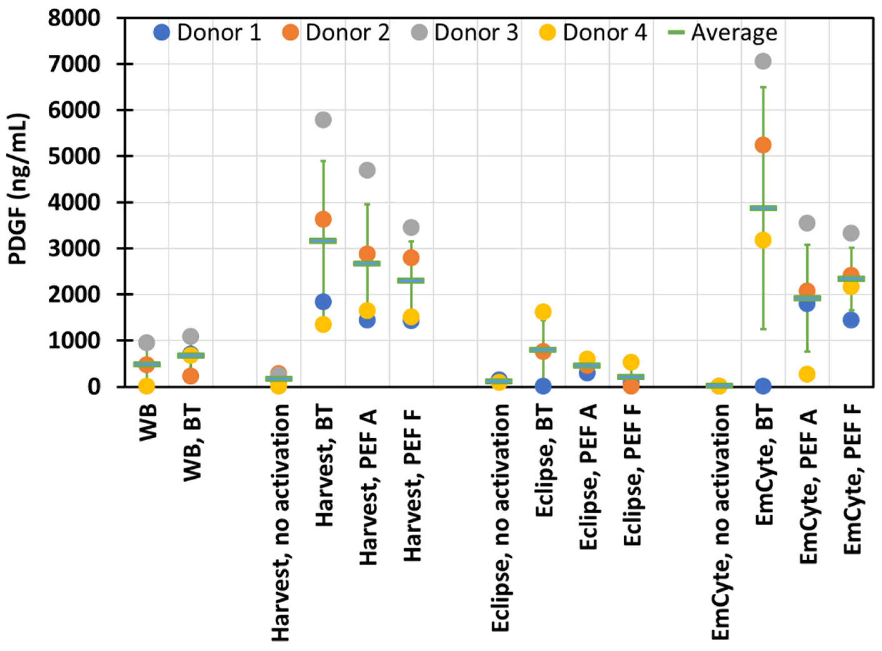
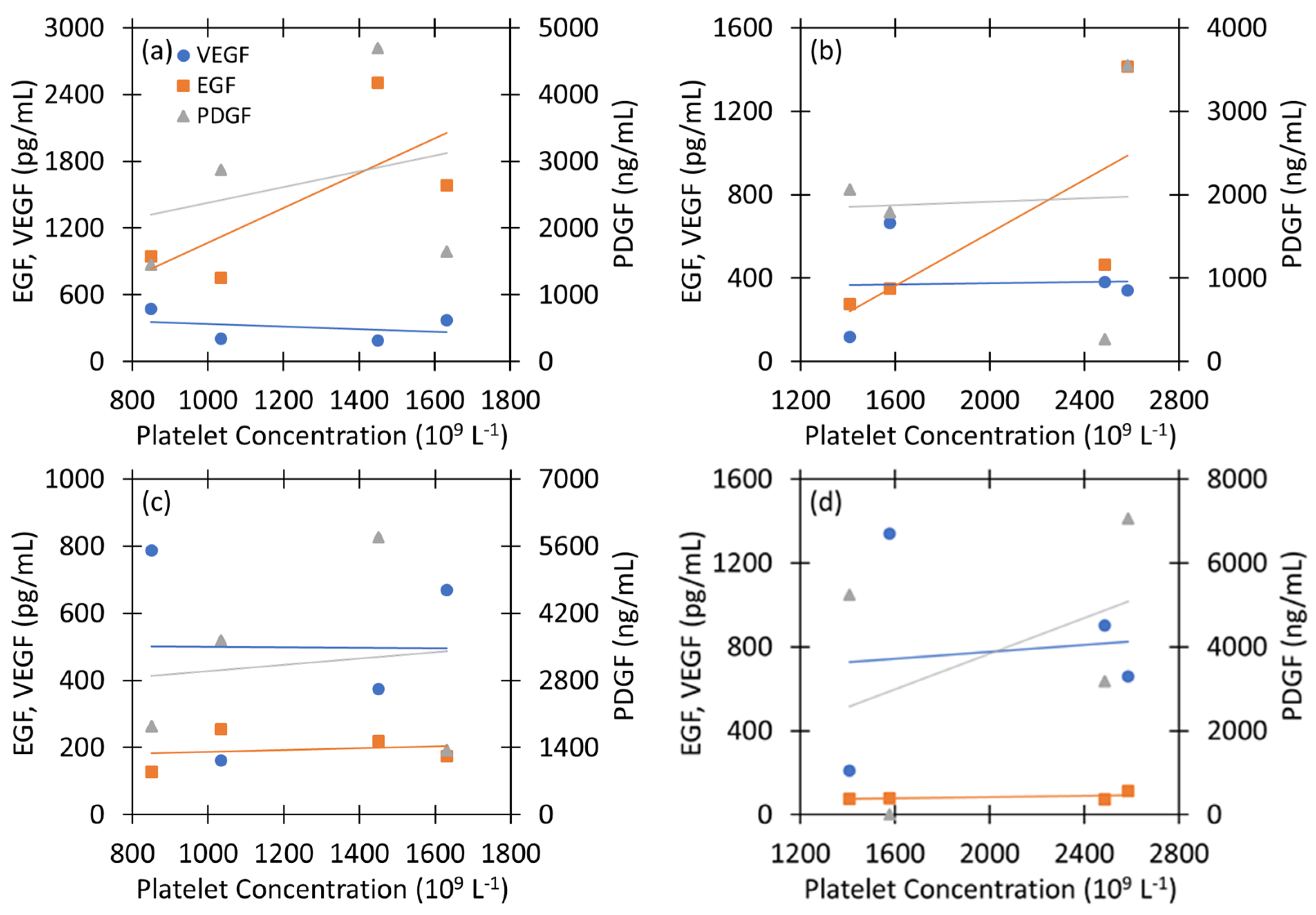
Data were analyzed using a four-parameter logistic curve (4PL) using MyAssays software (MyAssays Ltd., Brighton, UK). The average and standard deviation for each data point was calculated and plotted.

3. Results

Table 1 shows the average composition of the whole blood for all donors and the average PRP composition for each of the three systems examined in this study. While Harvest PRP is “red”, indicating a significant concentration of red blood cells, Eclipse and EmCyte PRPs are “white”, indicating minimum RBC content. In terms of platelet concentration compared to whole blood, EmCyte PRP had the highest concentration while Eclipse PRP had the lowest concentration from the three PRPs.
Figure 1 shows representative voltage and current waveforms for the two PEF waveforms used in this study for electrical stimulation: PEF A (peak voltage ~4.3 kV and pulse duration 2 μs) and PEF F (peak voltage ~3.5 kV and pulse duration 5 μs).
Figure 2, Figure 3 and Figure 4 show the release of epidermal growth factor (EGF), vascular endothelial growth factor (VEGF), and platelet-derived growth factor (PDGF), respectively. For each type of PRP, growth factors are presented for PEFs A and F compared to negative control (no activation) and positive control (activation with bovine thrombin). Also, growth factor release for whole blood under baseline (no activation) and bovine thrombin activation are included for comparison.
Finally, we note that the Eclipse growth factor release data include measurements only for donors 1, 2 and 4—donor 3 measurements were not included here due to challenges with PRP separation for this donor.
We next assess the correlation of the various growth factors with the donor platelet levels and each other. Figure 5 assesses the linear correlation of EGF, VEGF, and PDGF release for Harvest and EmCyte PRPs treated with either PEF A or bovine thrombin (BT) with platelet count for the four donors. We selected PEF A because Figure 2, Figure 3 and Figure 4 show that the general trends in the growth factors are relatively similar for both PEF waveforms. We did not consider Eclipse PRPs because we only had data for three donors rather than the complete set of four donors that we had for the other two types of PRP. Table 2 reports the correlation coefficients and p-values for the growth factors with the donor platelet levels and each other.
For the PEF A combinations, the strongest positive correlation is with EGF, while PDGF slightly increases and VEGF slightly decreases for Harvest and remains approximately constant for EmCyte. Particularly interesting, Table 2 suggests that PDGF and VEGF are constant with increasing donor platelet concentration for EmCyte. Table 2 also indicates that PDGF and VEGF are negatively correlated, while PDGF and EGF are positively correlated for Harvest; EGF and VEGF are negatively correlated, but less strongly than PDGF with either VEGF or EGF. For EmCyte with PEF A, the correlations are generally weaker with only EGF and donor platelet concentration and EGF and PDGF exhibiting strong positive correlation. Although there is insufficient data to make a strong conclusion, as indicated by the p-values in Table 2, this data suggests that one may be able to select a platelet concentration method to tune growth factors relative to each other and with donor platelet concentration when using PEF activation.
For the BT treatment of the two PRPs highlighted in Figure 5, EGF appears fairly constant with increasing platelet concentration for both Harvest (a slope of 0.027 pg/mL EGF/(109 L−1 platelets) and R2 = 0.0306) and EmCyte (0.0166 pg/mL/(109 L−1 platelets) and R2 = 0.3122) PRPs; Table 2 shows that it has a lower positive correlation with Harvest than with EmCyte. VEGF is nearly constant for both PRP based on Figure 5 and the correlation factor in Table 2. Similarly, PDGF has slight positive correlation factors for both PRPs. All three growth factors have higher positive correlations with donor platelet concentrations with the EmCyte PRP than the Harvest PRP when using BT activation. EGF and VEGF have a strong negative correlation (correlation factor of −0.99) for Harvest PRP and BT, while both Harvest and EmCyte PRPs have a strong negative correlation between VEGF and PDGF and positive correlation between EGF and PDGF. As in the PEF case, only the EGF and VEGF case with Harvest PRP and BT was statistically significant, suggesting that additional studies are required to achieve statistical significance to make more definitive claims concerning these observations.

4. Discussion

This experimental study explored the effect of PEF treatment for three different types of PRPs. While our previous platelet activation experiments using electrical stimulation primarily focused on the Harvest PRP, with some initial results on whole blood [31], this paper introduces PEF activation of PRPs generated using commercially available separation kits from Eclipse and EmCyte. PRP from Harvest, Eclipse and EmCyte cover a relevant range of platelet concentration from 2× to 5.8×, include both high red blood cell content (red) PRP (Harvest) and low red blood cell content (white) PRP (Eclipse, EmCyte), and scan also a sizeable range of WBC content. While the experiments described here have specific limitations, particularly the consideration of blood samples from a limited number of donors (four) and growth factors, they are the first to consider growth factor differences across these three types of PRP. A published study from our research used only one type of PRP but measured a larger number of growth factors [26]. Future investigations could consider more donors, measure additional growth factors, and consider in vivo wound healing using electrically stimulated PRP.
Our previous electrical activation of PRPs induced just growth factor release with no clotting when no CaCl2 or specific quantities of CaCl2 were added to the PRP prior to exposure to PEFs [29]. In this study, we did not add CaCl2 prior to PEFs, and we did not visually observe any clotting post PEF treatment.
PEFs A and F triggered growth factor release for all PRPs, indicating that PEFs may be used for electrical activation for any of these PRP systems. While EmCyte PRP has almost 1.5× in platelet concentration compared to Harvest, we do not observe similar scaling of growth factor release from these two PRPs. While BT clotted all three PRPs, the PEFs used here did not trigger visible clotting in any PRP, confirming prior results that using PEFs with no added calcium facilitates only growth factor release, but without clotting [29]. This finding confirms that PEF treatment may be utilized for injectable PRP clinical applications, where non-activated PRP is largely used today.
EGF release is much higher here with PEF for EmCyte and Harvest compared to BT, confirming prior results obtained in Harvest PRP activation experiments. PDGF and VEGF release for these two PRPs is more comparable between BT and PEF. To the best of our knowledge, no published study compares the three types of PRP considered here in a multi-donor study, even for the state-of-the-art BT activation. Our PRP composition data and BT activation results here for Harvest, EmCyte and Eclipse are not associated with potential clinical efficacy since our study did not involve therapeutic applications and comparisons.
Interestingly, Harvest and EmCyte PRPs under PEF stimulation exhibited a narrower range between the minimum and maximum concentrations of PDGF and VEGF for all donors compared to BT release, indicating a smaller donor to donor variability in terms of growth factor release. This narrower range of growth factor release may favor reproducibility of clinical outcomes for PEF-activated PRP compared to BT-activated PRP. Growth factor release is more modest for the Eclipse PRP with BT and PEF; platelet concentration is also lower for Eclipse compared to Harvest and EmCyte. Generally, EGF increased more strongly with donor platelet concentration under PEF activation than with BT activation, VEGF had a weak correlation with donor platelet concentration under both PEF and BT activation, and PDGF had a slightly higher positive correlation for BT activation than PEF activation. Comparing the correlation matrices for PEF A and BT in Table 2 shows that selecting activation methods (i.e., PEF A or BT) or concentration (i.e., Harvest or EmCyte) can tune the correlations between the growth factors. For instance, all four cases reported in Table 2 exhibited positive correlation between PDGF and EGF. Interestingly, only EmCyte and PEF A has a weak negative correlation between VEGF and PDGF, while the other three combinations exhibited strong negative correlations. Similarly, the EmCyte with either PEF A and BT had weak correlation between VEGF and EGF, while the Harvest PRP with both PEF A and BT had strong negative correlations. Even then, the correlation between VEGF and EGF was much stronger for BT (r = −0.99) than for PEF A (r = −0.40). Thus, the relationships between certain growth factors after activation can depend strongly on both the platelet concentration and activation methods. While more data is necessary to make a complete assessment based on statistics, these results motivate future work to acquire more data to better assess the statistical significance of this data. Moreover, the overall significance of platelet concentration, or an “optimum” platelet concentration, for achieving desirable clinical outcomes for specific applications remains a topic of active research [42].
This proof-of-concept study using biomaterial from four human donors demonstrates that electrical stimulation may be utilized to induce growth factor release without clotting for PRP obtained from various commercially available kits. Future work could entail refining or tailoring the PEF parameters (pulse width, pulse amplitude, number of pulses, and/or repetition rate) for each PRP system to optimize growth factor release. While no CaCl2 was added prior to electrical activation, our prior studies using the Harvest PRP system [25,32] suggest that one could experiment with PEFs and limited amounts of added CaCl2 [28,29,32] to further explore growth factor release without clotting for PRP obtained using EmCyte, Eclipse, or other commercial systems with clinical value. While typical injectable clinical workflows utilize non-activated PRPs, PEF activation introduces the opportunity of releasing growth factors without clotting, essentially generating upgraded biological matrices with potential clinical relevance. Moreover, PEF activation facilitates the potential for standardization of activation (researchers can use the same PEF parameters), which could have significant benefits for achieving common efficacy of growth factor release for patient treatment since no activation standard for PRP currently exists.
PRP is a complex biological matrix containing numerous cell types besides platelets. The unique tunability of PEF treatments [33] may be leveraged to adjust favorably the impact of PEFs on other cell types in PRP [32,43,44]. For example, when treating the “red” Harvest PRP, electric pulse parameters may be tuned to minimize RBC lysis and hemoglobin release [32].
PEF activated PRP has produced promising results for in vitro cell proliferation assays and animal wound healing testing. These will be followed by human clinical trials, which have been delayed by the COVID-19 pandemic. Therapeutic applications of injectable PRP, with growth factors released and no clotting, are one of the target areas for these first pilot clinical trials. It is important that these trials and, generally, any other trials involving PRPs record and share the following information with the scientific community: (i) the composition of PRP utilized, (ii) the growth factors released in the PRPs immediate post activation and right before treatment, and even more comprehensive PRP characterization before treatment [26,28,32,33], and (iii) the clinical outcomes of these trials. Wider adoption of PRP in clinical workflows depends critically on documenting and sharing these three key elements with the community.

5. Conclusions

This study demonstrates that PEF can release growth factors from three types of PRP that cover a wide range of cell content and platelet concentrations. The described experiments used bovine thrombin activation for the positive control and non-activated PRP for the negative control. Data also include whole blood measured growth factor release with and without bovine thrombin. While previous studies [21,22,23,24,25,26,27,28,29,30,32,33] have examined electrical stimulation of the Harvest PRP and the resulting levels of EGF, VEGF and PDGF-AA post electrical stimulation, no published study has performed a multi donor comparison for these three different types of PRP (Harvest, EmCyte, and Eclipse) in terms of composition and activation via bovine thrombin. Growth factor release for all PRPs with PEF is on the same order of magnitude as bovine thrombin activation for PDGF-AA and VEGF, but significantly higher than bovine thrombin for EGF. The study does not reveal dramatic differences between growth factor release for pulses A (2 μs) and F (5 μs). Previous studies have shown robust growth factor release for Harvest PRP with 5 μs pulses [28]; therefore, reducing pulse durations (e.g., 2 μs) at higher voltages may not provide practical benefits.

Author Contributions

Conceptualization, B.N.; methodology, B.N.; formal analysis, B.N., A.L.G., S.K. and E.A.L.; investigation, S.K.; resources, B.N. and S.K. writing—original draft preparation, B.N. and A.L.G.; writing—review and editing, B.N., A.L.G., S.K. and E.A.L.; supervision, B.N. and A.L.G.; project administration, B.N.; funding acquisition, B.N. All authors have read and agreed to the published version of the manuscript.

Funding

This research was funded by Santiste Medical and GE.

Institutional Review Board Statement

Not applicable.

Informed Consent Statement

This study was conducted at the Pennsylvania Biotechnology Center and all subjects provided informed consent.

Data Availability Statement

The data presented in this study are available on request from the corresponding author.

Acknowledgments

The authors of this paper thank Sanjay Batra, Justin Batra, Aejaz Sayeed, Mark Eisenach and Peace Nwankwo for contributing to this work. Also, the authors thank Andrew L. Frelinger III and Alan D. Michelson for important suggestions and comments. The support, encouragement, and critical feedback for the research of in vitro electrical activation of platelets from John Burczak and James Rothman are very much appreciated.

Conflicts of Interest

The funders had no role in the design of the study; in the collection, analyses, or interpretation of data; in the writing of the manuscript, or in the decision to publish the results.

Appendix A

The study in this paper also measured soluble P-selectin, as shown in Figure A1. For all three PRPs, non-activated and BT activated averaged levels of soluble P-selectin are nearly similar. PEF seems to increase the average P-selecting values; the highest increase is for Harvest PRP. The whole blood readings show increase of P-selectin when BT is applied compared to base line cases. The averaged soluble P-selectin for non-activated Eclipse PRP is similar to whole blood; non-activated Harvest and EmCyte PRP have higher levels of this biomarker compared to whole blood. P-selectin is usually associated with the platelet membrane because a portion of it crosses the membrane. However, P-selectin can be cleaved by a protease releasing soluble P-selectin from its membrane anchor. It should be noted that soluble P-selectin is also present within platelet alpha granules. Therefore, soluble P-selectin is released from platelets during activation and degranulation.
Figure A1. Soluble P-selectin release for: whole blood (WB)—no activation and bovine thrombin (BT) activation; Harvest PRP (no activation, bovine thrombin—BT—activation, and activation using electric stimulation with pulses A and F); Eclipse PRP (no activation, bovine thrombin—BT—activation, and activation using electric stimulation with pulses A and F); EmCyte PRP (no activation, bovine thrombin—BT—activation, and activation using electric stimulation with pulses A and F).
Applsci 12 04965 g0a1
Reference [45] concludes: “These data clearly suggest that the anticoagulant matrix and blood collection procedures may significantly influence the plasmatic P-selectin levels. Furthermore, in different clinical conditions, elevation of this marker may reflect endogenous platelet activation”. Since the three PRPs utilized in our study include the usage of different anticoagulant solutions, it may be challenging in this context to compare the soluble P-selectin among Harvest, Eclipse and EmCyte. There is a significant body of research about the role of soluble P-selectin [46,47]. Finally, previous studies of electrical activation of (Harvest) PRP have quantified platelet surface P-selectin via flow cytometry [26,28].

References

  1. Klement, G.L.; Shai, S.; Varon, D. The role of platelets in angiogenesis. In Platelets, 3rd ed.; Michelson, A.D., Ed.; Elsevier; Academic Press: San Diego, CA, USA, 2013; pp. 487–502. [Google Scholar]
  2. Lacci, K.M.; Dardik, A. Platelet-rich plasma: Support for its use in wound healing. Yale J. Biol. Med. 2010, 83, 1–9. [Google Scholar] [PubMed]
  3. Driver, V.R.; Hanft, J.; Fylling, C.P.; Beriou, J.M. Autologel Diabetic Foot Ulcer Study Group. A prospective, randomized, controlled trial of autologous platelet-rich plasma gel for the treatment of diabetic foot ulcers. Ostomy Wound Manag. 2006, 52, 68–74. [Google Scholar]
  4. Gunaydin, S.; McCusker, K.; Sari, T.; Onur, M.; Gurpinar, A.; Sevim, H.; Atasoy, P.; Yorgancioglu, C.; Zorlutuna, Y. Clinical impact and biomaterial evaluation of autologous platelet gel in cardiac surgery. Perfusion 2008, 23, 179–186. [Google Scholar] [CrossRef] [PubMed]
  5. Bielecki, T.M.; Gazdzik, T.S.; Arendt, J.; Szczepanski, T.; Krol, W.; Wielkoszynski, T. Antibacterial effect of autologous platelet gel enriched with growth factors and other active substances: An in vitro study. J. Bone Joint Surg. 2007, 89-B, 417–420. [Google Scholar] [CrossRef] [PubMed]
  6. Everts, P.; Onishi, K.; Jayaram, P.; Lana, J.F.; Mautner, K. Platelet-Rich Plasma: New Performance Understandings and Therapeutic Considerations in 2020. Int. J. Mol. Sci. 2020, 21, 7794. [Google Scholar] [CrossRef] [PubMed]
  7. Mlynarek, R.A.; Kuhn, A.W.; Bedi, A. Platelet-Rich Plasma (PRP) in Orthopedic Sports Medicine. Am. J. Orthop. 2016, 45, 290–326. [Google Scholar]
  8. Hausauer, A.K.; Jones, D.H. Evaluating the Efficacy of Different Platelet-Rich Plasma Regimens for Management of Androgenetic Alopecia: A Single-Center, Blinded, Randomized Clinical Trial. Dermatol. Surg. 2018, 44, 1191–1200. [Google Scholar] [CrossRef]
  9. Gentile, P.; Garcovich, S.; Bielli, A.; Scioli, M.G.; Orlandi, A.; Cervelli, V. The Effect of Platelet-Rich Plasma in Hair Regrowth: A Randomized Placebo-Controlled Trial. Stem Cells Transl. Med. 2015, 4, 1317–1323. [Google Scholar] [CrossRef]
  10. Gentile, P.; Garcovich, S. Systematic Review of Platelet-Rich Plasma Use in Androgenetic Alopecia Compared with Minoxidil®, Finasteride®, and Adult Stem Cell-Based Therapy. Int. J. Mol. Sci. 2020, 21, 2702. [Google Scholar] [CrossRef] [Green Version]
  11. Alves, R.; Grimalt, R. A Review of Platelet-Rich Plasma: History, Biology, Mechanism of Action, and Classification. Skin Appendage Disord. 2018, 4, 18–24. [Google Scholar] [CrossRef]
  12. Emer, J. Platelet-Rich Plasma (PRP): Current Applications in Dermatology. Skin Ther. Lett. 2019, 24, 1–6. [Google Scholar] [PubMed]
  13. Diesen, D.L.; Lawson, J.H. Bovine thrombin: History, use, and risk in the surgical patient. Vascular 2008, 16 (Suppl. 1), S29–S36. [Google Scholar] [CrossRef] [PubMed]
  14. Ofosu, F.A.; Crean, S.; Reynolds, M.W. A safety review of topical bovine thrombin-induced generation of antibodies to bovine proteins. Clin. Ther. 2009, 31, 679–691. [Google Scholar] [CrossRef] [PubMed]
  15. Chapman, W.C.; Singla, N.; Genyk, Y.; McNeil, J.W.; Renkens, K.L., Jr.; Reynolds, T.C.; Murphy, A.; Weaver, F.A. A phase 3, randomized, double-blind comparative study of the efficacy and safety of topical recombinant human thrombin and bovine thrombin in surgical hemostasis. J. Amer. Coll. Surg. 2007, 205, 256–265. [Google Scholar] [CrossRef] [PubMed]
  16. Cada, D.J.; Levien, T.; Baker, D.E. Thrombin, Topical (Recombinant). Hosp. Pharm. 2008, 43, 577–585. [Google Scholar] [CrossRef]
  17. Textor, J.A.; Tablin, F. Activation of equine platelet-rich plasma: Comparison of methods and characterization of equine autologous thrombin. Vet. Surg. 2012, 41, 784–794. [Google Scholar] [CrossRef] [PubMed]
  18. Nuccitelli, R. Application of pulsed electric fields to cancer therapy. Bioelectricity 2019, 1, 30–34. [Google Scholar] [CrossRef]
  19. Garner, A.L. Pulsed electric field inactivation of microorganisms: From fundamental biophysics to synergistic treatments. Appl. Microbiol. Biotechnol. 2019, 103, 7917–7929. [Google Scholar] [CrossRef]
  20. Zhang, J.; Blackmore, P.F.; Hargrave, B.Y.; Xiao, S.; Beebe, S.J.; Schoenbach, K.H. Nanosecond pulse electric field (nanopulse): A novel non-ligand agonist for platelet activation. Arch. Biochem. Biophys. 2008, 471, 240–248. [Google Scholar] [CrossRef]
  21. LaPlante, N.; Neculaes, V.B.; Lee, B.D.; Torres, A.S.; Conway, K.; Klopman, S.; Caiafa, A. Ex-vivo Platelet Activation using Electric Pulse Stimulation. In Proceedings of the 6th International Conference on Biomedical Electronics and Devices (BIODEVICES 2013), Barcelona, Spain, 11–14 February 2013; pp. 202–208. [Google Scholar] [CrossRef] [Green Version]
  22. Caiafa, A.; Neculaes, V.B.; Garner, A.L.; Jiang, Y.; Klopman, S.; Torres, A.; LaPlante, N. Compact solid state pulsed power architecture for biomedical workflows: Modular topology, programmable pulse output and experimental validation on Ex vivo platelet activation. In Proceedings of the 2014 IEEE International Power Modulator and High Voltage Conference (IPMHVC), Santa Fe, MO, USA, 1–5 June 2014; pp. 35–40. [Google Scholar] [CrossRef]
  23. Torres, A.S.; Caiafa, A.; Garner, A.L.; Klopman, S.; LaPlante, N.; Morton, C.; Conway, K.; Michelson, A.D.; Frelinger, A.L.; Neculaes, V.B. Platelet activation using electric pulse stimulation: Growth factor profile and clinical implications. J. Trauma Acute Care Surg. 2014, 77 (Suppl. 2), S94–S100. [Google Scholar] [CrossRef]
  24. Neculaes, V.B.; Torres, A.; Caiafa, A.; Morton, C.; Larriera, A.; Klopman, S.; Conway, K.; Garner, A.L. Ex vivo platelet activation with extended duration pulse electric fields for autologous platelet gel applications. EWMA J. 2015, 15, 15–19. [Google Scholar]
  25. Neculaes, V.B.; Torres, A.S.; Caiafa, A.; Lee, B.D.L.; Garner, A.L. Platelet Activation and Growth Factor Release Using Electric Pulses. U.S. Patent 9,452,199, 27 September 2016. [Google Scholar]
  26. Frelinger, A.L., III; Torres, A.S.; Caiafa, A.; Morton, C.A.; Berny-Lang, M.A.; Gerrits, A.J.; Carmichael, S.L.; Neculaes, V.B.; Michelson, A.D. Platelet-rich plasma stimulated by pulse electric fields: Platelet activation, procoagulant markers, growth factor release and cell proliferation. Platelets 2016, 27, 128–135. [Google Scholar] [CrossRef]
  27. Garner, A.L.; Caiafa, A.; Jiang, Y.; Klopman, S.; Morton, C.; Torres, A.S.; Loveless, A.M.; Neculaes, V.B. Design, characterization and experimental validation of a compact, flexible pulsed power architecture for ex vivo platelet activation. PLoS ONE 2017, 12, e0181214. [Google Scholar] [CrossRef] [PubMed] [Green Version]
  28. Frelinger, A.L., III; Gerrits, A.J.; Neculaes, V.B.; Gremmel, T.; Torres, A.S.; Caiafa, A.; Carmichael, S.L.; Michelson, A.D. Tunable activation of therapeutic platelet-rich plasma by pulse electric field: Differential effects on clot formation, growth factor release, and platelet morphology. PLoS ONE 2018, 13, e0203557. [Google Scholar] [CrossRef] [PubMed]
  29. Garner, A.L.; Frelinger, A.L.; Gerrits, A.J.; Gremmel, T.; Forde, E.E.; Carmichael, S.L.; Michelson, A.D.; Neculaes, V.B. Using extracellular calcium concentration and electric pulse conditions to tune platelet-rich plasma growth factor release and clotting. Med. Hypotheses 2019, 125, 100–105. [Google Scholar] [CrossRef]
  30. Neculaes, B.; Garner, A.L.; Klopman, S.; Morton, C.A.; Torres, A.S. A multi-donor ex vivo platelet activation and growth factor release study using electric pulses with durations up to 100 microseconds. IEEE Access 2021, 9, 31340–31349. [Google Scholar] [CrossRef]
  31. Garner, A.L.; Torres, A.S.; Klopman, S.; Neculaes, B. Electrical stimulation of whole blood for growth factor release and potential clinical implications. Med. Hypotheses 2020, 143, 110105. [Google Scholar] [CrossRef]
  32. Neculaes, B.; Frelinger, A.L., III; Gerrits, A.J.; Gremmel, T.; Forde, E.E.; Klopman, S.; Carmichael, S.L.; Michelson, A.D. Activation of platelet-rich plasma by pulse electric fields: Voltage, pulse width and calcium concentration can be used to control and tune the release of growth factors, serotonin and hemoglobin. PLoS ONE 2021, 16, e0249209. [Google Scholar] [CrossRef]
  33. Frelinger, A.L., III; Gerrits, A.J.; Garner, A.L.; Torres, A.S.; Caiafa, A.; Morton, C.A.; Berny-Lang, M.; Carmichael, S.L.; Neculaes, V.B.; Michelson, A.D. Modification of pulsed electric field conditions results in distinct activation profiles of platelet-rich plasma. PLoS ONE 2016, 11, e0160933. [Google Scholar] [CrossRef] [Green Version]
  34. Hargrave, B.Y. Autologous platelet rich plasma (platelet gel): An appropriate intervention for salvaging cardiac myocytes under oxidative stress after myocardial infarction. Anat. Physiol. 2014, 4, 134. [Google Scholar] [CrossRef] [Green Version]
  35. Hargrave, B.; Li, F. Nanosecond pulse electric field activated-platelet rich plasma enhances the return of blood flow to large and ischemic wounds in a rabbit model. Physiol. Rep. 2015, 3, e12461. [Google Scholar] [CrossRef] [PubMed] [Green Version]
  36. Hargrave, B.; Varghese, F.; Barabutis, N.; Catravas, J.; Zemlin, C. Nanosecond pulsed platelet-rich plasma (ns PRP) improves mechanical and electrical cardiac function following myocardial reperfusion injury. Physiol. Rep. 2016, 4, e12710. [Google Scholar] [CrossRef] [PubMed]
  37. Rego, D.; Redondo, L.; Casaleiro, S.; Sousa, A.P.; Abreu, S.; Serra, M.; Santo, V.E. Application of pulsed electric fields for the valorization of platelets with no therapeutic value for transfusion medicine. Technology 2019, 7, 40–45. [Google Scholar] [CrossRef]
  38. Bradley, J.P.; Lawyer, T.J.; Ruef, S.; Towers, J.D.; Arner, J.W. Platelet-rich plasma shortens return to play in National Football League players with acute hamstring injuries. Orthop. J. Sports Med. 2020, 8, 2325967120911731. [Google Scholar] [CrossRef]
  39. Blanco-Rivera, J.; Elizondo-Rodríguez, J.; Simental-Mendía, M.; Vilchez-Cavazos, F.; Peña-Martínez, V.M.; Acosta-Olivo, C. Treatment of lateral ankle sprain with platelet-rich plasma: A randomized clinical study. Foot Ankle Sur. 2020, 26, 750–754. [Google Scholar] [CrossRef] [PubMed]
  40. Keene, D.J.; Alsousou, J.; Harrison, P.; Hulley, P.; Wagland, S.; Parsons, S.R.; Thompson, J.Y.; O’Connor, H.M.; Schlüssel, M.M.; Dutton, S.J.; et al. Platelet rich plasma injection for acute Achilles tendon rupture: PATH-2 randomised, placebo controlled, superiority trial. BMJ 2019, 367, 16132. [Google Scholar] [CrossRef] [PubMed] [Green Version]
  41. Souza, D. Platelet-rich plasma. In Ultrasound for Interventional Pain Management; Peng, P., Finlayson, R., Lee, S.H., Bhatia, A., Eds.; Springer: Cham, Switzerland, 2020; pp. 317–324. [Google Scholar]
  42. Straum, O.K. The optimal platelet concentration in platelet-rich plasma for proliferation of human cells in vitro—diversity, biases, and possible basic experimental principles for further research in the field: A review. PeerJ 2020, 8, e10303. [Google Scholar] [CrossRef]
  43. Castle, J.; Smith, R.; Davis, B.; Klopman, S.; Torres, A.; Robinson, V.S.; Neculaes, V.B.; Garner, A.L. Pilot study assessing the impact of platelet activation electric stimulation protocols on hematopoietic and mesenchymal stem cell proliferation. In Proceedings of the 2016 IEEE International Power Modulator and High Voltage Conference (IPMHVC), San Francisco, CA, USA, 6–9 July 2016; pp. 88–90. [Google Scholar] [CrossRef]
  44. Castle, J.; Garner, A.L.; Smith, R.; Davis, B.; Klopman, S.; Dinn, S.R.; Torres, A.S.; Neculaes, V.B. Hematopoietic stem cell and fibroblast proliferation during electrically stimulated platelet activation. IEEE Access 2019, 6, 56395–56401. [Google Scholar] [CrossRef]
  45. Amin, H.M.; Ahmad, S.; Walenga, J.M.; Hoppensteadt, D.A.; Leitz, H.; Fareed, J. Soluble P-selectin in human plasma: Effect of anticoagulant matrix and its levels in patients with cardiovascular disorders. Clin. Appl. Thromb. Hemost. 2000, 6, 71–76. [Google Scholar] [CrossRef]
  46. Ferroni, P.; Martini, F.; Riondino, S.; La Farina, F.; Magnapera, A.; Ciatti, F.; Guadagni, F. Soluble P-selectin as a marker of in vivo platelet activation. Clin. Chim. Acta. 2009, 399, 88–91. [Google Scholar] [CrossRef]
  47. Katayama, M.; Handa, M.; Araki, Y.; Ambo, H.; Kawai, Y.; Watanabe, K.; Ikeda, Y. Soluble P-selectin is present in normal circulation and its plasma level is elevated in patients with thrombotic thrombocytopenic purpura and haemolytic uraemic syndrome. Br. J. Haematol. 1993, 84, 702–710. [Google Scholar] [CrossRef] [PubMed]
Figure 1. Representative voltage and current waveforms for pulsed electric field conditions (a) peak voltage ~4.3 kV and pulse duration 2 μs (PEF A) and (b) peak voltage ~3.5 kV and pulse duration 5 μs (PEF F). Electric stimulation of PRP used a single electric pulse.
Applsci 12 04965 g001
Figure 2. EGF release for: whole blood (WB)—no activation and bovine thrombin (BT) activation; Harvest PRP (no activation, bovine thrombin—BT-activation, and activation using electric stimulation with pulses A and F); Eclipse PRP (no activation, bovine thrombin—BT-activation, and activation using electric stimulation with pulses A and F); EmCyte PRP (no activation, bovine thrombin—BT-activation, and activation using electric stimulation with pulses A and F). Note that there is no Donor 3 data for Eclipse.
Applsci 12 04965 g002
Figure 3. VEGF release for: whole blood (WB)—no activation and bovine thrombin (BT) activation; Harvest PRP (no activation, bovine thrombin—BT-activation, and activation using electric stimulation with pulses A and F); Eclipse PRP (no activation, bovine thrombin—BT-activation, and activation using electric stimulation with pulses A and F); EmCyte PRP (no activation, bovine thrombin—BT-activation, and activation using electric stimulation with pulses A and F). Note that there is no Donor 3 data for Eclipse.
Applsci 12 04965 g003
Figure 4. PDGF release for: whole blood (WB)—no activation and bovine thrombin (BT) activation; Harvest PRP (no activation, bovine thrombin—BT-activation, and activation using electric stimulation with pulses A and F); Eclipse PRP (no activation, bovine thrombin—BT-activation, and activation using electric stimulation with pulses A and F); EmCyte PRP (no activation, bovine thrombin—BT-activation, and activation using electric stimulation with pulses A and F). Note that there is no Donor 3 data for Eclipse.
Applsci 12 04965 g004
Figure 5. Assessment of the linear correlation of EGF, VEGF, and PDGF release with platelet concentration for the four donors for (a) the Harvest PRP and PEF A, (b) the EmCyte PRP and PEF A, (c) the Harvest PRP and BT, and (d) the EmCyte PRP and BT. Generally, EGF increased more strongly with donor platelet concentration under PEF activation than with BT activation, while PDGF and VEGF exhibited more variability.
Applsci 12 04965 g005
Table 1. Composition of whole blood, Harvest platelet rich plasma (PRP), Eclipse PRP and EmCyte PRP composition averaged for all four donors with uncertainty determined using standard deviation. WBC = white blood cells; RBC = red blood cells; HCT = hematocrit; PLT = platelets.
AverageConcentration Ratio (Compared to Whole Blood)
Whole Blood
WBC (×109 L−1)7.0 ± 1.26
RBC (×1012 L−1)4.06 ± 0.78
HCT (%)35.4 ± 6.70
PLT (×109 L−1)358 ± 94
Harvest
WBC (×109 L−1)10.8 ± 9.301.5 ± 1.28
RBC (×1012 L−1)0.86 ± 0.410.21 ± 0.09
HCT (%)6.9 ± 2.420.19 ± 0.09
PLT (×109 L−1)1242 ± 3613.8 ± 0.54
Eclipse
WBC (×109 L−1)1.8 ± 1.110.3 ± 0.13
RBC (×1012 L−1)0.05 ± 0.060.01 ± 0.02
HCT (%)0.3 ± 0.420.02 ± 0.01
PLT (×109 L−1)662 ± 4802.0 ± 1.18
EmCyte
WBC (×109 L−1)17.5 ± 6.502.5 ± 0.63
RBC (×1012 L−1)0.20 ± 0.090.05 ± 0.02
HCT (%)1.5 ± 0.580.04 ± 0.02
PLT (×109 L−1)2014 ± 6085.8 ± 0.80
Table 2. Correlation matrix comparing the growth factors with donor platelet concentration and each other for the various treatments with correlation factor r and p values.
PLTVEGFEGFPDGF
Harvest PRP and PEF Arprprprp
VEGF−0.300.70 −0.400.60−0.860.14
EGF0.720.28−0.400.60 0.700.30
PDGF0.290.72−0.860.140.700.30
EmCyte PRP and PEF A
VEGF0.040.96 −0.050.96−0.150.85
EGF0.730.27−0.050.96 0.720.28
PDGF0.050.95−0.150.850.720.28
Harvest PRP and BT
VEGF−0.010.99 −0.990.02−0.690.1
EGF0.180.83−0.990.02 0.660.34
PDGF0.120.88−0.690.10.660.34
EmCyte PRP and BT
VEGF0.110.89 −0.160.84−0.800.20
EGF0.560.44−0.160.84 0.680.32
PDGF0.430.58−0.800.200.680.32
Publisher’s Note: MDPI stays neutral with regard to jurisdictional claims in published maps and institutional affiliations.

