Article

Design of a Low-Resolution Gamma-ray Spectrometer for Monitoring Radioactive Levels of Wastewater

1
Radiation Safety Section, Korea Institute of Radiation and Medical Sciences, Seoul 01812, Korea
2
Proton Therapy Center, Research Institute and Hospital, National Cancer Center, Goyang 10408, Korea
3
Department of Physics, Korea University Sejong Campus, Sejong 30019, Korea
*
Author to whom correspondence should be addressed.
Appl. Sci. 2022, 12(11), 5613; https://doi.org/10.3390/app12115613
Submission received: 18 March 2022 / Revised: 28 May 2022 / Accepted: 29 May 2022 / Published: 1 June 2022

Abstract

:
Wastewater containing radioactive materials is stored in decay tanks, which are typically installed in basements to restrict public access. Specific activity is measured by a gamma spectroscopy system or a gamma counter for several hours per sample before discharge. To simplify the sample collection and measurement procedures before draining, the specific activity conversion coefficient for converting a measured value from a 1.5″ NaI (Tl) detector installed on a tank into specific activity was calculated. The experimental calibration was performed in a test water tank with a volume of 14 L filled with diluted reference source 18F, which is most commonly used in hospitals. The specific activity of the test water was measured with a gamma spectroscopy system using a high-purity germanium detector calibrated with certified reference materials. The change in the conversion coefficient according to the change in the volume of the water tank was corrected based on a Monte Carlo calculation. Finally, it was determined whether the minimum detectable activity (MDA) of this system was lower than that of the draining standard. The counts for the specific activity conversion factor and MDA were evaluated as 5.5389 × 10¹⁰ and 2.09 × 10⁵ for 18F and for the one-hour count, respectively. The MDA was lower than the standard value. Thus, this system can be used for discharge determination.

1. Introduction

Nuclear radiation has a wide range of applications [1,2]. Because radiation is handled in radiation control zones, people who work and visit these areas are at risk of exposure. Radiation safety management encompasses all activities that limit radiation exposure to within acceptable levels [3,4,5].
Radiation safety management comprises three components: source management, human management, and area management. Source management focuses on maintaining the soundness of radiation sources, such as leak tests and radiation surveys. Human management enforces legal obligations for those entering and exiting radiation-controlled zones; these obligations include awareness, blood tests, and personal dosimetry. Area management includes activities that verify whether the legal standards for radiation dose rates inside and outside the radiation-controlled area are satisfied, whether the area is contaminated, and whether radioactive materials deviate from the radiation-controlled area [5,6,7].
In practice, radiation safety management depends on the type of radiation source. Radiation sources are classified as radiation generators or radioisotopes, and radioisotopes are further classified into sealed and unsealed radioisotopes [5].
The use of unsealed radioisotopes for experiments or medical treatments produces radioactive wastewater. From the perspective of area management in radiation safety management, radioactive wastewater should be stored separately in a decay tank and drained into the general environment only after it has been ascertained that it poses little risk [4,5,6]. Because radioactivity decreases with time, the half-life of the radioisotopes must be considered, and the drainage standards must be satisfied. If the specific activity of the wastewater sample, including that of the stored radioisotopes, does not meet the criteria, the storage, sampling, and measurement processes should be repeated until the standards are satisfied.
The specific activity of wastewater is primarily measured using gamma spectroscopy systems with a semiconductor detector, which can analyze nuclides and quantities simultaneously. Another method is to use a gamma counter, including a proportional counter, which can only count the amount of radiation emitted by a sample.
The sample collection of radioactive wastewater from decay tanks to analyze the nuclides and specific activity of radioisotopes is a time-consuming process because decay tanks are typically installed in basements to restrict public access, and the level of specific activity that escapes into the natural environment is similar to the minimum detectable activity (MDA), requiring several hours of measurement time per sample.
To reduce this inconvenience, a regional monitoring system was used in this study to measure count rates in a fixed place continuously, and the applicability of this system was evaluated.

2. Materials and Methods

To the best of our knowledge, no study has continuously measured the radioactivity of radioactive wastewater decay tanks, and the continuous detection system proposed in this article is similar to underwater radiation monitoring systems that measure radioactivity in seawater or groundwater using NaI(Tl), LaBr3:Ce, Ce:GAGG, or CeBr3 detectors [8,9,10,11,12,13,14] and in lakes using NaI(Tl) detectors [15,16,17,18]. A scintillation detector capable of examining radionuclides by analyzing the gamma radiation energy was used in all these studies. Considering the field characteristics—a semiconductor detector that requires cooling cannot be used—a scintillation detector is the best choice in situations that require direct installation in the field. High-purity germanium (HPGe) radiation detectors are not suitable for in situ monitoring because of their high cost and high consumption requirements [19].
In addition, because water is a good shielding material, the efficiency decreases as the distance increases. If the distance exceeds a certain limit, measurement is not possible. In other words, a virtual sphere around the detector in water affects the detector. Owing to this shielding effect, the detection efficiency of the detector in water is extremely low. Therefore, the measurement error value increases, and the reliability of the measurement data decreases. Thus, it is necessary to increase the measurement time [16]. Fortunately, because radioactive wastewater in a decay tank is stored for weeks or months to reduce radioactivity owing to the decay of radionuclides, there is no practical limitation on the measurement time when a detector is installed on top of the tank.

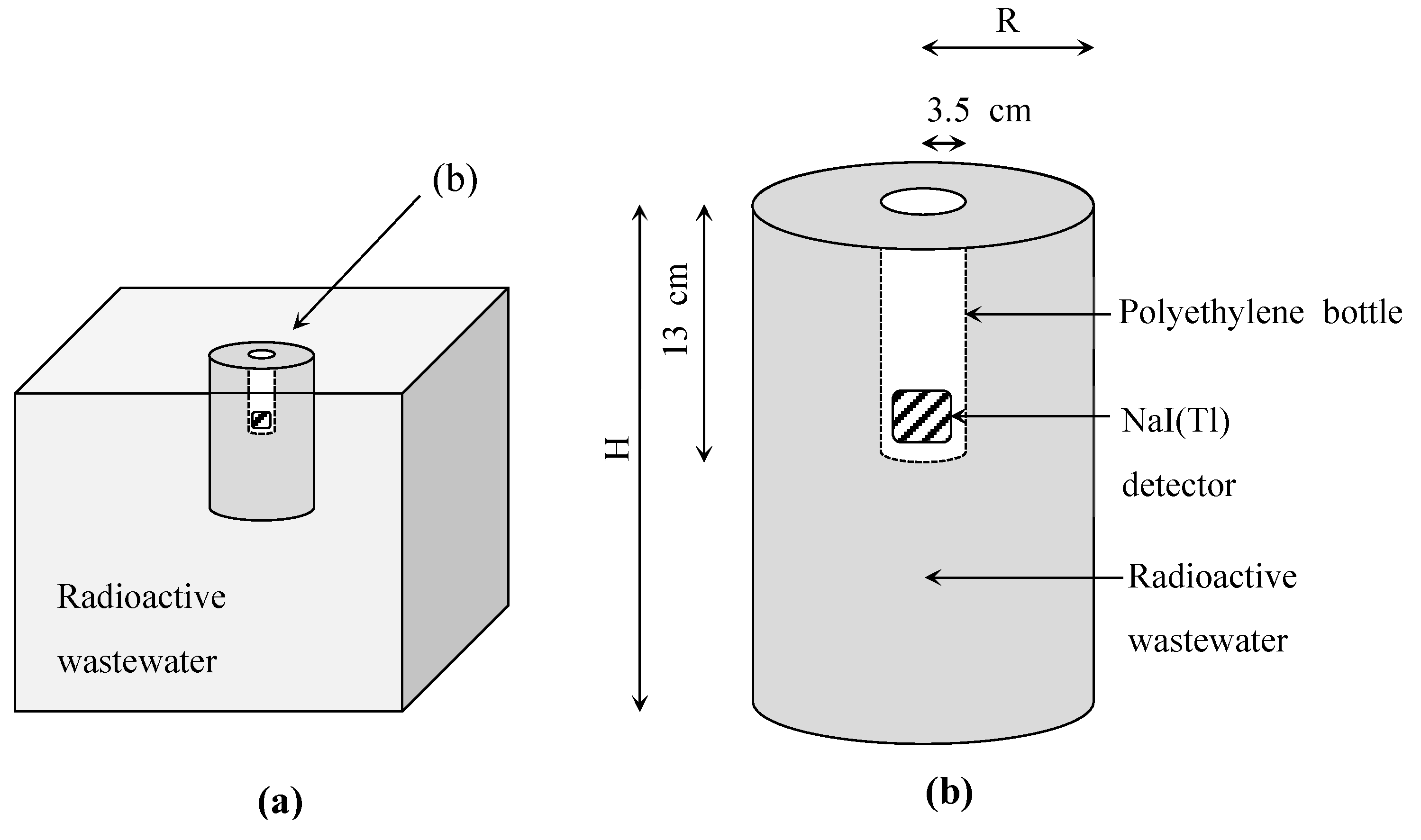
2.1. System Description

In this study, a 38-mm-diameter and 38-mm-height NaI(Tl) scintillation detector (GS-1515-NaI, Bee Research Pty, Ltd., Mascot, Australia) connected to a photomultiplier tube, preamplifier, and power supplier was used to measure the amount of radiation. The output of the preamplifier was connected to a shaping amplifier in a multichannel analyzer (MCA; GS-USB-PRO, Bee Research Pty, Ltd., Mascot, Australia).
Free gamma spectroscopy software PRA 24.0.2.0 (Marek Dolleiser, Sydney University, Sydney, Australia) [19] was used as a data logger for Windows 10 (Microsoft Ltd., Raymond, WA, USA). This software controls an analog-to-digital converter (ADC), including an MCA, through the USB communication interface, analyzes pulses received from the shaping amplifier, and records them in real time.
The specifications of the continuous radioactivity monitoring system for the radioactive wastewater decay tank are listed in Table 1.

2.2. Experimental Calibration

The experimental efficiency calibration was performed in a test water tank with a volume of 14 L, filled with water and reference source 18F, which is most commonly used in positron emission tomography/computed tomography. It has a half-life of only 110 min and emits two 511 keV gamma rays.
The absolute detection efficiency (ε) for 18F was obtained using the following equation from previous research [19,20]:
ε = N/(A·Iγ·T),
where N denotes the net count. Here, A denotes the specific activity of the specific radionuclide obtained using a gamma spectroscopy system including an MCA (DSA-1000 Digital Spectrum Analyzer, Canberra Industries, Meriden, CT, USA), analyzing software (Genie 2000, Miron technologies, Atlanta, GA, USA) [21], and a high-purity germanium detector (GEM25P-PLUS, Canberra Industries, Atlanta, GA, USA) calibrated by the certified reference materials in a cylinder-type source (Table 2) certified by the Korea Research Institute of Standards and Science (KRISS), in units of Bq·m−3. In addition, Iγ denotes the emission probability of the gamma ray, and T denotes the live time of measurement.

2.3. Monte Carlo Simulation

In this study, the activity conversion factor (ACF) was applied to convert the counts measured by the detector into specific activity. Although the ACF has the same physical meaning as detection efficiency, it was used to clearly distinguish the purpose of use.
Because the ACF is determined using the intrinsic characteristics of the scintillation detector and MCA, it cannot be obtained only by simulation; therefore, it must be obtained only experimentally or calculated by experiments and simulations [15]. Different departments use different isotopes in hospitals. The 18F has been commonly used for nuclear medicine outpatients due to its short half-life and production convenience among proton-emitting nuclides. In our study, 18F was used, and the sample was placed in the tank, as shown in Figure 1b. The count rates of the sample and background count rates were measured using a NaI(Tl) detector for one hour under the same experimental conditions. The ACF was calculated by comparing the simulation results obtained under the same conditions.
The decay tanks used in the field store tens to hundreds of tons of wastewater. In the experiment, the periphery of the detector, as shown in Figure 1b, was constructed and used instead of that in Figure 1a, and the detector was installed at the top of an actual-sized decay tank. A NaI(Tl) detector was installed inside a polyethylene bottle to prevent the detector from being immersed.
The ACF was calculated by comparing the counters in the full energy peak measured with a NaI(Tl) detector, and the specific activity was measured with a gamma spectrometry system using diluted 18F as radioactive wastewater. A gamma spectrometry system using a semiconductor detector was calibrated using a radioactive certified standard material. The ACF was calculated using the following equation, and the counts measured by the NaI(Tl) detector were converted to specific activities measured by a gamma spectroscopy system with an HPGe detector. In this equation, the units of ACF, NNaI, and CHPGe were Bq·s·m−3·counts−1, counts·s−1, and Bq·m−3, respectively:
ACF = CHPGe/NNaI(Tl),
The radius (R) and height (H) of the water tank used in the experiment were 12.5 and 29.5 cm, respectively, and 14 L of radioactive wastewater was used in the experiment. Because practical water tanks are usually much larger, the ACF was calculated using a Monte Carlo simulation.
Because water is a good shielding material, its efficiency varies according to the measurement radius [15]. Thus, by changing the radius (R) and height (H) of the water tank, as shown in Table 1, the change in ACF was calculated by the MCNP 6.2 (Los Alamos, NM, USA) with the Monte Carlo code using the F8 tally with the GEB option [22].
In this simulation, the geometry of the scintillation detector was designed based on the drawing provided by the manufacturer, and the density of the NaI(Tl) detector was 3.667 g·cm−3 [23]. In this case, the volume of radioactive wastewater used in the calculation ranged from 14 to 56.1 L.

2.4. Calculation of MDA in Water

The MDA is the minimum detectable activity that can be distinguished from the background radioactivity and is calculated using Equation (3), called “Currie’s method” [24,25]:
MDA = LD/(ε·Iγ·T),
where ε denotes the absolute detection efficiency, Iγ denotes the gamma-ray emission probability, and T denotes the live measurement time. Here, LD is the detection limit in counts with a 95% confidence level, and it is calculated as:
LD = 2.71 + 4.653 σB,
where σB denotes the standard deviation of background radioactivity [24,25,26].

3. Results

3.1. Full Energy Peak Efficiency

The count of the full energy peak for one hour, N, was measured, using a NaI(Tl) detector, to be 3318, and the count of 18F in the sample was calculated by MCNP 6.2 to be 7.5372 × 10⁷. Using Equation (1), the efficiency value ε was calculated to be 4.402 × 10⁻⁵.

3.2. Determination of 18F ACF in Water

In this study, the count rates of wastewater stored in a decay tank were measured using a NaI(Tl) scintillation detector and then converted into specific activity. To this end, a radioactive wastewater sample containing 18F radioisotopes was prepared. Because our experimental data were used to determine whether radioactive wastewater could be discharged safely, the specific activity of test water containing 18F should be less than 1 × 10⁷ Bq·m−3, which is the discharge standard for radioactive wastewater provided by law.
The count rates measured for the sample including 18F and the background count rates are shown in Figure 2a,b, respectively. Figure 2a confirms that the 511 keV gamma rays are emitted by 18F. Because the background count rate was only approximately 5.2% around the 511 keV peak, that of the sample including 18F, the graph subtracting the background count rate from Figure 2a was almost the same as in Figure 2a; therefore, it was not separately indicated in the graph.
To estimate the ACF that can change the count rates (counts·s−1) of the 18F sample measured with a NaI(Tl) detector to the specific activity (Bq·m−3), 500 mL of the wastewater sample used in the above experiment was selected. It was measured using a gamma spectroscopy system for one hour. The results are shown in Figure 3. The gamma spectroscopy system used in this measurement was calibrated using a certified reference material (Table 2); thus, the background radioactivity was automatically removed.
The count rates of the full energy peak at 511 keV, obtained by subtracting the background counts from the counts of the wastewater sample measured using the NaI(Tl) detector, was 0.9217 counts·s−1, and the specific activity of the same sample measured using the gamma spectroscopy system was 1.15031 × 10⁶ Bq·m−3. Thus, the ACF value calculated using Equation (2) was 4.6415 × 10¹⁰ Bq·s·m−3·counts−1.
To determine the change in ACF for the variation in tank size, a calculation was implemented by a Monte Carlo simulation using MCNP 6.2 with the same geometry and physical conditions as in the above experiment. Figure 4a was obtained by subtracting the background count rates shown in Figure 2b from the count rates of the wastewater sample measured using the NaI(Tl) detector shown in Figure 2a.
In Figure 4a, a small peak appears at approximately 440 keV. This peak might be attributed to the difference between the Monte Carlo geometry in our calculation and the actual geometry of the material used in our experiment. This is because there was no peak around 440 keV when measuring the same sample using the gamma spectroscopy system, as shown in Figure 3.
As shown in Table 3, the ACF calculated in computer simulations continued to increase and was fixed at 5.5389 × 10¹⁰ Bq·s·m−3·counts−1 when the volume exceeded 38 L. It was concluded from those that the larger the water tank, the larger the value measured using the NaI(Tl) detector, and it is fixed when the volume exceeds 38 L. Because the decay tank for radioactive wastewater used in the field was at least 38 L, the ACF of 5.5389 × 10¹⁰ Bq·s·m−3·counts−1 was suitable as an ACF for wastewater containing 18F.

3.3. Calculation of MDA of 18F in Water

Figure 2b shows the background count rates measured for one hour in a 14 L water tank containing tap water, and the counts in the full energy peak of 18F were 186 counts. Using Equation (4), the LD for the one-hour measurement was calculated as 6.62 × 101 counts. Then, this value was transferred to 2.09 × 10⁵ Bq·m−3, the MDA for the same measurement live time, using Equation (3) with an absolute detection efficiency (ε) of 511 keV. This efficiency was obtained by dividing the counts at 511 keV in NaI(Tl) by the counts at 511 keV in the gamma spectroscopy system, and it was 4.40 × 10⁻⁵. This value was smaller than 1 × 10⁷ Bq·m−3, which is the amount prescribed by law in Korea, where the public is predicted to reach the dose limit owing to the draining of 18F-containing wastewater into the public environment. Using this system, it was possible to determine whether the specific activity of radioactive wastewater containing 18F could be measured for one hour.
The change in MDA at various measurement times and assuming that the background radioactivity was constant is shown in Table 4. When the measurement time was one hour, the MDA calculated using Equation (3) was 2.09 × 10⁵ Bq·m−3; however, when the measurement time was increased to 24 h, the MDA decreased to 4.12 × 10⁴ Bq·m−3. This means that, by increasing the measurement time from one hour to one day, the measurement ability was improved by five times.

4. Discussion

Measuring the specific activity before draining radioactive wastewater is a resource- and time-intensive process. In this study, a method of continuously measuring radioactivity by installing a detector on a decay tank was used to reduce time and cost.
For this purpose, a NaI(Tl) detector, which is commonly used in studies measuring radioactivity in seawater, groundwater, or lakes, was used to measure the count rates in a tank containing radioactive wastewater, and the ACF was calculated. The specific activities in Bq·m−3 (CHPGe) were obtained by multiplying the counts per second (NNaI) measured using a NaI(Tl) detector by the ACF. Using the ACF, a radiation safety manager can observe the specific activity of wastewater stored in the wastewater tank in real time and can immediately decide whether to drain the tank or not.
For this experiment, a NaI(Tl) detector with a diameter of 38 mm and a height of 38 mm was used. It is inexpensive and has low efficiency among the products sold. The ACF to transfer counts obtained using a NaI(Tl) detector to the specific activity was calculated using a gamma spectroscopy system with an HPGe detector calibrated with standard reference materials.
When underwater radioactivity is measured, water acts as a shield and affects the measurement radius [13]; thus, a change in ACF was observed while increasing the size of the tank containing radioactive wastewater. The change in the counted value for various sizes of the tank containing radioactive wastewater was calculated using MCNP 6.2. As a result of the calculation, when the volume of the tank exceeded 38 L, the ACF was fixed at 5.5389 × 10¹⁰ Bq·s·m−3·counts−1. Because the decay tank for radioactive wastewater used in the field is at least 38 L, this conversion factor is suitable for radioactive wastewater containing 18F.
The MDA is the minimum detectable activity that can be separated from natural radioactivity. Because the system used in this study must prove that the specific activity of radioactive wastewater is less than the draining standard, the MDA of this measurement system must be less than that of radioactive wastewater. Otherwise, this system cannot determine whether the radioactive wastewater can be drained or not. To prove this, the MDA of the measurement system was calculated to be 2.09 × 10⁵ Bq·m−3 for one hour of measurement, which was smaller than the draining standard of 1 × 10⁷ Bq·m−3 as prescribed by law in Korea, where the public is predicted to reach the dose limit owing to the draining of 18F-containing wastewater into the public environment. Thus, it is possible to use this system to determine whether the specific activity of radioactive wastewater containing 18F can be measured for one hour.
However, the following limitations exist when applying the results of this study to the field. First, 18F is a nuclide that emits 511 keV gamma rays and has sufficient energy to pass through the aluminum outer wall of the NaI(Tl) detector. Therefore, radioisotopes emitting low-energy gamma rays that do not pass through the outer wall of the detector cannot be analyzed. This is a common problem with general radiation counters, and this type of radioisotope should be used with a specialized instrument, such as a liquid scintillation counter.
Another limitation is that the ACF is determined by the measurement system, and the MDA is determined by the background radioactivity of the measurement environment. In other words, the ACF should be determined for each measurement system, and the MDA should be determined by measuring the background level after installation. For a lower MDA, the shielding of the measurement system can be considered.
Finally, the cause of the peak around 440 keV in Figure 4 could be the difference between the MCNP geometry in the calculation and the real geometry of the material used in the experiment. This is because there was no peak at approximately 440 keV when the same sample was measured using a gamma spectroscopy system with HPGe.
Despite these limitations, 18F and 131I, the most commonly used unsealed radioisotopes in hospitals, emit radiation with sufficient energy to be measured with this system—18F used for cancer diagnosis and 131I used for thyroid cancer treatment emit 511 and 971 keV gamma rays, respectively. In particular, many hospitals use only 18F and 131I; therefore, the possibility of applying this system is high.
The results of this study indicate that specific activity measurements for the draining of radioactive wastewater into the public environment can be conducted using a relatively inexpensive NaI(Tl) detector without relying on an expensive HPGe detector or a gamma counter.
When measuring radioactivity using a NaI(Tl) scintillation detector with a diameter of 38 mm and height of 38 mm, which is the smallest commercially available detector, the MDA was found to be smaller than the value that is the draining standard for radioactive wastewater provided by law. This system can be improved by using a larger scintillation detector or installing a shield on the detector to reduce background radioactivity, which is planned for further research. In this system, a detector is fixed on the top of a decay tank for continuous measurement; therefore, the measurement time can be extended to several hours or days; in such cases, the MDA decreases. These properties are not applicable to radioactive wastewater containing 18F, which has a short half-life. However, it can be used for wastewater with a long half-life. In addition, this system is very similar to the underwater, in situ spectrometer system for measuring the radioactivity contained in rivers or seas, and it has potential applications for measuring radioactivity in ballast tanks or live fish transport containers because it measures the radioactivity of stored water. We expect that the proposed monitoring system can be used to monitor the radioactivity of the water contained in a ship’s ballast tanks [27] and live fish containers. The proposed system can help identify radioactivity because many people are concerned about the marine pollution possibility by radioactive materials after the Fukushima nuclear disaster.

Author Contributions

Investigation, T.K.; methodology, T.K. and S.K.; simulation and experiment, S.K.; writing—original draft, S.K.; writing—review and editing, T.K. and H.Y. All authors have read and agreed to the published version of the manuscript.

Funding

This study was supported by the Research Foundation of Korea University (K1915361) and a grant from the Korea Institute of Radiological and Medical Sciences (KIRAMS), funded by the Ministry of Science and ICT (MSIT), Republic of Korea (No. 50422-2022).

Institutional Review Board Statement

Not applicable.

Informed Consent Statement

Not applicable.

Data Availability Statement

Not applicable.

Conflicts of Interest

The authors declare no conflict of interest.

References

  1. Adrovic, F. Gamma radiation. In Gamma Radiation; InTech: Rijeka, Croatia, 2012; pp. 43–54. [Google Scholar]
  2. Vandergriff, K.U. Guidelines for radiation effects on organic materials. In Designing Equipment for Use in Gamma Radiation Environments; National Information Service: Alexandria, VA, USA, 1990; pp. 45–103. [Google Scholar]
  3. The International Commission on Radiological Protection. The framework of radiological protection. In Radiological Protection and Safety in Medicine; Elsevier Science Inc.: New York, NY, USA, 1996; pp. 9–10. [Google Scholar]
  4. The International Commission on Radiological Protection. The aims and scope of the recommendations. In The 2007 Recommendations of the International Commission on Radiological Protection; Valentin, J., Ed.; Elsevier Ltd.: Kidlington, UK, 2007; pp. 41–47. [Google Scholar]
  5. Johnson, T.E. Review of physical principles. In Introduce to Health Physics, 5th ed.; McGraw Hill Education: New York, NY, USA, 2017; pp. 3–56. [Google Scholar]
  6. International Atomic Energy Agency. Preamble: Principles and fundamental objectives. In International Basic Safety Standards for Protection against Ionizing Radiation and for the Safety of Radiation Sources; IAEA: Vienna, Austria, 1996; pp. 1–9. [Google Scholar]
  7. Lee, J.W.; Lee, Y.J.; Lee, H.J.; Jeong, G.S.; Lee, S.B. Radiation Safety Management for RT Facilities (2015~2017); Korea Atomic Energy Research Institute: Daejeon, Korea, 2018. [Google Scholar]
  8. Bagatelas, C.; Tsabaris, C.; Kokkoris, M.; Papadopoulos, C.; Vlastou, R. Determination of marine gamma activity and study of the minimum detectable activity (MDA) in 4pi geometry based on Monte Carlo simulation. Environ. Monit. Assess. 2010, 165, 159–168. [Google Scholar] [CrossRef] [PubMed]
  9. Kim, J.H.; Park, K.H.; Joo, K.S. Development of low-cost, compact, real-time, and wireless radiation monitoring system in underwater environment. Nucl. Eng. Technol. 2018, 50, 801–805. [Google Scholar] [CrossRef]
  10. Park, H.M.; Joo, K.S. Development of a real-time radiation level monitoring sensor for building an underwater radiation monitoring system. J. Sens. Sci. 2015, 24, 96–100. [Google Scholar]
  11. Tsabaris, C.; Bagatelas, C.; Dakladas, T.; Papadopoulos, C.; Vlastou, R.; Chronis, G. An autonomous in situ detection system for radioactivity measurements in the marine environment. Appl. Radiat. Isot. 2008, 66, 1419–1426. [Google Scholar] [CrossRef] [PubMed]
  12. Wang, J.; Zhang, Y.; Liu, D.; Wu, B.; Zhang, Y.; Jiang, H. Automated spectra analysis of in situ radioactivity measurements in the marine environment using NaI (Tl) detector. Appl. Radiat. Isot. 2018, 141, 88–94. [Google Scholar] [CrossRef] [PubMed]
  13. Zhang, Y.; Li, C.; Liu, D.; Zhang, Y.; Liu, Y. Monte Carlo simulation of a NaI (Tl) detector for in situ radioactivity measurements in the marine environment. Appl. Radiat. Isot. 2015, 98, 44–48. [Google Scholar] [CrossRef]
  14. Tsabaris, C.; Androulakaki, E.G.; Prospahopoulos, A.; Alexakis, S.; Eleftheriou, G.; Patiris, D.L.; Pappa, F.K.; Sarantakos, K.; Kokkoris, M.; Vlastou, R. Development and optimization of an underwater in-situ cerium bromide spectrometer for radioactivity measurements in the aquatic environment. J. Environ. Radioact. 2019, 204, 12–20. [Google Scholar] [CrossRef]
  15. Park, J.G.; Jung, S.H.; Moon, J.H.; Oh, D.; Kang, S.W.; Kim, Y.S. Determination of effective detection distance and minimum detectable activity for radiation monitoring system in water. J. Instrum. 2018, 12, 11–14. [Google Scholar]
  16. Park, J.G.; Jung, S.H.; Moon, J.; Kim, J.; Oh, D.; Kang, S.; Kim, Y. Determination of minimum detectable activity for underwater radiation detection system. J. Instrum. 2019, 14, P10038. [Google Scholar] [CrossRef]
  17. Moon, J.; Park, J.; Jung, S.; Kim, Y. Development and Performance Evaluation of Underwater Radiation Monitoring System. J. Instrum. 2020, 14, 213–218. [Google Scholar]
  18. Ji, Y.; Toung, S.; Min, B.I.; Suh, K. Development and performance of underwater gamma-ray spectrometry for riverbed monitoring. J. Coast. Res. 2021, 114, 330–334. [Google Scholar] [CrossRef]
  19. GammaSpectacular.com. Available online: http://www.gammaspectacular.com/software-downloads/pra-spectrometry-software (accessed on 17 March 2022).
  20. Han, S.Y.; Maeng, S.; Lee, H.Y.; Lee, S.H. Preliminary study on the detection efficiency and estimation of minimum detectable activity for a NaI (Tl)-based seawater monitoring system. J. Environ. Radioact. 2020, 218, 106222. [Google Scholar] [CrossRef]
  21. Canberra Industries, Inc. GenieTM 2000 Spectroscopy Software Operations; Canberra Industries, Inc.: Meriden, CT, USA, 2006; pp. 1–281. [Google Scholar]
  22. Werner, C.J.; Bull, J.S.; Solomon, C.J.; Brown, F.B.; McKinney, G.W.; Rising, M.E.; Dixon, D.A.; Martz, R.L.; Hughes, H.G.; Cox, L.J.; et al. MCNP6.2 Release Notes; Los Alamos National Laboratory: Santa Fe, NM, USA, 2018. [Google Scholar]
  23. Saito, K.; Moriuchi, S. Monte Carlo calculation of accurate response functions for a NaI (Tl) detector for gamma rays. Nucl. Instrum. Methods Phys. Res. 1981, 185, 299–308. [Google Scholar] [CrossRef]
  24. Currie, L.A. Limits for qualitative detection and quantitative determination. Application to radiochemistry. Anal. Chem. 1968, 40, 586–593. [Google Scholar] [CrossRef]
  25. Knoll, G.F. Limits of detectability. In Radiation Detection and Measurement, 4th ed.; John Wiley & Sons, Inc.: Hoboken, NJ, USA, 2010; pp. 94–98. [Google Scholar]
  26. Tsoulfanidis, N.; Landsberger, S. Errors of radiation counting. In Measurement and Detection of Radiation, 4th ed.; CRC Press: Boca Raton, FL, USA, 2015; pp. 61–63. [Google Scholar]
  27. Abdul, K.L.; Wo, Y.M.; Muhamat, O. Monitoring of Radionuclides in Ship Ballast Water Following the Fukushima Nuclear Accident. 2011. Available online: https://inis.iaea.org/collection/NCLCollectionStore/_Public/44/122/44122660.pdf (accessed on 20 May 2022).
Figure 1. Outline of real wastewater tank (a) and calculation model (b).
Applsci 12 05613 g001
Figure 2. Count rates of 18F source including background (a) and background (b) obtained by experiment using NaI(Tl) detector.
Applsci 12 05613 g002
Figure 3. Count rates of 18F source obtained by experiment using HPGe detector.
Applsci 12 05613 g003
Figure 4. Specific activities measured by HPGe and count rates measured by NaI(Tl): count rates (a), (b) and full energy peaks (c), (d) obtained by experiment and calculation, respectively.
Applsci 12 05613 g004
Table 1. Specifications of the monitoring system.
ParametersDetails
Scintillation crystal1.5″ × 1.5″ NaI(Tl)
EnclosureAluminum case with cylinder shape
Resolution6.5% to 8% at 660 keV
Gamma efficiencyApprox. 60% at 662 keV
Input power+5 V, USB 2.0
Current draw<150 mA
ADC sample rate and bit rate48 kHz, 16 bit
Pulse lengthAdjustable
Signal-to-noise ratio73.97 dB
High-voltage connectorSHV
Signal in connectorBNC
Table 2. Specifications of certified reference materials in cylinder-type source, certified by KRISS.
NuclideHalf-Life (day)Activity (Bq)γ-ray Energy
(keV)
γ-ray Emission
Probability (%)
Half-LifeUncert.ActivityUncert.Prob.Uncert.
241Am158,00421918447459.5435.920.17
109Cd461.90.410,25741088.033.660.05
57Co271.810.0448920122.06
136.47
85.49
10.71
0.14
0.15
139Ce137.6410.02059424165.8679.900.04
51Cr27.7040.00463,6352600320.089.890.02
113Sn115.090.03151561391.7064.970.17
85Sr64.8500.007200981514.0098.50.4
137Cs10,9762990937661.6684.990.20
60Co1925.230.291251501173.23
1332.49
99.85
99.9826
0.03
0.0006
88Y106.630.053156130898.04
1836.05
93.7
99.346
0.3
0.025
Table 3. Activity conversion factors (ACFs) for various water tank volumes in Figure 1b.
R (cm)H (cm)Volume (ℓ)Calculated NNaI(Tl)
(counts·s−1)
ACF
(Bq·s·m−3·counts−1)
12.529.514.02.4783 × 1044.6415 × 1010
14.431.420.02.2132 × 1045.1975 × 1010
16.033.026.02.1122 × 1045.4460 × 1010
17.434.232.02.0775 × 1045.5370 × 1010
18.635.438.02.0768 × 1045.5389 × 1010
19.736.544.02.0768 × 1045.5389 × 1010
20.737.550.02.0768 × 1045.5389 × 1010
21.638.656.12.0768 × 1045.5389 × 1010
Table 4. Minimum detectable activity (MDA) for reading time.
Reading Time (h)MDA (Bq·m−3)Reading Time (h)MDA (Bq·m−3)
12.09 × 105135.62 × 104
21.46 × 105145.41 × 104
31.18 × 105155.23 × 104
41.02 × 105165.06 × 104
59.12 × 104174.91 × 104
68.32 × 104187.77 × 104
77.69 × 104194.64 × 104
87.19 × 104204.52 × 104
96.77 × 104214.41 × 104
106.42 × 104224.31 × 104
116.11 × 104234.21 × 104
125.85 × 104244.12 × 104
Publisher’s Note: MDPI stays neutral with regard to jurisdictional claims in published maps and institutional affiliations.

