Article

Pulsed Electric Fields for Valorization of Platelets with No Therapeutic Value towards a High Biomedical Potential Product—A Proof of Concept

1
iBET, Instituto de Biologia Experimental e Tecnológica, 2780-157 Oeiras, Portugal
2
ITQB-NOVA, Instituto de Tecnologia Química e Biológica António Xavier, Universidade Nova de Lisboa, 2780-157 Oeiras, Portugal
3
EPS, EnergyPulse Systems,1600-546 Lisboa, Portugal
4
IPST, Instituto Português do Sangue e da Transplantação, 1749-005 Lisboa, Portugal
5
GIAAPP/ISEL, Pulsed Power Advanced Applications Group, Lisbon Engineering Superior Institute, 1959-007 Lisbon, Portugal
*
Author to whom correspondence should be addressed.
Appl. Sci. 2022, 12(12), 5773; https://doi.org/10.3390/app12125773
Submission received: 20 April 2022 / Revised: 28 May 2022 / Accepted: 3 June 2022 / Published: 7 June 2022
(This article belongs to the Special Issue Biomedical Applications of Pulsed Power and Plasmas)

Abstract

:

Featured Application

Biomedical product development from PEF application into platelets.

Abstract

Nowadays, the standard media used in clinical-scale mesenchymal stem cell (MSC) production to supply hundreds of clinical trials uses animal-derived components as supplements, which raises several health concerns. Consequently, the development of xeno-free media supplements has emerged. In the current study, the effect of pulse electric field (PEF) application to platelet concentrates (PC) with no therapeutic value for producing platelet releasates (PR) able to sustain the ability of bone marrow-MSCs (BM-MSCs) to self-renew and differentiate was tested. It was demonstrated that PEF application to PC induces platelet activation and growth factor (GF) release, namely PDGF, FGF, IGF, and TGF-β. The highest GF release was observed for TGF-β, achieving similar levels to those attained in platelet lysates (PL). BM-MSCs expanded in the presence of PR obtained by the application of PEF (7 pulses of 10 and 12.5 kV/cm) to PC (PR PEF) retained the characteristic MSC cell-surface markers, and the ability to proliferate and differentiate into osteogenic, adipogenic, and chondrogenic lineages. In this study, evidence is provided that PR PEF represents a suitable alternative to fetal bovine serum (FBS) for use in MSC production.

1. Introduction

Tissue regeneration is a physiological process that is important to maintain organ homeostasis and to restore organs after an injury. This process is intrinsic of several human tissues, however some organs such as the heart, liver, and central nervous system present a very limited regenerative capacity [1]. Studies have demonstrated that mesenchymal stem/stromal cells (MSCs) have an important role in tissue regeneration, not only because they have the ability to differentiate into mesodermal lineage cells but also due to their rich secretome [1]. Today, MSCs isolated from different tissues have been tested in more than 300 clinical trials (www.clinicaltrials.gov, accessed on 1 February 2022) with promising results, and several MSC-based products have already received regulatory approval [2]; however, the advanced manufacturing of these cells still needs to overcome several issues, such as the use of animal-derived components such as fetal bovine serum (FBS) to supplement cell-culture media [3,4]. The use of FBS raises several concerns; not only ethical concerns related to the animal welfare used to produce it, but also due to immune reactions and the transmission of bovine pathogens to humans [5].
An alternative to FBS that has been shown to be efficient in MSC expansion is the group of human platelet derivatives [6,7,8].
Platelets—when activated—release a wide range of bioactive factors, including growth factors (GF) and cytokines, which are known to have an important role in cell expansion and survival [9]. Platelet concentrates (PCs) are regularly produced in blood banks, and due to their short-shelf life (5–7 days), around 5% of them are discarded before clinical use [10,11].
Giving new use to these PCs with no therapeutic value, using them as a source of cell culture media supplements has become a very attractive tool; however, the methodology used to induce GF release from platelets affects platelet derivatives’ features [6,12]. The most common methods used are hypo-osmotic shock (freeze and thaw cycles) and bovine thrombin [13,14]. In the first methodology, the ideal number of cycles remains to be established, since a high number of cycles might lead to GF damage and a final lysate containing other platelet debris [14,15]. Using bovine thrombin to induce GF release from platelets presents the same problems associated with animal-derived components [16]. Thus, more efficient strategies to activate platelet pools and consequently generate releasates with an improved biological activity for MSC expansion are needed.
Pulse electric field (PEF) is a technology characterized by inducing electroporation on cell membranes, with the application of ns–µs range pulses and high electric fields kV/cm, allowing proteins to flow into and out of the cells, with a low thermal load [17,18]. In a previous work, the potential for the application of PEF in the valorization of expired PCs into a biomedical product with therapeutic value was demonstrated [19]. In particular, it was shown that PCs subjected to a single PEF treatment can release PDGF into the supernatant, and a proof-of-concept study was performed to confirm the biological activity of this releasate to support MSC culture in vitro. In the current study, the impact of key PEF parameters, namely the pulse width, electric field, and pulse number, on platelet activation from expired PCs was further investigated in order to prove the concept, aiming at modulating the release of specific GFs to improve their bioactivity for MSC expansion while maintaining cells’ quality attributes.

2. Materials and Methods

2.1. Platelet Concentrates

Blood was collected from healthy donors in compliance with the current Portuguese Institute for Blood and Transplantation (IPST, Lisbon Portugal) standard operating procedure. All donations were screened for HIV, HBV, HCV, HTLV, and treponema pallidum.
Whole blood donations were centrifuged, and the buffy coats from four group-specific donations were pooled with platelet additive solution (PAS) and stored for 5–7 days (if submitted to pathogen reduction technologies) at 20–24 °C in continuous agitation. Conventional quality assurance tests were performed, including platelet content, product volume, and residual white blood cell count. All tested units met the standards of the Council of Europe’s guide to the preparation, use, and quality assurance of blood components. Microbiological control was performed by the BacTAlert system when PCs were not submitted to pathogen reduction. Gamma irradiation of the platelet concentrates was used to prevent transfusion-associated graft versus host disease (TA-GvHD) in patients who may develop TA-GvHD.
In the current study, 90 PCs with 1–2 days after the expiring day (5 days post-production) were analyzed, and the six PCs with lower levels of platelet activation were used for further analysis.

2.2. Pulse Electric Fields Application

The pulse electric fields were applied in batch mode using a cuvette connected to a pulse generator. A total of 1 mL of PC was added into the cuvette chamber and pulses from a pulse generator, with a frequency of 1 Hz, were applied through the cuvette electrodes, as seen in Figure 1a,b. The pulse voltage and current was measured using a digital oscilloscope (LeCroy WaveAce 224), with a sampling rate of 1 GS/s, connected to a current transformer (Pearson 110) and a high voltage probe (North Start High Voltage PVM-1). As an operation example, a 2 µs pulse with 4.5 kV and 150 A is presented in Figure 1c, giving about 11 kV/cm.
The commercial pulse generator EPULSUS-PM1B-10 (from EnergyPulse Systems, Lisbon, Portugal), was used. This generator is able to deliver positive pulses up to 10 kV and 400 A. The pulses can last 1 µs to 200 µs, with repetition rates of 1 Hz to 200 Hz, at a maximum average output power of 3 kW. The pulse generator is based on a solid-state Marx generator, where various n capacitors are first charged in parallel from a relatively low voltage Udc, and then connected in series with the load, applying about nUdc [20]. As this generator is based on a direct capacitive discharge, it can deliver almost rectangular pulses, independent of the load impedance, as long as the generator’s stored energy is much higher than the pulse energy, which is the case in this application.
A standard electroporation cuvette was used, with a 4 mm distance between electrodes and an 800 µL volume.
For PEF applications the most important parameters are the electric field amplitude, E (kV/cm), and the specific energy, W s   ( kJ / kg ) , given by
E = U d
where U refers to the pulse voltage amplitude applied in kV , and d the distance between the electrodes of the treatment chamber in cm .
W s = W t m
where W t is the total applied energy, and m is the mass of the sample inside the treatment chamber in kg . The total applied energy is given, in J, by
W t = U I t o n N
where I is the pulse current amplitude in A, ton the pulse width in µs, and N the number of pulses applied.
The main objective of using PEF is to avoid the thermal impact, so the increase in temperature, ∆T in °C, from the application of PEF, is important, given by
Δ T = W s C p
where Cp is the specific heat capacity ( kJ / ( kg · K ) ) of the treated material, which was assumed to be similar to water: 4.18 kJ / ( kg · K ) .
Prior to PEF application, PCs were initially centrifuged at 1000× g for 10 min. The resulting platelet pellet was resuspended in plasma additive solution (PAS; InterSol) to a final concentration of 1 × 10⁹ platelets/mL. Platelets in PAS (1 mL) were then added to the cuvettes and exposed to 9 PEF conditions, comprising a range of 10–15 kV/cm and 1–15 pulses of 2 µs duration.

2.3. Preparation of Platelet Lysates

Platelet lysates (PL F&T) were obtained as previously described [19]. Briefly, PCs were exposed to three cycles of freeze (liquid nitrogen) and thaw (37 °C). At the end, samples were centrifuged to remove cell debris and obtain the platelet lysates.

2.4. Platelet Activation Measurement

Platelet activation was evaluated by measuring platelet surface markers (CD61 and CD62P) via flow cytometry [21]. Briefly, platelets exposed to PEF were resuspended in tyrode buffer containing albumin and glycose (1 × 10⁸ platelets/mL), and then added to a mixture containing peridinin-chlorophyll-protein (PerCP)-conjugated CD61 and phycoerythrin (PE)-conjugated CD62P (P-selectin; BD Biosciences, Franklin Lakes, NJ, USA). The samples were incubated for 15 min in the dark, at room temperature. The samples were acquired in Cytometer BD FACSCaliburTM equipment, and platelets distinguished by forward angle light scatter (FSC) and CD61 markers.
In parallel, platelets were stimulated for 15 min with thrombin (0.05 U/mL) in tyrode buffer containing albumin and glycose, in the presence of Gly-Pro-Arg-Pro (GPRP; BD Biosciences) to avoid platelet aggregation, and analyzed by flow cytometry.

2.5. Growth Factor Quantification

Untreated PCs, PCs subjected to PEF, and PCs exposed to freeze and thaw cycles (PL F&T) were centrifuged (1000× g for 10 min, at room temperature) and the supernatants, also termed the releasate (PR PEF) or lysate (PL F&T), were collected and stored at −20 °C until further use. Platelet derived growth factor (PDGF), vascular endothelial growth factor (VEGF), insulin-like growth factor (IGF), and transforming growth factor (TGF) release was measured in PR PEF using the Human PDGF-BB Quantikine ELISA Kit (R&D Systems, Minneapolis, MN, USA), Human VEGF Quantikine ELISA Kit (R&D Systems), Human TGF-β1 Quantikine ELISA Kit (R&D Systems), and Human IGF Quantikine ELISA Kit (R&D Systems). ELISA was performed according to the manufacturer’s protocol.

2.6. Human BM-MSC Culture

The releasate obtained from PEF exposure was tested for bioactivity using hMSCs isolated from bone marrow (BM-MSC, donor 00055; RoosterBio, Frederick, MD, USA). For routine cell culture, BM-MSCs were thawed and propagated in MesencultTM human MSC media according to the manufacturer’s instructions.
For bioactivity experiments, MSCs were grown in (i) standard culture medium composed of DMEM, low glucose, pyruvate, no glutamine, no phenol red (Gibco™, ThermoFisher, Waltham, MA, USA) supplemented with 10% (v/v) FBS, 1× GlutaMAX™ supplement (Gibco™, ThermoFisher), 2 IU/mL heparin (Stem cell technologies, Vancouver, BC, Canada), 100 U/mL penicillin, and 100 µg/mL streptomycin (Gibco™, ThermoFisher); (ii) medium supplemented with PR PEF composed of DMEM supplemented with 6% (v/v) PR, 1× GlutaMAX™ supplement (Gibco™, ThermoFisher), 2 IU/mL heparin (Stem cell technologies), 100 U/mL penicillin, and 100 µg/mL streptomycin (Gibco™, ThermoFisher), supplemented with either 6% PR PEF or PL F&T. For all conditions, MSCs were inoculated at a density of 4500 cell/cm2 in adherent static culture systems (12- and 48-well plates) and cultured for up to 7 days at 37 °C in a humidified atmosphere of 5% CO2 and 95% air. MSCs were cultured inside of an incubator at 37 °C, 5% CO2. The medium was exchanged every 3 days and cultures were monitored as follows:
Cell adhesion: Cell adhesion was evaluated by determining the number of cells 24 h after inoculation via the CellToxTM green cytotoxity assay (Promega, Madison, WI, USA) using the manufacturer’s instructions.
Cell proliferation: Cell number was determined using the CellToxTM green cytotoxity assay (Promega) according to the manufacturer’s protocol for live cells. Briefly, CellToxTM green was added to cells and incubated for 15 min before fluorescence measurement, using an Incucyte® Zoom, Essen BioScience, Ann Arbor, MI, USA, 10× objective. At each time point, the initial number of positive CellToxTM green cells was acquired, followed by the fluorescence measurement of the total CellToxTM green positive cells (total cell number) determined 10 min after 0.5% (v/v) Triton X-100 in PBS treatment. The number of live cells over time was obtained by subtracting (Initial CellToxTM green positive cells) from (total CellToxTM green positive cells).
Cell viability: MSC viability was determined by double-staining the MSCs with 20 μg/mL fluorescein diacetate (FDA; Sigma, St. Louis, MO, USA) and 10 μg/mL propidium iodide (PI; Sigma), which stains viable and non-viable cells, respectively. Briefly, the MSCs were incubated for 5 min with FDA/PI in PBS, and cell imaging was acquired using an Incucyte® Zoom, Essen BioScience.
MSC Immunophenotype: MSC phenotype was evaluated by measuring the expression of cell surface antigens through flow cytometry. After 7 days of culture in different medium formulations, cells were detached using TrypLE™ Select Enzyme (Gibco™, ThermoFisher), and 2 × 10⁵ were resuspended in PBS with 5% (v/v) FBS. Cells were incubated with the following antibodies: CD73 (Biolegend, San Diego, CA, USA), CD90 (Biolegend), CD105 (Miltenyi Biotec), CD29 (Biolegend), CD34 (Miltenyi Biotec, Cologne, Germany), CD45 (BD Pharmingen™, Franklin Lakes, NJ, USA), and HLA-DR (Biolegend) for 30 min. Data were acquired using a BD FACSCelesta™ Cell Analyzer—BD Biosciences. A total of 5000 events were acquired per sample.
Multilineage differentiation potential of BM-MSC: BM-MSC cells were seeded in 48-well plates at 4500 cells/cm2 and expanded for 6 days in DMEM supplemented with 6% PR or 10% FBS, as mentioned above. After this period, cells were analyzed for their potential to differentiate into adipocytes, osteocytes, and chondrocytes. Differentiation into adipocytes and osteocytes was performed by changing the culture medium to an adipogenic or osteogenic differentiation medium (Sigma-Aldrich), respectively, and cells were cultured for 21 days according to the manufacturer’s instructions. For chondrogenic differentiation, BM-MSC were detached and resuspended in 50 µL of DMEM supplemented with 6% PR or 10% FBS; droplets of 5 µL were inoculated in a low adherence plate to form cell aggregates. After 20 min, DMEM supplemented with 6% PR or 10% FBS was added to the cells, and the next day replaced with chondrogenic differentiation medium (Sigma-Aldrich). Cells were differentiated for 21 days and the medium was exchanged every 3–4 days. Cell differentiation was performed inside an incubator at 37 °C, 5% CO2. At the end of all differentiated cultures, cells were washed with PBS fixed with 4% (w/v) paraformaldehyde containing 4% (w/v) sucrose in PBS, for 15 min. Adipocytic, osteogenic, and chondrogenic differentiated cultures were stained with oil red O (Sigma-Aldrich), alizarin red S (Carl Roth, Karlsruhe, Germany), and Alcian blue (Sigma-Aldrich) staining solution, respectively, as previously described [22].

3. Results

3.1. Effects of PEF on Platelet Activation and Growth Factor Release

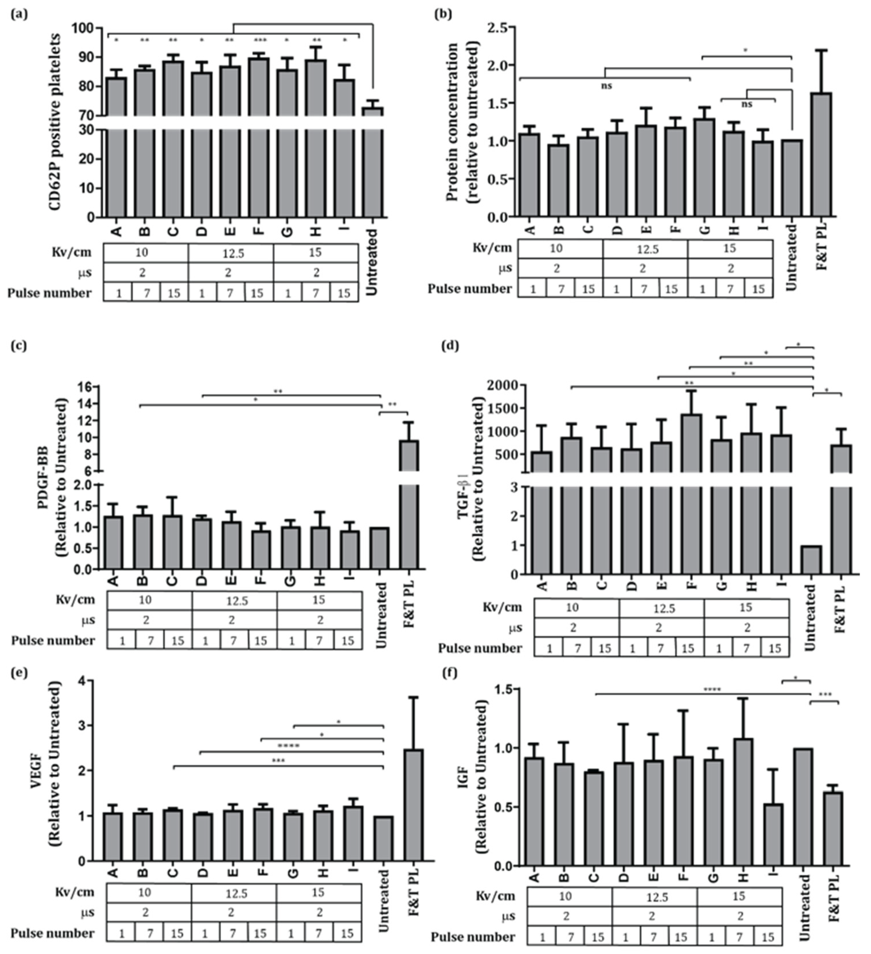
PCs were subjected to nine different PEF treatments, varying the electric field (10–15 kV/cm) and pulse number (1, 7, and 15) with 1 Hz frequency. The pulse duration was the same in all conditions (2 µs/pulse). Platelet activation was evaluated by flow cytometry, defining the activation by the percentage of CD62P+ platelets [21,23]. The data showed a significant increase in CD62P+ platelets in comparison to the untreated group for all PEF conditions tested. In particular, platelet activation was enhanced with the increase in pulse number, whereas no differences were observed between the different electric fields applied (Figure 2a).
The proteins released from platelets upon PEF stimulation were evaluated and compared to untreated PR. No differences in the total protein present in PR PEF were observed for the majority of PEF conditions applied when compared to untreated PR or PL F&T; moreover, one pulse of 15 kV/cm (PR PEF G) leads to a significant increase in the total protein in comparison to untreated PR (Figure 2b).
Following this, the levels of GF described to be stored in platelets [24,25] and relevant for MSC expansion, such as PDGF-BB, TGF-β1, VEGF, and IGF [26,27,28,29,30], were measured in the PR of all conditions tested. Our data demonstrate that PEF application leads to the release of GF to the PR, with higher release levels observed for TGF-β1, achieving similar levels to PL F&T (Figure 2c–f).
Our data demonstrate that the PEF conditions with 10 (PR PEF A, B, and C) and 12.5 kV/cm (PR PEF D, E, and F) pulses led to an increase in the PDGF concentration in the PR PEF when compared to the untreated group (Figure 2c).
In addition, the levels of TGF-β1 were similar to those obtained in the PL F&T condition, which were significantly higher than the untreated PR. Moreover, comparing the PR PEF with untreated PR, we could observe that lower pulse intensities require a higher pulse number to achieve a significant increase in comparison to the untreated PR. Thus, a significant enhancement of TGF-β1 was observed for 7 pulses of 10 and 12.5 kV/cm (PR PEF B and E), and 15 pulses of 12.5 and 15 kV/cm (PR PEF F and I). The VEGF concentration in the PR PEF was similar to the untreated condition; however, PEF application led to a higher VEGF release if 1 pulse of 12.5 and 15 kV/cm (PR PEF D and G), and 15 pulses of 10 kV/cm (PR PEF C) were applied to cells. However, PEF application to platelets led to a significant reduction in IGF levels in the PR after the application of 15 pulses of 10 and 15 kV/cm.
Of particular note, the levels of PDGF-BB or VEGF released from platelets after the application of PEF did not achieve PL F&T levels, which indicates that PEF application induces a controlled released of GF (Figure 2c,d).

3.2. BM-MSC Expansion on PR PEF Supplemented Media

BM-MSCs were cultured in DMEM supplemented with a selected PR PEF condition (PR PEF E and H), PL F&T, or FBS, the standard media condition used to expand these cells. Cells were cultured in these medium formulations for 6 days to understand the impact of the different medium supplements on BM-MSC growth features, namely adhesion, proliferation, and viability, as well as on the cell quality attributes including immunophenotype and multilineage differentiation potential.
Our results showed that cell adhesion was not altered by culture in PR PEF E and H supplemented media, as adhesion levels were similar to FBS and PL F&T (Figure 3a). Moreover, no differences in cell proliferation were observed between cells expanded in PR PEF, and FBS or PL F&T. Of note, cell viability assays demonstrated that cells expanded in FBS, PR PEF E, and PL F&T exhibited the highest cell viability, but PR PEF H showed a significantly lower cell viability than FBS. Nevertheless, the lowest cell survival was observed for untreated PR (Figure 3c,d).
Untreated PR supplement cells had a significantly lower proliferative capacity and viability than FBS, indicating that the GF release from platelets induced by PEF are essential to maintain BM-MSC proliferation (Figure 3b). Our data demonstrate that PR PEF supplemented media is an alternative to FBS, as cells maintain their proliferative and adhesion capacity, as well as viability.

3.3. BM-MSC Differentiation of Cells Expanded in PR PEF Supplemented Media

Subsequently, we characterized BM-MSCs expanded in PR PEF and FBS according to their potential to maintain the quality attributes of BM-MSCs, including immunophenotype and differentiation capability. MSC cell-surface markers were analyzed by flow cytometry. As shown in Figure 4a, no differences in MSC markers were observed for cells cultured in PR PEF or FBS. The BM-MSCs expressed a cell-surface profile positive for CD29, CD73, CD90, and CD105, and negative for CD34, CD45, and HLA-DR (Figure 4a).
We examined the potential of these cells to differentiate into adipogenic, osteogenic, and chondrogenic lineages. We observed that BM-MSCs cultured in all conditions had the ability to differentiate into these lineages, as confirmed by the positive staining of oil red O (adipogenic), Alcian blue (chondrogenic), and alizarin red S (osteogenic) (Figure 4b).

4. Discussion

In this work, we evaluated the use of several PEF protocols applied to platelets with no therapeutic value to produce a platelet derivative component that could be used as an alternative to FBS and PL as a media supplement for MSC cultures. Taken together, our data show that the application of PEF to expired PCs leads to a controlled released of GFs from platelets (Figure 2), and this PR PEF supports BM-MSC renewal and differentiation, while cell characteristics are maintained, as seen in Figure 3 and Figure 4.
It was observed that PR features are dependent on the PEF parameters applied, as different levels of GFs are released upon PEF application, as well as the percentage of platelet activation (Figure 2). These data are in accordance with previous studies demonstrating that PEF parameters influence the platelet rich plasma characteristics [19,31,32,33,34]. The application of PEF to cells induces the opening of pores on cell membranes leading to a release of proteins into and out of the cells [17,18]. Depending on PEF conditions, it can permeabilize internal membranes, such as mitochondria or endoplasmic reticulum membrane [35,36,37]. Consequently, the PEF parameters applied can regulate the bioactive components that are released from the cells, due to their protein size and location within the cell compartments. The performed analysis of PDGF, VEGF, TGF-β1, and IGF in the PR obtained after PEF stimulation demonstrates that TGF-β1 was released in a higher amount than VEGF, PDGF, or IGF, when compared with the basal levels characterized by the untreated PR (Figure 2c–e). Interestingly, only TGF-β1 achieved similar levels to PL F&T, which causes the breakage of all platelets and the release of all proteins stored in the cell (Figure 2d). Previously, Frelinger et al. characterized the GF release from platelet rich plasma upon PEF application and correlated it with the expression of several platelet granule markers, observing that GFs were differently localized in the platelets [31]. PDGF and VEGF distribution correlates with α-granules and T granules, indicating a similar distribution in the cell [31]. In this study, an analogous release of PDGF and VEGF was observed, while TGF-β1 was highly released, which might indicate that these GFs were stored differently in the platelets, and that different PEF parameters affect platelets differently. Besides this, TGF-β1 is the GF with the lowest protein size analyzed in the present study, thus it requires small pores in the cell membrane to be able to cross the cell membrane [38]. Previously, studies have shown that PDGF-BB and TGF-beta are stored in the α-granules of platelets; however, it might be differently distributed within the granules, as TGF-beta is highly released upon PEF application in comparison to PDGF [31,39]. Moreover, the TGF-beta protein is smaller than PDGF which also could lead to these differences in protein release from platelets.
In addition, our data show that the IGF levels obtained in the PR PEF I and C, and PL F&T were significantly lower than in the untreated condition, which might indicate IGF degradation caused by the freezing cycles or by the application of PEF [14,40]. Currently, there are multiple ongoing clinical trials using MSC which have raised concerns about the use of animal-derived components as a medium supplement for cell expansion in vitro [4]. One alternative proposed is the use of chemical-defined media; however, studies have demonstrated that it fails to sustain the MSC features [41]. Consequently, the need to develop a xeno-free media supplement which is able to retain the biologic activity of MSCs emerged. Our data demonstrate that PEF application leads to the release of GFs that are important for MSCs, such as the TGF-β1 and FGF which are known to regulate osteogenic and adipogenic differentiation, as well as cell proliferation [42]. Therefore, in the current study, we compared PRs obtained by the application of 7 pulses of 10 and 12.5 kV/cm to FBS or PL F&T as a supplement for BM-MSC cultures. Our data demonstrate that the basic features of MSC cells were maintained in comparison to FBS or PL F&T, namely cell adhesion to the cell-culture plate and cell proliferation (Figure 3). Moreover, our data reveal the importance of stimulating PC platelets to create an efficient MSC media supplement, as the untreated condition showed a significantly lower cell proliferation and survival in comparison to PR PEF. Studies have demonstrated that cells expanded in PL proliferate faster than FBS during initial passages [15,43]. Significant differences in cell proliferation were not observed between PR PEF, FBS, or PL F&T; however, our study was performed for a short period of time (6 days).
Furthermore, an important characteristic of MSCs is their ability to differentiate into other cell types, namely adipocytes, chondrocytes, and osteoblasts. With this in mind, evaluating the established MSC surface markers and their differentiation capacity after expansion in the presence of PR PEF is crucial for the MSC clinical applicability. In the current study, we observed that cells expanded in PR PEF retained the cell-surface markers described for BM-MSCs (Figure 4a), as well as their ability to differentiate into osteogenic, adipogenic, and chondrogenic phenotypes (Figure 4b). This is consistent with data previously published using PL to expand MSCs, showing a low variability within an MSC sample cohort expanded in PL or PRP, and in comparison, to MSCs expanded in FBS [15,44].

5. Conclusions

In conclusion, PEF parameters can be used to regulate GF release from platelets obtained from expired PC, and tune PR characteristics. The PR PEF had the required components to maintain BM-MSC features in vitro, indicating its clinical advantage for MSC production. Further studies are required to characterize the MSC features after longer periods of expansion with PR PEF, as well as the impact of PR PEF on the MSC secretome.

Author Contributions

Conceptualization D.S., H.A., M.S., P.M., A.P.S. and L.R.; methodology, D.S., H.A. and D.R.; formal analysis, D.S.; investigation, D.S., H.A., D.R., M.S. and L.R.; writing—original draft preparation, D.S.; writing—review and editing, D.S., H.A., A.P.S., M.S. and L.R. All authors have read and agreed to the published version of the manuscript.

Funding

This research was funded by the Fundação para a Ciência e Tecnologia, grant number: PTDC/BTM-ORG/32187/2017.

Institutional Review Board Statement

Not applicable.

Informed Consent Statement

Not applicable.

Acknowledgments

The authors thank the institutions that made the development of this work possible, namely iBET, ISEL, IPST, and EPS.

Conflicts of Interest

The authors declare no conflict of interest.

References

  1. Dörnen, J.; Dittmar, T. The Role of MSCs and Cell Fusion in Tissue Regeneration. Int. J. Mol. Sci. 2020, 22, 10980. [Google Scholar] [CrossRef]
  2. Serra, M.; Cunha, B.; Peixoto, C.; Gomes-Alves, P.; Alves, P.M. Advancing manufacture of human mesenchymal stem cells therapies: Technological challenges in cell bioprocessing and characterization. Curr. Opin. Chem. Eng. 2018, 22, 226–235. [Google Scholar] [CrossRef]
  3. Bieback, K.; Fernandez-Muñoz, B.; Pati, S.; Schäfer, R. Gapsin the knowledge of human platelet lysate as a cell culture supplement for cell therapy: A joint publication from the AABB and the International Society for Cell & Gene Therapy. Transfusion 2019, 59, 3448–3460. [Google Scholar]
  4. Kouchakian, M.R.; Baghban, N.; Moniri, S.F.; Baghban, M.; Bakhshalizadeh, S.; Najafzadeh, V.; Safaei, Z.; Izanlou, S.; Khoradmehr, A.; Nabipour, I.; et al. The Clinical Trials of Mesenchymal Stromal Cells Therapy. Stem Cells Int. 2021, 2021, 1634782. [Google Scholar] [CrossRef] [PubMed]
  5. Karnieli, O.; Friedner, O.M.; Allickson, J.G.; Zhang, N.; Jung, S.; Fiorentini, D.; Abraham, E.; Eaker, S.S.; Chan, A.; Griffiths, S.; et al. A consensus introduction to serum replacements and serum-free media for cellular therapies. Cytotherapy 2017, 19, 155–169. [Google Scholar] [CrossRef]
  6. Pawitan, J.A. Platelet Rich Plasma in Xeno-Free Stem Cell Culture: The Impact of Platelet Count and Processing Method. Curr. Stem Cell Res. Ther. 2012, 7, 329–335. [Google Scholar] [CrossRef]
  7. Doucet, C.; Ernou, I.; Zhang, Y.; Llense, J.R.; Begot, L.; Holy, X.; Lataillade, J.J. Platelet lysates promote mesenchymal stem cell expansion: A safety substitute for animal serum in cell-based therapy applications. J. Cell. Physiol. 2005, 205, 228–236. [Google Scholar] [CrossRef] [PubMed]
  8. Schallmoser, K.; Henschler, R.; Gabriel, C.; Koh, M.B.C.; Burnouf, T. Production and Quality Requirements of Human Platelet Lysate: A Position Statement from the Working Party on Cellular Therapies of the International Society of Blood Transfusion. Trends Biotechnol. 2019, 38, 13–23. [Google Scholar] [CrossRef] [PubMed]
  9. Bieback, K. Platelet lysate as replacement for fetal bovine serum in mesenchymal stromal cell cultures. Transfus. Med. Hemother. 2013, 40, 326–335. [Google Scholar] [CrossRef] [Green Version]
  10. Luhar, R.K.; Shah, R.J. Discard rate in blood transfusion service-A critical tool to support blood inventory management. Int. J. Med. Sci. Public Health 2020, 9, 426–430. [Google Scholar] [CrossRef]
  11. Lee, H.J.; Oh, S.H.; Jo, S.Y.; Kim, I.S. Platelet Inventory Management Program: Development and Practical Experience. Ann. Lab. Med. 2020, 41, 95–100. [Google Scholar] [CrossRef]
  12. Giusti, V.D.I.; D’Ascenzo, S.; Macchiarelli, G. In vitro evidence supporting applications of platelet derivatives in regenerative medicine. Blood Transfus. 2020, 18, 117–129. [Google Scholar] [PubMed]
  13. Lacoste, E.; Martineau, I.; Gagnon, G. Platelet Concentrates: Effects of Calcium and Thrombin on Endothelial Cell Proliferation and Growth Factor Release. J. Periodontol. 2003, 74, 10. [Google Scholar] [CrossRef] [PubMed]
  14. Strandberg, G.; Sellberg, F.; Sommar, P.; Ronaghi, M.; Lubenow, N.; Knutson, F.; Berglund, D. Standardizing the freeze-thaw preparation of growth factors from platelet lysate. Transfusion 2017, 57, 1058–1065. [Google Scholar] [CrossRef]
  15. Kocaoemer, A.; Kern, S.; Klüter, H.; Bieback, K. Human AB Serum and Thrombin-Activated Platelet-Rich Plasma Are Suitable Alternatives to Fetal Calf Serum for the Expansion of Mesenchymal Stem Cells from Adipose Tissue. Stem Cells 2007, 25, 1270–1278. [Google Scholar] [CrossRef] [Green Version]
  16. Clark, J.; Crean, S.; Reynolds, M.W. Topical bovine thrombin and adverse events: A review of the literature. Curr. Med. Res. Opin. 2008, 24, 2071–2087. [Google Scholar] [CrossRef] [PubMed]
  17. Sweeney, D.C.; Weaver, J.C.; Davalos, R.V. Characterization of cell membrane permeability in vitro part I: Transport behavior induced by single-pulse electric fields. Technol. Cancer Res. Treat. 2018, 17, 1533033818792491. [Google Scholar] [CrossRef] [Green Version]
  18. Pakhomov, A.G.; Gianulis, E.; Vernier, P.T.; Semenov, I.; Xiao, S.; Pakhomova, O.N. Multiple nanosecond electric pulses increase the number but not the size of long-lived nanopores in the cell membrane. Biochim. Biophys. Acta-Biomembr. 2015, 1848, 958–966. [Google Scholar] [CrossRef] [PubMed] [Green Version]
  19. Rego, D.; Redondo, L.; Casaleiro, S.; Sousa, A.P.; Abreu, S.; Serra, M.; Santo, V.E. Application of pulsed electric fields for the valorization of platelets with no therapeutic value for transfusion medicine. Technology 2019, 7, 40–45. [Google Scholar] [CrossRef]
  20. Redondo, L.M.; Silva, J.F. Repetitive high-voltage solid-state marx modulator design for various load conditions. IEEE Trans. Plasma Sci. 2009, 37, 1632–1637. [Google Scholar] [CrossRef]
  21. Michelson, A.D.; Barnard, M.R.; Krueger, L.A.; Frelinger, A.L.; Furman, M.I. Evaluation of Platelet Function by Flow Cytometry. Methods 2000, 270, 259–270. [Google Scholar] [CrossRef]
  22. Cunha, B.; Peixoto, C.; Silva, M.M.; Carrondo, M.J.T.; Serra, M.; Alves, P.M. Filtration methodologies for the clarification and concentration of human mesenchymal stem cells. J. Memb. Sci. 2015, 478, 117–129. [Google Scholar] [CrossRef]
  23. Kicken, H.; Roest, M.; Henskens, Y.M.C.; de Laat, B. Application of an optimized flow cytometry- based quantification of Platelet Activation (PACT): Monitoring platelet activation in platelet concentrates. PLoS ONE 2017, 12, e0172265. [Google Scholar] [CrossRef] [PubMed] [Green Version]
  24. Ziegler, C.G.; Van Sloun, R.; Gonzalez, S.; Whitney, K.E.; DePhillipo, N.N.; Kennedy, M.I.; Dornan, G.J.; Evans, T.A.; Huard, J.; LaPrade, R.F. Characterization of Growth Factors, Cytokines, and Chemokines in Bone Marrow Concentrate and Platelet-Rich Plasma A Prospective Analysis. Am. J. Sports Med. 2019, 47, 2174–2187. [Google Scholar] [CrossRef] [PubMed]
  25. El-Sharkawy, H.; Kantarci, A.; Deady, J.; Hasturk, H.; Liu, H.; Alshahat, M.; Van Dyke, T.E. Platelet-rich plasma: Growth factors and pro- and anti-inflammatory properties. J. Periodontol. 2017, 78, 661–669. [Google Scholar] [CrossRef] [PubMed] [Green Version]
  26. Nazari, M.; Ni, N.C.; Lüdke, A.; Li, S.H.; Guo, J.; Weisel, R.D.; Li, R.K. Mast cells promote proliferation and migration and inhibit differentiation of mesenchymal stem cells through PDGF. J. Mol. Cell. Cardiol. 2016, 94, 32–42. [Google Scholar] [CrossRef]
  27. Grafe, I.; Alexander, S.; Peterson, J.R.; Snider, T.N.; Levi, B.; Lee, B.; Mishina, Y. TGF-β Family Signaling in Mesenchymal Differentiation. Cold Spring Harb. Perspect. Biol. 2018, 10, a022202. [Google Scholar] [CrossRef]
  28. Brown, P.T.; Squire, M.W.; Li, W. Characterization and evaluation of mesenchymal stem cells derived from human embryonic stem cells and bone marrow. Cell Tissue Res. 2014, 358, 149–164. [Google Scholar] [CrossRef] [PubMed] [Green Version]
  29. Pons, J.; Huang, Y.; Arakawa-Hoyt, J.; Washko, D.; Takagawa, J.; Ye, J.; Grossman, W.; Su, H. Biochemical and Biophysical Research Communications VEGF improves survival of mesenchymal stem cells in infarcted hearts. Biochem. Biophys. Res. Commun. 2008, 376, 419–422. [Google Scholar] [CrossRef]
  30. Longobardi, L.; O’Rear, L.; Aakula, S.; Johnstone, B.; Shimer, K.; Chytil, A.; Horton, W.A.; Moses, H.L.; Spagnoli, A. Effect of IGF-I in the Chondrogenesis of Bone Marrow Mesenchymal Stem Cells in the Presence or Absence of TGF-β Signaling. J. Bone Miner. Res. 2006, 21, 626–636. [Google Scholar] [CrossRef] [PubMed]
  31. Frelinger, A.L., III; Gerrits, A.J.; Neculaes, V.B.; Gremmel, T.; Torres, A.S.; Caiafa, A.; Carmichael, S.L.; Michelson, A.D. Tunable activation of therapeutic platelet-rich plasma by pulse electric field: Differential effects on clot formation, growth factor release, and platelet morphology. PLoS ONE 2018, 13, e0203557. [Google Scholar] [CrossRef] [PubMed]
  32. Neculaes, B.; Frelinger, A.L., III; Gerrits, A.J.; Gremmel, T.; Forde, E.E.; Klopman, S.; Carmichael, S.L.; Michelson, A.D. Activation of platelet-rich plasma by pulse electric fields: Voltage, pulse width and calcium concentration can be used to control and tune the release of growth factors, serotonin and hemoglobin. PLoS ONE 2021, 16, e0249209. [Google Scholar] [CrossRef] [PubMed]
  33. Frelinger, A.L., III; Gerrits, A.J.; Garner, A.L.; Torres, A.S.; Caiafa, A.; Morton, C.A.; Berny-Lang, M.A.; Carmichael, S.L.; Neculaes, V.B.; Michelson, A.D. Modification of pulsed electric field conditions results in distinct activation profiles of platelet-rich plasma. PLoS ONE 2016, 11, e0160933. [Google Scholar] [CrossRef] [PubMed] [Green Version]
  34. Garner, A.L.; Frelinger, A.L., III; Gerrits, A.J.; Gremmel, T.; Forde, E.E.; Carmichael, S.L.; Michelson, A.D.; Neculaes, V.B. Using extracellular calcium concentration and electric pulse conditions to tune platelet-rich plasma growth factor release and clotting. Med. Hypotheses 2019, 125, 100–105. [Google Scholar] [CrossRef]
  35. Smith, K.C.; Weaver, J.C. Microdosimetry for conventional and supra-electroporation in cells with organelles. Biochem. Biophys. Res. Commun. 2006, 341, 1266–1276. [Google Scholar]
  36. Hanna, H.; Denzi, A.; Liberti, M.; André, F.M.; Mir, L.M. Electropermeabilization of Inner and Outer Cell Membranes with Microsecond Pulsed Electric Fields: Quantitative Study with Calcium Ions. Sci. Rep. 2017, 7, 13079. [Google Scholar] [CrossRef]
  37. Napotnik, D.M.T.B.; Wu, Y.; Gundersen, M.A.; Vernier, P.T. Nanosecond Electric Pulses Cause Mitochondrial Membrane Permeabilization in Jurkat Cells. Bioelectromagnetics 2012, 264, 257–264. [Google Scholar] [CrossRef]
  38. Rols, M.P.; Teissié, J. Electropermeabilization of mammalian cells to macromolecules: Control by pulse duration. Biophys. J. 1998, 75, 1415–1423. [Google Scholar] [CrossRef] [Green Version]
  39. Fava, R.A.; Casey, T.T.; Wilcox, J.; Pelton, R.W.; Moses, H.L.; Nanney, L.B. Synthesis of transforming growth factor-beta 1 by megakaryocytes and its localization to megakaryocyte and platelet alpha-granules. Blood 1990, 76, 1946–1955. [Google Scholar] [CrossRef] [Green Version]
  40. Mcclain, A.K.; Mccarrel, T.M. The effect of four different freezing conditions and time in frozen storage on the concentration of commonly measured growth factors and enzymes in equine platelet-rich plasma over six months. BMC Vet. Res. 2019, 4, 292. [Google Scholar] [CrossRef] [PubMed]
  41. Al-Saqi, S.H.; Saliem, M.; Asikainen, S.; Quezada, H.C.; Ekblad, Å.; Hovatta, O.; Le Blanc, K.; Jonasson, A.F.; Götherström, C. Defined serum-free media for in vitro expansion of adipose-derived mesenchymal stem cells. Cytotherapy 2014, 16, 915–926. [Google Scholar] [CrossRef] [PubMed]
  42. Chen, Q.; Shou, P.; Zheng, C.; Jiang, M.; Cao, G.; Yang, Q.; Cao, J.; Xie, N.; Velletri, T.; Zhang, X.; et al. Fate decision of mesenchymal stem cells: Adipocytes or osteoblasts? Cell Death Differ. 2016, 23, 1128–1139. [Google Scholar] [CrossRef] [PubMed] [Green Version]
  43. Kirsch, M.; Rach, J.; Handke, W.; Seltsam, A.; Pepelanova, I.; Strauß, S.; Vogt, P.; Scheper, T.; Lavrentieva, A. Comparative Analysis of Mesenchymal Stem Cell Cultivation in Fetal Calf Serum, Human Serum, and Platelet Lysate in 2D and 3D Systems. Front. Bioeng. Biotechnol. 2021, 8, 1499. [Google Scholar] [CrossRef] [PubMed]
  44. Reis, M.; McDonald, D.; Nicholson, L.; Godthardt, K.; Knobel, S.; Dickinson, A.M.; Filby, A.; Wang, X.N. Global phenotypic characterisation of human platelet lysate expanded MSCs by high-throughput flow cytometry. Sci. Rep. 2018, 8, 3907. [Google Scholar] [CrossRef]
Figure 1. (a) Schematic design of PEF application to platelets. (b) Details of the cuvette treatment chamber, with electrical connections. (c) Voltage and current pulse example of the experiments, (green) voltage 1 kV/div, (yellow) current 50 A/div, 1 µs/div.
Applsci 12 05773 g001
Figure 2. Characterization of platelet activation and PR after PEF application to PCs. (a) Percentage of CD62P positive platelets after PEF treatment. (b) Total PR protein after PEF application. (c) PDGF-BB, (d) TGF-β1, (e) VEGF, and (f) IGF released to PR after application of PEF. Results plotted relative to the untreated condition. Mean ± SD is shown. n = 3. ns = non-significant. * p < 0.05, ** p < 0.01, *** p < 0.001, **** p < 0.0001.
Applsci 12 05773 g002
Figure 3. BM-MSC expansion in media supplemented with PR obtained upon PEF treatment, PL F&T, and FBS. (a) BM-MSC adhesion one day after cell seeding in media supplemented with PR PEF, untreated PR, or FBS. Results plotted relative to the untreated condition. (b) BM-MSC proliferation of cells cultured in media containing PR obtained by the application of PEF B, E and H, PL F&T, and FBS. (c) MSC viability after 6 days in culture in the presence of PR, PL F&T, and FBS. (d) Live (FDA) and dead (Pi) staining of MSCs for 1 and 6 days in culture with PR. Mean ± SD is shown. n = 3. ns = non-significant. * p < 0.05, ** p < 0.01.
Applsci 12 05773 g003
Figure 4. Differentiation potential of BM-MSCs cultured in media supplemented with PR obtained after PEF application and FBS. (a) Flow cytometry analysis of MSC markers, CD29, CD73, CD90, CD105, CD34, CD45, and HLA-DR. (b) Differentiation potential to adipogenic, osteogenic, and chondrogenic lineages by oil red O, alizarin red S, and Alcian blue staining, respectively.
Applsci 12 05773 g004aApplsci 12 05773 g004b
Publisher’s Note: MDPI stays neutral with regard to jurisdictional claims in published maps and institutional affiliations.