Review

Technique Evolutions for Microorganism Detection in Complex Samples: A Review

1
Université de Reims Champagne-Ardenne, EA7506-BioSpectroscopie Translationnelle (BioSpecT), UFR de Pharmacie, 51 rue Cognacq-Jay, 51097 Reims, France
2
Université de Reims Champagne-Ardenne, EA 4691-Biomatériaux et Inflammation en Site Osseux (BIOS), UFR de Pharmacie, SFR Cap Santé, 51 rue Cognacq-Jay, 51096 Reims, France
3
Université de Bourgogne Franche-Comté, FEMTO-ST Institute, CNRS UMR-6174, 15B Av. des Montboucons, 25030 Besançon, France
*
Authors to whom correspondence should be addressed.
Appl. Sci. 2022, 12(12), 5892; https://doi.org/10.3390/app12125892
Submission received: 10 May 2022 / Revised: 5 June 2022 / Accepted: 7 June 2022 / Published: 9 June 2022

Abstract

:
Rapid detection of microorganisms is a major challenge in the medical and industrial sectors. In a pharmaceutical laboratory, contamination of medical products may lead to severe health risks for patients, such as sepsis. In the specific case of advanced therapy medicinal products, contamination must be detected as early as possible to avoid late production stop and unnecessary costs. Unfortunately, the conventional methods used to detect microorganisms are based on time-consuming and labor-intensive approaches. Therefore, it is important to find new tools to detect microorganisms in a shorter time frame. This review sums up the current methods and represents the evolution in techniques for microorganism detection. First, there is a focus on promising ligands, such as aptamers and antimicrobial peptides, cheaper to produce and with a broader spectrum of detection. Then, we describe methods achieving low limits of detection, thanks to Raman spectroscopy or precise handling of samples through microfluids devices. The last part is dedicated to techniques in real-time, such as surface plasmon resonance, preventing the risk of contamination. Detection of pathogens in complex biological fluids remains a scientific challenge, and this review points toward important areas for future research.

1. Introduction

Rapid and sensitive detection of pathogenic microorganisms is of paramount importance in numerous fields, such as food industry [1], biological warfare [2], or medicine [3]. In the industrial field, the detection of contaminants is necessary for production controls but also to validate the sterility of health products such as advanced therapy medicinal products (ATMP) used as cell therapy. These ATMP samples are complex biological media composed by the presence of multiple analytes, including cells, which make the detection of microorganisms particularly difficult as the sensors used must be compatible and adapted. If some conventional and alternative methods are already presented in the Standardized Pharmacopoeia (European, Japanese, USA) they are time consuming and no longer in line with some ATMP. Microorganisms must be detected as soon as possible, and if possible, within one hour in order to stop any industrial production or to treat a patient with ATMP.
This review is focused on the detection of microorganisms rather than their identification. Detection is the ability to indicate the presence or absence of a microorganism in an environment. Identification is the ability to highlight a set of characteristics that allow to know the genus and species of a pathogen or a bacterium. Even if some techniques combine these two aspects, they are different issues with different evaluation criteria.
Improvements in the detection of microorganisms are needed in medical and pharmaceutical domains [4,5,6]. Advanced therapy medicinal products such as gene therapy, cell therapy, and tissue engineering, constitute new medical important advances, specifically concerning the manipulation of living structures and reconstruction of the human body. Tissue engineering, sometimes referred to as regenerative medicine, includes applications that repair or replace, partially or completely, tissues (bone, skin, and muscle). Nevertheless, these therapies can increase microbial transmission risks to recipients, usually by bacteria but also yeasts and fungi. Cell products must be controlled during their production for microbial contamination as it can lead to life-threatening complications during transplantation, such as bacteremia causing septic shock. However, conventional tools are often bulky, expensive, and require several days to analyze a set of culture. These disadvantages are mainly related to the fact that these methods are mostly based on microbial growth [7]. These long response times are a real problem and have potentially fatal consequences. Therefore, the interest of developing new rapid and efficient tools for the detection of pathogens is increasingly important for pharmaceutical industries and regulatory agencies.
The economic and health issues require reducing the detection delay to a few hours or even minutes, while having portable devices (easily transportable). However, it is difficult to combine sensitivity, cost-effectiveness, high selectivity, and simplicity in a portable and compact device. As a result, there is an increasing interest to produce and apply real-time detection devices with a high sensitivity but a rather low specificity allowing the detection of a wide panel of microorganisms. Given the large number of possible applications, this challenging task has drawn the interest of researchers [8]. In recent years, many articles have reported on the design and implementation of elaborate microbiological protocols [1,3,8,9,10] trying to provide more accurate, faster, analytic-specific, and robust results. Here, we conducted a literature review of technologies currently being developed for rapid pathogen detection in biological samples.

2. Current Detection Methods

Several detection methods of microorganisms are routinely used in industry. The choice of method or combination of methods depends on the characteristics of the medium of interest. Indeed, its composition can physically hinder some methods. Similarly, culture medium and incubation times must be chosen accordingly to promote the growth of specific microorganisms (molds, yeasts, or bacteria). The most commonly used technique for detection of microorganisms in complex biological samples is microbiological growth, either on solid agar plate or in liquid medium, i.e., blood culture, which refers to automated methods based on the growth of microorganisms in liquid media. This historical model tends to be replaced by faster methods.
Several alternatives exist to confirm growth of a microorganism population (Section 2.1). Other methods aim at detecting pathogens cells individually (Section 2.2) or the cellular components of microorganisms (Section 2.3). Section 2 briefly summarizes existing and proven techniques. The list is not meant to be exhaustive and we refer the reader to previous reviews for a more complete presentation [1,3,11], or to previous papers cited for detailed descriptions of each method.

2.1. Methods Based on Growth Monitoring

The following methods need a growth step, either to have a detectable signal or a change in signal due to microorganism growth.

2.1.1. Measurement of Gas Production

Microorganisms in an active multiplication and metabolism phase can produce or consume gas. One of the possible approaches is to monitor changes in the composition of the gaseous state in closed culture flasks, using pressure transducers that react to the production (e.g., CO2) or to the consumption of a gas (e.g., O2). Blood culture is an example of application of this method and is the reference method for microorganism detection in patient blood samples [12]; it is widely used at the hospital in sepsis diagnosis, but also for all sorts of biomedical culture such as cell therapy products [7]. The blood culture technique only allows to detect the presence or absence of a pathogen through the release of CO2. A false negative can occur if the bottle is not filled with enough medium. The method only works for culturable microorganisms. The detection of viable but non-culturable (VBNC) pathogens [13], as well as non-viable ones, requires a complementary method [14]. While non-viable microorganisms are less hazardous than viable ones, they can be associated with the presence of pathogenic metabolites, toxins, or membrane debris.

2.1.2. Electrochemical Methods

When microorganisms multiply, their metabolism transforms weakly charged organic nutrients into highly charged ionic metabolites. This process modifies different electrical properties of the culture medium, such as its electrical potential, conductance, capacitance, or impedance. Impedance-based methods are the most numerous, in which an alternative voltage is applied to the medium using two electrodes, and the resulting current is measured. The binding of bacteria to ligands (see Section 3.1) can also be detected through impedance-based measurement as it changes the surface potential of the substrate. For further details we refer the reader to following reviews [15,16].

2.1.3. Bioluminescence

This method makes use of the luciferase enzyme, which actively emits light and depends on the presence of adenosine triphosphate (ATP). The bioluminescence light is proportional to the amount of ATP and is used as a marker of microorganism viability [17]. By lysing the cells, ATP is released which allows to decrease the limit of detection. This method cannot detect a low level of contamination without an incubation step to increase the number of microorganisms. In addition, filtration is required to distinguish bacterial ATP from any other ATP source.

2.1.4. Microcalorimetry

Microbial catabolism generates heat that can be measured accurately by microcalorimetry [18]. A minimum number of microorganisms is required to generate thermal measurements above the baseline, which is generally achieved using an enrichment medium.

2.1.5. Turbidimetry

The more microorganisms there are, the more opaque the medium is. This change in optical density can be measured by a spectrophotometer at a wavelength usually between 420 and 615 nm to detect microorganism growth. This method requires a calibration step to be quantitative. Applied to the monitoring of a cell therapy product, measuring the optical density spectrum allows to distinguish between an increase in optical density due to cell growth or an increase due to contamination [19]. Recent works have also shown the possibility of developing portable systems suitable for in situ measurements [20,21].
To be detectable, these techniques often require the presence of a minimum number of target cells to obtain a measurable signal, which implies enrichment.
The choice of the method is guided by the suspected microorganisms. Indeed, the sensitivity of each method depends on the type of microorganisms, whether they produce gas, heat, or significantly change the electrical impedance or the opacity of the medium. All these methods are only semi-quantitative because the relationship between the measured physical cue (light emission, pressure, heat, and impedance) and the number of microorganisms depend on the type of microorganism and experimental conditions. Therefore, the initial number of microorganisms cannot always be accurately quantified. In addition, the complexity of analytical medium can lead to a bias in the results. As these methods rely on the growth of microorganisms, they are limited to culturable bacteria and the detection time is limited by the growth step, even if considerable improvements have been achieved over the years, with or without labelling as detailed in [22,23].

2.2. Individual Cells Detection Methods

If single cells can be detected, a growth step is no longer needed, and the detection time can be shorter. This was the improvement step of cytometry applications.

2.2.1. Solid Phase Cytometry

Microorganisms are trapped on a filter membrane and stained with a fluorophore that only emits light for viable cells. Viable microorganisms are detected by epifluorescence, with a single-cell resolution. Due to this high sensitivity, the usual incubation step is not required. Microbial contaminants can be detected within a few hours, even the viable and non-culturable ones. A wider field of view allows a faster scanning of the membrane [24]. Appropriate software is required to distinguish between viable microorganisms and auto-fluorescent particles. Otherwise, the confusion leads to false positives. The more general version of this method is named direct epifluorescent filtration technique (DEFT), in which other fluorescent dyes can be used (DAPI, CTC, etc.).

2.2.2. Flow Cytometry

The principle is similar to solid phase cytometry, except microorganisms are in suspension [25]. Using a viability-activated fluorophore, viable and nonviable cells are sorted into different channels from their epifluorescence detection. This method allows fast counting and cells can be characterized by multiple fluorophores simultaneously. The development of more specific or intense fluorescent probes (e.g., quantum dots) has improved the sensitivity and specificity of the method [26]. However, it is not as sensitive as solid phase cytometry and an incubation step in culture medium is often required [27]. This brings the method back to the category of growth-based methods. Moreover, agglomeration of bacteria can be problematic. More details about current developments in flow cytometry can be found in the review published by Zand et al. [26].
In theory, a single cell can be detected, but in practice the signal is weak and can be missed or confused with auto fluorescent particles. The former decreases the sensitivity while the latter decreases the specificity.

2.3. Cellular Components Detection and Analytical Methods

Instead of detecting cells as a whole, their specific components can be detected by the different methods described below. This specificity improvement allows to distinguish the signal coming from eukaryotic cells, which are part of the ATMP, from the signal coming from microorganisms, which are contaminants.

2.3.1. Immunological Methods

Microorganisms can be detected or identified by their specific antigens using antibodies. The antibody-antigen reactions can result in agglutination, colorimetric or fluorimetric changes, allowing both qualitative and quantitative detection. A good example is Enzyme-linked immunosorbent assays (ELISA) [28].

2.3.2. Infrared Spectroscopy

The infrared spectrum of all microorganism components is a specific pattern that can be recognized by comparison with a library of spectra of known species [29]. Detection of microorganisms directly in blood samples is also possible with the latest technical advances currently being developed [30]. For the pattern to match the library, a high degree of standardization is required. Simultaneous identification of multiple microorganisms is possible, but measurements are often not quantitative.

2.3.3. Mass Spectrometry

By exposing microbial cells to a laser beam in a vacuum, its molecules are ionized and vaporized. Recording the time of flight of the different molecules provides a mass spectrum, distinctive of the species. As for infrared spectroscopy, the mass spectrum can be compared with known spectra for identification [31]. Matrix-assisted laser desorption-ionization time of flight (MALDI-TOF) mass spectroscopy allows for minimal fragmentation during the ionization. A growth phase on agar may be required before the mass spectrum acquisition and the culture conditions must be standardized. The method is destructive but quantitative.

2.3.4. Nucleic Acid Amplification Techniques

This method consists in detecting the presence or the absence of a specific nucleic acid fragment. The targeted nucleic acid is amplified exponentially by repeating DNA polymerization. The most widely used method is the polymerase chain reaction (PCR), in which a thermostable DNA polymerase copies the fragment using nucleotides primers [32]. The result can be analyzed through DNA sequencing, fragment size analysis in gel electrophoresis, or fluorescent-labelled probes. Depending on the method of analysis chosen, the amplification technique may be qualitative, semi-quantitative, or quantitative. False negatives may occur if inhibitors of the DNA polymerase are present. False positives are also prone to happen because of cross-contamination from background DNA. PCR is a proven and robust technique that is currently widely used for the detection of COVID-19 in particular [33,34].
PCR has been a reference technique in molecular biology for a long time, and a new technique seems promising: isothermal amplification [35,36]. This method seems to be robust and allows to amplify nucleic acids in an exponential way at a constant temperature. Isothermal amplification is a technique adapted to the monitoring of pathogens and in situations of low quantities of DNA [37,38,39]. We can briefly quote different techniques such as: nucleic acid sequence-based amplification (NASBA), loop-mediated isothermal amplification (LAMP), helicase-dependent amplification (HDA), and recombinase polymerase amplification (RPA).
These methods do not necessarily differentiate between viable and non-viable microorganisms. Contrary to the methods presented in the previous sections, cellular component analysis requires prior knowledge on the microorganisms. Specific antigen, spectral signature, or a DNA fragment must be used to detect the corresponding pathogen. While this is adapted for identification purposes, multiple specific analyses are necessary for broad-spectrum detection. Cultivation of microorganisms is often necessary to obtain a detectable signal.
To conclude on the current detection methods, they are routinely used in laboratories as a valuable tool for controlling biological complex medium and ensuring their microbiological safety. However, most of them are considered as slow, results being delivered only after an incubation time up to several days or with too many preparation steps not in accordance with the final use of the sample. Therefore, conventional microbiological controls rarely allow for proactive corrective action. The culture step is usually needed for reasons of sensitivity of the methods. In complex media, a small number of bacteria is hard to detect because the signal-to-noise ratio is low. Recently, innovative detection methods proposed solutions to this issue.

3. Developments in Innovative Detection Methods

In the last decades, new microbiological quality control methods have emerged, some of which have been shown to deliver results in shorter periods than conventional ones, opening the possibility of earlier corrective actions. In particular, removing the culture step provides faster or even real-time results. Moreover, these new innovative techniques can improve the sensitivity, correlated with the limit of detection (LOD), and/or the specificity, i.e., reducing false positive. Other developments include facilitating automation or reducing either the size or the cost of the device.
In this section, we present state-of-the-art of techniques and their applications in the sterility control of complex biological media. The first part deals with ligands that allow better isolation of microorganisms for easier detection. Ligands can improve the sensitivity and especially the specificity of an analysis. For some techniques they are indispensable. The second and third parts present methods allowing a faster detection, either on regularly taken samples, or directly in real-time monitoring. The specific characteristics of each innovative method are summarized in the synthesis table at the end of this review.

3.1. Ligands for Classical Detection Techniques Improvement

A ligand is a molecule that reversibly binds to a targeted macromolecule, protein, or nucleic acid. Ligands are extensively used for pathogen detection. By binding specifically to microorganisms, they can be used to obtain a specific and easy to detect signal. They can also be used to isolate and concentrate pathogens, easing their detection. In both cases, the sensitivity of the method associated is increased, allowing an earlier detection. In this section, we start with the current challenges of ligands for microorganism detection, and then detail two innovative solutions: aptamers and antimicrobial peptides.

3.1.1. Broad-Spectrum Ligand

The choice of the appropriate ligand is an important issue in detection methods [40]. A multitude of ligands seems potentially useful for the detection of bacteria, such as phage, aptamers, anti-microbial peptides, or antibodies (Figure 1). Ligands can be used for both detection and identification. For the subject of this review, the goal is to detect the presence or absence of any kind of bacteria. This is enough in some situations, such as monitoring the sterility of a therapeutic product or an industrial ATMP bioreactor. A ligand is usually very specific to a bacterial species. As a result, general detection requires numerous ligands at the risk of increasing the analysis time and cost. A ligand that can bind to several bacterial species or genera is thus more relevant for detection and appears as an objective to achieve. A search for ligands with a broader recognition spectrum was therefore undertaken. Their role would be to interact with several potential microorganisms responsible for the contamination of samples and to allow the detection of any bacterial species. This is however challenging because there is a trade-off between the affinity of the ligand, and its ability to bind to a broad spectrum of bacteria.
Antibodies are the more widely used ligands and can be good candidates. Antibodies form immune complexes with antigens. The long-standing use of antibodies provides an extended literature on their engineering and use for bacterial detection. Moreover, a large variety of antibodies recognizing a wide range of antigens is commercially available. However, the specificity of the antibodies makes it difficult to detect a large number of different bacterial species simultaneously. Although some antibodies are capable of recognizing conserved epitopes in many bacteria, in practice, due to bacterial proteolytic capabilities, these antibodies have not proven conclusive for broad-spectrum detection [40].
Bacteriophages, or phages, are also interesting ligands. Phages are viruses that specifically infect bacteria. They are abundant on Earth and are involved in the regulation of bacterial species. Using a phage as a ligand makes it possible to detect, fix, and eliminate bacteria. The recognition of bacteria by bacteriophages is performed via binding proteins with strong affinities to structures on the surface of the bacterial host. Modifications of these proteins can allow biotechnological adaptation to specific detection requirements [40,41]. Whole bacteriophages or fragments of phage proteins can be grafted onto biosensing surfaces. In addition, the size of whole bacteriophages limits the sensitivity of some detection methods. While there is a growing interest in phages as ligands [22,42], it is mainly used for identification application as the binding is very specific to the species of the bacteria.
Enzymes are another example of molecules used for recognition. They are catalytic proteins that facilitate certain chemical reactions. They can be used for detection, taking advantage of their highly specific affinity for their substrates. In the case of pathogens, enzymes directed to specific proteins have been used for detection [43]. Upon recognition of the target, enzyme activity can be detected by optical (fluorescence, colorimetry) or electrochemical means. Redox enzymes have been widely used in biosensors as their redox activity can be monitored relatively easily with electrodes [44]. The specificity panel of the different enzymes makes them suitable for detection of broad-spectrum detection (esterases) or of single species of bacteria (bacterial enzyme considered as one of their virulent factor), and in specific cases for identification. While enzymes can be used as receptors, they are more frequently used to produce a signal after the binding to another type of receptor [45], as shown in the examples provided in the following sections.
The fabrication of ligands is largely inspired by nature, but synthetic ligands are gradually emerging. The performance of these alternative ligands is often compared with that of antibodies, which remain a reference probe. Technologies using aptamers and Anti-Microbial Peptides (AMPs) are increasingly emerging (Figure 1). In the following section these two new ligands and the technological advances concerning them are developed.

3.1.2. The Most Promising Ligands

Aptamer and DNAzyme

Aptamers are short oligonucleotide sequences with a specific affinity for various targets: peptides, proteins, even whole cells, bacteria, or viruses [46]. They have received increasing attention in the last two decades as a complement or alternative to antibodies. Aptamers are cheaper and easier to produce than antibodies and can be more easily dried for storage. They can also be selected against small very small molecules (MW > 1000 g.mol−1) [47] or toxins [48].
Aptamers can be isolated by in vitro selection from a random-sequence DNA pool [49], using the systematic evolution of ligands by exponential enrichment (SELEX) method [50]. The SELEX method is an in vitro screening method to determine binding affinities of molecules, peptides, or proteins to DNA or RNA strands. For the detection of pathogenic bacteria, aptamers specific to different genus have already been isolated: Vibrio, Staphylococcus, Streptococcus, Haemophilus, or Escherichia [51]. Detection methods classically used with antibodies are also practicable with aptamers [52] such as fluorescence, optical, or electrochemical [53] methods.
Recently, a detection method more specific to aptamers emerged, RNA-cleaving fluorescent DNAzyme (RFD) [54,55,56]. The principle of operation of RFDs is illustrated in Figure 2. DNAzymes are a special class of functional nucleic acids that are artificial single-stranded DNA molecules with catalytic abilities. RFD combines a DNAzyme with a pair of donor and acceptor fluorophores or a fluorophore and a quencher, which emits a fluorescent signal upon activation of the DNAzyme. Coupled with an aptamer, the inactive form of the RFD is allosterically converted to an active form upon interaction with the target molecule. Ali et al. demonstrated the detection of Escherichia coli at a single-cell sensitivity [57]. Kang et al. further elaborated the method by coupling RFD with droplet microencapsulation and 3D particle counter system [58]. The final system detects E. coli cells in blood with a concentration as low as 1 bacterium per mL.
Some aptamers can also be used after being split into two or three fragments. In the presence of a specific target, they can assemble and activate or inhibit a fluorescent signal. In comparison to whole aptamers, split ones are easier to synthesize, and less prone to form unwanted secondary structures, leading to a false-positive [59].
In conclusion, aptamers represent a new class of ligand that can target new elements in bacteria with a cheaper cost of production. They are increasingly used in biosensors as new aptamers are regularly synthesized. To detect a large spectrum of bacteria, it would be interesting to isolate aptamers specific to common elements in Gram-positive or Gram-negative cells. For example, this is what Shin et al. undertook with aptamers targeted against outer membrane vesicles of Gram-negative bacteria [60].

Antimicrobial Peptides

Antimicrobial peptides (AMPs) are another promising possibility for broad-spectrum ligands. AMPs are short (usually less than fifty amino acids) and natural peptides found in most living animals, both eukaryotic and prokaryotic organisms. They are powerful broad-spectrum antibiotics. They can destabilize biological membranes to form trans-membrane channels. In addition, they are effective against biofilms, where antibiotics are often ineffective [61,62,63].
Apart from antibiotic applications, their strong affinity to bacterial membranes makes them interesting ligands for biosensing applications [64,65]. Through molecular engineering, the bactericidal portion of the AMP can be removed, allowing the bacteria to bind without being destroyed so that a signal can be produced for later detection. Furthermore, AMPs are highly stable in storage and relatively easy and cheap to produce [66]. Surprisingly, their use for biosensors is only recent. So far, several examples of surface-immobilized AMPs used for detection have been described in the literature [67,68]. Lillehoj et al. devised a microfluid chip for multiplexed detection of bacterial using AMPs [69]. Demonstrated on Streptococcus and Pseudomonas, bacteria were detected at minimum concentrations of 105 CFU/mL within 25 min, due to electrical detection. Using an AMP called Nisin and an impedimetric detection, Malvano et al. successfully detected Salmonella with an LOD of 15 CFU/mL with a 30 min incubation period [70]. This represents a significant improvement in the detection limit compared with [69].
The broad-spectrum affinity of AMPs can enable biosensors to detect multiple bacteria strains at once. To have an exhaustive detection, few AMPs are sufficient instead of a panel of antibodies that are more specific to a bacterial genus. It would be interesting to evaluate the effectiveness of AMPs inside blood or other complex environment and evaluate the possibility of profiling bacteria with only a small set of AMPs.

3.2. Improvements and Developments in Analytical Methods Requiring Sampling

In this section, we present different other analysis methods that require a sampling step. While sampling allows a very fine and precise analysis, it must be performed with care. It involves a breach of containment, and it must be representative of the total volume of the biological medium of interest. However, due to robotics sampling it can be automated, allowing multiple controls throughout the industrial process of manufacturing.

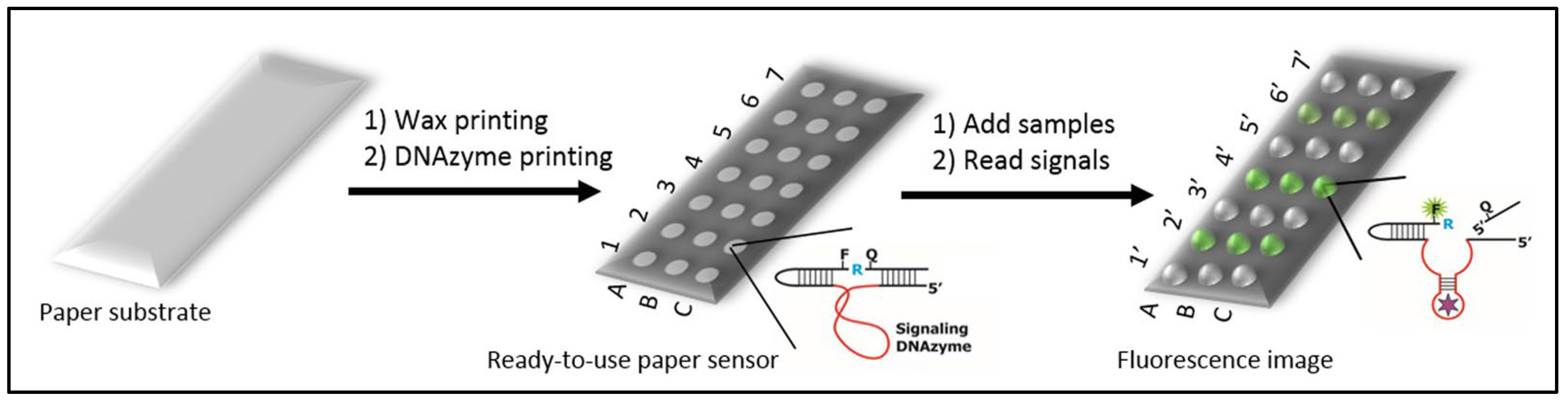
3.2.1. Paper Sensors

Paper sensors are strips of paper on which reagents, molecules, or ligands, can be grafted to make sensors. They can be used to detect the presence or absence of an analyte and can be useful in some diagnoses. The best-known example of the use of paper sensors is the pregnancy test. Paper sensors were initially intended for use in the field in developing countries where the installation of expensive and cumbersome equipment is impossible. However, their many advantages make them good laboratory tools too. With their low cost, ease of use, and portability (not very cumbersome and heavy) it becomes possible to multiply sampling for pathogen detection. During an industrial process using cell bioreactors or in the manufacturing of innovative therapy drugs, one can now easily imagine more controls at all critical steps of the process. Moreover, paper sensors can be integrated in a more complex device in order to automate the task by a robot and allow a quick reading of the result afterwards. Indeed, sampling takes time, it requires an operator and can lead to contamination. These constraints make it impossible to repeat the controls while they should be regular and representative of the whole sample. With the help of an automaton and paper sensors, these problems can be solved, in particular the problem of sterility breakage by the operator. Several techniques are possible, and several different ligands can be used with paper sensors. We review the main techniques.
Paper sensors are affordable devices for food [71,72,73] or environmental quality control [74]. They can also be used for the detection of pathogens or the rapid and accurate detection of diseases [75,76,77]. Their versatility makes them useful for virus diagnosis [78] or detection of bacterial spores [79].
Hossain et al. reported an approach for the selective and sensitive multiplexed detection of E. coli using a lab-on-paper test strip based on the activity of an intracellular enzyme. The objective is to trigger a rapid enzymatic hydrolysis of the substrate, causing a color change from colorless to blue or from yellow to red depending on the bacteria. With a step of preconcentration using immunomagnetic nanoparticles, the detection limit was about 10 CFU/mL for E. coli in 30 min and without cell culture. Thus, these paper test strips may be suitable for the detection of viable pathogens in water, blood, or cell therapy products. In addition, if a culture step is added, it allows the detection of less than 1 CFU in 100 mL in 8 h, making the paper test strips useful for the detection of multiple pathogens in complex samples. However, a filtration step is required to remove enzymes from the medium before lysis of the microorganism. This allows selective detection of viable bacteria. The test strips can be adapted to different bacteria, or different intracellular enzyme markers by varying the nature of the reagents printed on the paper test strip.
Ali et al. presented a simple all-in-one paper-based sensor for the detection of E. coli using a composite ink [77]. The paper consists of a fluorogenic DNAzyme probe for bacterial recognition and signal generation. It uses lysozymes that lyse the bacteria and sugars that stabilize the printed bioactive molecules. A fluorescent signal can be detected within 5 min of contact between the bacteria and the paper probe. The detection limit is 102 cells per mL, in different sample matrices, without sample enrichment (Figure 3).
In recent years, many efforts have been made to develop easy-to-use and inexpensive detection methods using paper sensors. Paper is readily available and can be modified by printing several types of reagents. Biorecognition items can be antibodies, nucleic acids, or proteins to detect a variety of analytes. The availability of high-throughput printers combined with microfabrication techniques allows the precise design of small diagnostic devices. The field of early detection of infectious organisms in clinical samples is the preferred area for the use of paper-based sensors. The cited works used E. coli as a target. The same approach can be extended to the detection of other bacteria. This is possible by selecting specific DNAzyme probes from available libraries. These scientific papers focused on food control, but an adaptation to ATMP control is largely feasible.
Several research avenues are promising; paper sensors can be combined with other techniques such as acoustophoresis [80,81] for analyte sorting, discussed in Section 3.2.2, and Raman spectroscopy [82] to improve accuracy (Section 3.3.1). The system and sampling can also be automated to operate under sterile conditions. A final avenue to explore would be to integrate the paper sensor or DNAzyme directly into the industrial culture process to approach a real-time reading of the ATMP bioreactor.

3.2.2. Microfluidics

Recent advances in the microfluidic field makes it the most promising technique and place it at the crossroads of sample-based and real-time techniques. The major limitation of microfluidics remains the low volumes processed, but due to other sorting techniques (acoustophoresis) or multiplexing, microfluidics can become a near real-time technique. It offers many possibilities adapted to our problem, which we can see in this section.
Microfluidics is the science of fluid manipulation at the micrometer scale. This field of research is inspired by the nature that already mastered these techniques perfectly, such as blood capillaries or sap transport in plants. Microfluidics can also be considered as a technology for manufacturing “lab-on-a-chip” with innovative applications in the field of chemistry, biology [83], medicine [84], and the food industry [85]. Microfluidic systems include a set of miniaturized components that can replace bulky and very expensive instruments [86,87]. This true “microprocessor for biology” represents a revolution for biology and chemistry similar to that brought by microprocessors to electronics and computers.

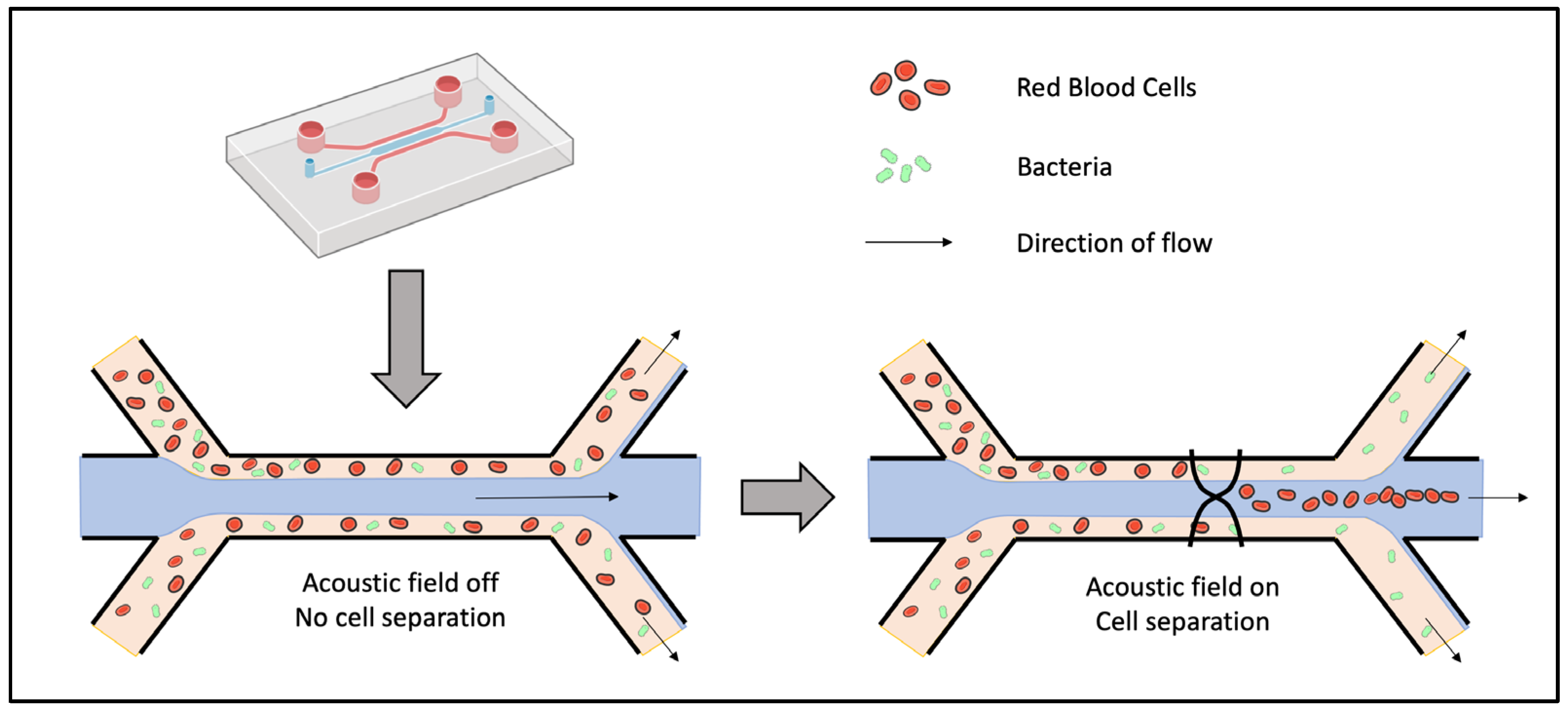
Sorting by Acoustophoresis

Combined with a classical detection method, microfluidics can be used as a first step to sort pathogens from the rest of the complex environment. This can greatly help the detection of the signal of pathogens that would be otherwise hidden by the strong signal of other components. Several microfluidic methods exist to separate bacteria from a sample [88,89]; here we present the acoustophoresis method in more detail. By applying intense sound waves on micro channels, flowing particles are sorted according to their size because the acoustically induced velocity of a suspended object scales with the square of their radius. For example, small bacteria can be separated from the larger blood cells as depicted in Figure 4. Blood flows in the periphery of the channel and when the acoustic field is turned on, it applies a force on blood cells. They are thereby moved into the central stream, which then flows in a channel separated from the rest of the blood. On the contrary, small bacteria are much less affected by the acoustic field and stay in the peripheric streams.
Li et al. successfully sorted a mixture of E. coli and human blood cells using acoustophoresis [80]. The resulting solution containing the bacteria showed a purity of more than 96% bacteria (less than 4% blood cells). This made the following electrochemical detection more sensitive as the non-specific signals due to blood cells were eliminated. As for most of microfluidic systems, their device has the advantages of being cost-effective, automated, and miniaturized. Those characteristics are well-suited for bacterial detection in industrial production.
Similarly, Ohlsson et al. invented a device for acoustic separation of bacteria from blood combined with PCR detection for rapid sepsis diagnostics [90]. The acoustic separation of whole blood was performed as in Figure 4, then bacteria were trapped onto suspended polystyrene particles. Finally, a PCR was performed on the bacteria trapped in a polymer microchip containing dried PCR reagents. Tested with Pseudomonas putida, they showed a detection under two hours with an LOD of 103 bacteria/mL. Compared with blood culture on samples from septic patients, their system detected half of the septic cases, indicating the real need for further improvement to compete with the reference method. Consequently, they presented a new microfluidics design two years later [81] to increase the throughput of bacteria separation from whole blood. By matching the acoustic impedance of the central stream to that of the blood sample, they could process 1 mL of undiluted blood within 12.5 min, with a bacteria recovery rate of 90% and a blood cell removal rate above 99%. As shown in Table 1, flow rate and dilution factor can be tuned differently to optimize either the bacteria recovery rate (up to 99.7%) or the blood cell removal (up to 99.99%).
Dow et al. combined a similar acoustophoresis system with a bacteriophage-based luminescence assay [91]. Tested on Pseudomonas aeruginosa, E. coli, and Staphylococcus aureus, they demonstrated an LOD of six bacteria from blood with initial concentration of 102 CFU/mL. Acoustophoresis greatly improved the LOD compared with unpurified samples.
In conclusion, acoustophoresis can be used to separate bacteria from blood cells with excellent bacterial recovery. The high throughput enables the processing of clinical samples in a short period of time. It can also allow real-time monitoring without the need to collect samples during cell therapy production. Rate flow can further be increased through the use of higher blood cell concentrations, larger channels, or multiple parallel separation channels to monitor all types of ATMPs. In combination with downstream bacterial detection, this separation method paves the way for the potential development of fast detection methods. More detection methods should be tested with acoustophoresis in order to find the most suited and reduce the LOD and the time of response. This method can also be used to remove bacteria from a sample for sterilization purposes and not just for detection. The manufacturing of cell or gene therapies can be facilitated.
As for previous methods, acoustophoresis should be validated on a larger number of bacterial species and strains. Other individual bacteria should behave in a similar way during acoustic separation, because the size difference with blood cells is always large. However, pathogenic bacteria are known to form clumps, to reside within white blood cells, or to adhere to blood cells or platelets. The risk is therefore that bacteria are sorted out with the large cells. One solution could be to chemically prevent the adhesion of bacteria to blood cells during the assay with the risk of denaturing the ATMP sample.

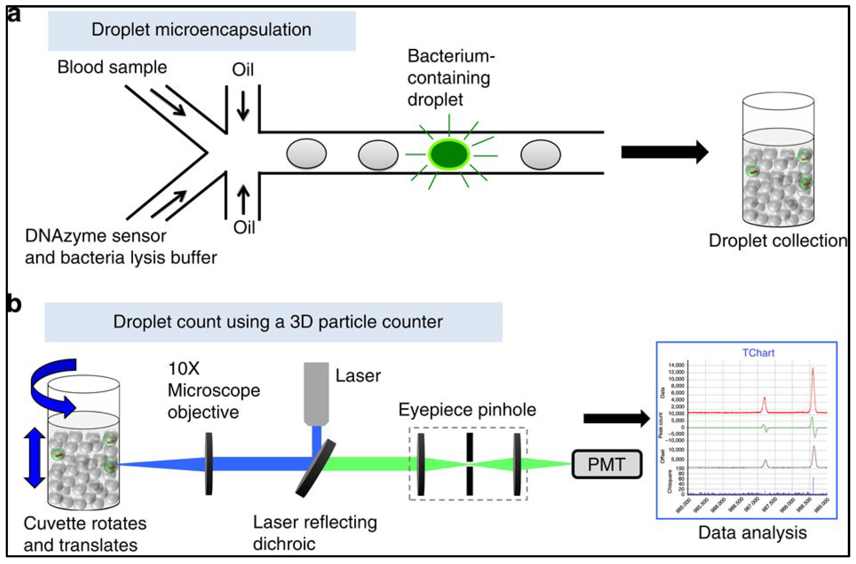
Microdroplets and 3D Particle Counter

Droplet microfluidics is another microfluidic method that can improve the detection of pathogens. The idea is to analyze a solution by decomposing it into a microdroplet surrounded by oil. By doing so, every microliter of the solution is screened individually, allowing a finer detection. By sorting the droplets of interest, it is as if the initial solution was concentrated, or enriched, by a factor of several hundred [92].
While droplet microfluidics can improve detection sensitivity and specificity, initial flow rates limited its application to sample with a volume in the microliter range. However, recent developments resulted in a 3D particle counter able to process, within minutes, samples of several milliliters, as usually required for clinical relevance. Kang et al. presented a technology termed Integrated Comprehensive Droplet Digital Detection (IC 3D) based on the microencapsulation of bacteria and DNAzyme-based detection [58,93]. Blood samples are mixed in a microfluidic channel with the DNAzyme sensor solution comprising a bacterial lysis buffer (Figure 5). This mixture is then immediately encapsulated in hundreds of millions of individual droplets. The confinement of the bacteria in the droplets increases the concentration of the bacteria’s target molecules. This allows individual bacteria to be detected more quickly by DNAzyme’s sensors. The droplets are then collected and analyzed using a high-throughput particle counting system. Tested with E. coli, their system could detect the bacteria from milliliters of diluted blood with an LOD of 1 bacteria/mL. The detection does not need culture or amplification steps and can thus provide a result within 1.5–4 h. It can also detect slow growing organisms such as Mycobacterium tuberculosis. Key performance specifications are summarized in Table 2.
This set of technologies can also detect other pathogens, genetic material, or tumor markers [94]. Hedde et al. also showed how 3D particle sorting enables the detection and isolation of E. coli from whole blood within minutes for clinically relevant fluid volumes (1–10 mL) [92]. Other combinations of microsystems coupling high throughput bacterial immunomagnetic capture to non-contact cell lysis using an alternating current magnetic field allow bacterial detection in the range of 102 CFU/mL with a flow rate of 50 mL/h [95].
In conclusion, microfluidics combined with sorting methods, microdroplets, innovative particle counters, and the use of good ligands is becoming the most efficient and promising method to address the problem of broad-spectrum detection. It allows to process larger volumes and thus to overcome the low throughput problem encountered by flow cytometry. Future perspectives are the development of an automated, compact, and multiplexed device allowing the rapid detection of several bacterial species in any sample, in particular innovative cell therapies. Furthermore, these microfluidic techniques can be coupled to a variety of biosensors for detection and identification.

3.3. Development of Physical and Computer Analysis Methods

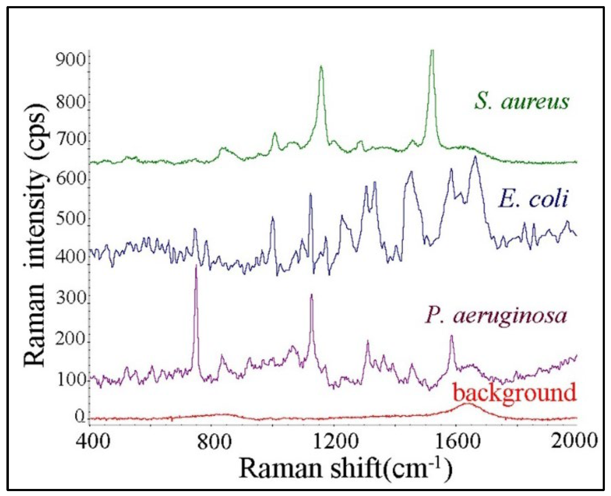
3.3.1. Raman Spectroscopy

Among instrumental analytical methods, vibrational spectroscopy techniques have long been established; they allow the identification and quantification of the chemical composition of samples in a non-destructive manner. Raman spectroscopy exploits the physical phenomenon in which molecular vibrations in a sample are excited by incoming monochromatic light. This energy transfer results in a small shift in the frequency of the subsequent scattered light. Consequently, measuring the spectrum of the Raman scattered light provides molecular information about the sample. The preparation time is zero or almost zero and samples can be of different types: solid, liquid, or gaseous. Raman spectroscopy offers the possibility of in situ measurements.
In the study of bacteria, Raman spectroscopy is particularly well-suited for identification. Multiple bacteria can be identified simultaneously from the Raman spectrum of the analyzed sample by comparison with reference spectra of library [96] (see Figure 6). This point is similar to what exists for infrared spectroscopy. For example, Rebrošová et al. showed that 16 different species of Staphylococcus can be distinguished using Raman spectroscopy [97]. Although many studies deal with the identification of bacteria, we focused here on their detection, in accordance with the scope of this review. However, one can keep in mind that Raman spectroscopy offers the possibility to perform both at the same time.
Combined with microscopy, Raman spectroscopy allows the detection of a single cell [98]. This avoids the time-consuming step of incubation, and thus increases greatly the rapidity of the analysis. This is similar to solid phase cytometry, but the Raman spectrum allows for a more specific detection. In particular, auto-fluorescent particles can no longer be confused with pathogens. One drawback of the high resolution of microscopy is the time that is required to scan the sample looking for bacteria. Strola et al. [99] overcame this issue by using lens-free imaging instead of a classical microscope, providing a wide field of view. Moreover, lens-free imaging is cheaper and allows for more compact systems. With such a setup, they could localize and detect single bacteria under 1 min, though the concentration of bacteria in the solution, after drying, was high (108 CFU/mL).
Figure 6. Averaged Raman spectra of different bacteria species. This figure is a partial reproduction from reference [100], licensed under CC BY-NC-ND 3.0.
Applsci 12 05892 g006
For bacterial detection applications, other approaches used Surface-Enhanced Raman Spectroscopy (SERS) to enhance the spontaneous Raman signal, which is normally of low intensity [101]. For example, Yang et al. combined an aptamer, a DNA walker on magnetic nanoparticles, and SERS detection to reach an LOD as low as 4 CFU/mL for S. typhimurium [102]. This limit was made possible both by SERS and the enzymatic augmentation performed by the DNA walker. However, the method did not seem specific at this concentration. As another promising example of SERS-based detection, Cheng et al. [100] used electrokinetic microfluidics to isolate and concentrate bacteria on a SERS substrate for detection directly from blood samples. They demonstrated the detection of S. aureus, E. coli, and P. aeruginosa under 5 min, with a concentration of 5 × 10³ CFU/mL.
In conclusion, Raman spectroscopy allows for the detection and identification of pathogens by looking for their specific spectrum inside samples. Compared with infrared spectroscopy, Raman spectroscopy is less hindered by the signal of water and is thus more suited for biological complex media. Consequently SERS-based biosensors are a growing field. There are already many databases of pathogens spectra, but applications to detection in complex biological media are rare. Spectra are difficult to interpret and time consuming to analyze but this technique can be greatly improved by artificial intelligence as described in the next paragraph.

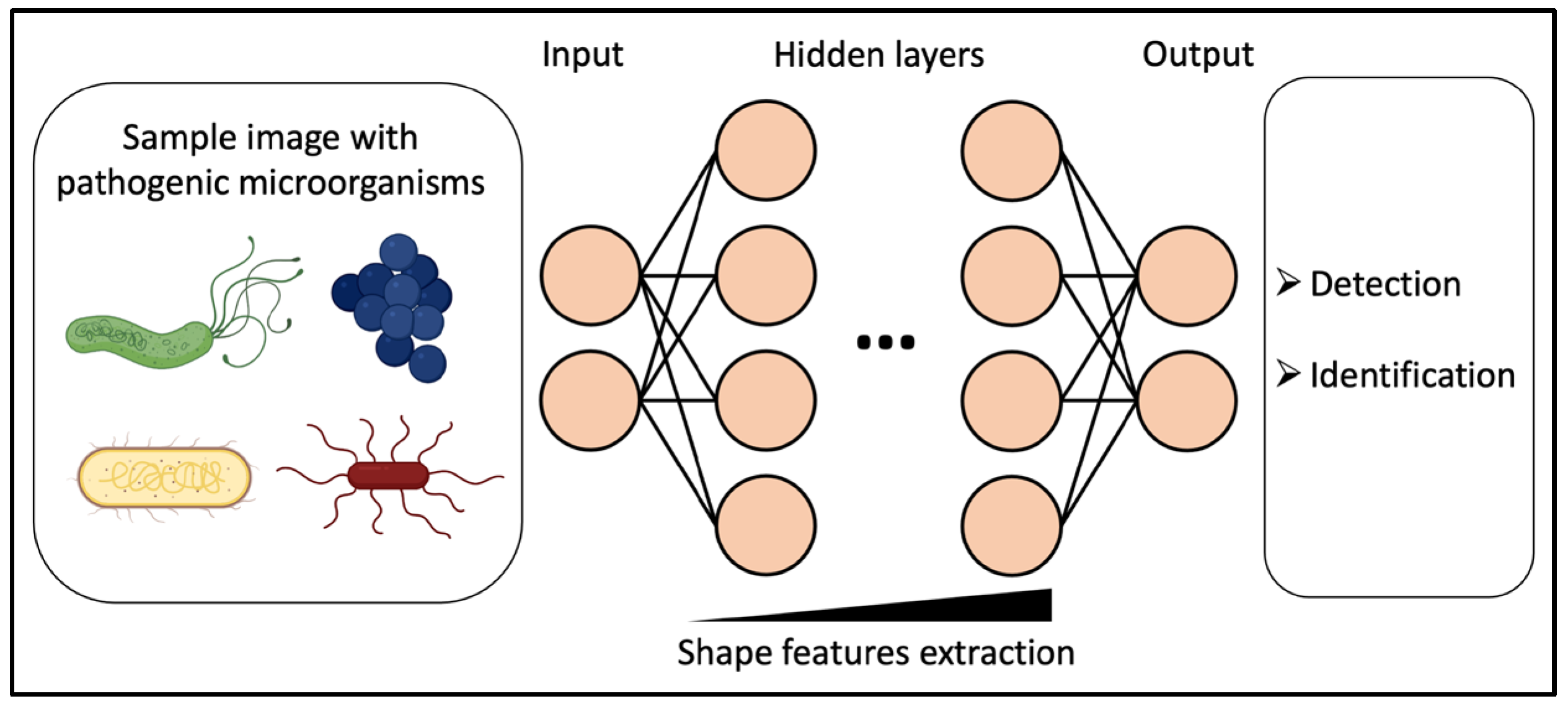
3.3.2. Deep Learning for Microscopy-Based Sampling Methods

Recent progress in artificial intelligence, and more specifically deep learning, opened the way to an automatic and robust analysis of images. This is also true for the analysis of microscopic images, and deep learning was applied to the detection and identification of microorganisms [103,104].
Deep learning is part of the larger family of artificial neural networks (ANNs). ANNs were inspired by the functioning of the brain, with neurons organized in successive layers connected between them (Figure 7), from the input (e.g., images) to the output (e.g., bacteria presence). By training them on labelled data (e.g., image of known bacteria), they can find patterns allowing them to recognize unknown data afterwards. Deep learning refers to ANNs with many consecutive layers and corresponding ANNs are called deep neural networks (DNNs). Such structure was only recently enabled from the development of computational power.
In the case of image analysis, DNNs are capable of extracting shape features of images to classify them according to pre-defined labels (e.g., different species of bacteria), or to extract a quantity of an image (e.g., a concentration in bacteria). In practically all fields of image analysis, deep learning has proved to be more accurate than other methods, and even more accurate than human eyes for some applications [105]. Deep learning is mainly used for classification of objects, and in the field of microbiology, to identify various species of virus [106], bacteria [107,108], fungi [109], or parasites [110]. Those studies include the analysis of different modalities of microscopy such as electron microscopy (for viruses), fluorescence microscopy, brightfield microscopy, or even vibrational microspectroscopy. Trained on labelled datasets, deep neural networks are then able to identify microorganisms based on their shape or spectrum for spectroscopic images.
For detection purposes, DNNs can detect automatically bacteria over a large field of view, and distinguish them against other particles. Kang et al. combined several DNNs to detect and identify single cells of five strains of bacteria (Campylobacter, E. coli, Listeria, Salmonella, and Staphyloccocus) from visible spectroscopy images [111]. Wang et al. [112] used a DNN to detect bacterial growth inside an agar plate from coherent microscopy images taken every 30 min. Three strains (E. coli, Klebsiella aerogenes, and Klebsiella pneumoniae) were tested for detection and subsequent identification, with an LOD of ~1 CFU/L in less than 9 h.
Deep learning allows for a finer analysis of images for increased sensitivity and specificity in detection. Public databases of different types of microorganisms are emerging [103], which allows a broader use of deep learning for microbiology. For now, studies are mainly carried out on isolates, but it would be interesting to extend on bacteria within complex environments.
In conclusion, while Raman spectroscopy took a long time to analyze and process data in the past, it is becoming fast and very sensitive thanks to deep learning. If we witness the birth of an official reference system for microorganisms, the Raman method will become a must in the quality control of ATMP. However, its purchase cost may remain an obstacle to its acquisition for some laboratories.

3.4. Improvements and Developments in Real-Time and Online Analysis Techniques

In methods requiring sampling, each breach of containment can potentially lead to a risk of contamination of the sample, whether in the production of advanced therapy drugs, or the industrial production of complex media. On the other hand, not enough sampling may lead to delayed or unrepresentative results. For this reason, methods allowing real-time monitoring of biological samples were developed.

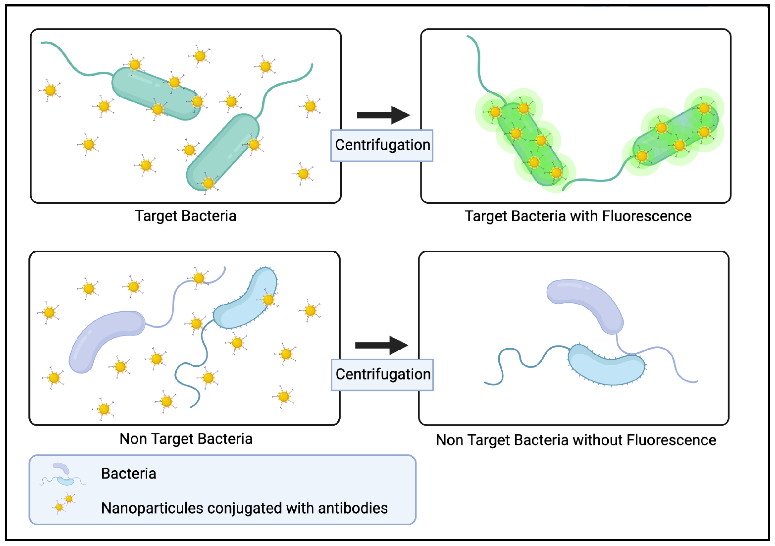
3.4.1. Bio-Conjugated Nanoparticles

Conjugating nanoparticles (NPs) with ligands such as antibodies (see Section 2.1), make them interesting tags for pathogens detection [113]. Zhao et al. demonstrated the use of bio-conjugated NP as fluorescent tag in bioassays for the detection of bacteria [114]. Silica NPs were mixed with the sample to be analyzed and bounded to presumed bacteria thanks to specific antibodies (Figure 8). Then, unbound NPs were eliminated by centrifugation, and finally, the fluorescence of the sample was measured either in well plates by a fluorometer or by flow cytometry. As the NPs encapsulated a thousand fluorophores, they provided a fluorescence intensity a thousand times greater than what a single antibody-conjugated fluorophore could provide. Tested on E. coli, Salmonella, or Bacillus cereus in ground beef samples suspended in a 10 mL solution, the high fluorescence signal allowed for the detection of a single bacterium under 20 min. Moreover, the measurement was quantitative. Their method also works for DNA detection.
NPs can also be used as SERS tags for pathogen detection, as Paul et al. showed with Dengue and West Nile viruses [115]. Conjugating NPs with selective antibodies, they reached an LOD of 10 plaque-forming units/mL in less than 30 min. Using magnetic bio-conjugated NPs, it is also possible to selectively isolate bacteria or aptamers specifics to bacteria [102].
In conclusion, bio-conjugated nanoparticles, combined with various detection systems, allow sensitive and specific pathogen detection. They can be used for real-time detection in industrial cell bioreactors or in the manufacture of innovative therapy drugs. However, the question arises as to whether the nanoparticles are safe for humans or cells, and the quantity required for high-volume applications allowing rapid detection of several bacterial species.

3.4.2. Surface Plasmon Resonance

Surface Plasmon Resonance (SPR) has been extensively used in the past decade for optical biosensors [116,117]. Its principle works as follows: when a beam of light illuminates an interface between two media, a portion of the incident light is reflected on the interface and the other portion of the light is refracted through the surface. If a thin layer of metal, rich in free electrons, is deposited at the interface, they come into resonance with the photons of the incident beam. This phenomenon is called Surface Plasmon Resonance. The resonance is visible through a loss of intensity in the reflected beam at a given angle. Any deposit on the surface can be detected because it modifies the angle at which the plasmon resonance occurs. In biosensors, this sensitive detection is used to detect the binding of species present in solutions to ligands grafted to the surface (Figure 9) [118,119]. By grafting ligands specific to different types of pathogens on different positions on the surface, it is possible to perform a multiplex assay. This method is called SPR imaging (SPRi) as the surface is imaged to cover all ligand positions. The identification of the pathogens can be performed simultaneously to detection, depending on the specificity of the ligands used.
The technology of an SPR biosensor has been successfully used for the detection of various analytes such as biomolecules [120] and proteins, bacterial toxins [121], or to monitor the formation of a bacterial biofilm and its degradation [122]. In the case of microorganisms, SPRi have been used for their detection in various media. Nair el al. detected E. coli in PBS and human urine in under 35 min for concentrations ranging from 103 to 109 CFU/mL and an LOD of 102 CFU/mL [123]. In food samples, Chen et al. demonstrated the detection of Salmonella, E. coli and Listeria with a SPRi-based immunoassay. Their system had an LOD around 106 CFU/mL for direct measurement on a chicken carcass rinse or 1 CFU/mL with overnight enrichment [124].
Pardoux et al. [41,68,125,126,127] proposed a method to make the detection of bacteria by SPRi as fast as possible where they no longer analyze a bacterial suspension after a growth step. The suspension to be tested is injected directly in contact with the microarray. The possible growth of the bacteria and their detection is thus performed at the same time (Figure 10). The detection limits claimed for standard SPRi in sample-based detection are often less relevant. Thus, by combining the growth of microorganisms and their detection, the time saving is real. This technique has similarities with blood cultures with comparable analysis times. The advantage here is real-time monitoring, whereas blood culture bottles provide a positive response without allowing quantification.
In this method, the analysis time is governed by the growth rate of the microorganism; the faster the growth, the sooner the detection threshold of the sensor can be reached. (Figure 10) The method has been validated on various strains of Salmonella, Listeria, Staphylococcus, and Escherichia.
SPRi is suitable for bacteria detection in whole blood or cellular products. With few probes, it is possible to detect a wide range of species within less than 18 h from a low concentration sample. The method can be used in real-time monitoring without the need of sampling and the associated contamination risks.
SPRi has a multitude of advantages in bacterial detection. There is no need for multiple sampling and no breach of containment. Each breach of containment can potentially lead to a risk of contamination of the sample, whether in the production of advanced therapy drugs or industrial production of complex media. The analysis is performed in real-time and is extremely interesting in the quality control of health products.
Despite this progress, the conditions required for applications in the health and pharmaceutical industries are not yet in place. Advances in sensor and device design, as well as improvements in ligands, are future prospects in this area. Future research in SPRi can provide this technique a promising role in the pharmaceutical and food industries.
Other techniques are under development, in particular the Localized Surface Plasmon Resonance (LSPR) technique [128] with very promising results in the detection of viruses [129] or in the detection of biomarkers in cancer [130].
If broad-spectrum probes are validated, they can be combined with more specific probes within SPRi sensors. This provides broad-spectrum detection on the one hand, and specific identification of selected relevant strains on the other. A significant advantage of SPRI is that it makes it possible to probe the sample without altering it and then inject the patient with the cell therapy. Another possibility would be to copy the automatic detection model of blood culture bottles and apply it with an SPRi chip. One could proceed to an automated analysis of the final product of ATMP while leaving the possibility of complementary analysis with other conventional techniques from the same sample. Originally the main limitation of SPRi was the limited recognition spectrum due to the ligands used, the antibodies. The use of antimicrobial peptides in complex biological media provides future perspectives in research and development of microorganism detection methods.
As described along this review and summarized in this Table 3, there are many techniques for microorganism detection. Nevertheless, choosing the right technique for complex samples depends on different factors such as the sensibility toward bacteria and the limit of detection (LOD), the analysis time, the specificity of the technique, its cost, and its advantages and disadvantages.

4. Conclusions

Conventional detection methods have demonstrated a reliable, reproducible, and resourceful tool for over a century in determining pathogenic contamination. The current state-of-the-art techniques demonstrate reproducible sensitivity and are generally much faster than conventional techniques, as shown in Table 3, synthesizing this review. However, since no single approach satisfies all or even most of the emerging criteria for quick, effective, reproducible, and sensitive results; there is a knowledge gap in this research field.
The use of microfluidics combined with DNAzyme or Raman spectroscopy have excellent analysis time but the need for sampling and the absence of a real-time follow-up are important weak aspects. Analytical methods requiring sampling have many advantages such as speed of analysis and unequalled precision. The major disadvantage is the inescapable break in sterility and the small volume processed. A breach of containment provides the possibility of intrusion of micro-organisms during the manufacture of ATMPs. These methods can be more appropriate for quality control on final product, without reinjection to patients but the possibility to perform other analyses (conformity, identification, etc.) as they are non-destructive. A major possible improvement is undoubtedly the automation of this task due to robotics which allow multiple controls throughout the industrial process of manufacturing ATMPs.
The new methods of analysis requiring sampling are therefore very promising, but the time needed for the examination can also delay the corrective actions. The multiplication of control steps poses many problems of organization, cost, sterility break, and loss of raw material. Thus, the need for real-time monitoring is needed in an industrial manufacturing process to accelerate the detection process in order to save time and money. The aspect of real-time control has therefore become a concern for researchers.
SPRi has these qualities of online and real-time analysis. Even if the analysis time remains quite long, it allows to process large volumes.
There is an notable duality, either the technique is very fast but requires sampling, or the technique is slower but allows real-time monitoring without breaking sterility.
The notion of representativeness thus intervenes in the balance of the advantage/inconvenience of each technique. For techniques with sampling, is the sample representative of the whole of the complex environment analyzed? For in-line techniques, is the probability that a bacterium in the whole medium meets the probe high enough to make the technique significantly robust? These notions are not addressed in the review but deserve further investigation in the future.
In conclusion, the ideal solution to control the sterility of complex biological samples is therefore to combine several techniques chosen in accordance with the sample to be analyzed and analysis context. SPRi can be used for real-time monitoring to stop production if there is a potential contamination risk. It could be interesting to combine SPRi with a technique such as Raman spectroscopy for control of the final product.

Author Contributions

Conceptualization, P.P.; Methodology, P.P., B.B.; Writing—preparation of original version, P.P., B.B.; Writing—revision and editing, P.P., B.B., F.R., S.C.G.; Supervision, F.R., S.C.G. All authors have read and agreed to the published version of the manuscript.

Funding

This research received no external funding.

Conflicts of Interest

The authors declare no conflict of interest.

References

  1. Hameed, S.; Xie, L.; Ying, Y. Conventional and Emerging Detection Techniques for Pathogenic Bacteria in Food Science: A Review. Trends Food Sci. Technol. 2018, 81, 61–73. [Google Scholar] [CrossRef]
  2. Green, M.S.; LeDuc, J.; Cohen, D.; Franz, D.R. Confronting the Threat of Bioterrorism: Realities, Challenges, and Defensive Strategies. Lancet Infect. Dis. 2019, 19, e2–e13. [Google Scholar] [CrossRef]
  3. Rajapaksha, P.; Elbourne, A.; Gangadoo, S.; Brown, R.; Cozzolino, D.; Chapman, J. A Review of Methods for the Detection of Pathogenic Microorganisms. Analyst 2019, 144, 396–411. [Google Scholar] [CrossRef] [PubMed]
  4. Bone, R.C.; Balk, R.A.; Cerra, F.B.; Dellinger, R.P.; Fein, A.M.; Knaus, W.A.; Schein, R.M.H.; Sibbald, W.J. Definitions for Sepsis and Organ Failure and Guidelines for the Use of Innovative Therapies in Sepsis. Chest 1992, 101, 1644–1655. [Google Scholar] [CrossRef] [Green Version]
  5. Chun, K.; Syndergaard, C.; Damas, C.; Trubey, R.; Mukindaraj, A.; Qian, S.; Jin, X.; Breslow, S.; Niemz, A. Sepsis Pathogen Identification. J. Lab. Autom. 2015, 20, 539–561. [Google Scholar] [CrossRef]
  6. Gotts, J.E.; Matthay, M.A. Sepsis: Pathophysiology and Clinical Management. BMJ 2016, 353, i1585. [Google Scholar] [CrossRef] [Green Version]
  7. Hocquet, D.; Sauget, M.; Roussel, S.; Malugani, C.; Pouthier, F.; Morel, P.; Gbaguidi-Haore, H.; Bertrand, X.; Grenouillet, F. Validation of an Automated Blood Culture System for Sterility Testing of Cell Therapy Products. Cytotherapy 2014, 16, 692–698. [Google Scholar] [CrossRef]
  8. Váradi, L.; Luo, J.L.; Hibbs, D.E.; Perry, J.D.; Anderson, R.J.; Orenga, S.; Groundwater, P.W. Methods for the Detection and Identification of Pathogenic Bacteria: Past, Present, and Future. Chem. Soc. Rev. 2017, 46, 4818–4832. [Google Scholar] [CrossRef]
  9. Monteiro, A.C.; Fortaleza, C.M.; Ferreira, A.M.; Cavalcante Rde, S.; Mondelli, A.L.; Bagagli, E.; da Cunha Mde, L. Comparison of Methods for the Identification of Microorganisms Isolated from Blood Cultures. Ann. Clin. Microbiol. Antimicrob. 2016, 15, 45. [Google Scholar] [CrossRef] [Green Version]
  10. Buszewski, B.; Rogowska, A.; Pomastowski, P.; Złoch, M.; Railean-Plugaru, V. Identification of Microorganisms by Modern Analytical Techniques. J. AOAC Int. 2017, 100, 1607–1623. [Google Scholar] [CrossRef]
  11. Kumar, H.; Bhardwaj, K.; Kaur, T.; Nepovimova, E.; Kuča, K.; Kumar, V.; Bhatia, S.K.; Dhanjal, D.S.; Chopra, C.; Singh, R.; et al. Detection of Bacterial Pathogens and Antibiotic Residues in Chicken Meat: A Review. Foods 2020, 9, 1504. [Google Scholar] [CrossRef] [PubMed]
  12. Thorpe, T.C.; Wilson, M.; Turner, J.; DiGuiseppi, J.; Willert, M.; Mirrett, S.; Reller, L. BacT/Alert: An Automated Colorimetric Microbial Detection System. J. Clin. Microbiol. 1990, 28, 1608–1612. [Google Scholar] [CrossRef] [PubMed] [Green Version]
  13. Ramamurthy, T.; Ghosh, A.; Pazhani, G.P.; Shinoda, S. Current Perspectives on Viable but Non-Culturable (VBNC) Pathogenic Bacteria. Front. Public Health 2014, 2, 103. [Google Scholar] [CrossRef] [PubMed]
  14. Wideman, N.E.; Oliver, J.D.; Crandall, P.G.; Jarvis, N.A. Detection and Potential Virulence of Viable but Non-Culturable (VBNC) Listeria Monocytogenes: A Review. Microorganisms 2021, 9, 194. [Google Scholar] [CrossRef]
  15. Furst, A.L.; Francis, M.B. Impedance-Based Detection of Bacteria. Chem. Rev. 2018, 119, 700–726. [Google Scholar] [CrossRef]
  16. Kaya, H.O.; Cetin, A.E.; Azimzadeh, M.; Topkaya, S.N. Pathogen Detection with Electrochemical Biosensors: Advantages, Challenges and Future Perspectives. J. Electroanal. Chem. 2021, 882, 114989. [Google Scholar] [CrossRef]
  17. Spaeth, S.; Tran, Q.; Liu, Z. Evaluation of an ATP-Bioluminescence Rapid Microbial Screening Method for In-Process Biologics. PDA J. Pharm. Sci. Technol. 2018, 72, 574–583. [Google Scholar] [CrossRef]
  18. Fricke, C.; Harms, H.; Maskow, T. Rapid Calorimetric Detection of Bacterial Contamination: Influence of the Cultivation Technique. Front. Microbiol. 2019, 10, 2530. [Google Scholar] [CrossRef] [Green Version]
  19. Wacogne, B.; Legrand, D.; Azzopardi, C.-L.; Pieralli, C.; Frelet-Barrand, A. Optical Spectroscopy Methods to Monitor Cells and Bacteria Concentrations and to Detect Contamination During Cell Culture: Application to the Fabrication of ATMPs. In Biomedical Engineering Systems and Technologies; Ye, X., Soares, F., De Maria, E., Gómez Vilda, P., Cabitza, F., Fred, A., Gamboa, H., Eds.; Communications in Computer and Information Science; Springer International Publishing: Cham, Switzerland, 2021; Volume 1400, pp. 53–75. ISBN 978-3-030-72378-1. [Google Scholar]
  20. Hatiboruah, D.; Devi, D.Y.; Namsa, N.D.; Nath, P. Turbidimetric Analysis of Growth Kinetics of Bacteria in the Laboratory Environment Using Smartphone. J. Biophotonics 2020, 13, e201960159. [Google Scholar] [CrossRef]
  21. Chen, Z.; Yang, T.; Yang, H.; Li, T.; Nie, L.; Mou, X.; Deng, Y.; He, N.; Li, Z.; Wang, L.; et al. A Portable Multi-Channel Turbidity System for Rapid Detection of Pathogens by Loop-Mediated Isothermal Amplification. J. Biomed. Nanotechnol. 2018, 14, 198–205. [Google Scholar] [CrossRef]
  22. Paczesny, J.; Richter, Ł.; Hołyst, R. Recent Progress in the Detection of Bacteria Using Bacteriophages: A Review. Viruses 2020, 12, 845. [Google Scholar] [CrossRef] [PubMed]
  23. Péter, B.; Farkas, E.; Kurunczi, S.; Szittner, Z.; Bősze, S.; Ramsden, J.J.; Szekacs, I.; Horvath, R. Review of Label-Free Monitoring of Bacteria: From Challenging Practical Applications to Basic Research Perspectives. Biosensors 2022, 12, 188. [Google Scholar] [CrossRef] [PubMed]
  24. Sibilo, R.; Pérez, J.M.; Tebbenjohanns, F.; Hurth, C.; Pruneri, V. Surface Cytometer for Fluorescent Detection and Growth Monitoring of Bacteria over a Large Field-of-View. Biomed. Opt. Express 2019, 10, 2101–2116. [Google Scholar] [CrossRef] [PubMed]
  25. Kennedy, D.; Wilkinson, M.G. Application of Flow Cytometry to the Detection of Pathogenic Bacteria. Curr. Issues Mol. Biol. 2017, 23, 21–38. [Google Scholar] [CrossRef] [PubMed] [Green Version]
  26. Zand, E.; Froehling, A.; Schoenher, C.; Zunabovic-Pichler, M.; Schlueter, O.; Jaeger, H. Potential of Flow Cytometric Approaches for Rapid Microbial Detection and Characterization in the Food Industry—A Review. Foods 2021, 10, 3112. [Google Scholar] [CrossRef] [PubMed]
  27. Lemarchand, K.; Parthuisot, N.; Catala, P.; Lebaron, P. Comparative Assessment of Epifluorescence Microscopy, Flow Cytometry and Solid-Phase Cytometry Used in the Enumeration of Specific Bacteria in Water. Aquat. Microb. Ecol. 2001, 25, 301–309. [Google Scholar] [CrossRef] [Green Version]
  28. Lequin, R.M. Enzyme Immunoassay (EIA)/Enzyme-Linked Immunosorbent Assay (ELISA). Clin. Chem. 2005, 51, 2415–2418. [Google Scholar] [CrossRef] [Green Version]
  29. Lecellier, A.; Gaydou, V.; Mounier, J.; Hermet, A.; Castrec, L.; Barbier, G.; Ablain, W.; Manfait, M.; Toubas, D.; Sockalingum, G. Implementation of an FTIR Spectral Library of 486 Filamentous Fungi Strains for Rapid Identification of Molds. Food Microbiol. 2015, 45, 126–134. [Google Scholar] [CrossRef]
  30. Kochan, K.; Bedolla, D.E.; Perez-Guaita, D.; Adegoke, J.A.; Chakkumpulakkal Puthan Veettil, T.; Martin, M.; Roy, S.; Pebotuwa, S.; Heraud, P.; Wood, B.R. Infrared Spectroscopy of Blood. Appl. Spectrosc. 2021, 75, 611–646. [Google Scholar] [CrossRef]
  31. Hou, T.-Y.; Chiang-Ni, C.; Teng, S.-H. Current Status of MALDI-TOF Mass Spectrometry in Clinical Microbiology. J. Food Drug Anal. 2019, 27, 404–414. [Google Scholar] [CrossRef]
  32. Zhu, H.; Zhang, H.; Xu, Y.; Laššáková, S.; Korabečná, M.; Neužil, P. PCR Past, Present and Future. BioTechniques 2020, 69, 317–325. [Google Scholar] [CrossRef] [PubMed]
  33. Benevides Lima, L.; Mesquita, F.P.; Brasil de Oliveira, L.L.; Andréa da Silva Oliveira, F.; Elisabete Amaral de Moraes, M.; Souza, P.F.N.; Montenegro, R.C. True or False: What Are the Factors That Influence COVID-19 Diagnosis by RT-QPCR? Expert Rev. Mol. Diagn. 2022, 22, 157–167. [Google Scholar] [CrossRef] [PubMed]
  34. Gdoura, M.; Abouda, I.; Mrad, M.; Ben Dhifallah, I.; Belaiba, Z.; Fares, W.; Chouikha, A.; Khedhiri, M.; Layouni, K.; Touzi, H.; et al. SARS-CoV2 RT-PCR Assays: In Vitro Comparison of 4 WHO Approved Protocols on Clinical Specimens and Its Implications for Real Laboratory Practice through Variant Emergence. Virol. J. 2022, 19, 54. [Google Scholar] [CrossRef]
  35. Li, J.; Macdonald, J.; Stetten, F. von Review: A Comprehensive Summary of a Decade Development of the Recombinase Polymerase Amplification. Analyst 2018, 144, 31–67. [Google Scholar] [CrossRef] [PubMed] [Green Version]
  36. Glökler, J.; Lim, T.S.; Ida, J.; Frohme, M. Isothermal Amplifications—A Comprehensive Review on Current Methods. Crit. Rev. Biochem. Mol. Biol. 2021, 56, 543–586. [Google Scholar] [CrossRef]
  37. Leonardo, S.; Toldrà, A.; Campàs, M. Biosensors Based on Isothermal DNA Amplification for Bacterial Detection in Food Safety and Environmental Monitoring. Sensors 2021, 21, 602. [Google Scholar] [CrossRef]
  38. Etchebarne, B.E.; Li, Z.; Stedtfeld, R.D.; Nicholas, M.C.; Williams, M.R.; Johnson, T.A.; Stedtfeld, T.M.; Kostic, T.; Khalife, W.T.; Tiedje, J.M.; et al. Evaluation of Nucleic Acid Isothermal Amplification Methods for Human Clinical Microbial Infection Detection. Front. Microbiol. 2017, 8, 2211. [Google Scholar] [CrossRef] [Green Version]
  39. Zanoli, L.M.; Spoto, G. Isothermal Amplification Methods for the Detection of Nucleic Acids in Microfluidic Devices. Biosensors 2013, 3, 18–43. [Google Scholar] [CrossRef] [Green Version]
  40. Templier, V.; Roupioz, Y. On the Challenges of Detecting Whole Staphylococcus Aureus Cells with Biosensors. J. Appl. Microbiol. 2017, 123, 1056–1067. [Google Scholar] [CrossRef] [Green Version]
  41. Templier, V.; Livache, T.; Boisset, S.; Maurin, M.; Slimani, S.; Mathey, R.; Roupioz, Y. Biochips for Direct Detection and Identification of Bacteria in Blood Culture-Like Conditions. Sci. Rep. 2017, 7, 9457. [Google Scholar] [CrossRef]
  42. Xu, J.; Chau, Y.; Lee, Y. Phage-Based Electrochemical Sensors: A Review. Micromachines 2019, 10, 855. [Google Scholar] [CrossRef] [PubMed] [Green Version]
  43. Pala, L.; Sirec, T.; Spitz, U. Modified Enzyme Substrates for the Detection of Bacteria: A Review. Molecules 2020, 25, 3690. [Google Scholar] [CrossRef] [PubMed]
  44. Bollella, P.; Katz, E. Enzyme-Based Biosensors: Tackling Electron Transfer Issues. Sensors 2020, 20, 3517. [Google Scholar] [CrossRef] [PubMed]
  45. Yunus, G. Chapter 42—Biosensors: An Enzyme-Based Biophysical Technique for the Detection of Foodborne Pathogens. In Enzymes in Food Biotechnology; Kuddus, M., Ed.; Academic Press: Cambridge, MA, USA, 2019; pp. 723–738. ISBN 978-0-12-813280-7. [Google Scholar]
  46. Jayasena, S.D. Aptamers: An Emerging Class of Molecules That Rival Antibodies in Diagnostics. Clin. Chem. 1999, 45, 1628–1650. [Google Scholar] [CrossRef] [Green Version]
  47. Melaine, F.; Coilhac, C.; Roupioz, Y.; Buhot, A. A Nanoparticle-Based Thermo-Dynamic Aptasensor for Small Molecule Detection. Nanoscale 2016, 8, 16947–16954. [Google Scholar] [CrossRef]
  48. Jafari, M.; Rezaei, M.; Kalantari, H.; Tabarzad, M.; Daraei, B. DNAzyme-Aptamer or Aptamer-DNAzyme Paradigm: Biochemical Approach for Aflatoxin Analysis. Biotechnol. Appl. Biochem. 2018, 65, 274–280. [Google Scholar] [CrossRef]
  49. Ellington, A.D.; Szostak, J.W. In Vitro Selection of RNA Molecules That Bind Specific Ligands. Nature 1990, 346, 818–822. [Google Scholar] [CrossRef]
  50. Kaur, H. Recent Developments in Cell-SELEX Technology for Aptamer Selection. Biochim. Biophys. Acta BBA-Gen. Subj. 2018, 1862, 2323–2329. [Google Scholar] [CrossRef]
  51. McConnell, E.M.; Morrison, D.; Rincon, M.A.R.; Salena, B.J.; Li, Y. Selection and Applications of Synthetic Functional DNAs for Bacterial Detection. TrAC Trends Anal. Chem. 2020, 124, 115785. [Google Scholar] [CrossRef]
  52. Zhang, K.; Li, H.; Wang, W.; Cao, J.; Gan, N.; Han, H. Application of Multiplexed Aptasensors in Food Contaminants Detection. ACS Sens. 2020, 5, 3721–3738. [Google Scholar] [CrossRef]
  53. Jamal, R.B.; Shipovskov, S.; Ferapontova, E.E. Electrochemical Immuno-and Aptamer-Based Assays for Bacteria: Pros and Cons over Traditional Detection Schemes. Sensors 2020, 20, 5561. [Google Scholar] [CrossRef] [PubMed]
  54. Ali, M.M.; Aguirre, S.D.; Mok, W.W.; Li, Y. Developing Fluorogenic RNA-Cleaving DNAzymes for Biosensing Applications. In Ribozymes; Springer: Totowa, NJ, USA, 2012; pp. 395–418. [Google Scholar]
  55. Micura, R.; Höbartner, C. Fundamental Studies of Functional Nucleic Acids: Aptamers, Riboswitches, Ribozymes and DNAzymes. Chem. Soc. Rev. 2020, 49, 7331–7353. [Google Scholar] [CrossRef] [PubMed]
  56. Ma, X.; Ding, W.; Wang, C.; Wu, H.; Tian, X.; Lyu, M.; Wang, S. DNAzyme Biosensors for the Detection of Pathogenic Bacteria. Sens. Actuators B Chem. 2021, 331, 129422. [Google Scholar] [CrossRef]
  57. Ali, M.M.; Aguirre, S.D.; Lazim, H.; Li, Y. Fluorogenic DNAzyme Probes as Bacterial Indicators. Angew. Chem. Int. Ed. 2011, 50, 3751–3754. [Google Scholar] [CrossRef] [PubMed]
  58. Kang, D.-K.; Ali, M.M.; Zhang, K.; Huang, S.S.; Peterson, E.; Digman, M.A.; Gratton, E.; Zhao, W. Rapid Detection of Single Bacteria in Unprocessed Blood Using Integrated Comprehensive Droplet Digital Detection. Nat. Commun. 2014, 5, 5427. [Google Scholar] [CrossRef] [Green Version]
  59. Debiais, M.; Lelievre, A.; Smietana, M.; Müller, S. Splitting Aptamers and Nucleic Acid Enzymes for the Development of Advanced Biosensors. Nucleic Acids Res. 2020, 48, 3400–3422. [Google Scholar] [CrossRef]
  60. Shin, H.-S.; Gedi, V.; Kim, J.-K.; Lee, D. Detection of Gram-Negative Bacterial Outer Membrane Vesicles Using DNA Aptamers. Sci. Rep. 2019, 9, 1–8. [Google Scholar] [CrossRef]
  61. Reffuveille, F.; de la Fuente-Núñez, C.; Mansour, S.; Hancock, R.E.W. A Broad-Spectrum Antibiofilm Peptide Enhances Antibiotic Action against Bacterial Biofilms. Antimicrob. Agents Chemother. 2014, 58, 5363–5371. [Google Scholar] [CrossRef] [Green Version]
  62. De la Fuente-Núñez, C.; Reffuveille, F.; Mansour, S.C.; Reckseidler-Zenteno, S.L.; Hernández, D.; Brackman, G.; Coenye, T.; Hancock, R.E.W. D-Enantiomeric Peptides That Eradicate Wild-Type and Multidrug-Resistant Biofilms and Protect against Lethal Pseudomonas Aeruginosa Infections. Chem. Biol. 2015, 22, 196–205. [Google Scholar] [CrossRef]
  63. de la Fuente-Núñez, C.; Reffuveille, F.; Haney, E.F.; Straus, S.K.; Hancock, R.E.W. Broad-Spectrum Anti-Biofilm Peptide That Targets a Cellular Stress Response. PLoS Pathog. 2014, 10, e1004152. [Google Scholar] [CrossRef] [Green Version]
  64. Templier, V.; Roux, A.; Roupioz, Y.; Livache, T. Ligands for Label-Free Detection of Whole Bacteria on Biosensors: A Review. TrAC Trends Anal. Chem. 2016, 79, 71–79. [Google Scholar] [CrossRef]
  65. Silva, R.R.; Avelino, K.Y.; Ribeiro, K.L.; Franco, O.L.; Oliveira, M.D.; Andrade, C.A. Optical and Dielectric Sensors Based on Antimicrobial Peptides for Microorganism Diagnosis. Front. Microbiol. 2014, 5, 443. [Google Scholar] [CrossRef] [PubMed] [Green Version]
  66. Lachowicz, J.I.; Szczepski, K.; Scano, A.; Casu, C.; Fais, S.; Orrù, G.; Pisano, B.; Piras, M.; Jaremko, M. The Best Peptidomimetic Strategies to Undercover Antibacterial Peptides. Int. J. Mol. Sci. 2020, 21, 7349. [Google Scholar] [CrossRef] [PubMed]
  67. Hoyos-Nogués, M.; Gil, F.J.; Mas-Moruno, C. Antimicrobial Peptides: Powerful Biorecognition Elements to Detect Bacteria in Biosensing Technologies. Molecules 2018, 23, 1683. [Google Scholar] [CrossRef] [PubMed] [Green Version]
  68. Pardoux, É.; Boturyn, D.; Roupioz, Y. Antimicrobial Peptides as Probes in Biosensors Detecting Whole Bacteria: A Review. Molecules 2020, 25, 1998. [Google Scholar] [CrossRef] [PubMed]
  69. Lillehoj, P.B.; Kaplan, C.W.; He, J.; Shi, W.; Ho, C.-M. Rapid, Electrical Impedance Detection of Bacterial Pathogens Using Immobilized Antimicrobial Peptides. J. Lab. Autom. 2014, 19, 42–49. [Google Scholar] [CrossRef] [PubMed]
  70. Malvano, F.; Pilloton, R.; Albanese, D. A Novel Impedimetric Biosensor Based on the Antimicrobial Activity of the Peptide Nisin for the Detection of Salmonella Spp. Food Chem. 2020, 325, 126868. [Google Scholar] [CrossRef]
  71. Kim, H.J.; Kwon, C.; Lee, B.S.; Noh, H. One-Step Sensing of Foodborne Pathogenic Bacteria Using a 3D Paper-Based Device. Analyst 2019, 144, 2248–2255. [Google Scholar] [CrossRef]
  72. Díaz-Amaya, S.; Zhao, M.; Lin, L.-K.; Ostos, C.; Allebach, J.P.; Chiu, G.T.-C.; Deering, A.J.; Stanciu, L.A. Inkjet Printed Nanopatterned Aptamer-Based Sensors for Improved Optical Detection of Foodborne Pathogens. Small Weinh. Bergstr. Ger. 2019, 15, e1805342. [Google Scholar] [CrossRef]
  73. Jiang, H.; Yang, J.; Wan, K.; Jiang, D.; Jin, C. Miniaturized Paper-Supported 3D Cell-Based Electrochemical Sensor for Bacterial Lipopolysaccharide Detection. ACS Sens. 2020, 5, 1325–1335. [Google Scholar] [CrossRef]
  74. Bisha, B.; Adkins, J.; Jokerst, J.; Chandler, J.; Pérez-Méndez, A.; Coleman, S.; Sbodio, A.; Suslow, T.; Danyluk, M.; Henry, C.; et al. Colorimetric Paper-Based Detection of Escherichia Coli, Salmonella Spp., and Listeria Monocytogenes from Large Volumes of Agricultural Water. J. Vis. Exp. JoVE 2014, 88, 51414. [Google Scholar] [CrossRef] [Green Version]
  75. Singh, A.; Lantigua, D.; Meka, A.; Taing, S.; Pandher, M.; Camci-Unal, G. Paper-Based Sensors: Emerging Themes and Applications. Sensors 2018, 18, 2838. [Google Scholar] [CrossRef] [PubMed] [Green Version]
  76. Ali, M.M.; Wolfe, M.; Tram, K.; Gu, J.; Filipe, C.D.M.; Li, Y.; Brennan, J.D. A DNAzyme-Based Colorimetric Paper Sensor for Helicobacter Pylori. Angew. Chem. Int. Ed. Engl. 2019, 58, 9907–9911. [Google Scholar] [CrossRef] [PubMed]
  77. Ali, M.M.; Brown, C.L.; Jahanshahi-Anbuhi, S.; Kannan, B.; Li, Y.; Filipe, C.D.M.; Brennan, J.D. A Printed Multicomponent Paper Sensor for Bacterial Detection. Sci. Rep. 2017, 7, 12335. [Google Scholar] [CrossRef] [PubMed]
  78. Pardee, K.; Green, A.A.; Takahashi, M.K.; Braff, D.; Lambert, G.; Lee, J.W.; Ferrante, T.; Ma, D.; Donghia, N.; Fan, M.; et al. Rapid, Low-Cost Detection of Zika Virus Using Programmable Biomolecular Components. Cell 2016, 165, 1255–1266. [Google Scholar] [CrossRef] [Green Version]
  79. Zhou, C.; You, T.; Jang, H.; Ryu, H.; Lee, E.-S.; Oh, M.-H.; Huh, Y.S.; Kim, S.M.; Jeon, T.-J. Aptamer-Conjugated Polydiacetylene Colorimetric Paper Chip for the Detection of Bacillus Thuringiensis Spores. Sensors 2020, 20, 3124. [Google Scholar] [CrossRef]
  80. Li, S.; Ma, F.; Bachman, H.; Cameron, C.E.; Zeng, X.; Huang, T.J. Acoustofluidic Bacteria Separation. J. Micromech. Microeng. 2017, 27, 015031. [Google Scholar] [CrossRef]
  81. Ohlsson, P.; Petersson, K.; Augustsson, P.; Laurell, T. Acoustic Impedance Matched Buffers Enable Separation of Bacteria from Blood Cells at High Cell Concentrations. Sci. Rep. 2018, 8, 9156. [Google Scholar] [CrossRef]
  82. Hilton, S.H.; Hall, C.; Nguyen, H.T.; Everitt, M.L.; DeShong, P.; White, I.M. Phenotypically Distinguishing ESBL-Producing Pathogens Using Paper-Based Surface Enhanced Raman Sensors. Anal. Chim. Acta 2020, 1127, 207–216. [Google Scholar] [CrossRef]
  83. Narang, R.; Mohammadi, S.; Ashani, M.M.; Sadabadi, H.; Hejazi, H.; Zarifi, M.H.; Sanati-Nezhad, A. Sensitive, Real-Time and Non-Intrusive Detection of Concentration and Growth of Pathogenic Bacteria Using Microfluidic-Microwave Ring Resonator Biosensor. Sci. Rep. 2018, 8, 15807. [Google Scholar] [CrossRef]
  84. Srinivasan, V.; Pamula, V.K.; Fair, R.B. An Integrated Digital Microfluidic Lab-on-a-Chip for Clinical Diagnostics on Human Physiological Fluids. Lab. Chip 2004, 4, 310. [Google Scholar] [CrossRef] [PubMed]
  85. Ng, H.-Y.; Lee, W.-C.; Kung, C.-T.; Li, L.-C.; Lee, C.-T.; Fu, L.-M. Recent Advances in Microfluidic Devices for Contamination Detection and Quality Inspection of Milk. Micromachines 2021, 12, 558. [Google Scholar] [CrossRef] [PubMed]
  86. Mark, D.; Haeberle, S.; Roth, G.; von Stetten, F.; Zengerle, R. Microfluidic Lab-on-a-Chip Platforms: Requirements, Characteristics and Applications. Chem. Soc. Rev. 2010, 39, 1153. [Google Scholar] [CrossRef] [PubMed] [Green Version]
  87. Haeberle, S.; Zengerle, R. Microfluidic Platforms for Lab-on-a-Chip Applications. Lab. Chip 2007, 7, 1094. [Google Scholar] [CrossRef]
  88. Burklund, A.; Zhang, J.X.J. Microfluidics-Based Organism Isolation from Whole Blood: An Emerging Tool for Bloodstream Infection Diagnosis. Ann. Biomed. Eng. 2019, 47, 1657–1674. [Google Scholar] [CrossRef]
  89. Luo, F.; Li, Z.; Dai, G.; Lu, Y.; He, P.; Wang, Q. Simultaneous Detection of Different Bacteria by Microchip Electrophoresis Combined with Universal Primer-Duplex Polymerase Chain Reaction. J. Chromatogr. A 2020, 1615, 460734. [Google Scholar] [CrossRef]
  90. Ohlsson, P.; Evander, M.; Petersson, K.; Mellhammar, L.; Lehmusvuori, A.; Karhunen, U.; Soikkeli, M.; Seppä, T.; Tuunainen, E.; Spangar, A.; et al. Integrated Acoustic Separation, Enrichment, and Microchip Polymerase Chain Reaction Detection of Bacteria from Blood for Rapid Sepsis Diagnostics. Anal. Chem. 2016, 88, 9403–9411. [Google Scholar] [CrossRef] [Green Version]
  91. Dow, P.; Kotz, K.; Gruszka, S.; Holder, J.; Fiering, J. Acoustic Separation in Plastic Microfluidics for Rapid Detection of Bacteria in Blood Using Engineered Bacteriophage. Lab. Chip 2018, 18, 923–932. [Google Scholar] [CrossRef]
  92. Hedde, P.N.; Bouzin, M.; Abram, T.J.; Chen, X.; Toosky, M.N.; Vu, T.; Li, Y.; Zhao, W.; Gratton, E. Rapid Isolation of Rare Targets from Large Fluid Volumes. Sci. Rep. 2020, 10, 12458. [Google Scholar] [CrossRef]
  93. Kang, D.-K.; Gong, X.; Cho, S.; Kim, J.; Edel, J.B.; Chang, S.-I.; Choo, J.; deMello, A.J. 3D Droplet Microfluidic Systems for High-Throughput Biological Experimentation. Anal. Chem. 2015, 87, 10770–10778. [Google Scholar] [CrossRef]
  94. Zhang, K.; Kang, D.-K.; Ali, M.M.; Liu, L.; Labanieh, L.; Lu, M.; Riazifar, H.; Nguyen, T.N.; Zell, J.A.; Digman, M.A.; et al. Digital Quantification of MiRNA Directly in Plasma Using Integrated Comprehensive Droplet Digital Detection. Lab. Chip 2015, 15, 4217–4226. [Google Scholar] [CrossRef] [PubMed]
  95. Burklund, A.; Petryk, J.D.; Hoopes, P.J.; Zhang, J.X.J. Microfluidic Enrichment of Bacteria Coupled to Contact-Free Lysis on a Magnetic Polymer Surface for Downstream Molecular Detection. Biomicrofluidics 2020, 14, 034115. [Google Scholar] [CrossRef] [PubMed]
  96. AlMasoud, N.; Muhamadali, H.; Chisanga, M.; AlRabiah, H.; Lima, C.A.; Goodacre, R. Discrimination of Bacteria Using Whole Organism Fingerprinting: The Utility of Modern Physicochemical Techniques for Bacterial Typing. Analyst 2021, 146, 770–788. [Google Scholar] [CrossRef] [PubMed]
  97. Rebrošová, K.; Šiler, M.; Samek, O.; Růžička, F.; Bernatová, S.; Holá, V.; Ježek, J.; Zemánek, P.; Sokolová, J.; Petráš, P. Rapid Identification of Staphylococci by Raman Spectroscopy. Sci. Rep. 2017, 7, 14846. [Google Scholar] [CrossRef] [PubMed] [Green Version]
  98. Lorenz, B.; Wichmann, C.; Stöckel, S.; Rösch, P.; Popp, J. Cultivation-Free Raman Spectroscopic Investigations of Bacteria. Trends Microbiol. 2017, 25, 413–424. [Google Scholar] [CrossRef] [PubMed]
  99. Strola, S.A.; Baritaux, J.-C.; Schultz, E.; Simon, A.C.; Allier, C.; Espagnon, I.; Jary, D.; Dinten, J.-M. Single Bacteria Identification by Raman Spectroscopy. J. Biomed. Opt. 2014, 19, 111610. [Google Scholar] [CrossRef]
  100. Cheng, I.-F.; Chang, H.-C.; Chen, T.-Y.; Hu, C.; Yang, F.-L. Rapid (<5 Min) Identification of Pathogen in Human Blood by Electrokinetic Concentration and Surface-Enhanced Raman Spectroscopy. Sci. Rep. 2013, 3, 2365. [Google Scholar] [CrossRef]
  101. Liu, Y.; Zhou, H.; Hu, Z.; Yu, G.; Yang, D.; Zhao, J. Label and Label-Free Based Surface-Enhanced Raman Scattering for Pathogen Bacteria Detection: A Review. Biosens. Bioelectron. 2017, 94, 131–140. [Google Scholar] [CrossRef]
  102. Yang, E.; Li, D.; Yin, P.; Xie, Q.; Li, Y.; Lin, Q.; Duan, Y. A Novel Surface-Enhanced Raman Scattering (SERS) Strategy for Ultrasensitive Detection of Bacteria Based on Three-Dimensional (3D) DNA Walker. Biosens. Bioelectron. 2021, 172, 112758. [Google Scholar] [CrossRef]
  103. Zhang, Y.; Jiang, H.; Ye, T.; Juhas, M. Deep Learning for Imaging and Detection of Microorganisms. Trends Microbiol. 2021, 29, 569–572. [Google Scholar] [CrossRef]
  104. Xu, J.; Yi, X.; Jin, G.; Peng, D.; Fan, G.; Xu, X.; Chen, X.; Yin, H.; Cooper, J.M.; Huang, W.E. High-Speed Diagnosis of Bacterial Pathogens at the Single Cell Level by Raman Microspectroscopy with Machine Learning Filters and Denoising Autoencoders. ACS Chem. Biol. 2022, 17, 376–385. [Google Scholar] [CrossRef] [PubMed]
  105. Ardila, D.; Kiraly, A.P.; Bharadwaj, S.; Choi, B.; Reicher, J.J.; Peng, L.; Tse, D.; Etemadi, M.; Ye, W.; Corrado, G.; et al. End-to-End Lung Cancer Screening with Three-Dimensional Deep Learning on Low-Dose Chest Computed Tomography. Nat. Med. 2019, 25, 954–961. [Google Scholar] [CrossRef] [PubMed]
  106. Matuszewski, D.J.; Sintorn, I.-M. Reducing the U-Net Size for Practical Scenarios: Virus Recognition in Electron Microscopy Images. Comput. Methods Programs Biomed. 2019, 178, 31–39. [Google Scholar] [CrossRef]
  107. Zieliński, B.; Plichta, A.; Misztal, K.; Spurek, P.; Brzychczy-Włoch, M.; Ochońska, D. Deep Learning Approach to Bacterial Colony Classification. PLoS ONE 2017, 12, e0184554. [Google Scholar] [CrossRef] [PubMed] [Green Version]
  108. Ho, C.-S.; Jean, N.; Hogan, C.A.; Blackmon, L.; Jeffrey, S.S.; Holodniy, M.; Banaei, N.; Saleh, A.A.; Ermon, S.; Dionne, J. Rapid Identification of Pathogenic Bacteria Using Raman Spectroscopy and Deep Learning. Nat. Commun. 2019, 10, 1–8. [Google Scholar] [CrossRef] [PubMed]
  109. Tahir, M.W.; Zaidi, N.A.; Rao, A.A.; Blank, R.; Vellekoop, M.J.; Lang, W. A Fungus Spores Dataset and a Convolutional Neural Network Based Approach for Fungus Detection. IEEE Trans. Nanobiosci. 2018, 17, 281–290. [Google Scholar] [CrossRef]
  110. Li, S.; Yang, Q.; Jiang, H.; Cortés-Vecino, J.A.; Zhang, Y. Parasitologist-Level Classification of Apicomplexan Parasites and Host Cell with Deep Cycle Transfer Learning (DCTL). Bioinformatics 2020, 36, 4498–4505. [Google Scholar] [CrossRef] [PubMed]
  111. Kang, R.; Park, B.; Eady, M.; Ouyang, Q.; Chen, K. Single-Cell Classification of Foodborne Pathogens Using Hyperspectral Microscope Imaging Coupled with Deep Learning Frameworks. Sens. Actuators B Chem. 2020, 309, 127789. [Google Scholar] [CrossRef]
  112. Wang, H.; Koydemir, H.C.; Qiu, Y.; Bai, B.; Zhang, Y.; Jin, Y.; Tok, S.; Yilmaz, E.C.; Gumustekin, E.; Rivenson, Y.; et al. Early Detection and Classification of Live Bacteria Using Time-Lapse Coherent Imaging and Deep Learning. Light Sci. Appl. 2020, 9, 1–17. [Google Scholar] [CrossRef]
  113. Mocan, T.; Matea, C.T.; Pop, T.; Mosteanu, O.; Buzoianu, A.D.; Puia, C.; Iancu, C.; Mocan, L. Development of Nanoparticle-Based Optical Sensors for Pathogenic Bacterial Detection. J. Nanobiotechnol. 2017, 15, 1–14. [Google Scholar] [CrossRef] [Green Version]
  114. Zhao, X.; Hilliard, L.R.; Mechery, S.J.; Wang, Y.; Bagwe, R.P.; Jin, S.; Tan, W. A Rapid Bioassay for Single Bacterial Cell Quantitation Using Bioconjugated Nanoparticles. Proc. Natl. Acad. Sci. USA 2004, 101, 15027–15032. [Google Scholar] [CrossRef] [PubMed] [Green Version]
  115. Paul, A.M.; Fan, Z.; Sinha, S.S.; Shi, Y.; Le, L.; Bai, F.; Ray, P.C. Bio-Conjugated Gold Nanoparticle Based SERS Probe for Ultrasensitive Identification of Mosquito-Borne Viruses Using Raman Fingerprinting. J. Phys. Chem. C Nanomater. Interfaces 2015, 119, 23669–23775. [Google Scholar] [CrossRef] [PubMed] [Green Version]
  116. Ahmed, A.; Rushworth, J.V.; Hirst, N.A.; Millner, P.A. Biosensors for Whole-Cell Bacterial Detection. Clin. Microbiol. Rev. 2014, 27, 631–646. [Google Scholar] [CrossRef] [PubMed] [Green Version]
  117. Bocková, M.; Slabỳ, J.; Špringer, T.; Homola, J. Advances in Surface Plasmon Resonance Imaging and Microscopy and Their Biological Applications. Annu. Rev. Anal. Chem. 2019, 12, 151–176. [Google Scholar] [CrossRef] [PubMed]
  118. Piliarik, M.; Vaisocherová, H.; Homola, J. Surface Plasmon Resonance Biosensing. Methods Mol. Biol. Clifton N.J. 2009, 503, 65–88. [Google Scholar] [CrossRef]
  119. Dudak, F.C.; Boyaci, I.H. Rapid and Label-Free Bacteria Detection by Surface Plasmon Resonance (SPR) Biosensors. Biotechnol. J. 2009, 4, 1003–1011. [Google Scholar] [CrossRef]
  120. Desmet, C.; Vindas, K.; Alvarado Meza, R.; Garrigue, P.; Voci, S.; Sojic, N.; Maziz, A.; Courson, R.; Malaquin, L.; Leichle, T.; et al. Multiplexed Remote SPR Detection of Biological Interactions through Optical Fiber Bundles. Sensors 2020, 20, 511. [Google Scholar] [CrossRef] [Green Version]
  121. Wang, B.; Park, B.; Chen, J.; He, X. Rapid and Label-Free Immunosensing of Shiga Toxin Subtypes with Surface Plasmon Resonance Imaging. Toxins 2020, 12, 280. [Google Scholar] [CrossRef]
  122. Filion-Côté, S.; Melaine, F.; Kirk, A.G.; Tabrizian, M. Monitoring of Bacterial Film Formation and Its Breakdown with an Angular-Based Surface Plasmon Resonance Biosensor. Analyst 2017, 142, 2386–2394. [Google Scholar] [CrossRef]
  123. Nair, S.; Gomez-Cruz, J.; Manjarrez-Hernandez, Á.; Ascanio, G.; Sabat, R.G.; Escobedo, C. Rapid Label-Free Detection of Intact Pathogenic Bacteria in Situ via Surface Plasmon Resonance Imaging Enabled by Crossed Surface Relief Gratings. Analyst 2020, 145, 2133–2142. [Google Scholar] [CrossRef]
  124. Park, B.; Wang, B.; Chen, J. Label-Free Immunoassay for Multiplex Detections of Foodborne Bacteria in Chicken Carcass Rinse with Surface Plasmon Resonance Imaging. Foodborne Pathog. Dis. 2021, 18, 202–209. [Google Scholar] [CrossRef] [PubMed]
  125. Pardoux, É.; Roux, A.; Mathey, R.; Boturyn, D.; Roupioz, Y. Antimicrobial Peptide Arrays for Wide Spectrum Sensing of Pathogenic Bacteria. Talanta 2019, 203, 322–327. [Google Scholar] [CrossRef] [PubMed]
  126. Melaine, F.; Saad, M.; Faucher, S.; Tabrizian, M. Selective and High Dynamic Range Assay Format for Multiplex Detection of Pathogenic Pseudomonas Aeruginosa, Salmonella Typhimurium, and Legionella Pneumophila RNAs Using Surface Plasmon Resonance Imaging. Anal. Chem. 2017, 89, 7802–7807. [Google Scholar] [CrossRef]
  127. Bouguelia, S.; Roupioz, Y.; Slimani, S.; Mondani, L.; Casabona, M.G.; Durmort, C.; Vernet, T.; Calemczuk, R.; Livache, T. On-Chip Microbial Culture for the Specific Detection of Very Low Levels of Bacteria. Lab. Chip 2013, 13, 4024–4032. [Google Scholar] [CrossRef] [Green Version]
  128. Shimanoe, K.; Endo, S.; Matsuyama, T.; Wada, K.; Okamoto, K. Localized Surface Plasmon Resonance in Deep Ultraviolet Region below 200 Nm Using a Nanohemisphere on Mirror Structure. Sci. Rep. 2021, 11, 5169. [Google Scholar] [CrossRef]
  129. Takemura, K. Surface Plasmon Resonance (SPR)- and Localized SPR (LSPR)-Based Virus Sensing Systems: Optical Vibration of Nano- and Micro-Metallic Materials for the Development of Next-Generation Virus Detection Technology. Biosensors 2021, 11, 250. [Google Scholar] [CrossRef]
  130. Azzouz, A.; Hejji, L.; Kim, K.-H.; Kukkar, D.; Souhail, B.; Bhardwaj, N.; Brown, R.J.C.; Zhang, W. Advances in Surface Plasmon Resonance–Based Biosensor Technologies for Cancer Biomarker Detection. Biosens. Bioelectron. 2022, 197, 113767. [Google Scholar] [CrossRef] [PubMed]
Figure 1. Schematic representation of the binding of bacteria to a surface that can be coated (or functionalized) with different molecules acting as ligands such as antibodies, bacteriophages, antimicrobial peptides (AMPs), or DNAzyme.
Applsci 12 05892 g001
Figure 2. Representation of RFD-based sensor. The targets produced by the bacterium bind to the inactive DNAzyme sequence, which undergoes a conformational change to activate the DNAzyme. Then, activated DNAzyme catalyzes the cleavage of the fluorogenic substrate at the ribonucleotide junction (R), leading to the separation of the fluorophore (F) and the quencher (Q) to produce a fluorescence signal.
Applsci 12 05892 g002
Figure 3. Representation of paper sensors principle. In each microzone, DNAzyme sensors are printed. Then, biological samples are deposited into test zones. If a sample contains the target bacteria, the DNAzyme produces a fluorescent signal. This figure is a partial reproduction from reference [77], licensed under CC BY 4.0.
Applsci 12 05892 g003
Figure 4. Schematic description of acoustic separation principle. The acoustic field separates blood cells by pushing them in the central stream while bacteria stay in the side streams.
Applsci 12 05892 g004
Figure 5. Schematic description of the IC 3D technology. This figure is a reproduction from reference [58], licensed under CC BY 4.0. (a) DNAzyme and blood samples are mixed and then encapsulated in microdroplets. DNAzyme sensors produce a fluorescent signal in the droplets with bacteria. (b) The 3D particle counter scans several milliliters worth of droplets within minutes and detects precisely single-fluorescent droplets.
Applsci 12 05892 g005
Figure 7. Diagram of deep learning applied to the detection and identification of microorganisms in microscopic images.
Applsci 12 05892 g007
Figure 8. Drawing of bacterial cells with nanoparticles conjugated with antibody. The fluorescence intensity is strong, enabling single-bacterium cell identification in aqueous solution.
Applsci 12 05892 g008
Figure 9. Schematic representation of SPRi. Analytes flowing in the channel bind to ligands grafted on a gold surface. This interaction is detected by a change in the intensity of the light reflected on the other side of the gold surface.
Applsci 12 05892 g009
Figure 10. Principle of the method “Culture-Capture-Measurement” for the detection of bacteria by SPRi. The bacteria initially present in small numbers in the sample multiply and end up interacting with the AMPs placed on the surface provided that some have an affinity for them. These interactions are visualized either on the differential images where a difference of color intensity with respect to the initial levels is carried out, or on the kinetics of variation in reflectivity (ΔR) as a function of time.
Applsci 12 05892 g010
Table 1. Comparison of label-free microfluidic continuous flow methods for separation of bacteria from red blood cells. All numbers are per channel, adapted from [81], licensed under CC BY 4.0.
Flow RateDilution FactorTime to Process 1 mLRed Blood Cells RemovalBacteria RecoveryOptimized for
400 µL/min1004 h>99.9%99.7%Bacteria recovery
100 µL/min550 min99.99%75%Blood cell removal
400 µL/min512.5 min>99%90%Throughput
Table 2. Major specifications of the IC 3D test in comparison with blood culture for bacteria detection. Compilation adapted from [58], licensed under CC BY 4.0.
SpecificationsIC 3D SystemBlood Culture
Specimen typesDiluted bloodBlood
Sample volumeMicroliters to millilitersMilliliters
Culture enrichmentNoYes
Time to results<90 min, yes or no
<4 h, quantitative
10 h–20 h
Limit of detection (CFU mL−1)1–10~100
SelectiveYesNo
QuantitativeYesNo
Table 3. Synthesis of different analysis techniques (AMP: antimicrobial peptide; SPRi: Surface Plasmon Resonance imaging) (Cost scale: from less expensive (+) to more expensive (+++)).
TechniquesLOD
(UFC/mL)
Analysis TimeNeeded for SamplingReal-Time AnalysisSpecificityAdvantageDisadvantageCost
Blood culture10–10020 hYesNoNot specific
-
Easy-to-use
-
Automated
-
Good detection limit
-
Long analysis time
-
Non-specific, non-qualitative
+
Raman spectroscopy110 minYesNoDatabase
-
Quick analysis
-
Very sensitive
-
Very specific
-
Software learning
-
Microorganism database
+++
Paper Sensor1001–8 hYesNoDesign Ligand
-
Easy-to-use
-
Portability
-
Low cost
-
Long analysis time
+
Microfluidics and DNAzyme190 minYesNoDesign DNAzyme
-
Quick analysis
-
Universal analysis
-
Low volume sampling
++
Bio-conjugated nanoparticles1–1020 minNoYesDesign Antibody
-
No need for sampling
-
Immediate visibility if contamination
-
Wide range of antibodies
-
Non-qualitative
-
Free particles in solution
++
SPRi1015 hNoYesDesign AMP
-
Real-time, qualitative, and quantitative
-
Without circulating ligand
-
Long analysis time
++
Publisher’s Note: MDPI stays neutral with regard to jurisdictional claims in published maps and institutional affiliations.

