Article

Novel Dual-Threaded Pedicle Screws Provide Fixation Stability That Is Comparable to That of Traditional Screws with Relative Bone Preservation: An In Vitro Biomechanical Study

1
Department of Orthopedic Surgery, Spine Section, Bone and Joint Research Center, Chang Gung Memorial Hospital, Taoyuan 333, Taiwan
2
Department of Biomedical Engineering, Chang Gung University, Taoyuan 333, Taiwan
3
Department of Mechanical Engineering, Chang Gung University, Taoyuan 333, Taiwan
*
Authors to whom correspondence should be addressed.
Appl. Sci. 2022, 12(12), 6172; https://doi.org/10.3390/app12126172
Submission received: 1 June 2022 / Revised: 12 June 2022 / Accepted: 15 June 2022 / Published: 17 June 2022

Abstract

:
Replacement with larger diameter screws is always used in pedicle screw loosening but carries a risk of pedicle wall violation. A pedicle screw with more preserved bone stock is the preferred primary fixation choice. The purpose of this study was to evaluate whether a newly designed proximal-conical dual-thread screw with less bone occupancy provides fixation strength comparable to that of a traditional screw. Six types of pedicle screws based on three different shapes (cylindrical, conical, and proximal-conical) and two thread profiles (single-thread and dual-thread) were grouped. Conical and proximal-conical screws differed mainly in the slope of the outer diameter from the hub to the tip. Conical screws had an outer diameter (6.5 mm) that differed from the hub and tapered by 30% to an outer diameter (4.5 mm) at the tip and proximal-conical screws had the same outer diameter from the hub and tapered by 30% (4.5 mm) at 20 mm from the hub and then maintained the outer diameter (45 mm) to the tip. A total of 36 L4 Sawbones® vertebrae were used in the study and six trials for each screw group. The results of the imaging, screw volume in bone, insertion torque, and pullout force were analyzed. For screws with the same shape, insertion torque and pullout force were significantly higher for those in the dual-thread groups than for those in the single-thread groups (p < 0.05). For screws with the same thread profile, there was no significant difference in either biomechanical test between the different screw shapes (p > 0.05). Our results demonstrated that these proximal-conical dual-thread screws, with the property of relative bone stock preservation, display a comparable biomechanical performance to traditional dual-thread screws and a better performance than single-thread screws. This screw design could serve as the primary pedicle screw choice to reduce revision difficulty.

1. Introduction

Pedicle screw fixation and fusion is the standard procedure commonly used to treat a variety of spinal disorders, including degeneration, fracture, deformity, malignancy, and infection [1,2,3,4,5,6]. However, screw loosening is still one of the common complications that requires additional revision for long-term clinical satisfaction [7,8,9,10,11]. In cases of screw loosening, replacement with larger diameter screws is always performed [12,13,14,15,16]. The pullout force imposed by the pedicle reaches 60% in normal density vertebrae and 57% in osteoporotic vertebrae [13,15]; unfortunately, this salvaging procedure potentially increases the risk of pedicle wall violation with a subsequent loss of fixation and potential neural injury [12,17,18,19,20].
Dual-thread screws with coarse threads in the vertebral body and fine threads in the pedicle have been proven to be more useful fixation screws than traditional screws; notably, conical shaped dual-thread screws possess more robust pullout strength than traditional cylindrical screws [21,22,23,24,25]. A pedicle screw with more preserved bone stock could become the primary methodological choice as it decreases the risk of pedicle breach when exchanging a screw of the same core size in revision surgery. In a previous study, dual-thread screws were designed by equipping a fine thread adjacent to the standard coarse thread on the proximal shaft and proven to possess improved pullout force [24]. In the current study, a newly designed proximal-conical dual-thread screw was designed as a proximal-conical-shaped shaft with fine threads and a distal cylindrical shaft with coarse threads. It is geometrically reasonable to expect the proximal-conical shape to preserve more pedicle bone stock than other designed screws. The purpose of this biomechanical study was to determine whether a newly designed proximal-conical dual-thread screw provides fixation strength comparable to that of a traditional screw.
The purpose of the study was to evaluate whether a newly designed proximal-conical dual-thread screw with less bone occupancy provides fixation strength comparable to that of a traditional screw and whether it would be suitable for use in clinical practice. The insertion torque and pullout force of six different geometric configurations of screws composed of three screw shapes (cylindrical, conical, and proximal-conical) and two thread profiles (single and dual) were compared.

2. Materials and Methods

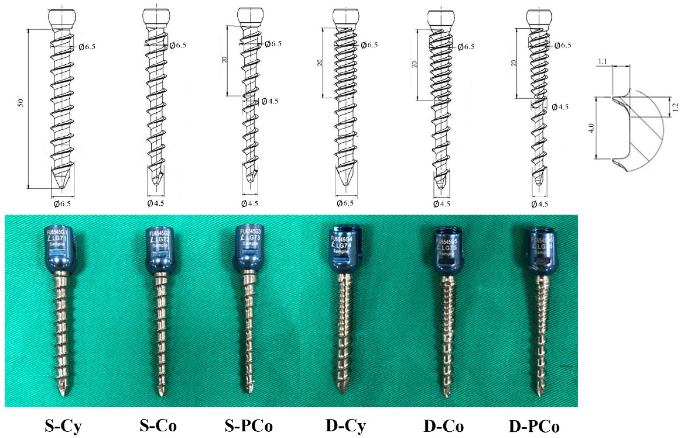
2.1. Pedicle Screw Design

Six types of pedicle screws were made through the same materials and processes based on three different shape (cylindrical, conical, and proximal-conical) and two thread profiles (single-thread and dual-thread). The screws were grouped as follows: single-thread and cylindrical shape (S-Cy), single-thread and conical shape (S-Co), single-thread and proximal-conical shape (S-PCo), dual-thread and cylindrical shape (D-Cy), dual-thread and conical shape (D-Co), and dual-thread and proximal-conical shape (D-Pco). Cylindrical, conical, and proximal-conical screws differed mainly in the outer diameter from the hub to the tip. Cylindrical screws maintained a constant outer diameter (6.5 mm) from hub to tip, conical screws had an outer diameter (6.5 mm) that tapered from the hub to the tip by 30% (to an outer diameter 4.5 mm), and proximal-conical screws had an outer diameter (6.5 mm) from hub and tapered by 30% to an outer diameter (4.5 mm) at 20 mm from hub, then maintained the outer diameter (45 mm) to the tip. The proximal high slope design of the proximal-conical screws theoretically preserves more pedicle bone stock than traditional cylindrical or conical screw. For the single-thread screw, coarse threads covered the entire length; for the dual-thread screw, fine threads covered the region 20 mm from the hub, followed by coarse threads to the tip. The thread pitch and depth of the coarse thread were designed as 4.0 mm and 1.1 mm, respectively, and 2.0 mm and 1.1 mm of the fine thread, respectively. The six groups of pedicle screws with various designed geometries are illustrated in Figure 1.

2.2. Specimen Preparation and Screw Insertion

A total of 36 commercially available synthetic L4 Sawbones® vertebrae (Model #3429-4-2, Pacific Research Laboratory, Inc., Vashon Island, WA, USA) were used to model normal spinal vertebrae. Sawbones® vertebrae have an inner cancellous core and outer cortex layer, which simulate healthy human vertebrae, including normal bone properties and morphometry. A pilot hole was drilled using a 2.5 mm “twist” metric drill bit attached to a Dremel 4000 rotary tool that was mounted on a Dremel WorkStation Model 220–01. This trajectory was selected based on previously reported morphometric characteristic data [13,15]. The pilot track was followed with a standard straight pedicle probe to a depth of 50 mm. Six groups of pedicle screws were randomly implanted into each pedicle of the vertebrae by an experienced surgeon, and six specimens were used in each group.

2.3. Image Analysis

Axial and sagittal views were examined via X-ray (GE DX300 X-ray machine, Salt Lake City, UT, USA) for all specimens prior to performing the pullout test to confirm an appropriate screw trajectory and insertion depth (Figure 2). The specimens were also examined thoroughly to rule out any fractures or defects caused by screw insertion.

2.4. Biomechanical Testing

The maximal insertion torque was measured with an electronic torque wrench (OLY 921/6NB, New Taipei City, Taiwan) at the last thread of screw insertion. The same insertional depth was confirmed using X-ray imaging prior to the pullout test using a material testing machine. Following screw insertion, the instrumented vertebral specimens were embedded in acrylate resin (#20–3568; Buehler, Lake Bluff, IL, USA) to allow clamping during the screw pullout test. The method for the screw pullout test was identical to that used in our previous study [15,24]. Each prepared specimen was secured to a custom-made grip mounted on the platform of the testing machine (E10000/E10BMTB19359., Instron Com., Norwood MA, USA) to conduct axial pullout tests with the screws. The polyaxial screw head was fixed to a 10-mm diameter cylindrical rod with an outer thread that matched the inner thread of the screw head cup. The cylindrical rod was then clamped to the upper wedge grip of the Instron testing machine. The potted specimen was clamped on a lower custom-made grip capable of x–y plane translation and rotation to achieve the coaxial alignment of the pedicle screw with the pullout ram (Figure 3). After the specimens were mounted, pullout force was applied at a constant crosshead rate of 5 mm/min [15,24]. During the pullout test, the relation between the applied force and displacement was simultaneously recorded in 0.1 mm increments until failure. The peak force recorded was defined as the maximum pullout strength for comparison.

2.5. Screw Volume in Bone (SV)

The SV in bone was approximately quantified by analyzing the illustrated image of Figure 1 and summation of thread volume and inner core volume.

2.5.1. Thread Volume of Screws

  • Thread volume in cylindrical screw = A × C × N
    where
    A denotes cross-section area of the thread and equals 1.2 × 1.1 × 0.5.
    C denotes circumference of screw and equals 6.5 × π .
    N denotes thread number, and the thread number in the single-thread screw is 10 and that in the dual-thread screw is 15.
  • Thread volume in conical screw = A   ×   C ¯   ×   N
    where
    A denotes cross-section area of the thread and equals 1.2 × 1.1 × 0.5
    C ¯ denotes mean circumference of screw and equals 6.5 + 4.5 2 × π
    N denotes thread number, and the thread number in the single-thread screw is 10 and that in the dual-thread screw is 15.
  • Thread volume in proximal-conical screw = ( A × Cp ¯ × Np ) + (A ×   Cd   ×   Nd )
    where
    A denotes cross-section area of the thread and equals 1.2 × 1.1 × 0.5
    Cp ¯ denotes the mean circumference of the proximal-conical region and equals 6.5 + 4.5 2 × π
    Np denotes thread number at proximal-conical region, and the thread number in a single-thread screw is 5 and that in the dual-thread screw is 10.
    Cd denotes the circumference of the distal-cylindrical region and equals 4.5 × π
    Nd denotes thread number at distal-cylindrical region and the thread number is 5 in both single-thread and dual-thread screws.

2.5.2. Inner Core Volume of Screws

  • Inner core volume in S-Cy/D-Cy groups = π × R × L
    where
    R denotes the radius of the screw core and equals ( 6.5 2.2 2 )
    L denotes the length of the screw and equals 50
  • Inner core volume in S-Co/D-Co groups = [ R × r + R 2 + r 2 ] × π L 3
    where
    R denotes the radius of the screw core at the hub and equals ( 6.5 2.2 2 )
    r denotes the radius of the screw core at the tip and equals ( 4.5 2.2 2 )
    L denotes the length of the screw and equals 50
  • Inner core volume in S-PCo/D-PCo groups = [ R × r + R 2 + r 2 ] × π 20 3 + π r 2 × 30
    where
    R denotes the radius of the screw core at the hub and equals ( 6.5 2.2 2 )
    r denotes the radius of the screw core at the tip and equals ( 4.5 2.2 2 )

2.6. Statistical Analysis

To evaluate the biomechanical performance of the six groups of screws, the magnitudes of the insertional torque and ultimate pullout force were compared. All of the measurements are expressed as the mean ± standard deviation (SD). Statistical software (SPSS for Windows version 12.0, SPSS, Inc., Chicago, IL, USA) was used to analyze the pullout strength and insertional torque of all specimens. Analysis of variance (ANOVA) with post hoc analyses was performed to evaluate the differences among groups. Differences were considered to be significant at p < 0.05.

3. Results

3.1. Biomechanical Performance

The insertion torques of screws in the S-Cy, S-Co, S-PCo, D-Cy, D-Co, and D-PCo groups were 1.07 ± 0.25, 1.15 ± 0.27, 1.0 ± 0.17, 1.4 ± 0.35, 1.68 ± 0.22, and 1.44 ± 0.44 N·m, respectively (Figure 4). For screws with the same shape (cylindrical, conical, or proximal-conical), insertion torque was significantly higher in the dual-thread groups than in the single-thread groups. For screws with the same thread profile (single- or dual-thread), there was no significant difference between the different screw shapes.
A typical force–displacement curve and the mean maximum pullout strength for various screws are shown in Figure 5 and Figure 6, respectively. The pullout forces of screws in the S-Cy, S-Co, S-PCo, D-Cy, D-Co, and D-Pco groups were 689.45 ± 128.25, 760.08 ± 145.03, 739.05 ± 128.72, 911.37 ± 132.77, 1007.99 ± 196.1, and 939.72 ± 160.4 N, respectively (Figure 6). For screws with the same screw shape, the ultimate pullout force was significantly higher in the dual-thread groups than in the single-thread groups. For screws with the same thread profile, there is no significant difference among different screw shapes. Dual-thread screws were proven to have better biomechanical performance than single-thread screws, and the increased percentage of insertion torque was 31% in cylindrical screws, 46% in conical screws, and 62% in proximal-conical shape screws (Figure 4). The increased percentage of pullout force from the single- to dual-thread groups was 32% in cylindrical screws, 33% in conical screws, and 27% in proximal-conical screws (Figure 6).

3.2. Screw Volume in Bone (SV)

The calculated SV of screws in the S-Cy, S-Co, S-PCo, D-Cy, D-Co, and D-PCo groups were 860.43, 580.27, 414.69, 927.75, 637.26, and 471.68 mm3, respectively. The SVs of proximal-conical screws were lower than those of cylindrical and conical screws; nevertheless, there was no significant biomechanical difference between the screw shapes, which implied no compromised fixation stability in the proximal-conical groups, and even less bone stock was purchased.

4. Discussion

Pedicle screw fixation has been broadly used in patients with various spinal disorders in recent decades [3,4,5]. Screw loosening is one of the most frequently reported complications of pedicle screw fixation and is associated with the necessity of revision surgery [9,11]. Poor bone quality followed by cyclic caudocephalad toggling and rotational stress cause micromotion between bone and screws finally results in screw loosening [26]. Several revision strategies, including various cement augmentations, extended fixations, and replacements of loosened screws with larger diameter screws have been studied [10,13,24,27]. Replacing loose screws with larger diameter screws potentially increases the risk of pedicle wall violation, which reasonably suggests a pronounced trend of pedicle bone stock preservation in primary surgery [12]. Our biomechanical results showed that in the dual-thread groups, when using a smaller volume screw (D-PCo), even less bone stock was purchased, providing similar stability when compared with traditional screws (D-Cy and D-Co), which decreased the necessity of upsizing the screw diameter in revision surgery. In cases of metastatic spinal cord compression, rigid pedicle screws fixation is important. However, postoperative iatrogenic neurological deterioration due to the extravasation of the tumor or the fragility of the pedicle bone reached 0.6–7% [28,29,30], and complications were rare but could still be prevented. Extended clinical application could be used in these diseases, which requires immediate postoperative stability without risking the extravasation of intravertebral lesions.
Synthetic bone models, such as Sawbones® and polyurethane foam, are widely accepted due to their regularity and reproducibility compared to the characteristics of human or porcine cadaveric vertebrae; moreover, they are well-established vertebral surrogates for biomechanical testing [15,31,32]. Considering the anatomical simulation and distribution of pedicle bone density, Sawbones®, instead of a polyurethane test block, was chosen for this in vitro biomechanical testing [15]. Our biomechanical data showed that dual-thread screws led to higher insertion torques and pullout forces than single-thread screws, which was consistent with other research. Dual-thread screws had been proven to increase insertional torque and pullout force in healthy/osteoporotic human cadaveric vertebrae and testing foam blocks [22,23]. Increased fatigue cyclic loading was observed in dual-thread screws compared with single-thread screws in human vertebrae with reduced bone mineral density [25]. Additive biomechanical performance was observed when combined with conical shape, which progressively allowed pedicle compression with advancing screws [24,33]. An additive trend could also be observed in our data. The reasons why proximal-conical dual-thread screws (D-PCo) decreased screw volume and reached similar insertion torque and pullout force compared to the other two screws (D-Co and D-Cy) are as follows: (1) The screw pullout force was a major factor in the ability of the pedicle to achieve 57–60% performance; thus, the effect of the vertebral body was far less influential [13]. The distal portion of the screw plays a less important role in pedicle screw pullout performance. (2) The geometric matching of the proximal-conical shape to the pedicle. Conical screws have been reported to possess superior anchoring power compared to cylindrical screws [24,33] and a higher slope conical angle (D-PCo) embedded more bone in threads when screws advanced. The proximal-conical screws tapered by 30% from the hub to the proximal 20 mm; however, the traditional conical screws tapered by 30% from the hub to the tip (50 mm); the supposedly decreased pullout force resulting from the downsized distal portion of the screw was balanced by this proximal enhancement.
In our study, compared to single-thread designs (S-Co, S-PCo), dual-thread designs (D-Co and D-PCo) were proven to increase insertion torque more than pullout force, which corresponded to other studies [22,23]. The screws in the D-PCo groups afforded 62% more mean insertion torque than the S-PCo groups; however, only 27% more mean axial pullout force. It appears that the proximity of the fine threads in the dual-threaded design might have interfered with more bone purchase and then created a more compressive force than coarse threads in the single-threaded screw, thus obviously increasing the insertion torque to 62%. Instead of the tapper compression mechanism in insertion torque, the performance of axial pullout force was achieved through bone-thread embedded volume and resistance, which are far less affected by thread design [22,23,34].
The present study had some limitations. First, standard L4 Sawbones® were used as substitutes for cadaveric vertebrae to eliminate the effects of the variability of bone density and anatomical morphometry but may not represent the real clinical situation. Second, the effect of proximal-conical dual-thread screws should be evaluated with various screw sizes and densities of bone surrogates. Third, there was no significance between cylindrical and conical screws, whether in dual-thread or single-thread profiles, which is inconsistent with other studies [24,33]. This phenomenon might be due to the fixation stability of pedicle screws being influenced by multiple factors, including thread shape, core shape, outer diameter shape, pitch depth, insertional depth/trajectory, and pilot-hole size [35,36,37,38]. In our study, to highlight the effect of the proximal-conical shape, only the outer screw shape and thread type were controlled. Various inner/outer screw shapes, thread shapes and implantation techniques should be tested in future studies.

5. Conclusions

Dual-thread screws showed a significantly higher insertion torque and pullout force than single-thread screws in different screw shapes (p < 0.05). However, no significant difference was found between screws with different shapes with the same thread profiles (p > 0.05), suggesting that the supposed decreased fixation stability resulting from the downsized distal portion of the screw was balanced by the proximal enhancement design. Therefore, bone stock-preserved screws with comparable biomechanical performance could be recommended as the primary pedicle screw choice in future clinical practice.

Author Contributions

Conceptualization, M.-K.H., D.-M.L. and C.-L.T.; methodology, M.-K.H. and Y.-J.H.; software, Y.-J.H.; validation, M.-K.H., Y.-D.L., T.-T.T. and P.-L.L.; formal analysis, Y.-D.L., Y.-J.H., D.-M.L. and C.-L.T.; investigation, M.-K.H., Y.-D.L. and C.-L.T.; data curation: T.-T.T., P.-L.L. and D.-M.L.; supervision: C.-L.T.; writing—original draft preparation, M.-K.H.; writing—review and editing, Y.-D.L., D.-M.L. and C.-L.T.; funding acquisition, C.-L.T. All authors have read and agreed to the published version of the manuscript.

Funding

This work was supported by the Chang Gung Memorial Hospital (CMRPD2K0091, CMRPD2K0092).

Institutional Review Board Statement

Not applicable.

Informed Consent Statement

Not applicable.

Data Availability Statement

The data used to support the finding of this study are available from the corresponding author upon request.

Conflicts of Interest

The authors declare no conflict of interest.

References

  1. Kao, F.C.; Hsieh, M.K.; Yu, C.W.; Tsai, T.T.; Lai, P.L.; Niu, C.C.; Chen, L.H.; Chen, W.J. Additional vertebral augmentation with posterior instrumentation for unstable thoracolumbar burst fractures. Injury 2017, 48, 1806–1812. [Google Scholar] [CrossRef] [PubMed]
  2. Chang, C.W.; Fu, T.S.; Lin, D.Y.; Lai, P.L.; Chiu, P.Y.; Kao, F.C.; Tsai, T.T.; Hsieh, M.K. Percutaneous Balloon Kyphoplasty and Short Instrumentation Compared with Traditional Long Instrumentation for Thoracolumbar Metastatic Spinal Cord Compression. World Neurosurg. 2019, 130, e640–e647. [Google Scholar] [CrossRef] [PubMed]
  3. Hsieh, M.K.; Bowles, D.R.; Canseco, J.A.; Sherman, M.B.; Schroeder, G.D.; Vaccaro, A.R. Is open surgery for metastatic spinal cord compression secondary to lung cancer really beneficial? A systematic review. World Neurosurg. 2020, 144, e253–e263. [Google Scholar] [CrossRef] [PubMed]
  4. Liao, J.C.; Chen, W.P.; Wang, H. Treatment of thoracolumbar burst fractures by short-segment pedicle screw fixation using a combination of two additional pedicle screws and vertebroplasty at the level of the fracture: A finite element analysis. BMC Musculoskelet. Disord. 2017, 18, 262. [Google Scholar] [CrossRef] [Green Version]
  5. Su, K.C.; Chen, K.H.; Pan, C.C.; Lee, C.H. Biomechanical evaluation of cortical bone trajectoryfixation with traditional pedicle screw in the lumbar spine: A finite element study. Appl. Sci. 2021, 11, 10583. [Google Scholar] [CrossRef]
  6. Hsieh, M.K.; Chen, L.H.; Niu, C.C.; Fu, T.S.; Lai, P.L.; Chen, W.J. Combined anterior lumbar interbody fusion and instrumented posterolateral fusion for degenerative lumbar scoliosis: Indication and surgical outcomes. BMC Surg. 2015, 15, 26. [Google Scholar] [CrossRef] [Green Version]
  7. Tsuang, F.Y.; Chen, C.H.; Wu, L.C.; Kuo, Y.J.; Hsieh, Y.Y.; Chiang, C.J. Partial threading of pedicle screws in a standard construct increases fatigue life: A biomechanical analysis. Appl. Sci. 2021, 11, 1503. [Google Scholar] [CrossRef]
  8. Chan, A.; Parent, E.; Narvacan, K.; San, C.; Lou, E. Intraoperative image guidance compared with free-hand methods in adolescent idiopathic scoliosis posterior spinal surgery: A systematic review on screw-related complications and breach rates. Spine J. 2017, 17, 1215–1229. [Google Scholar] [CrossRef]
  9. Galbusera, F.; Volkheimer, D.; Reitmaier, S.; Berger-Roscher, N.; Kienle, A.; Wilke, H.J. Pedicle screw loosening: A clinically relevant complication? Eur. Spine J. 2015, 24, 1005–1016. [Google Scholar] [CrossRef]
  10. Hsieh, M.K.; Li, Y.D.; Li, Y.C.; Liu, M.Y.; Tsai, T.T.; Lai, P.L.; Tai, C.L. Improved fixation stability for repairing pedicle screw loosening using a modified cement filling technique in porcine vertebrae. Sci. Rep. 2022, 12, 2739. [Google Scholar] [CrossRef]
  11. Xu, F.; Zhou, S.; Zou, D.; Li, W.; Sun, Z.; Jiang, S. The relationship between S1 screw loosening and postoperative outcome in patients with degenerative lumbar scoliosis. BMC Musculoskelet. Disord. 2022, 23, 186. [Google Scholar] [CrossRef] [PubMed]
  12. Weegens, R.; Carreon, L.Y.; Voor, M.; Gum, J.L.; Laratta, J.L.; Glassman, S.D. Dual pitch screw design provides equivalent fixation to upsized screw diameter in revision pedicle screw instrumentation: A cadaveric biomechanical study. Spine J. 2022, 22, 168–173. [Google Scholar] [CrossRef] [PubMed]
  13. Hsieh, M.K.; Liu, M.Y.; Chen, J.K.; Tsai, T.T.; Lai, P.L.; Niu, C.C.; Tai, C.L. Use of longer sized screws is a salvage method for broken pedicles in osteoporotic vertebrae. Sci. Rep. 2020, 10, 10441. [Google Scholar] [CrossRef] [PubMed]
  14. Liu, D.; Shi, L.; Lei, W.; Wei, M.Q.; Qu, B.; Deng, S.L.; Pan, X.M. Biomechanical comparison of expansive pedicle screw and polymethylmethacrylate-augmented pedicle screw in osteoporotic synthetic bone in primary implantation: An experimental study. Clin. Spine Surg. 2016, 29, E351–E357. [Google Scholar] [CrossRef] [PubMed]
  15. Hsieh, M.K.; Liu, M.Y.; Chen, J.K.; Tsai, T.T.; Lai, P.L.; Niu, C.C.; Tai, C.L. Biomechanical study of the fixation stability of broken pedicle screws and subsequent strategies. PLoS ONE 2019, 14, e0219189. [Google Scholar] [CrossRef]
  16. Weiser, L.; Sehmisch, S.; Viezens, L.; Lehmann, W. Intraoperative revision of initially loosened pedicle screws. Oper. Orthop. Traumatol. 2019, 31, 293–300. [Google Scholar] [CrossRef] [Green Version]
  17. Isik, C.; Kose, K.C.; Inanmaz, M.E.; Tagil, S.M.; Sarman, H. The Mechanisms of Medial Pedicle Wall Violation: Insertion Method Is as Important as Correct Cannulation of the Pedicle. Adv. Orthop. 2014, 2014, 283783. [Google Scholar] [CrossRef] [Green Version]
  18. Goda, Y.; Higashino, K.; Toki, S.; Suzuki, D.; Kobayashi, T.; Matsuura, T.; Fujimiya, M.; Hutton, W.C.; Fukui, Y.; Sairyo, K. The pullout strength of pedicle screws following redirection after lateral wall breach or end-plate breach. Spine 2016, 41, 1218–1223. [Google Scholar] [CrossRef] [Green Version]
  19. Irie, T.Y.; Irie, T.; Espinoza Orías, A.A.; Segami, K.; Iwasaki, N.; An, H.S.; Inoue, N. Micro-computed tomography analysis of the lumbar pedicle wall. PLoS ONE 2021, 16, 2021. [Google Scholar] [CrossRef]
  20. Maeda, T.; Higashino, K.; Manabe, H.; Yamashita, K.; Hayashi, F.; Goda, Y.; Tsuruo, Y.; Sairyo, K. Pullout strength of pedicle screws following redirection after lateral or medial wall breach. Spine 2018, 43, E983–E989. [Google Scholar] [CrossRef]
  21. Takenaka, S.; Kaito, T.; Ishii, K.; Watanable, K.; Shinohara, A.; Harada, T.; Nakada, F.; Majima, Y.; Matsumoto, M. Influence of novel design alteration of pedicle screw on pull-out strength: A finite element study. J. Orthop. Sci. 2020, 25, 66–72. [Google Scholar] [CrossRef] [PubMed]
  22. Mehta, H.; Santos, E.; Ledonio, C.; Sembrano, J.; Ellingson, A.; Pare, P.; Murrell, B.; Nuckley, D.J. Biomechanical analysis of pedicle screw thread differential design in an osteoporotic cadaver model. Clin. Biomech. 2012, 27, 234–240. [Google Scholar] [CrossRef] [PubMed]
  23. Brasiliense, L.B.C.; Lazaro, B.C.R.; Reyes, P.M.; Reyes, P.M.; Newcomb, A.G.U.S.; Turner, J.L.; Crandall, D.G.; Crawford, N.R. Characteristics of immediate and fatigue strength of a dual-threaded pedicle screw in cadaveric spines. Spine J. 2013, 13, 947–956. [Google Scholar] [CrossRef]
  24. Liu, M.Y.; Tsai, T.T.; Lai, P.L.; Hsieh, M.K.; Chen, L.H.; Tai, C.L. Biomechanical comparison of pedicle screw fixation strength in synthetic bones: Effects of screw shape, core/thread profile and cement augmentation. PLoS ONE 2020, 15, e0229328. [Google Scholar] [CrossRef] [PubMed]
  25. Weiser, L.; Sellenschloh, K.; Püsche, L.K.; Morlock, M.M.; Viezens, L.; Lehmann, W.; Huber, G. Cortical threaded pedicle screw improves fatigue strength in decreased bone quality. Eur. Spine J. 2021, 30, 128–135. [Google Scholar] [CrossRef] [PubMed]
  26. Bokov, A.; Bulkin, A.; Aleynik, A.; Kutlaeva, M.; Mlyavykh, S. Pedicle Screws Loosening in Patients With Degenerative Diseases of the Lumbar Spine: Potential Risk Factors and Relative Contribution. Glob. Spine J. 2019, 9, 55. [Google Scholar] [CrossRef] [Green Version]
  27. Tai, C.L.; Tsai, T.T.; Lai, P.L.; Chen, Y.L.; Liu, M.Y.; Chen, L.H. A Biomechanical Comparison of Expansive Pedicle Screws for Severe Osteoporosis: The Effects of Screw Design and Cement Augmentation. PLoS ONE 2015, 10, e0146294. [Google Scholar] [CrossRef] [Green Version]
  28. Patil, C.G.; Lad, S.P.; Santarelli, J.; Boakye, M. National inpatient complications and outcomes after surgery for spinal metastasis from 1993–2002. Cancer 2007, 110, 625–630. [Google Scholar] [CrossRef]
  29. Dea, N.; Versteeg, A.; Fisher, C.; Kelly, A.; Hartig, D.; Boyd, M.; Paquette, S.; Kwon, B.; Dvorak, M.; Street, J. Adverse events in emergency oncological spine surgery: A prospective analysis. J. Neurosurg. Spine 2014, 21, 698–703. [Google Scholar] [CrossRef]
  30. Versteeg, A.L.; Verlaan, J.J.; de Baat, P.; Jiya, T.U.; Stadhouder, A.; Diekerhof, C.H.; van Solinge, G.B.; Oner, F.C. Complications After Percutaneous Pedicle Screw Fixation for the Treatment of Unstable Spinal Metastases. Ann. Surg. Oncol. 2016, 23, 2343. [Google Scholar] [CrossRef] [Green Version]
  31. Formiga, M.C.; Gehrke, A.F.; DeBortoli, J.P.; Gehrke, S.A. Can the design of the instruments used for undersized osteotomies influence the initial stability of implants installed in low-density bone? An in vitro pilot study. PLoS ONE 2021, 16, e0257985. [Google Scholar]
  32. Calvert, K.L.; Trumble, K.P.; Webster, T.J.; Kirkpatrick, L.A. Characterization of commercial rigid polyurethane foams used as bone analogs for implant testing. J. Mater. Sci. Mater. Med. 2010, 21, 1453–1461. [Google Scholar] [CrossRef] [PubMed]
  33. Abshire, B.B.; McLain, R.F.; Valdevi, A.; Kambic, H.E. Characteristics of pullout failure in conical and cylindrical pedicle screws after full insertion and back-out. Spine J. 2001, 1, 408–414. [Google Scholar] [CrossRef]
  34. Krenn, M.H.; Piotrowski, W.P.; Penzkofer, R.; Augat, P. Influence of thread design on pedicle screw fixation. Laboratory investigation. J. Neurosurg. Spine 2008, 9, 90–95. [Google Scholar] [CrossRef]
  35. Addevico, F.; Morandi, M.; Scaglione, M.; Solitro, G.F. Screw insertion torque as parameter to judge the fixation. Assessment of torque and pull-out strength in different bone densities and screw-pitches. Clin Biomech 2020, 72, 130–135. [Google Scholar] [CrossRef]
  36. Ricci, W.M.; Tornetta, P.; Petteys, T.; Gerlach, D.; Cartner, J.; Walker, Z.; Russell, T.A. A comparison of screw insertion torque and pullout strength. J. Orthop. Traum. A 2010, 24, 374–378. [Google Scholar] [CrossRef] [Green Version]
  37. Pfeiffer, F.M.; Abernathie, D.L. A comparison of pullout strength for pedicle screws of different designs: A study using tapped and untapped pilot holes. Spine 2006, 31, E867–E870. [Google Scholar] [CrossRef]
  38. Hsieh, M.K.; Li, Y.D.; Liu, M.Y.; Lin, C.X.; Tsai, T.T.; Lai, P.L.; Tai, C.L. Biomechanical Comparison of Fixation Stability among Various Pedicle Screw Geometries: Effects of Screw Outer/Inner Projection Shape and Thread Profile. Appl. Sci. 2021, 11, 9901. [Google Scholar] [CrossRef]
Figure 1. Schematic drawings (upper) and photographs (lower) of six types of pedicle screws with different thread profiles (single- or dual-thread) and projection shapes (cylindrical, conical, or proximal-conical shape). From left to right: single-thread and cylindrical shape (S-Cy), single-thread and conical shape (S-Co), single-thread and proximal conical shape (S-PCo), dual-thread and cylindrical shape (D-Cy), dual-thread and conical shape (D-Co), and dual-thread and proximal-conical shape (D-PCo).
Applsci 12 06172 g001
Figure 2. Axial (upper) and sagittal (lower) X-ray images of the vertebrae after the insertion of screws with various geometries. (A) S-Cy, (B) S-Co, (C) S-PCo, (D) D-Cy, (E) D-Co, and (F) D-PCo.
Applsci 12 06172 g002
Figure 3. Photographs showing the complete experimental setup for the screw pullout test. The polyaxial screw head was fixed to a 10-mm diameter cylindrical rod with an outer thread that matched the inner thread of the screw head cup. The cylindrical rod was then clamped to the upper wedge grip of the Instron testing machine. The potted specimen was clamped on a lower custom-made grip capable of x–y plane translation and rotation to achieve the coaxial alignment of the pedicle screw with the pullout ram.
Applsci 12 06172 g003
Figure 4. Mean maximal insertion torque for screws of various geometries. The insertion torques of screws in the S-Cy, S-Co, S-PCo, D-Cy, D-Co, and D-PCo groups were 1.07 ± 0.25, 1.15 ± 0.27, 1.0 ± 0.17, 1.4 ± 0.35, 1.68 ± 0.22, and 1.44 ± 0.44 Nm, respectively. For screws with the same shape, the insertion torque was significantly higher in the dual-thread groups than in the single-thread groups. For screws with the same thread profile, there was no significant difference between the different screw shapes. Groups with significant differences are indicated with the ‘‘*” symbol.
Applsci 12 06172 g004
Figure 5. Typical force–displacement curve for screws of various geometries. Dual-thread screws had a higher pullout force than single-thread screws.
Applsci 12 06172 g005
Figure 6. Mean maximal pullout force for screws of various geometries. For screws with the same shape, the ultimate pullout force was significantly higher in the dual-thread groups than in the single-thread groups. For the same thread profile, there was no significant difference between the different screw shapes. The increased percentage of pullout force from the single- to dual-thread groups was 32% in cylindrical screws, 33% in conical screws, and 27% in proximal-conical screws. Groups with significant differences are indicated with the ‘‘*” symbol.
Applsci 12 06172 g006
Publisher’s Note: MDPI stays neutral with regard to jurisdictional claims in published maps and institutional affiliations.

