Article

Bio_Fabricated Levan Polymer from Bacillus subtilis MZ292983.1 with Antibacterial, Antibiofilm, and Burn Healing Properties

1
Botany and Microbiology Department, Faculty of Science, Helwan University, Cairo 11795, Egypt
2
Department of Pharmaceutical Organic Chemistry, Faculty of Pharmacy, Cairo University, Cairo 11562, Egypt
3
Department of Microbiology and Immunology, National Research Centre, Dokki, Giza 12622, Egypt
4
Department of Biology, College of Science, Princess Nourah bint Abdulrahman University, P.O. Box 84428, Riyadh 11671, Saudi Arabia
*
Author to whom correspondence should be addressed.
Appl. Sci. 2022, 12(13), 6413; https://doi.org/10.3390/app12136413
Submission received: 28 May 2022 / Revised: 21 June 2022 / Accepted: 23 June 2022 / Published: 24 June 2022
(This article belongs to the Section Applied Microbiology)

Abstract

:
The biopolymer levan has sparked a lot of interest in commercial production and various industrial applications. In this study, a bacterial isolate with promising levan-producing ability was isolated from a soil sample obtained from Princess Nourah bint Abdulrahman University in Saudi Arabia. The isolate has been identified and submitted to GenBank as Bacillus subtilis MZ292983.1. The bacterial levan polymer was extracted using ethyl alcohol (75%) and CaCl2 (1%) and then characterized using several approaches, such as Fourier transform infrared spectrometry and nuclear magnetic resonance. The IR spectrum of the levan polymer showed characteristic peaks confirming characteristics of polysaccharides, including a broad stretching peak of OH around 3417 cm−1 and aliphatic CH stretching was observed as two peaks at 2943, and 2885 cm−1. In addition, the FTIR spectrum featured an absorption at 2121 cm−1, indicating the fingerprint of the β-glycosidic bond. Based on 1H and 13C NMR spectroscopy analysis, six unexchanged proton signals related to fructose as a forming monomer of levan were observed. Evaluation of levan’s antibacterial effect against two pathogenic bacteria, S. aureus (ATCC 33592) and E. coli (ATCC 25922), showed inhibition zones of 1 cm and 0.8 cm in diameter, respectively, with MICs of more than 256 μg mL−1 for both strains. Moreover, the antibiofilm property of the levan polymer was assessed and the results showed that the inhibition rate was positively proportional to the levan concentration, as the inhibition percentages were 50%, 29.4%, 29.4%, 26.5%, and 14.7% at concentrations of 2, 1, 0.5, 0.25, and 0.125 mg mL−1, respectively. Levan showed a significant role in burn healing properties since it accelerated the process of healing burn-induced areas in rats when compared with those either treated with normal saline or treated with the cream base only.

1. Introduction

Polysaccharides of microbial origin are carbohydrate polymers with a high molecular weight (MW) produced by microorganisms such as bacteria, fungi, yeast, and microalgae [1]. Polysaccharides in the cell may be found as intracellular polysaccharides, which accumulate in the cytoplasm as energy and carbon reserves (e.g., glycogen). They may be found in the cell walls, helping microbial cells to maintain their structural integrity (e.g., chitin) or polysaccharides that are produced outside the cell (exogenous polysaccharides) that either remain as capsular polysaccharides attached to the cell surface (CPS) or as a slime which is figuratively attached to the cell (EPS) [1]. Exopolysaccharides (EPS) are produced by a variety of microorganisms, where non-structural storage carbohydrates are produced by a few plant species. Homopolysaccharides and heteropolysaccharides are two different types of EPS, in which heteropolysaccharides are made up of various types of sugar monomers arranged in oligosaccharide repeating units, and homopolysaccharides are made up of only one type of constituting monosaccharide [2]. Levan is a fructose homopolymer that is naturally produced by a variety of microbes. Bacillus sp. are known as good producer of levan, including Bacillus subtilis, Bacillus licheniformis, Lactobacillus reuteri, BKAG21, Bacillus amyloliquefaciens, and Bacillus licheniformis BKAG21 [3]. Biofabrication of levan from B. subtilis has been reported in many studies. Santos et al. reported the production of the polymer levan from B. subtilis NATTO [4]. Hamdy et al. reported that two strains of B. subtilis exhibited in vitro promising probiotic characteristics [5]. Moreover, Bouallegue et al. reported the antioxidant activities of a levan produced from a new isolated B. subtilis AF17 [3]. Levansucrase is a fructosyltransferase enzyme that aids in the conversion of sucrose to glucose and fructose, resulting in -(2,6)-levan synthesis [4]. Levan is also known as -2,6-D-fructan because of its biodegradability, biocompatibility, and film-forming ability. Levan finds many uses in the medical, food, and cosmetics industries. In food manufacturing, levan is used as flavor- and color-stabilizing agent [3], while in the cosmetic industry it is used as a hair care and skin whitening agent. In addition, in the medical industry, it is used in wound and burn healing, an anti-inflammatory, an antimicrobial, an antioxidant, an anti-irritant, an anticancer agent, and as a blood plasma substitute [5]. The main function of intact skin is to maintain body homeostasis by acting as a mechanical barrier that prevents the entry of pathogens into the interior environment of the body and prevents the loss of electrolytes to the outer environment as well [6]. Skin damage due to burning, cutting, or trauma is a challenging problem in medicine, especially with the growing problem of antibiotic resistance. The disruption of skin integrity, particularly due to burns, damages the epidermal barrier and causes denaturation of structural proteins and lipids in the skin, which leaves the burned area susceptible to infection [7]. The use of topical agents, especially those with antimicrobial activity, has significantly decreased the mortality rate due to burn contamination. It is also a great advantage when the topical antimicrobial agent does not retard wound healing but accelerates the process of healing instead [8]. Furthermore, before employing levan in any application, it must first undergo characterization to establish its chemical and physical composition, its morphological, mechanical, and thermal characteristics, and its molecular weight and other chemical properties [9]. Several techniques, such as Fourier transform infrared (FT-IR), nuclear magnetic analysis (1H and 13C NMR spectroscopy), and others, were used for the characterization of levan polymers [10].
In this study, Bacillus subtilis MZ292983.1, a levan producer, was isolated from soil. Then, on a sucrose-containing medium, it was screened for levan production that was obtained, partially purified, and characterized by FTIR, 1H NMR, and 13C NMR spectroscopy. The produced levan was also tested for antibacterial, antibiofilm, and burn healing properties.

2. Materials and Methods

2.1. Isolation of Bacterial Strain

The bacterial strain was isolated from soil sample obtained from Princess Nourah bint Abdulrahman University, Saudi Arabia, according to [5], with some modifications. The following was the isolation method. In 90 mL of sterile distilled water, 10 g of soil was suspended. Then, 100 µL of soil suspension was screened on nutrient agar (Oxoid) medium. Mucoid colonies were chosen, purified on nutrient agar plates, and stored at −18 °C in 20% glycerol for testing.

2.2. Qualitative Screening of Levan Polymer

Bacterial strain was screened for levan production, as the follows: 24 h old bacterial culture was streaked on a medium containing (0.5 yeast extract, 1 tryptone, 1.5 agar, 10 NaCl, 0.25 K2HPO4, 20 sucrose) grams per 100 mL [11]. For 2 days, the inoculated plates were kept at 28 °C. With a slimy mucoid appearance, levan producer bacteria was identified.

2.3. Molecular Identification of Bacterial Strain

2.3.1. DNA Extraction and PCR

Molecular identification of bacterial strain was performed by extraction of genomic DNA, then performing polymerase chain reaction (PCR) using the InstaGeneTM Matrix Genomic DNA Kit. The 16S rRNA gene was amplified using the universal primers 785F 5′ (GGATTAGATACCCTGGTA) 3′ and 907R 5′ (CCGTCAATTCMTTTRAGTTT) 3′. 20 μL of the PCR reaction mixture containing 2 μL of 10X PCR buffer (KOMA BIOTECH, Gangseo-gu, Seoul, Korea), 2 μL of DNA template (20 ng µL−1), 1.0 µL KOMA Taq (2.5 µL−1), 2 µL (2.5 mM) dNTP mixture, and 1.6 µL of each 10 pmol primers (10 μmol L−1) and to adjust the reaction volume, HPLC-grade distilled water. The thermal cycling parameters were: 5 min. with initial denaturation at 95 °C; 30 cycles of 0.5 min denaturation at 95 °C, 55 °C annealing temperature for 2 min, and extension at 68 °C for 1.5 min; and the last polymerization extension at 68 °C for 10 min. The PCR products were finally stored at 4 °C. PCR was performed using a thermal cycler (TECHNE; Model Reference: TC-4000). All positive purified PCR products were checked by 1% agarose gel electrophoresis. Purification of the PCR products was carried out by Montage PCR Clean-up Kit (Millipore Sigma, Burlington, MA, USA).

2.3.2. DNA Sequence Analysis

The PCR purified product was sent to Macrogen, Inc., Seoul, Korea for sequencing. Sequencing results were read on an Applied Biosystems model 3730XL DNA sequencing device that is fully automated (Applied Bio-Systems, Waltham, MA, USA) at Macrogen, Inc. (Seoul, Korea). Geneious prime software Version 2020.0 was used to analyze the acquired sequences (available online at 26 May 2021: https://www.geneious.com) [12]. A combination of reverse and forward sequences was made to construct consensus sequences. The Basic Local Alignment Search Tool was used to align the sequences. (BLASTn) from the National Center of Biotechnology Information (NCBI) with nearest relatives’ DNA sequences available in the GenBank.

2.3.3. Phylogenetic Analyses

Phylogenetic tree was created by the alignment of achieved neighbor-joining analysis of nucleotide sequences [13] in the MEGA X software 10.1.8 (Philadelphia, PA, USA) [14]. The 16S rDNA gene sequence of the Pseudomonas gessardii AHB79N_LN866625.1 was used as an out-group.

2.4. Production and Extraction of Levan Polymer

For levan production, firstly, B. subtilis strain MZ292983.1 was cultured overnight in a liquid tryotic soy medium (Oxoid) at 28 °C and a 150-rpm shaking incubator. Then, 5% (v/v) of the obtained bacterial suspension was inoculated in a 250 mL Erlenmeyer flask containing a basic medium containing g L−1: 50.0 sucrose; 7.0 yeast extract; 2.5 KH2PO4; 1.6 (NH4)2SO4; and 1.0 MgSO4.7H2O; total pH was 5.0. It was then incubated statically at 28 °C for 24 h. Secondly, 5% of seeded medium with bacterial suspension was transferred to Erlenmeyer flasks containing 250 mL of levan production medium containing g L−1 (300.0 sucrose; 2.5 yeast extract; 1.0 KH2PO4; 1 (NH4)2SO4; 0.5 MgSO4.7H2O; and total pH 5.0), and the flasks were incubated statically at 28 °C for 72 h. After the incubation period, centrifugation for 10 min at 4 °C and 10,000 rpm was used to remove bacterial cells. The produced levan was obtained by adding three times (v/v) cold 75% ethanol (Sigma Aldrich, Burlington, MA, USA) to the supernatant after adjusting pH to 10 by using 0.1 N KOH. Additionally, 1 mL of CaCl2 (1%) was applied to each 10 mL of obtained supernatant. Then, the precipitated levan polymer was centrifuged at 4 °C and 10,000 rpm for 10 min and oven-dried at 60 °C for 4–6 h before further analysis [10], and then dry weight was calculated [15].

2.5. Determination of Total Soluble Carbohydrates

The total soluble carbohydrates assay was calculated as the following method [16]: 50 µL of levan sample at a concentration of 1 mg mL1 were moved to a glass vial and mixed with 100 µL concentrated sulfuric acid solution (75% v/v). Then, 200 µL of anthrone reagent (5 mg in 100 µL ethanol and 2.4 mL 75% v/v sulfuric acid) were added. In a dry oven, the vial was incubated for 15 min at 100 °C. The mixture was stood to cool for 5 min at room temperature after being heated. The analysis was carried out by transferring 100 µL of the sample mixture to a 96-well plate. The resulting green color was then measured spectrophotometrically using microplate reader FluoStar Omega at 578 nm and the total soluble carbohydrates for the sample were determined using the linear regression equation obtained from glucose standard curve and the concentration represented as µg glucose/mg sample. The data was represented using means and standard deviations.

2.6. Detection of Reducing Sugar by Dinitrosalicylic Acid Method (DNS)

The concentration of reducing sugar in levan polymer was estimated quantitatively by 3–5 dinitrosalicylic acid (DNS) as follows, according to [10]: 0.1 g of levan powder was dissolved for 1 h by 0.1 N HCl at 100 °C. After levan hydrolysis, 1 mL of freshly prepared DNS was added to two test tubes; one contained 1 mL of hydrolyzed levan sample and the second contained 1 mL of distilled H2O as a blank. The tubes were put in a water bath container for 10 min, and then it was permitted to cool at ambient temperature. To obtain an appropriate dilution from the sample, 10 mL of distilled H2O were added to individual tubes, and the absorbance was measured spectrophotometrically at 540 nm. Using the standard glucose curve, the unknown concentration of the reducing sugar in the sample was obtained. A glucose standard solution with a concentration range of 100–600 mg mL−1 was made in 0.05 M acetate buffer (pH 4.8).

2.7. Partial Purification of Levan Polymer

With minor modifications, the collected levan polymer was partially purified as follows [17]: the polymer was resuspended then dialyzed for 4 days using distilled water. After that, levan was collected by centrifugation at 4 °C for 10 min at 10,000 rpm. Levan was dried for 4–6 h in a dry-air oven at 60 °C. For further analysis, the purified product was frozen at −20 °C.

2.8. Characterization of Levan Polymer

2.8.1. Fourier Transform Infrared Spectroscopy (FTIR)

The functional groups in the partially purified polymer were investigated by infrared spectroscopy. The polymer was mixed and ground with KBr (1:10 ratio) and pressed into 1.0 mm disc. To record IR spectra, a spectrophotometer Shimadzu IR 435 (Shimadzu Corp., Kyoto, Japan) was used at the Faculty of Pharmacy, Cairo University, Cairo, Egypt. Values were expressed in cm−1.

2.8.2. Nuclear Magnetic Resonance (NMR) Spectroscopy

Nuclear magnetic resonance was used to characterize the partially purified levan polymer. Bruker 400 MHz (Bruker Corp., Billerica, MA, USA) spectrophotometer was used for 1H NMR spectra analysis at the Faculty of Pharmacy, Cairo University, Cairo, Egypt. The chemical shifts were measured in parts per million (ppm) on δ scale. The following are the multiplicities of the peak: s, singlet; d, doublet; t, triplet; m, multiplet. The Faculty of Pharmacy, Cairo University, Egypt, used a Bruker 100 MHz spectrophotometer to obtain 13C NMR spectra. Cold D2O (Sigma Aldrich) was used to dissolve the levan sample, which was then put into 5 mm tubes.

2.9. Antibacterial Activity of Levan

2.9.1. Microorganisms and Culture Conditions

Levan’s antibacterial activity was estimated in vitro against two pathogenic bacterial strains: Staphylococcus aureus (ATCC 33592) was used to represent Gram-positive strains, while Escherichia coli (ATCC 25922) was used to represent Gram-negative strains. The tested bacteria were kept at 4 °C on nutrient agar slant.

2.9.2. Well Diffusion Assay

Levan’s antibacterial properties were tested in vitro against Staphylococcus aureus (ATCC 33592) and Escherichia coli (ATCC 25922) using agar well diffusion method [18,19]. Using sterile swabs, 100 µL of 24 h old bacterial culture of the two reference strains with a concentration equivalent to 0.5 McFarland (1.5 × 10⁸ CFU mL−1) were streaked on the dry surface of the nutrient agar plate. Then, under aseptic conditions, a well of 0.8 mm was made on the nutrient agar surface by using a sterile cork borer and receiving 50 µL of levan solution at concentration 25 mg mL−1 DMSO. As positive controls, both Oxoid Sulfamethoxazole (25 µg/disc) and Ofloxacin (5 µg/disc) were used, while a negative control was DMSO. Then, incubation was carried out at 37 °C for 24 h to allow the growth of bacteria. Zones of inhibition were measured in mm using a ruler. The experiment was carried out in triplicate and the means of inhibition zones were measured in millimeters ± standard variation [20].

2.9.3. Minimum Inhibitory Concentration (MIC)

The MIC (minimum inhibitory concentration) was estimated by the broth microdilution method. The bacterial cultures of both S. aureus (ATCC 33592) and E. coli (ATCC 25922) were diluted to 106 CFU mL−1 in Muller–Hinton broth media (MHB). Serial dilution of levan solubilized in DMSO was prepared in a 96-well plate to reach final concentrations of (256, 128, 64, 32, 16, 8, 4, 2, 1, and 0.5 μg mL−1) in 100 μL of bacterial suspension in MHB (106 CFU mL−1). Then, the plates were incubated at 37 °C for 16–20 h, and the MIC was recorded as the lowest inhibitory concentration of the visible growth [21].

2.10. Antibiofilm Mechanism of Levan against Pseudomonas aeruginosa Strain MW846272 Using Crystal Violet Quantitative Assay

According to the method in [22], with some modifications, levan antibiofilm activity was determined as follows: 100 μL of P. aeruginosa strain MW846272 suspension containing approximately 2 × 10⁶ CFU mL−1 (OD~0.4) were added to different concentrations of levan polymer (0, 0.125, 0.25, 0.5, 1, and 2 mg mL−1) to each of the 96 wells of a polystyrene plate. Then, it was incubated statically at 37 °C for 48 h. After the incubation time, the wells were washed twice with sterile 1× phosphate buffer solution (PBS, pH 7.3) to eliminate the planktonic cells of bacteria; after air drying to fix the biofilm, methanol was added for 15 min, and the fixed biofilms were dyed with a 1% crystal violet solution for 10 min. Finally, each well was filled with pure ethanol, and at 630 nm the absorbance was measured using a microplate reader (ChroMate 4300, USA). The results were represented as a percentage of biofilm formation inhibition [23]. Statistical significance (p < 0.05) was estimated using ordinary one-way ANOVA using the GraphPad Prism software version 9.1.2 (226), Inc., San Diego, CA, USA.

2.11. Burn Healing Properties of Levan

2.11.1. Animals

Five adult albino Wistar rats, weighing 180 ± 20g, were obtained and housed in the animal house unit of the Faculty of Science, PNU, for a minimum pf 1 week before the investigations, in appropriate light and temperature conditions. All of the animals were fed a conventional laboratory diet that included (gL−1) a vitamin mixture (0.1), mineral mixture (0.4), maize oil (0.1), sucrose (0.2), cellulose (0.2), casein 95% pure (0.105), and starch (0.543). Water and food were supplied ad libitum throughout the duration of study.

2.11.2. Induction of Burn Wounds

The procedure for causing second-degree thermal injuries in rats was carried out according to [24] with minor modifications. Briefly, the rats were initially anesthetized using ether and the hair of the backside of the animals was removed. After that, the region was antisepticised with 1% Povidone-iodine. A solid aluminum bar (diameter = 10 mm) was heated to a temperature of nearly 97 °C and pressed to the shaved and disinfected back of the animals for 20 s to induce three burning positions on the back of each rat (diameter of each burn = 10 mm). The three burning positions were handled as follows: N = Negative control, the burn was treated daily with normal saline; C = Control, the burn was treated daily with the cream base (prepared by mixing lanolin and vaseline in equal amounts) only (reference group); and T = test group, the burn was treated daily with levan polymer mixed with cream base (used as a vehicle for levan) at concentration 0.25 g levan/5 g cream. All the treatments were applied once a day in a thin layer covering the burns’ surface using sterile cotton swabs. An analgesic was administered to all animals after the induction of burns to prevent animals from suffering. After that, each animal was put in its own cage until the end of the experiment to keep other animals from licking or biting the wound. On days 2, 4, 6, and 7 after burn injury, a digital camera was used to take color images of the wounds. On the last day (day 7) of the experiment, animal termination was performed by decapitation under anesthesia, and their debris was disposed of according to the guidelines of Princess Nourah bint Abdulrahman University, Saudi Arabia. (IRB, HAP-01-R-059).

2.11.3. Histopathological Evaluation of Burn Healing Ability of Levan

The samples for autopsy were collected from the skin of rats at day 7 in burn areas with normal tissue surrounding them, and for 24 h the specimen was immersed in 10% formol saline. After washing in tap water, dehydration was achieved using alcohol serial dilutions (ethyl, methyl, and absolute ethyl). Samples were cleaned in xylene before being inserted in paraffin at 56 °C in hot-air oven for 24 h, and 4 mm thick paraffin bees wax tissue blocks were constructed for sectioning by LEITZ ROTARY microtome. Tissue sections were taken and mounted on glass slides, deparaffinized, and stained with hematoxylin and eosin for usual examination by electric light microscope [25].

3. Results

3.1. Isolation and Molecular Identification of Bacterial Strain

The selected soil isolate with the characteristic of a slimy mucoid appearance of colonies on sucrose medium was subjected to molecular identification using 16S ribosomal DNA (16S rDNA). The 16S rDNA gene sequences of the isolate were placed in the GenBank of NCBI. The assigned accession number of the Bacillus strain is MZ292983.1. Phylogenetic analysis using the neighbor-joining method of 16S rDNA regions of the Bacillus subtilis strain Ba7 (accession numbers MZ292983.1) showed a significant degree of homology and strong grouping with closely related Bacillus strains obtained from the NCBI GenBank database with significant alignments of 96–99% (Figure 1).

3.2. Total Soluble Carbohydrates and Reducing Sugars

Slimy and mucoid growth of the B. subtilis strain was observed on the surface of medium containing sucrose sugar. Additionally, the amounts of both the total soluble carbohydrates and reducing sugars the of levan polymer were 90.28 ± 1.67 µg mg−1 and 290 µg mg−1, respectively.

3.3. Characterization of Levan Polymer

3.3.1. Infrared Spectroscopy

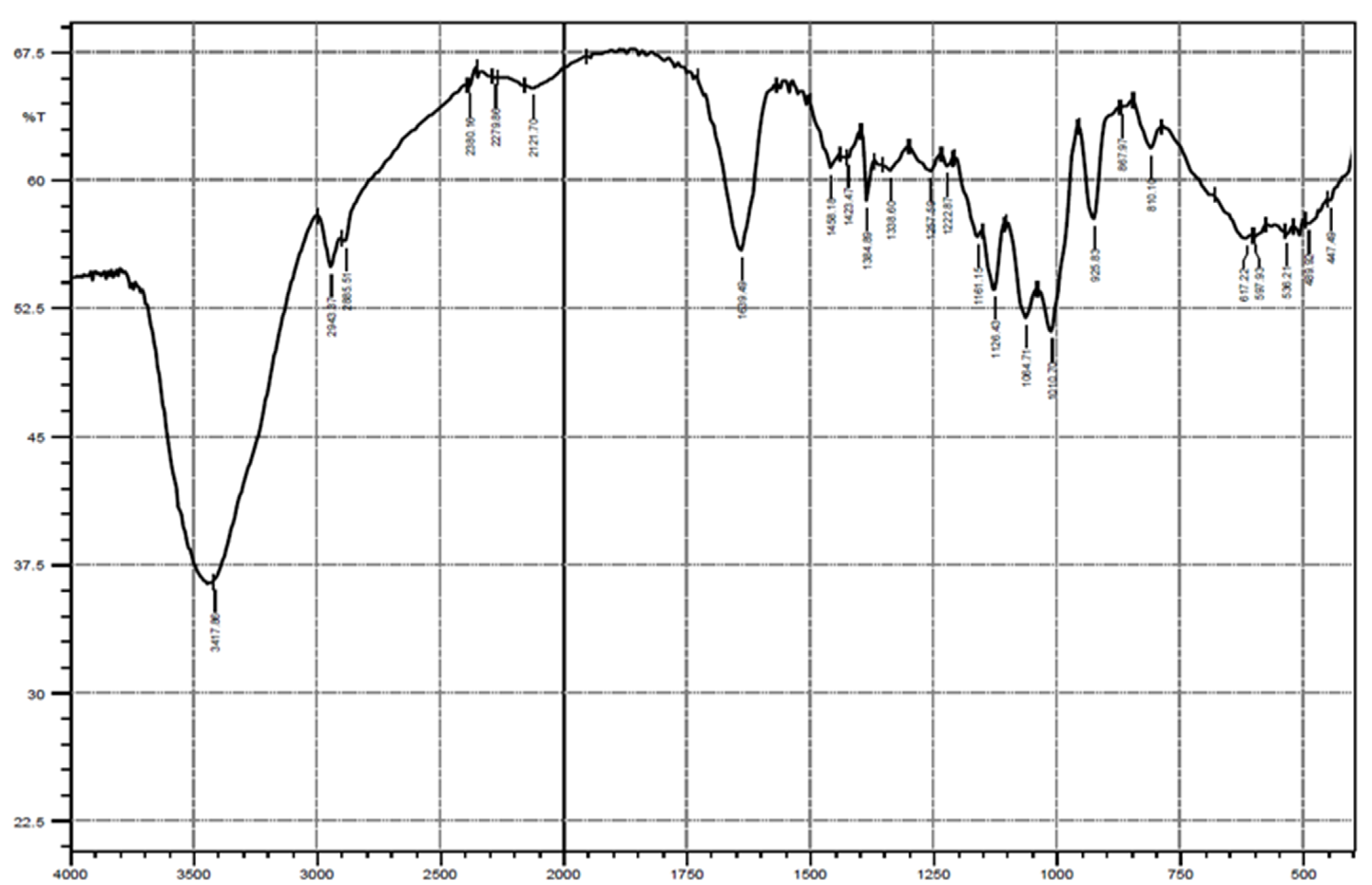
The IR spectrum of the levan from Bacillus subtilis strain MZ292983.1 showed a characteristic broad stretching peak of OH around 3417 cm1, which is characteristic of polysaccharides. Aliphatic CH stretching was observed as two peaks at 2943 and 2885 cm−1, and the peak at 1639 cm−1 indicates the stretching vibration of C=O. Peaks between 1126 and 900 cm−1 represent the fingerprint of polysaccharides. Finally, the FTIR spectrum featured an absorption at 2121 cm−1, indicating the fingerprint of the β-glycosidic bond (Figure 2).

3.3.2. Nuclear Magnetic Resonance (NMR) Spectroscopy

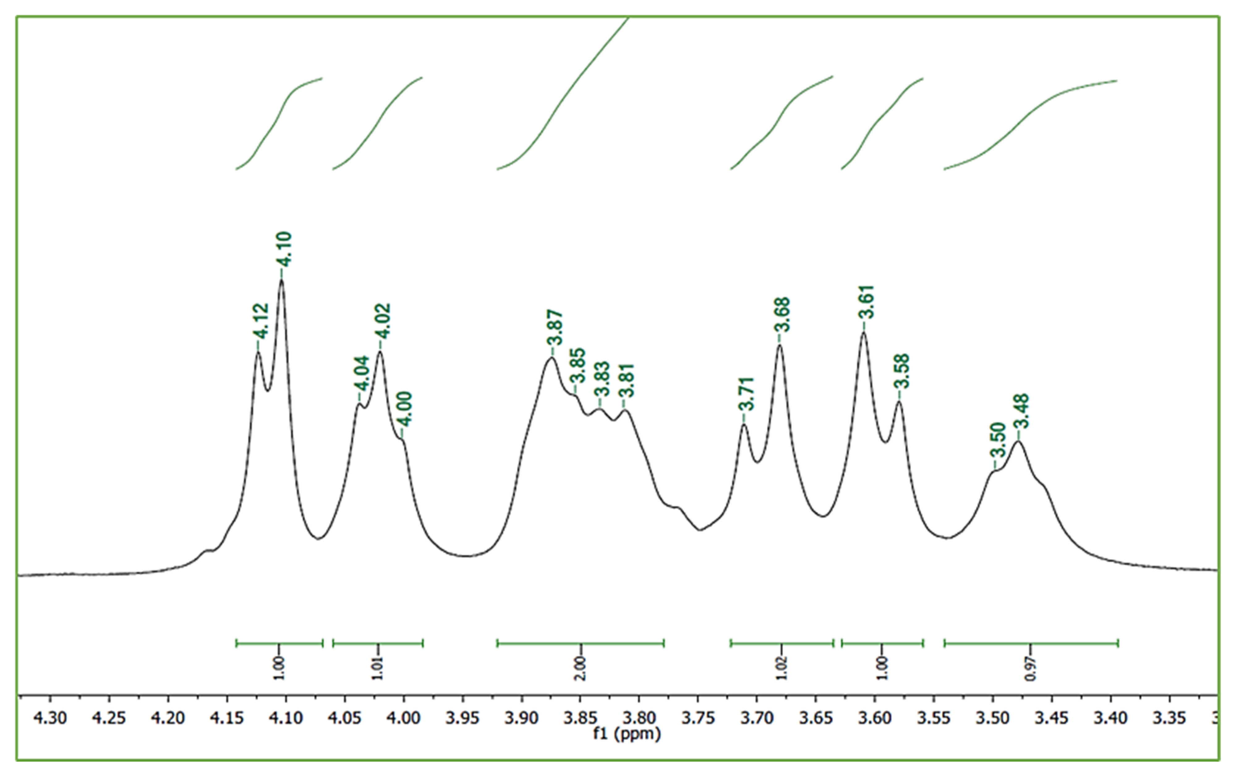
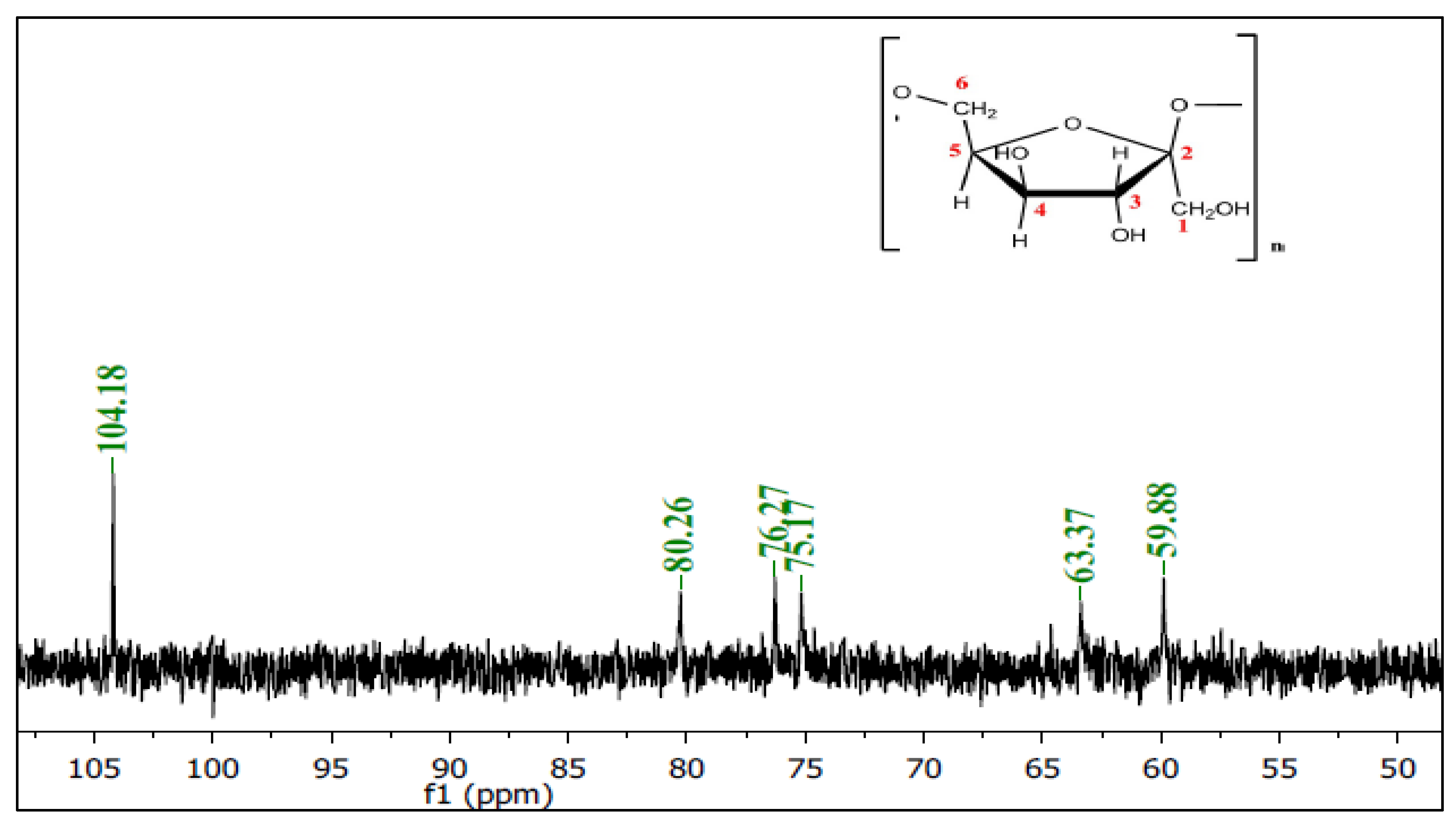
The 1H NMR spectrum of the partially purified levan (Figure 3) showed six unexchangeable proton signals related to fructose as the forming monomer of levan. 1H NMR (400 MHz, D2O) δ 4.11 ppm (d, 1H, H-3), 4.02 ppm (t, 1H, H-4), 3.87–3.81 ppm (m, 2H, H-5 & H-6a), 3.69 ppm (d, 1H, H-1a), 3.59 ppm (d, 1H, H-1b), and 3.48 ppm (t, 1H, H-6b). The spectrum of 13C NMR for the partially purified levan demonstrated six carbon signals at δ d 104.17, 80.26, 76.26, 75.17, 63.36, and 59.87 ppm; Figure 4. The spectrum of 13C NMR for the partially purified levan demonstrated six carbon signals at δ 104.18, 80.26, 76.27, 75.17, 63.37, and 59.88 ppm; Figure 4. The chemical changes in the carbon atoms are illustrated in Table 1.

3.4. Antibacterial Activity of Levan

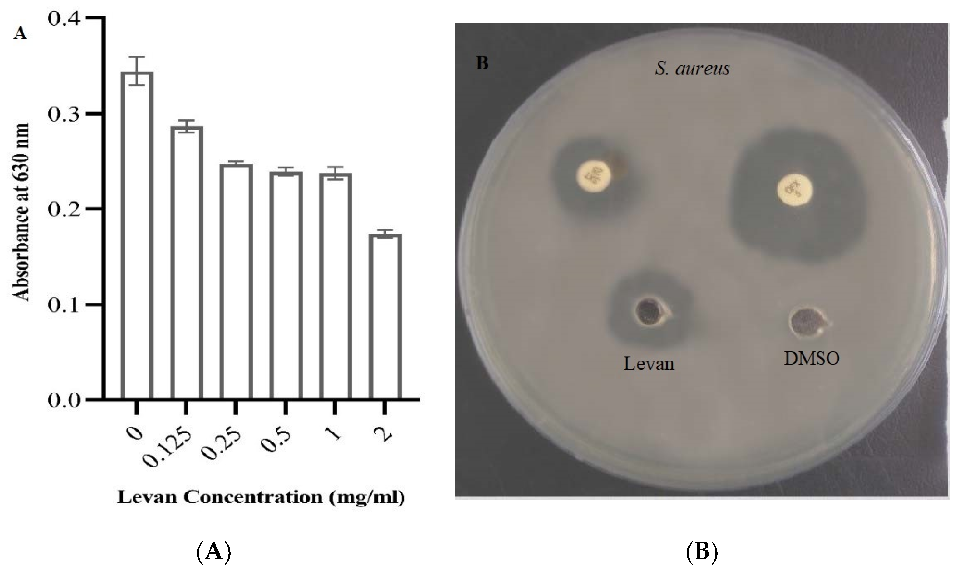
Levan’s antibacterial activity was evaluated in vitro against Gram-positive S. aureus (ATCC 33592) and Gram-negative E. coli (ATCC 25922). They scored inhibition zones of 1 cm and 0.8 cm, respectively. Additionally, both strains scored MICs of >256 μg mL−1.

3.5. Antibiofilm Activity of Levan

The inhibition of biofilm formation by levan was investigated quantitatively by crystal violet assay with different concentrations (0.125–2 mg mL−1) against the pathogenic biofilm forming P. aeruginosa strain MW846272. The obtained results revealed that the rate of biofilm formation was significantly (p < 0.0001) decreased with increasing levan concentration, as shown in Figure 5. This was shown when using concentrations 2, 1, 0.5, 0.25, and 0.125 mg mL−1, with the inhibition percentage found to be 50%, 29.4%, 29.4%, 26.5%, and 14.7%, respectively.

3.6. Burn Healing Properties of Levan

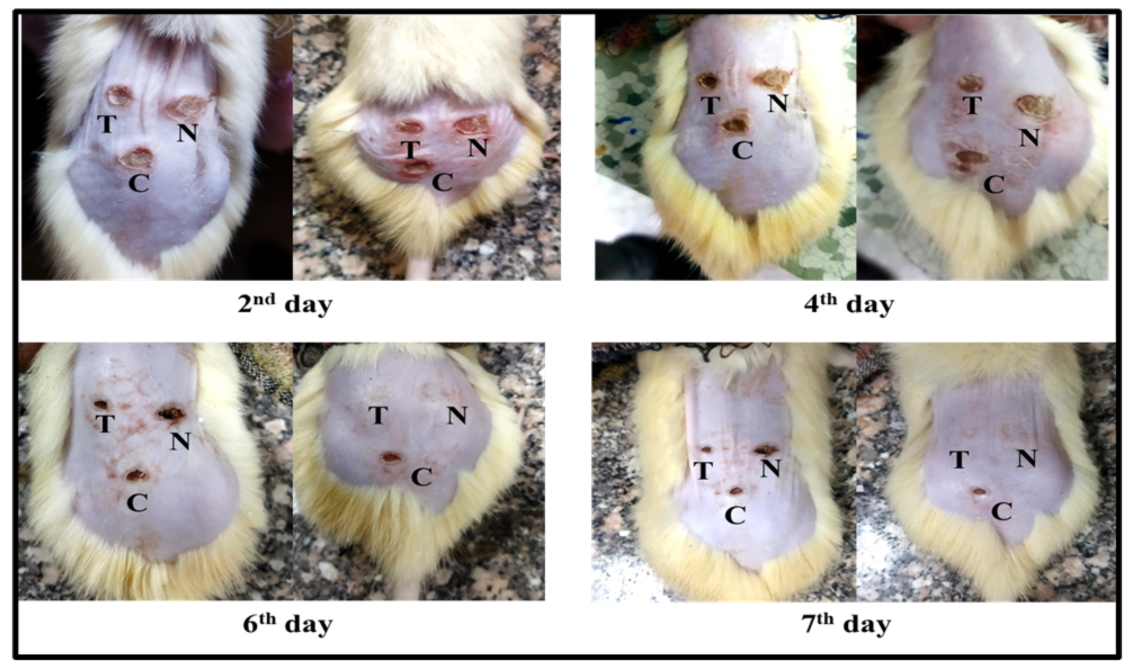
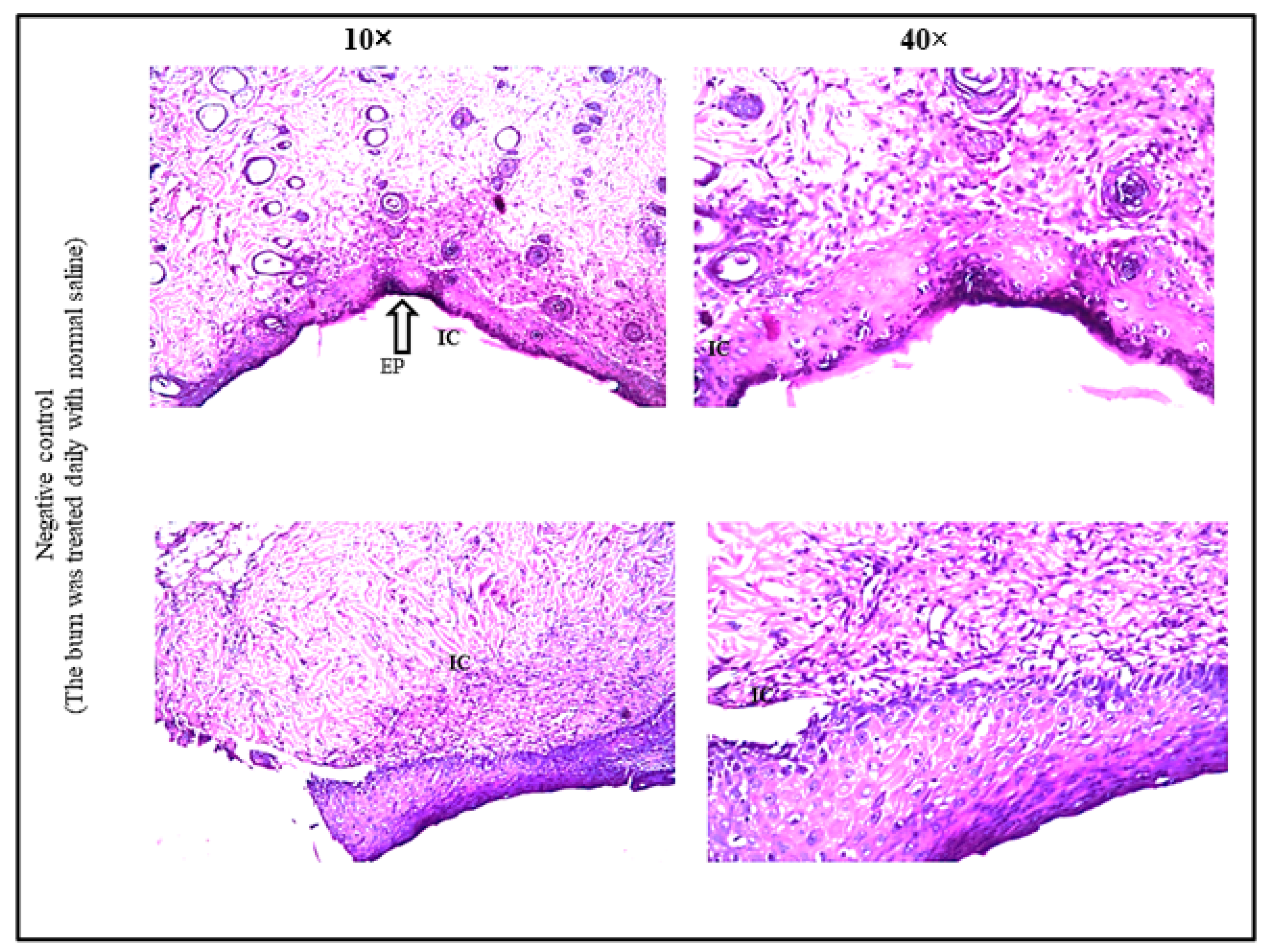
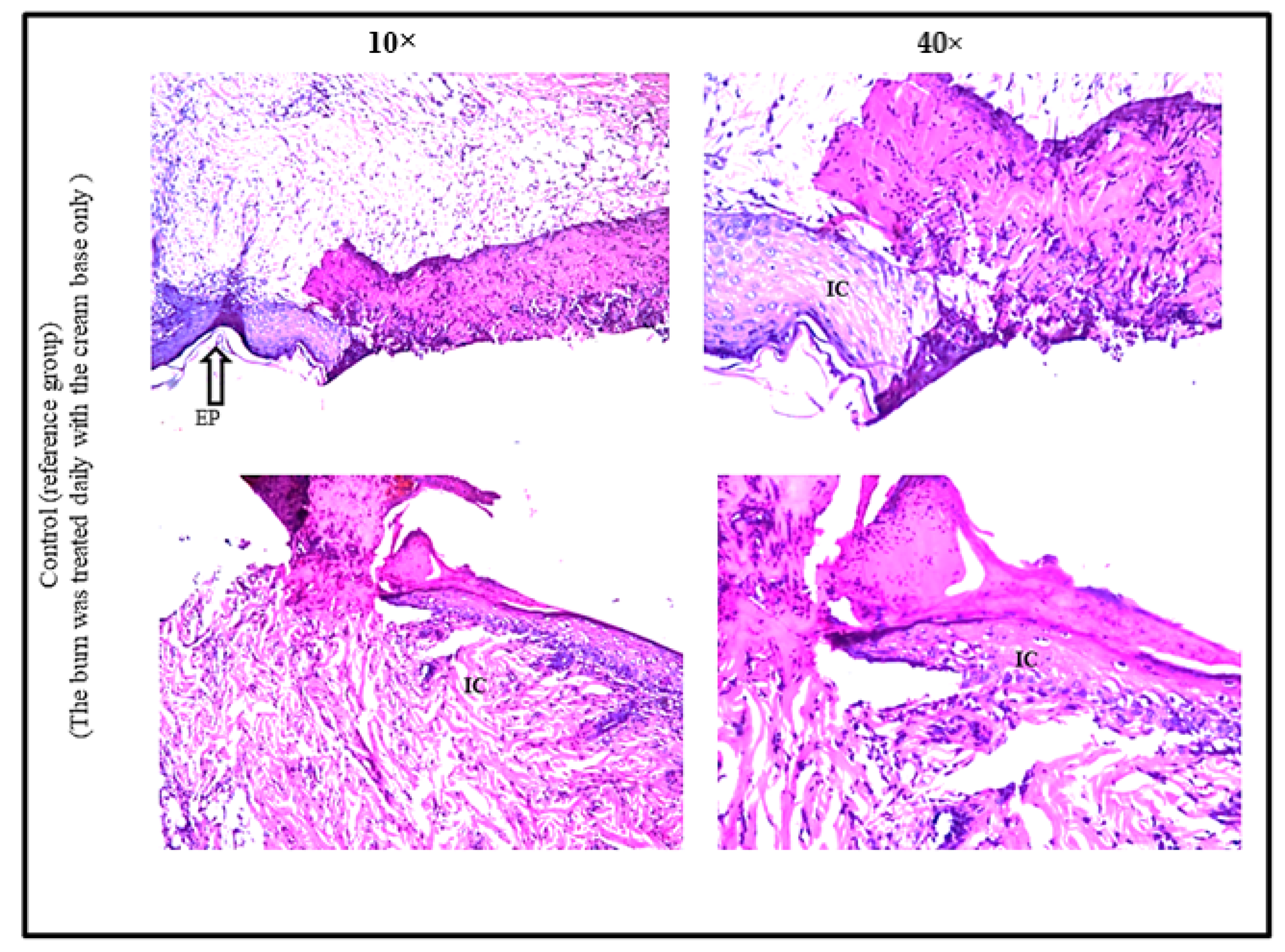
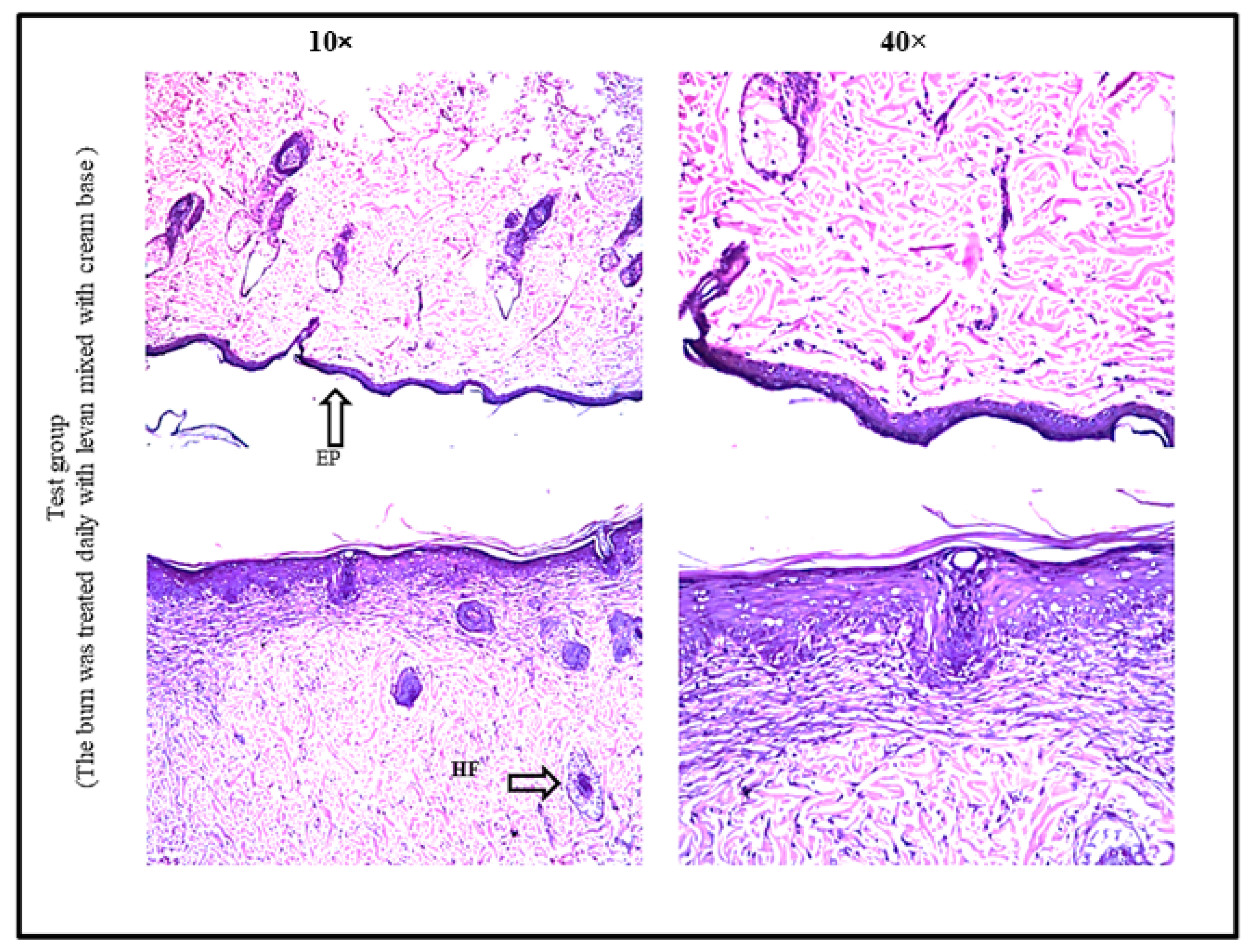
To assess the degree of burn healing in rats, color images of lesions were obtained on days 2, 4, 6, and 7 after burn induction using a digital camera. It was clear that levan accelerated the process of the healing of the burn areas when compared with the burn areas which were either treated with normal saline (N) or treated with the cream base only (C) (Figure 6). Visual observations showed similar appearance of burn areas on the first day of burn induction, with a characteristic dark red coloration indicating the formation of a blood clot. Starting from the fourth day, the clot was converted into a scab. On the sixth and seventh days, the retraction of the burn area was obvious, with more retraction in the burn areas treated with levan. Moreover, some of the mice showed complete healing of the burn areas in both levan-treated and normal saline–treated burn areas. To evaluate the percentage of burn healing in each group, we used a digital camera placed at a fixed distance from the rats to take digital images of the burn areas on days 2, 4, 6, and 7 after the induction of the burn. The burn areas in each group were then measured using Fiji image processing software. The percentage of burn contraction was then calculated (Table 2) based on the following equation: X = [(A2 − Ax)/A2] × 100 ([28]), where A2 is the surface area of the burn on the second day and Ax is x day. Histopathological examination confirmed the visual observations of the burn healing process. The burned areas treated with normal saline showed focal acanthosis, degeneration and parakeratosis in the epidermis layer. The underlying dermis layer showed inflammatory cell infiltration and granulation tissue formation (Figure 7). The burned areas treated with base cream only exhibited focal ulceration, necrosis, and acanthosis in the epidermis layer, with underlying inflammatory cell infiltration and necrosis in the dermis layer. (Figure 8). Finally, the burned areas treated with levan showed no histopathological alteration in one section and the other section showed acanthosis in the epidermis layer associated with fibrosis in the underlying dermis, as recorded in Figure 9. The severity of both the ulceration and inflammatory reaction were scored for the semi-quantitative assessment of the pathological changes in different groups (Table 3).

4. Discussion

Biopolymers are biodegradable polymers produced by different types of living organisms [29]. Exopolysaccharides (EPSs) are a fascinating biopolymer group formed by a diverse group of microorganisms. EPSs include homopolysaccharides such as dextran and levan, as well as heteropolysaccharides, including xanthans and gellans [5]. Levan is a homopolymer made up of ß-(2,6)-joined fructose residues, which can be generated by archaea, bacteria, and plants. Microorganisms grow faster than plants and algae, and their production processes are simple to manipulate to enhance yields and productivity. Furthermore, the manufacturing process is not affected by weather or seasons, and it can rely on low-cost wastes or by-products as raw materials [1]. Many bacteria, including B. subtilis, Z. mobilis, B. lentus, and Microbacterium laevaniformans, produce important metabolic products under both oxic and anoxic environments, such as levan polymer [30]. In this study, levan was produced, extracted, characterized, and partially purified from Bacillus subtilis strain MZ292983.1. The bacterial strain was isolated from soil obtained from Princess Nourah bint Abdulrahman University, Saudi Arabia. Screening of levan was performed on sucrose medium, and the addition of sucrose in the media as a substrate induces the bacterial secretion of the levansucrase enzyme [11]. Levansucrase is a fructosyltransferase enzyme that catalyzes ß-(2,6)-levan formed via the cleavage of sucrose to its fructose and glucose units, as well as fructooligosaccharide formation (FOS) [4]. Furthermore, the morphological results showed a slimy and mucoid appearance, in agreement with [11]. Levansucrase accounts for at least 25% of the released reducing sugars during bacterial growth in the existence of sucrose [31]. In our study, both the amount of total soluble carbohydrates and total reducing sugars were determined and recorded as 90.28 ± 1.67 µg mg−1 and 290 µg mg−1, respectively. The presence of uronic acids, amino sugars, and sulphate groups can all be determined using FTIR. Nonetheless, infrared spectroscopy is not suitable for structural analysis in detail [32]. Instead, in the field of polysaccharide structural determination, nuclear magnetic resonance (NMR) is the most useful tool. In both 1H and 13C spectra, anomeric resonances are frequently shown in a distinct region as carbinolic signals, assisting in establishing not just each anomeric configuration’s alpha or beta of individual residues but also establishing, with great confidence, the number and relative proportions [2]. Comparison of IR spectra of partially purified levan with previously reported IR data of levan confirmed the similarity of the spectra [32]. Our findings are consistent with previous studies which recommended that carbohydrates showed high absorbance in the fingerprint region of 1200–950 cm−1. The position and intensity of the bands of each polysaccharide are unique, allowing it to be identified. Spectroscopy of 1H and 13C NMR was also widely used in the characterization of levan polymers since it provides distinct and expected characteristics for each constituent, particularly in tiny compounds [33]. NMR analysis aids in the estimation of structural characteristics and saccharide composition within the mixture, as well as its ratio. It also distinguishes branching and linear levan from a plant and provides comprehensive details on the presence of certain “functional groups” in the specimen [34]. The spectrum of the 1H NMR spectrum of the partially purified levan from Bacillus subtilis strain MZ292983.1 was recorded in D2O and showed six peaks in the range of δ 3.48–4.11 ppm region, which revealed the existence of sugar protons. The spectrum was identical with previously reported levan polymers [5,35]. 1H NMR spectra showed the (2→6) fructofuranoside units of fructan [36]. All the recorded peaks were identical with the reported EPS levan and showed that the purified levan is β-(2→6)-linked fructan [17,37]. In accordance with previous recorded information from 13C NMR studies [10], six signals in the range of 60–110 ppm range were characteristic of ideal polysaccharide spectra. The chemical shift signals of 104.18 (C-2) and 63.37 ppm (C-6) are due to the presence of β-(2→6)-linked fructan [27]. Researchers and industries have been interested in microbial polysaccharides because of their non-toxic and biodegradable nature as well as their wide range of features that make them suitable for a variety of applications [38]. Commercial production and many industrial uses have sparked a lot of interest in levan due to its applications as a sweetener, gum, stabilizer, emulsifier, encapsulating agent, thickener, and an appropriate raw material for the manufacturing of green plastics. [31]. Many applications of levan have been documented in relation to its unique properties when compared to other polysaccharides. In our study, antibacterial, antibiofilm-forming, and burn healing properties of the produced levan were determined. Our results of levan as an antibiofilm-forming agent agreed with [37], which mentioned that the reduction of biofilm formation was related to increasing the concentration of levan. Additionally, they reported that the bactericidal effect of EPS is not responsible for the biofilm formation inhibition. It could be due to the inhibition of the attachment mechanism in the early stages of bacterial development as well as to changes to the cell surface that reduce auto-aggregation and bacterial cell interaction. Burns are traumatic injuries that cause the skin to be destroyed or broken. Burn injuries are associated with a high morbidity and mortality rate [39]. A burn becomes healed when a new epithelium forms over the injury, as evidenced by visual examination [40]. Synthetic drugs used to treat burn injuries are currently limited due to their high cost and potential side effects, such as allergy or drug resistance [41]. As a result, these problems must be solved by developing a therapy that promotes wound healing while simultaneously addressing therapeutic needs. Previous skin substitutes relied on naturally occurring components in the wound that could be stimulated during the healing process. Inflammation, proliferation, and modification of the extracellular matrix in the skin are the three stages of wound healing. Angiogenesis, collagen deposition, epithelialization, and wound contraction are among the events that occur during the proliferative phase [39]. Polysaccharides derived from plants have shown to have amazing impacts on human skin cells [42]. The significant increases in proliferation rates and cell viability in human or animal skin fibroblasts and keratinocytes suggest that various carbohydrates have a beneficial influence on skin regeneration [42]. Additionally, plant polysaccharides activate polymorphonuclear cells, the complement system, fibroblasts, and vascular endothelial cells during the inflammatory phase [43]. In the current research, levan accelerated the process of the healing of the burn area, which could be attributed to the ability of levan to activate matrix metalloproteinase (MMPs) enzymes. These enzymes are necessary for the degradation of collagen, a very important step in tissue repair [44]. Metalloproteinases play a vital role in the healing of burned or mechanically damaged tissues [45]. Furthermore, levan showed relatively weak antibacterial activity, and its ability to activate MMPs is very important in preventing wound and burn infection [46]. The antibacterial activity may help to accelerate the healing process. However, a variety of events and conditions can potentially interfere wound healing. The colonization of the wound bed by microorganisms is one issue that delays the progression of the wound healing effect [40]. The antibacterial mechanism of polysaccharide polymers can be attributed to a variety of factors, including changes in fluidity, which can increase membrane permeability. It may also cause metabolic problems, energy production inhibition, and cell death. Another explanation for many polysaccharides’ inhibitory effects is nutrient entrapment, which can impair nutritional bioavailability [47]. The histopathological examination was carried out on the seventh day after the induction of the burns. The histological evaluation revealed that, in general, focal acanthosis, degeneration, and parakeratosis were noticed in the epidermis layer. However, the underlying dermis layer showed inflammation cell infiltration and granulation tissue formation in the untreated group (control). In the reference group (treated only with base cream), focal ulceration, necrosis, and acanthosis in the epidermis layer, with underlying inflammatory cell infiltration and necrosis in the dermis layer, were detected. In contrast, when compared to the group that only received basic cream, the tissue in the levan-treated group showed total burn healing, proven by epithelial reformation and a structured tissue. Results indicated that, in the treated group with base cream mixed with levan polymer, a significant increase in connective tissue, collagen production, and organization was seen, and the obtained results nearly agreed with [42].

5. Conclusions

Levan, the biopolymer produced by Bacillus subtilis MZ292983.1, was partially purified, characterized, and investigated for various applications in this study. Different approaches were used to confirm levan composition, including (FTIR) and (NMR), which confirmed that it is mainly formed of D-fructofuranosyl residues. The partially purified polymer showed antibacterial effects toward pathogenic bacteria with MICs for both strains of >256 μg mL−1. Moreover, the biopolymer showed antibiofilm properties in which, with increasing levan concentration, the rate of biofilm formation decreased. In addition, using an animal model, levan showed burn healing properties in the burn-induced areas in rats, and the results were confirmed after histopathological examination. Therefore, our data revealed that the biopolymer levan produced by Bacillus subtilis MZ292983.1 is a promising polysaccharide that represents a natural alternative to the commercial synthetic chemicals used for many important applications in the market.

Author Contributions

Conceptualization, M.A.H., R.A.H., A.M.A., Y.M.E., A.S.A., H.S. and S.M.K.; methodology, M.A.H., R.A.H., A.M.A., Y.M.E., A.S.A., H.S. and S.M.K.; data analysis, M.A.H., R.A.H., A.M.A., Y.M.E., A.S.A., H.S. and S.M.K.; validation, M.A.H., R.A.H., A.M.A., Y.M.E., A.S.A., H.S. and S.M.K.; writing-original draft preparation, M.A.H., R.A.H., A.M.A., Y.M.E., A.S.A., H.S. and S.M.K.; editing, M.A.H., R.A.H., A.M.A., Y.M.E., A.S.A., H.S. and S.M.K.; visulalization, M.A.H., R.A.H., A.M.A., Y.M.E., A.S.A., H.S. and S.M.K. All authors have read and agreed to the published version of the manuscript.

Funding

This research was funded by Princess Nourah bint Abdulrahman University Researchers Supporting Project number (PNURSP2022R83), Princess Nourah bint Abdulrahman University, Riyadh, Saudi Arabia.

Institutional Review Board Statement

The animal study protocol was approved by the Ethics Committee of Princess Nourah bint Abdulrahman University, Saudi Arabia. (IRB, HAP-01-R-059).

Informed Consent Statement

Not applicable.

Data Availability Statement

All data generated or analyzed during this study are included in this article.

Acknowledgments

Princess Nourah bint Abdulrahman University Researchers Supporting Project number (PNURSP2022R83), Princess Nourah bint Abdulrahman University, Riyadh, Saudi Arabia.

Conflicts of Interest

The authors declare no conflict of interest.

References

  1. Freitas, F.; Torres, C.A.V.; Araújo, D.; Farinha, I. Functional Properties and Applications of Microbial Polysaccharides. In Biopolymers for Biomedical and Biotechnological Applications; Rehm, B.H.A., Fata Moradali, M., Eds.; WILEY-VCH GmbH: Weinheim, Germany, 2021; pp. 19–62. [Google Scholar]
  2. Lanzetta, R.; Parrilli, M.; Corsaro, M.M. Exopolysaccharides FrCasillo, Angelaom Marine and Marine Extremophilic Bacteria: Structures, Properties, Ecological Roles and Applications. Mar. Drugs 2018, 16, 69. [Google Scholar] [CrossRef] [Green Version]
  3. Bouallegue, A.; Casillo, A.; Chaari, F.; La Gatta, A.; Lanzetta, R.; Corsaro, M.M.; Bachoual, R.; Ellouz-Chaabouni, S. Levan from a New Isolated Bacillus subtilis AF17: Purification, Structural Analysis and Antioxidant Activities. Int. J. Biol. Macromol. 2020, 144, 316–324. [Google Scholar] [CrossRef] [PubMed]
  4. Vieira, A.M.; Zahed, F.; Crispim, A.C.; de Souza Bento, E.; de Freitas Oliveira França, R.; Pinheiro, I.O.; Pardo, L.A.; Carvalho, B.M. Production of Levan from Bacillus subtilis Var. Natto and Apoptotic Effect on SH-SY5Y Neuroblastoma Cells. Carbohydr. Polym. 2021, 273, 118613. [Google Scholar] [CrossRef] [PubMed]
  5. Ağçeli, G.K.; Cihangir, N. Nano-Sized Biopolymer Levan: Its Antimicrobial, Anti-Biofilm and Anti-Cancer Effects. Carbohydr. Res. 2020, 494, 108068. [Google Scholar] [CrossRef] [PubMed]
  6. Rezaie, F.; Momeni-moghaddam, M. Regeneration and Repair of Skin Wounds: Various Strategies for Treatment. Int. J. Low. Extrem. Wounds 2019, 18, 247–261. [Google Scholar] [CrossRef] [PubMed]
  7. Sevgi, M.; Toklu, A.; Vecchio, D.; Hamblin, M.R. Topical Antimicrobials for Burn Infections—An Update. Recent Pat. Antiinfect. Drug Discov. 2013, 8, 161–197. [Google Scholar] [CrossRef]
  8. Cambiaso-daniel, J.; Gallagher, J.J.; Norbury, W.B.; Celeste, C.; Herndon, D.N.; Culnan, D.M. 11—Treatment of Infection in Burn Patients, 5th ed.; Elsevier Inc.: Amsterdam, The Netherlands, 2018. [Google Scholar]
  9. Mendonça, C.M.N.; Oliveira, R.C.; Freire, R.K.B.; Piazentin, A.C.M.; Pereira, W.A.; Gudiña, E.J.; Evtuguin, D.V.; Converti, A.; Santos, J.H.P.M.; Nunes, C.; et al. Characterization of Levan Produced by a Paenibacillus sp. Isolated from Brazilian Crude Oil. Int. J. Biol. Macromol. 2021, 186, 788–799. [Google Scholar] [CrossRef]
  10. Korany, S.M.; El-Hendawy, H.H.; Sonbol, H.; Hamada, M.A. Partial Characterization of Levan Polymer from Pseudomonas fluorescens with Significant Cytotoxic and Antioxidant Activity. Saudi J. Biol. Sci. 2021, 28, 6679–6689. [Google Scholar] [CrossRef]
  11. Nasir, D.Q.; Wahyuningrum, D.; Hertadi, R. Screening and Characterization of Levan Secreted by Halophilic Bacterium of Halomonas and Chromohalobacter Genuses Originated from Bledug Kuwu Mud Crater. Procedia Chem. 2015, 16, 272–278. [Google Scholar] [CrossRef] [Green Version]
  12. Kearse, M.; Moir, R.; Wilson, A.; Stones-Havas, S.; Cheung, M.; Sturrock, S.; Buxton, S.; Cooper, A.; Markowitz, S.; Duran, C.; et al. Geneious Basic: An Integrated and Extendable Desktop Software Platform for the Organization and Analysis of Sequence Data. Bioinformatics 2012, 28, 1647–1649. [Google Scholar] [CrossRef]
  13. Saitou, N.; Nei, M. The Neighbor-Joining Method: A New Method for Reconstructing Phylogenetic Trees. Mol. Biol. Evol. 1987, 4, 406–425. [Google Scholar] [CrossRef] [PubMed]
  14. Kumar, S.; Stecher, G.; Li, M.; Knyaz, C.; Tamura, K. MEGA X: Molecular Evolutionary Genetics Analysis across Computing Platforms. Mol. Biol. Evol. 2018, 35, 1547. [Google Scholar] [CrossRef] [PubMed]
  15. Yoshida, Y.; Suzuki, R.; Yagi, Y. Production of Levan by a Zymomonas sp. J. Ferment. Bioeng. 1990, 70, 269–271. [Google Scholar] [CrossRef]
  16. Wood, W.A.; Krieg, N.R. Methods for General and Molecular Bacteriology. In American Society for Microbiology; ASM Press: Herndon, VA, USA, 1989. [Google Scholar]
  17. Abou-taleb, K.A.; Abdel-Monem, M.O.; Yassin, M.H.; Draz, A.A. Production, Purification and Characterization of Levan Polymer from Bacillus lentus V8 Strain. Microbiol. Res. J. Int. 2015, 5, 22–32. [Google Scholar] [CrossRef]
  18. Bauer, A.W.; Kirby, W.M.; Sherris, J.C.; Turck, M. Antibiotic Susceptibility Testing by a Standardized Single Disk Method. Am. J. Clin. Pathol. 1966, 45, 493–496. [Google Scholar] [CrossRef]
  19. Sgouras, D.; Maragkoudakis, P.; Petraki, K.; Eriotou, E.; Michopoulos, S.; Kalantzopoulos, G.; Tsakalidou, E. In Vitro and In Vivo Inhibition of Helicobacter pylori by Lactobacillus casei Strain Shirota. Appl. Environ. Microbiol. 2004, 70, 518–526. [Google Scholar] [CrossRef] [Green Version]
  20. Jiang, L.; Wang, F.; Han, F.; Prinyawiwatkul, W.; No, H.K.; Ge, B. Evaluation of Diffusion and Dilution Methods to Determine the Antimicrobial Activity of Water-Soluble Chitosan Derivatives. J. Appl. Microbiol. 2013, 114, 956–963. [Google Scholar] [CrossRef] [Green Version]
  21. Alqahtani, M.A.; Al Othman, M.R.; Mohammed, A.E. Bio Fabrication of Silver Nanoparticles with Antibacterial and Cytotoxic Abilities Using Lichens. Sci. Rep. 2020, 10, 16781. [Google Scholar] [CrossRef]
  22. Kang, J.; Jin, W.; Wang, J.; Sun, Y.; Wu, X.; Liu, L. Antibacterial and Anti-Biofilm Activities of Peppermint Essential Oil against Staphylococcus aureus. LWT 2019, 101, 639–645. [Google Scholar] [CrossRef]
  23. Kang, J.; Li, Q.; Liu, L.; Jin, W.; Wang, J.; Sun, Y. The Specific Effect of Gallic Acid on Escherichia coli Biofilm Formation by Regulating PgaABCD Genes Expression. Appl. Microbiol. Biotechnol. 2018, 102, 1837–1846. [Google Scholar] [CrossRef]
  24. Tavares Pereira, D.D.S.; Lima-Ribeiro, M.H.M.; de Pontes-Filho, N.T.; Carneiro-Leão, A.M.D.A.; Correia, M.T.D.S. Development of Animal Model for Studying Deep Second-Degree Thermal Burns. J. Biomed. Biotechnol. 2012, 2012, 460841. [Google Scholar] [CrossRef] [PubMed]
  25. Banchroft, J.D.; Stevens, A.; Turner, D.R. Theory and Practice of Histological Techniques, 4th ed.; Elsevier Health Sciences: Amsterdam, The Netherlands, 1996. [Google Scholar]
  26. Han, Y.W.; Clarke, M.A. Production and Characterization of Microbial Levan. J. Agric. Food Chem. 1990, 38, 393–396. [Google Scholar] [CrossRef]
  27. Ragab, T.I.M.; Shalaby, A.S.G.; Awdan, S.A.E.; El-Bassyouni, G.T.; Salama, B.M.; Helmy, W.A.; Esawy, M.A. Role of Levan Extracted from Bacterial Honey Isolates in Curing Peptic Ulcer: In Vivo. Int. J. Biol. Macromol. 2020, 142, 564–573. [Google Scholar] [CrossRef] [PubMed]
  28. Barzegari, A.A.; Hashemzaei, M.; Majdani, R.; Alihemmati, A.-R. Effects of Topical Treatment of Second-Degree Burn Wounds with Lactobacillus acidophilus on the Wound Healing Process in Male Rats. Pharm. Biomed. Res. 2017, 3, 23–30. [Google Scholar] [CrossRef] [Green Version]
  29. Gopi, S.; Amalraj, A. Effective Drug Delivery System of Biopolymers Based On Nanomaterials and Hydrogels—A Review. Drug Des. Open Access 2016, 5, 2169-0138. [Google Scholar] [CrossRef] [Green Version]
  30. Salman, J.A.S.; Ajah, H.A.; Khudair, A.Y. Analysis and Characterization of Purified Levan from Leuconostoc mesenteroides ssp. cremoris and its Effects on Candida albicans Virulence Factors. Jordan J. Biol. Sci. 2019, 12, 243–249. [Google Scholar]
  31. Nabot, M.; Guérin, M.; Sivakumar, D.; Remize, F.; Garcia, C. Variability of Bacterial Homopolysaccharide Production and Properties during Food Processing. Biology 2022, 11, 171. [Google Scholar] [CrossRef]
  32. Mathammal, R.; Sudha, N.; Guru Prasad, L.; Ganga, N.; Krishnakumar, V. Spectroscopic (FTIR, FT-Raman, UV and NMR) Investigation and NLO, HOMO-LUMO, NBO Analysis of 2-Benzylpyridine Based on Quantum Chemical Calculations. Spectrochim. Acta Part A Mol. Biomol. Spectrosc. 2015, 137, 740–748. [Google Scholar] [CrossRef]
  33. Srikanth, R.; Reddy, C.H.S.S.S.; Siddartha, G.; Ramaiah, M.J.; Uppuluri, K.B. Review on Production, Characterization and Applications of Microbial Levan. Carbohydr. Polym. 2015, 120, 102–114. [Google Scholar] [CrossRef]
  34. Hammer, H.; Morgenlie, S. Classification of Grass Fructans by 13C NMR Spectroscopy. Acta Chem. Scand. 1990, 44, 158–160. [Google Scholar] [CrossRef]
  35. Van Hijum, S.A.F.T.; Bonting, K.; Van Der Maarel, M.J.E.C.; Dijkhuizen, L. Purification of a Novel Fructosyltransferase from Lactobacillus reuteri Strain 121 and Characterization of the Levan Produced. FEMS Microbiol. Lett. 2001, 205, 323–328. [Google Scholar] [CrossRef]
  36. Xu, X.; Gao, C.; Liu, Z.; Wu, J.; Han, J.; Yan, M.; Wu, Z. Characterization of the Levan Produced by Paenibacillus bovis sp. Nov BD3526 and Its Immunological Activity. Carbohydr. Polym. 2016, 144, 178–186. [Google Scholar] [CrossRef] [PubMed]
  37. Taylan, O.; Yilmaz, M.T.; Dertli, E. Partial Characterization of a Levan Type Exopolysaccharide (EPS) Produced by Leuconostoc Mesenteroides Showing Immunostimulatory and Antioxidant Activities. Int. J. Biol. Macromol. 2019, 136, 436–444. [Google Scholar] [CrossRef] [PubMed]
  38. Giavasis, I. Production of Microbial Polysaccharides for Use in Food. In Microbial Production of Food Ingredients, Enzymes and Nutraceuticals; Woodhead Publishing: Sawston, UK, 2013; pp. 413–468. [Google Scholar] [CrossRef]
  39. Kim, E.-J.; Kim, S.-H. Anti-Inflammatory Effects of Low-Level Laser in Burn Wound Models in Rats. Phys. Ther. Rehabil. Sci. 2017, 6, 170–175. [Google Scholar] [CrossRef] [Green Version]
  40. Khedir, S.B.; Bardaa, S.; Chabchoub, N.; Moalla, D.; Sahnoun, Z.; Rebai, T. The Healing Effect of Pistacia Lentiscus Fruit Oil on Laser Burn. Pharm. Biol. 2017, 55, 1407–1414. [Google Scholar] [CrossRef] [Green Version]
  41. Zhang, H.; Chen, J.; Cen, Y. Burn Wound Healing Potential of a Polysaccharide from Sanguisorba officinalis L. in Mice. Int. J. Biol. Macromol. 2018, 112, 862–867. [Google Scholar] [CrossRef]
  42. Trabelsi, I.; Slima, S.B.; Ktari, N.; Bardaa, S.; Elkaroui, K.; Abdeslam, A.; Ben Salah, R. Purification, Composition and Biological Activities of a Novel Heteropolysaccharide Extracted from Linum usitatissimum L. Seeds on Laser Burn Wound. Int. J. Biol. Macromol. 2020, 144, 781–790. [Google Scholar] [CrossRef]
  43. Ktari, N.; Trabelsi, I.; Bardaa, S.; Triki, M.; Bkhairia, I.; Ben Slama-Ben Salem, R.; Nasri, M.; Ben Salah, R. Antioxidant and Hemolytic Activities, and Effects in Rat Cutaneous Wound Healing of a Novel Polysaccharide from Fenugreek (Trigonella foenum-graecum) Seeds. Int. J. Biol. Macromol. 2017, 95, 625–634. [Google Scholar] [CrossRef]
  44. Jabłońska-Trypuć, A.; Matejczyk, M.; Rosochacki, S. Matrix Metalloproteinases (MMPs), the Main Extracellular Matrix (ECM) Enzymes in Collagen Degradation, as a Target for Anticancer Drugs. J. Enzym. Inhib. Med. Chem. 2016, 31, 177–183. [Google Scholar] [CrossRef] [Green Version]
  45. Öner, E.T.; Hernández, L.; Combie, J. Review of Levan Polysaccharide: From a Century of Past Experiences to Future Prospects. Biotechnol. Adv. 2016, 34, 827–844. [Google Scholar] [CrossRef]
  46. Sturzoiu, C.; Petrescu, M.; Galateanu, B.; Anton, M.; Nica, C.; Simionca, G.I.; Dinischiotu, A.; Stoian, G. Zymomonas Mobilis Levan Is Involved in Metalloproteinases Activation in Healing of Wounded and Burned Tissues. Sci. Pap. Anim. Sci. Biotechnol. 2011, 44, 453–458. [Google Scholar]
  47. Zainulabdeen, S.M. Purification and Characterization of Levan from Lactobacillus Gasseri and Its Effect against Pseudomonas aeruginosa. NVEO Nat. Volatiles Essent. Oils J. NVEO 2021, 8, 5788–5808. [Google Scholar]
Figure 1. Constructed phylogenetic tree of partial 16S rDNA gene sequence of Bacillus isolate was constructed by neighbor-joining method with 1000 bootstraps using MEGAX. GenBank accession number of Bacillus isolate sequences are indicated in top of the branch. Pseudomonas gessardii AHB79N (GenBank accession number LN866625.1) was used as an outgroup. Bootstrap values (%) for 1000 repetitions are indicated by the numbers at nodes. The scale bar represents substitutions and nucleotide substitutions per site.
Applsci 12 06413 g001
Figure 2. FTIR spectrum of Bacillus subtilis strain MZ292983.1.
Applsci 12 06413 g002
Figure 3. 1H NMR spectrum of levan from Bacillus subtilis strain MZ292983.1. The recorded spectrum was recorded at 400 MHz for the sample in D2O.
Applsci 12 06413 g003
Figure 4. 13C NMR spectrum of levan from Bacillus subtilis strain MZ292983.1. The recorded spectrum was at 100 MHz for the sample in D2O.
Applsci 12 06413 g004
Figure 5. (A) Inhibition of P. aeruginosa biofilm formation using levan polymer and (B) antibacterial activity of levan against S. aureus (ATCC 33592).
Applsci 12 06413 g005
Figure 6. Burn induction in rats showing the healing process of burned areas of two representative rats at different time intervals, where N = negative control, the burn was treated daily with normal saline; C = control, the burn was treated daily with the cream base only (reference group); and T = test group, the burn was treated daily with levan mixed with cream base at concentration 0.25 g levan/5 g cream.
Applsci 12 06413 g006
Figure 7. The representative photomicrographs of burned areas from two different rats of negative control group (treated daily with normal saline) stained with hematoxyline eosin on the seventh day of burn induction. Photomicrographs were taken at a magnification of 10× and 40×. Ep, epidermis; IC, inflammatory cells.
Applsci 12 06413 g007
Figure 8. The representative photomicrographs of burned areas from two different rats of control group (treated daily with cream base only) stained with hematoxyline eosin on the seventh day of burn induction. Photomicrographs were taken at magnifications of 10× and 40×. Ep, epidermis; IC, inflammatory cells.
Applsci 12 06413 g008
Figure 9. The representative photomicrographs of burned areas from two different rats of test group (treated daily with levan mixed with cream base) stained with hematoxyline eosin on the seventh day of burn induction. Photomicrographs were taken at magnifications of 10× and 40×. Ep, epidermis; HF, hair follicle.
Applsci 12 06413 g009
Table 1. Comparison between 13C NMR chemical shift between the characterized levan from Bacillus subtilis strain MZ292983.1 and the previously reported and extracted levan.
Levan of Bacterial StrainChemical Shift (ppm)
C1C2C3C4C5C6
Standard Levan of
Z. mobilis [26]
60.76104.6477.6875.7580.7863.95
Standard Levan of
Bacillus sp. [27].
59.85104.1976.2775.7380.2863.33
Standard Levan of
Pseudomonas fluorescens [10]
59.87104.1776.2675.1780.2663.36
Levan of Bacillus subtilis strain MZ292983.159.88104.1876.2775.1780.2663.37
Table 2. Percentage of burn area contraction.
Rat NumberBurn Healing Percentage
Test Group (T)Negative Control Group (N)Control Group (C)
Day 4Day 6Day 7Day 4Day 6Day 7Day 4Day 6Day 7
159.879.191.427.253.170.136.045.077.9
232.0100.0100.023.089.596.314.746.078.4
361.272.490.429.556.376.339.447.071.6
455.275.386.854.861.469.434.266.081.7
554.668.588.144.255.673.240.353.478.9
Table 3. Semi-quantitative assessment of pathological changes in different groups.
Rat 1Rat 2
GroupsTest Group (T)Negative Control Group (N)Control Group (C)Test Group (T)Negative Control Group (N)Control Group (C)
Histopath
Alteration
Ulceration in epidermal layer with inflammatory reaction--++--+++
Non ulcerative epidermis and dermis with inflammatory reaction -+++++++
+++ Severe; ++ Moderate; + Mild; - Nil.
Publisher’s Note: MDPI stays neutral with regard to jurisdictional claims in published maps and institutional affiliations.