1. Introduction
Among the artefacts belonging to cultural heritage, wooden sculptures are among those most widespread in many churches of Europe. Most of them date from the Middle Ages, as well as during later times labeled as Renaissance and Baroque. Wooden crucifixes are a particular kind of sculpture, and are omnipresent in churches and in public places of Christian settlements.
Raman spectroscopy is an non-destructive technique which is widely used to study the materials of cultural heritage [
1,
2,
3,
4].
In this work, we present a micro-Raman spectroscopic investigation of the pigments used on a wooden crucifix present in the church of St. Mary Major in Acri, near Cosenza, in the region of Calabria.
The story of this cross is not very clear, and its dating is still uncertain, as well. According to some studies, it can be affirmed that the cross originates from the 15th century, although some recent studies reveal that the origin of the cross might be older than the previously mentioned period. During the following centuries, the cross has undergone several undocumented restorations, and in one of these, the decision was made to cover it with several papier mache layers, completely changing the aesthetics of the artwork.
In the last restoration in 2012 the papier-mache layers covering the crucifix were eliminated. After cleaning and removing the not-original parts from the cross, it became possible to perform a micro-spectroscopic analysis of the pigments used on the original artwork by taking several fragments extracted from various zones of the body, to identify the pigments using a para-destructive technique. This procedure was utilized to conduct spectroscopic tests without damaging the artwork.
2. Materials and Methods
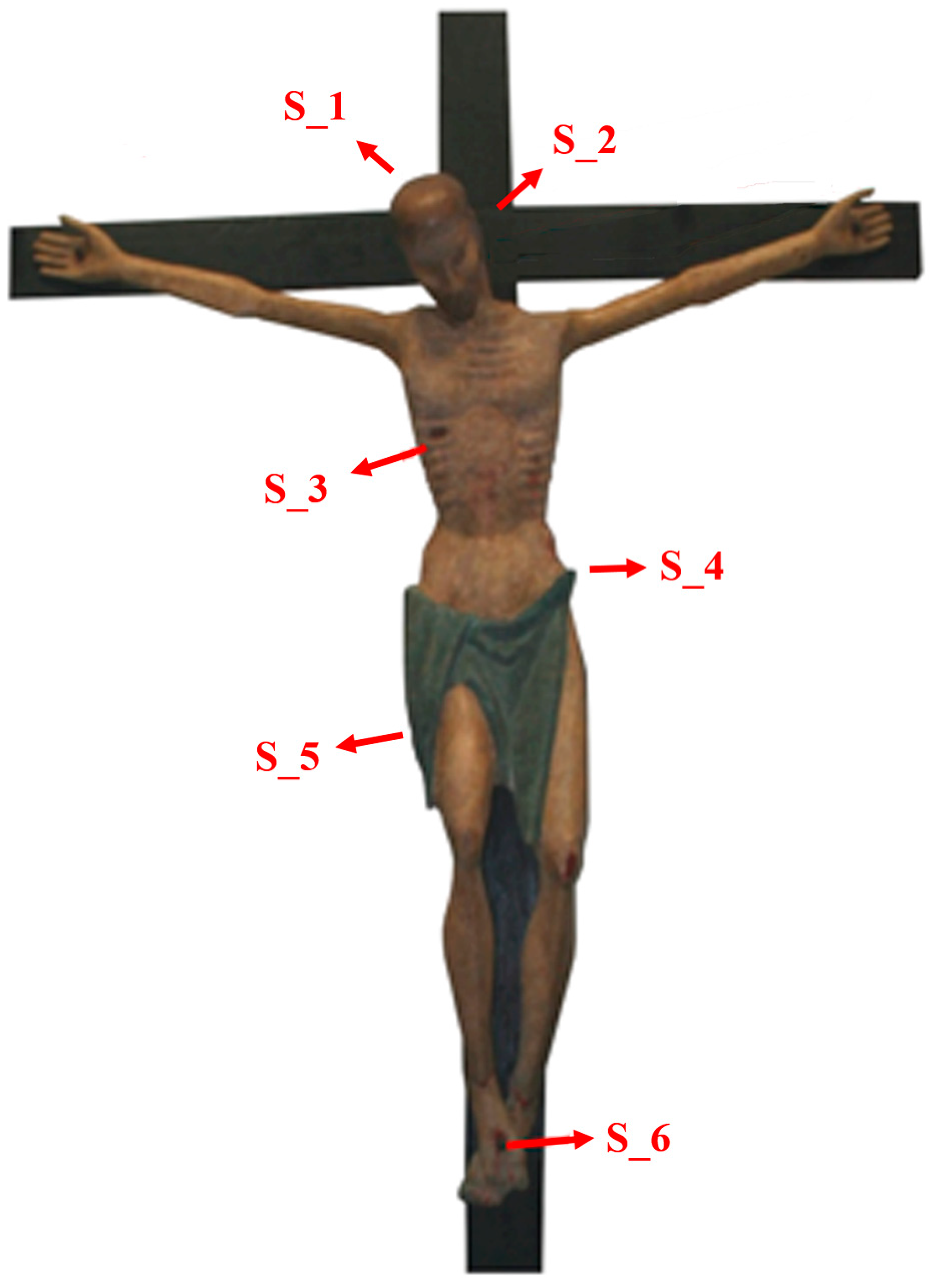
Very small, pigmented fragments were obtained during the last restoration work by using para-destructive methods, i.e., extracting them from zones that are not visible, or else from the most damaged part of the crucifix; six specimens were collected in this way from the different parts of the body, as shown in
Figure 1.
On each specimen, several Raman spectra were collected. The micro-Raman apparatus is a LabRAM spectrometer (Horiba Jobin Yvon, Piscataway, New Jersey, USA) consisting of a single grating spectrograph, interfaced with an Olympus microscope, an edge filter and a Peltier cooled CCD detector (256 × 1024 pixels). Different microscope objectives were used for visual analysis of the samples; for the collection of the spectra, carried out using backscattering geometry, the 50× long-working-distance objective was used. A He-Ne laser (633 nm) internal to the spectrometer was used as an excitation source. The grating used was with 1800 grooves/mm. The spectral resolution was about 1–2 cm−1. The Raman spectra shown in this work were baseline-corrected.
3. Results and Discussion
Figure 2 shows photos of the six specimens investigated using Raman spectroscopy.
For each specimen several points were analyzed, in order to provide detailed information on the pigments used. For instance, both samples S_4 and S_5, from the thong, appear green; however, the pigments found are not green, but yellow and blue.
In general, a strong luminescent background is present in all the spectra. In some cases, it totally covers the Raman signal. By using the He-Ne laser for excitation, we obtain a better ratio of Raman signal over luminescence. However, we performed many measurements at different points on the samples in order to obtain a reasonable Raman-signal-to-noise ratio.
Representative Raman spectra collected from the studies on different specimens are displayed in
Figure 3.
Because of the fine mixture of pigments present, in many cases, the spectral features originate from different materials.
A summary of the various colors found in the Crucifix and studied via micro-Raman analysis is given in
Table 1 below.
Some of the collected spectra are shown in the following section. Various pigments were identified using the databases provided [
5,
6,
7,
8,
9].
The first set of representative Raman spectra of the pigments found on the crucifix under examination is reported in
Figure 3. Shown are pigments of ancient use, which are expected in a sculpture made in the XV century, after the most reasonable estimate. The Raman micro-analysis, however, also reveals the presence of other pigments, introduced in use more recently: besides the titanium white, discussed above, the spectra of some modern pigments are shown separately in
Figure 3.
First at all, we report, in
Figure 3a, the spectrum of gypsum (calcium sulphate dihydrate), which was derived from the preparatory layer and was found in different samples, alone or in combination with other pigments. In addition, in the spectrum of gypsum, reported in
Figure 3a, the presence of titanium white (anatase) is also revealed: in fact, the strong, low frequency peak at 143 cm
−1 is derived from it, as are the shoulders at 395 cm
−1 and 636 cm
−1, which overlap with the typical internal mode of the sulphates.
In
Figure 3b, the spectrum of hematite can be seen—called red ochre by painters—which was found in the red regions of our samples. This pigment, largely used, has been well known since prehistoric times for painting red and red-like zones.
Another material used as a red color is cinnabar (mercury sulfide)—called vermillion in the artistic milieu—herein reported in
Figure 3c, found in some regions of our samples. Finally, in the darker zones of this painted crucifix, carbon black was found, and its typical spectrum is reported in
Figure 3d, corresponding to amorphous carbon. Some of the spectra associated with the modern pigments are reported in
Figure 4.
For instance, in the samples coming from the green zones of the sculpture, the occurrence of lithopone is revealed (see
Figure 4a). In other measurements on the dark green regions, the spectrum of Prussian blue was collected. This organometallic complex was discovered at the end of the 18th century, and its use in painting starts from the 19th century. Even in the presence of a strong, luminescent background, the characteristic Raman modes are clearly observed, particularly the one at 2152 cm
−1, which is quite a specific marker.
The complete list of the pigments found in this study is summarized in
Table 2.
4. Conclusions
In this work, the identification of different kinds of pigments on a Crucifix from the Church of St. Mary Major in Acri (Calabria), obtained via para-destructive micro-Raman analysis, was discussed. Gypsum, titanium white, lithopone, Siena earth, red ochre, vermilion, minium, chrome yellow, lead yellow, Prussian blue, and carbon black were found.
Considering their widespread applications in the past, from the middle of the 18th century to the 19th century, and even more recently, the presence of such pigments can support the hypothesis of several undocumented restoration works on this crucifix—probably during the 19th century and maybe one in the 20th century—before the application of “papier mache”, finally removed in the last restoration of 2012.
Author Contributions
Conceptualization, M.C. and E.C.; methodology, D.G.; software, A.N.; validation, M.C., C.R. and A.N.; formal analysis, D.G.; investigation, M.C.; resources, M.C.; data curation, D.G. and M.C.; writing—original draft preparation, M.C., R.C.B. and E.C.; writing—review and editing, E.C., C.R. and M.C.; visualization, C.R. and A.N.; supervision, M.C. and E.C.; project administration, M.C. and R.C.B.; funding acquisition, M.C. and R.C.B. All authors have read and agreed to the published version of the manuscript.
Funding
This restoration was financially supported by the Rotary club of Acri.
Institutional Review Board Statement
Not applicable.
Informed Consent Statement
Not Applicable.
Data Availability Statement
The data presented in this study are available on request from the corresponding author.
Acknowledgments
The authors thank Murat Cura, who carried out the restoration work on the crucifix, for his assistance and for providing the samples.
Conflicts of Interest
The authors declare no conflict of interest.
References
- Castriota, M.; Cosco, V.; Barone, T.; De Santo, G.; Carafa, P.; Cazzanelli, E. Micro-Raman Characterizations of Pompei’s mortars. J. Raman Spectrosc. 2008, 39, 295–301. [Google Scholar] [CrossRef]
- Castriota, M.; Meduri, E.; Barone, T.; De Santo, G.; Cazzanelli, E. Micro-Raman Investigations on the Fresco Trapasso Della Vergine in the Church of S. Giovanni Battista of Paterno Calabro in Southern Italy. J. Raman Spectrosc. 2008, 39, 284–288. [Google Scholar] [CrossRef]
- Cazzanelli, E.; Platania, E.; De Santo, G.; Fasanella, A.; Castriota, M. Micro-spectroscopic Raman Investigation on the Canvas Oil Painting ‘Rebecca at the Well’ of Neapolitan Anonymous. J. Raman Spectrosc. 2012, 43, 1694–1698. [Google Scholar] [CrossRef]
- Pingitore, G.; Cerchiara, T.; Chidichimo, G.; Castriota, M.; Gattuso, C.; Marino, D. Structural Characterization of Corrosion Product Layers on Archaeological Iron Artifacts from Vigna Nuova, Crotone (Italy). J. Cult. Herit. 2015, 16, 372–376. [Google Scholar] [CrossRef]
- Bell, I.M.; Clark, R.J.H.; Gibbs, P.J. Raman Spectroscopic Library of Natural and Synthetic Pigments (Pre- ≈ 1850 AD). Spectrochim. Acta. A. Mol. Biomol. Spectrosc. 1997, 53, 2159–2179. [Google Scholar] [CrossRef]
- Burgio, L.; Clark, R.J.H. Library of FT-Raman Spectra of Pigments, Minerals, Pigment Media and Varnishes, and Supplement to Existing Library of Raman Spectra of Pigments with Visible Excitation. Spectrochim. Acta Part A Mol. Biomol. Spectrosc. 2001, 57, 1491–1521. [Google Scholar] [CrossRef]
- Bersani, D.; Antonioli, G.; Lottici, P.P.; Casoli, A. Raman Microspectrometric Investigation of Wall Paintings in S. Giovanni Evangelista Abbey in Parma: A Comparison between Two Artists of the 16th Century. Spectrochim. Acta Part A Mol. Biomol. Spectrosc. 2003, 59, 2409–2417. [Google Scholar] [CrossRef]
- Bouchard, M.; Smith, D.C. Catalogue of 45 Reference Raman Spectra of Minerals Concerning Research in Art History or Archaeology, Especially on Corroded Metals and Coloured Glass. Spectrochim. Acta Part A Mol. Biomol. Spectrosc. 2003, 59, 2247–2266. [Google Scholar] [CrossRef]
- Caggiani, M.C.; Colomban, P. Testing of Raman Spectroscopy as a Non-Invasive Tool for the Investigation of Glass-Protected Pastels. J. Raman Spectrosc. 2011, 42, 790–798. [Google Scholar] [CrossRef]
| Publisher’s Note: MDPI stays neutral with regard to jurisdictional claims in published maps and institutional affiliations. |
© 2022 by the authors. Licensee MDPI, Basel, Switzerland. This article is an open access article distributed under the terms and conditions of the Creative Commons Attribution (CC BY) license (https://creativecommons.org/licenses/by/4.0/).


 ,
, 


