Article

Calculation of the Rearfoot Angle Representing Flatfoot from Comparison to the Cavanagh Arch Index

by
Yusuf Umardani
1,*,
Dwi Basuki Wibowo
1,*,
Wahyu Caesarendra
2,*,
Agus Suprihanto
1 and
Kirana Astari Pranoto
2
1
Department of Mechanical Engineering, Diponegoro University, Semarang 50275, Indonesia
2
Faculty of Integrated Technologies, Universiti Brunei Darussalam, Jalan Tungku Link, Gadong BE1410, Brunei
*
Authors to whom correspondence should be addressed.
Appl. Sci. 2022, 12(13), 6764; https://doi.org/10.3390/app12136764
Submission received: 5 March 2022 / Revised: 31 May 2022 / Accepted: 28 June 2022 / Published: 4 July 2022

Abstract

:
A type of flatfoot can be analyzed accurately using the footprint when a human is standing; however, this method cannot be applied when a human is wearing orthotic shoes. This study aims to analyze flatfoot using the rearfoot angle (RFA) measurement. The result is then compared to a footprint measurement known as Cavanagh’s Arch Index (AI). A total of 31 static footprints of the participants consisting of 14 males and 17 females aged 18 to 25 years were collected. According to Cavanagh’s AI as a gold standard, the correlation equation was obtained as RFA = 46.04AI − 6.41 and RFA = 45.32AI − 6.26 for left and right foot, respectively. The correlation coefficient R2 for the left and the right foot is 0.63 and 0.73, respectively. Other statistical analyses using ANOVA and t-tests are presented in this paper. The correlation results obtained from this study are necessary for estimating the reduction in the degree of flatfoot when using orthotic shoes, which is difficult to calculate using the typical AI method from footprint measurements.

1. Introduction

The human foot can be divided into three parts: rearfoot, arch of the foot, and forefoot [1]. When walking or running in the mid-stance phase, the body weight passes over the foot as the body comes forward. This aspect is where the foot supports the body weight, and the arch provides necessary shock absorption for the foot [2]. The pronator type of foot rolls far inward during the weight-bearing phase of the stride. This foot type is characterized by a very low or flat arch or commonly called flatfoot type [3]. The absorption of shock loads on this type of foot is low, so it easily tires during activities; however, in over-pronation conditions, most of the burden will be supported by the heel, which can cause heel pain [4] and/or plantar fasciitis [5].
Flatfoot soles can be further divided into two types, namely fixed flatfoot and flexible flatfoot [6]. The difference between these two types can be detected when the subject is standing and lying on their back foot (no weight carried by the subject). On the flatfoot subject with the previously mentioned treatment, the flatfoot remains detected when it is not burdened. In contrast, on the flexible flatfoot subject with similar treatment, the condition is significantly reduced or even becomes normal. Patients with flexible flatfoot are mostly children who generally can become normal as they age. Adolescents can also become normal after wearing orthotic shoes with a certain curvature of the medial arch for a certain period [7].
Previous studies have quantified flatfoot type by 2D footprints based on suitability for static standing [6]. The foot is measured from the width of the instep at its widest part. If the measurement is less than 1 cm, the foot type is classified as a flatfoot. The type of foot is expressed in terms of the Arch Index (AI) value, which is obtained by dividing the midfoot area by the total area of the foot, excluding the toes. The foot is categorized as flatfoot if AI ≥ 0.26 [8]. The measurement of the Clarke angle (α) is based on the tangent of the angle between the two lines, i.e., (1) the line connecting the most medial aspect of the heel to the most medial metatarsals and (2) the line connecting the most lateral point of the medial foot border to the most medial metatarsal. If the Clarke angle measurement α < 42°, it is categorized as a flatfoot [9]. In another method, flatfoot can be determined based on Plantar Arch Index (PAI). The PAI is a relationship between the central and posterior regions of the footprints. The PAI is calculated from the following steps: (1) a line is drawn tangentially to the medial forefoot edge and at the heel region; (2) the mean point of the line is calculated; (3) from this mean point, a perpendicular line is drawn crossing the footprint; (4) a similar procedure is repeated at the heel tangency; (5) measurements are obtained of the width of the central region of the footprint so-called A and of the heel region so-called B; (6) the PAI is then calculated from the ratio of A and B. If the PAI value is greater than 0.89, it is indicative of flatfoot [10]. Some authors recommend using the Chippaux–Smirak Index (CSI) as a screening instrument for flatfoot in preschool-aged children. The CSI is defined as the ratio of the maximum width at the middle arch of the footprint and the maximum width at the forefoot metatarsus region of the footprint. The criterion for determining the signs of flatfoot in preschool-aged children is CSI > 62 [11].
The pronation of the foot can also be seen from the rearfoot angle (RFA), which is the angle between the two lines that pass through the Achilles tendon and the posterior heel/calcaneus when a person stands upright [12]. The pronation is caused by the permanent angular movement of the ankle into the foot. It is clinically used to determine heel valgus and varus, i.e., the inclination of the heel bone. If the ankle leans inward, it is called flatfoot or valgus foot. Otherwise, if the ankle leans outward, it is called high arch or varus foot. RFA ≥ 5° valgus represents a flatfoot type, 4° valgus to 4° varus a neutral foot type, and RFA ≥ 5° varus a supinated foot type [13]. Heel valgus > 5° is considered to shift the axis of the foot and the normal distribution of pressure on the sole, which triggers pain [4]. The RFA, however, has not been reported obviously as a method for flatfoot detection.
This paper presents a calculation of the RFA as a simple alternative method to determine the flatfoot. It is motivated by the previous observation that shows flatfoot sufferers tend to have an abnormal RFA [12]. This study aims to examine whether RFA can be used to determine the condition of a person’s flatfoot based on the comparison with the Cavanagh AI method [14].
Prior to the RFA measurement and analysis, the authors conducted studies to calculate Cavanagh’s AI from confirmed flatfoot subjects. The AI result of the studies was then used as a comparable variable for the RFA measurement. Correlation methods such as linear correlation, ANOVA, and t-test were used as the analysis tools. The study revealed that the RFA is a potential simple method to determine the flatfoot. A more detailed description of the manual AI measurement method, digital AI measurement method, and the proposed method for flatfoot detection is presented in Section 2.

2. Materials and Methods

Determination of flatfoot conventionally uses the Cavanagh method by measuring the Arch Index (AI). The conventional Cavanagh’s AI measurement method is presented in Figure 1a. Cavanagh’s method was then developed in MATLAB program and embedded into the footprint 2D scanner. This embedded program enables users to automatically determine the Foot Length (FW), Foot Width (FW), Foot Area Contact (FAC), and AI. The footprint 2D scanning method is presented in Figure 1b. The RFA method is more straightforward because it only measures the angle of the foot’s backside when standing upright. The RFA method is presented in Figure 1c.

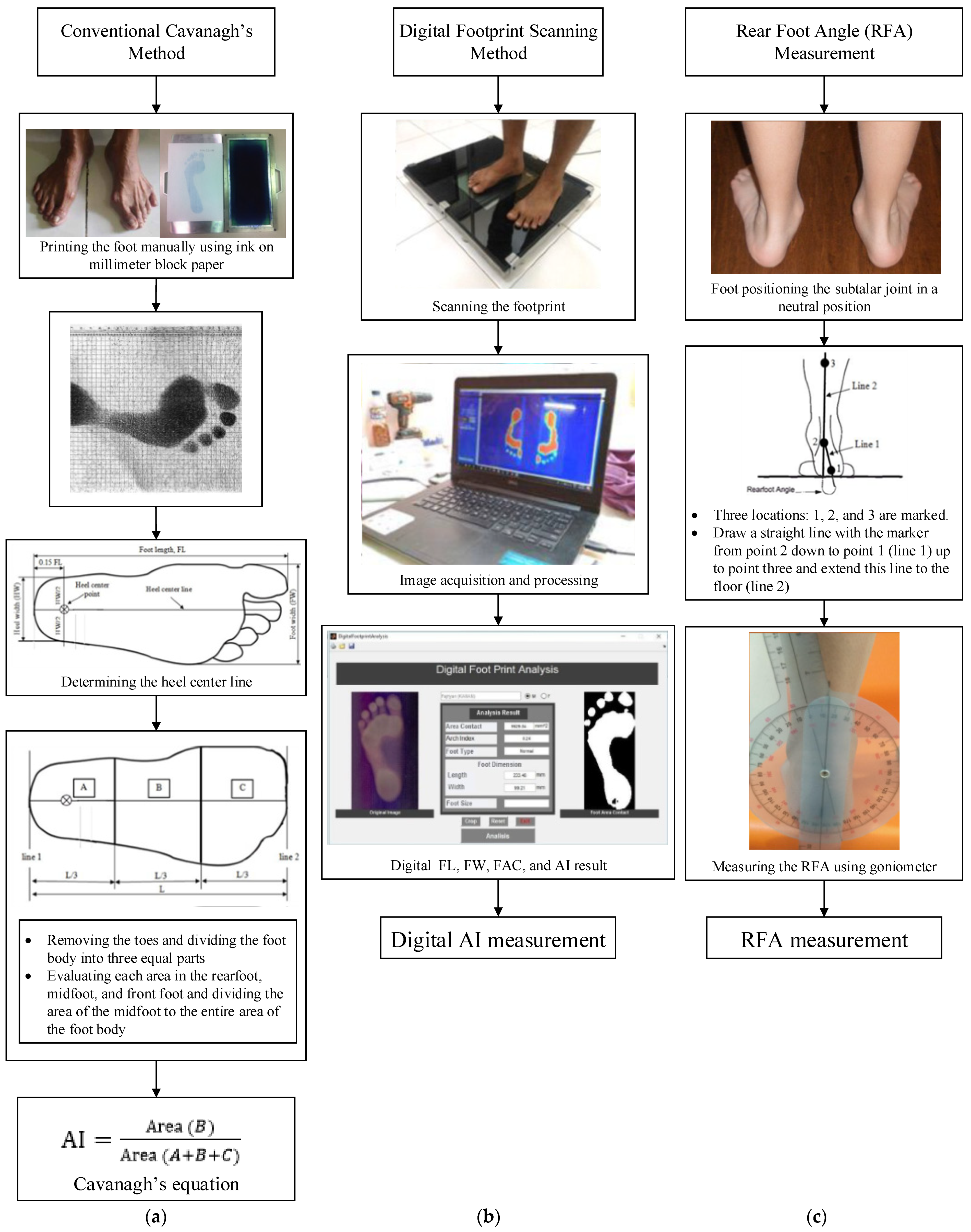
2.1. Conventional Cavanagh’s AI Measurement Method

The conventional Cavanagh’s AI measurement starts with printing the foot manually using ink and millimeter block paper, as presented in Figure 1a. The next three steps are (1) determining the heel center line, (2) removing the toes and dividing the foot body into three equal parts, and (3) evaluating each area in the rearfoot, midfoot, and front foot [15]. The final step is dividing the area of the midfoot into the entire area of the foot body to obtain the value of Cavanagh’s AI, as presented in Figure 1a.
In more detail, the heel centerline is a line drawn from the heel tip through the heel center point to the tip of the second toe. At the same time, the heel center point is a point at 0.15 FL from the heel tip and half the rearfoot width (heel width, HW) [16]. Line 1 in Figure 1a is drawn through the point of intersection between heel center lines with the heel tip, which is the lower limit to determine the division of the body of the sole without the toes into three equal parts.
The first step is dividing the sole into three equal parts. It starts by drawing a line perpendicular to the heel centerline, which tangents to the most anterior part of the outline of the main body of the footprint in front of the metatarsal heads as indicated in line 2 of Figure 1a. The next is deleting all the toes and dividing the length between line 1 and line 2 into equal thirds. This stage divides the foot without the toes into three areas; these are rearfoot (A), midfoot (B), and forefoot (C).
Evaluating each area in the rearfoot, midfoot, and front foot is the last step to determine the value of Cavanagh’s AI (hence, AI), which is dividing the area of the mid-foot to the entire body area of the sole without the toes as presented in Equation (1). It is classified to be normal if 0.26 > AI > 0.21 and flatfoot if AI ≥ 0.26.
AI = Area   ( B ) Area   ( A + B + C )

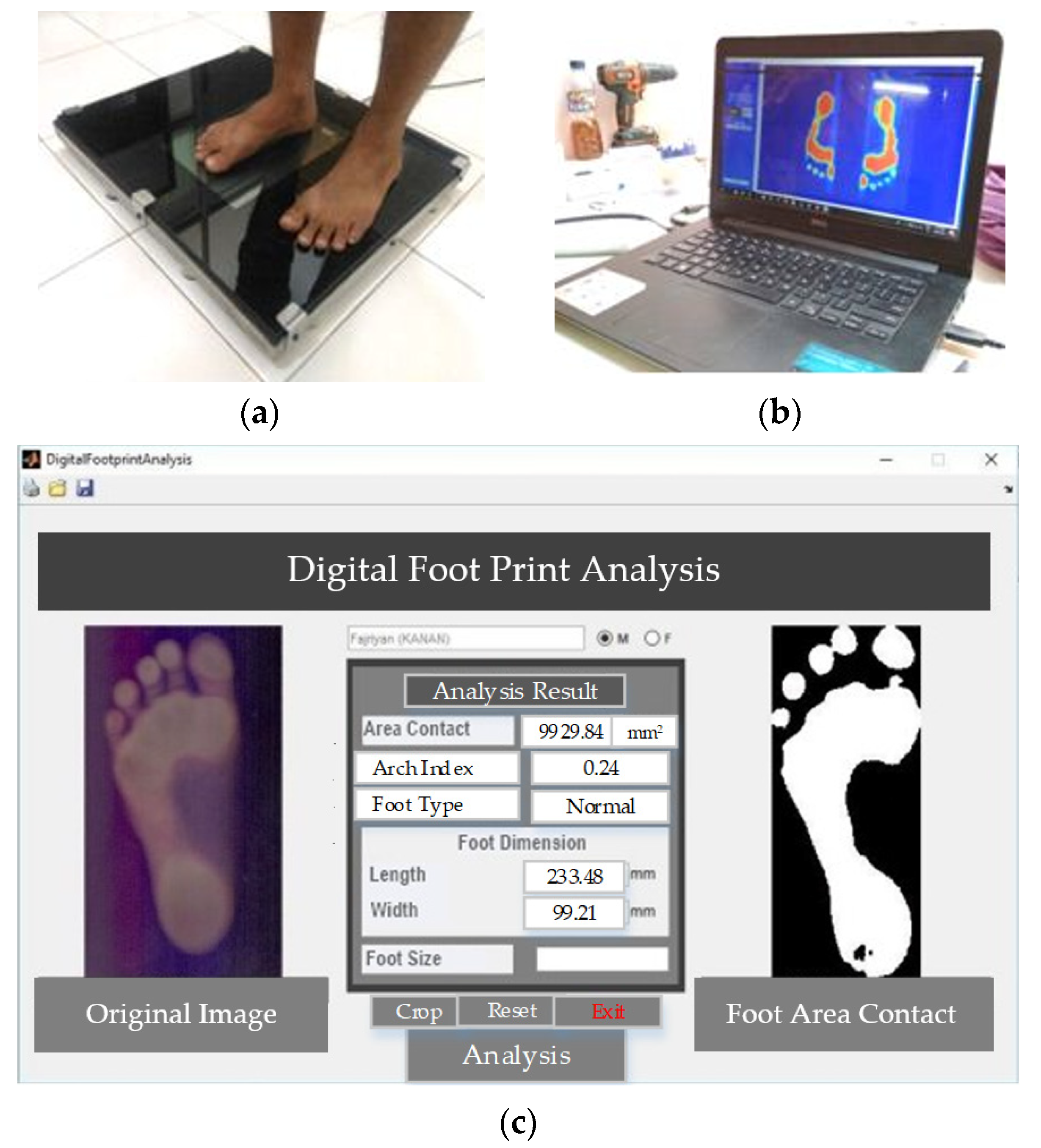
2.2. Digital AI Measurement Method

In this study, the instrument footprint scanner was custom-made [17]. The instrument is based on the HP Scanjet 200 A4 size flatbed scanner. During the development of the instrument, a clinician from the Department of Orthopedic, Tugurejo Hospital was involved in providing suggestions. Upon completion, the instrument is initially used for the study of in-shoe foot orthotic contours on heel pain due to calcaneal spurs [18]. The instrument is used to measure foot area and correlate the result with pronation. The result presented in [18] has been compared and validated with the result from another group of researchers in [19]. The instrument is further submitted for patent filling application and has been examined by experts in certain fields. The clinician also supervised the authors in terms of the experimental set-up, data acquisition, and data analysis in previous studies [17,18].
According to the above explanation, the instrument has been validated and the description of the validation is unnecessary to be presented in this paper. Interest readers are suggested to read the previous studies [17] that compared other research papers [19].
In the present study, the instrument is improved with the development of digital footprint analysis-based MATLAB software. The instrument, image acquisition, and image processing are presented in Figure 2. The original structure of the document scanner has not been changed, including the glass window. The only modification is the removal of the tray part. The scanner is placed directly under 10 mm thick clear glass as a foot scanner platform supported by a steel frame to withstand a mass of up to 100 kg.
To produce a clear image, the effect of light from the environment must be minimized and the foot must be clean. The scanning process occurs when the subject is standing in upright posture above the platform, which can be assisted by the operator. The digital footprint scanning process is presented in Figure 2a. A foot was placed over the scanner in a straight position. Then the image was processed using MATLAB software to produce Foot Length (FL), Forefoot Width (FW), Foot Area Contact (FAC), and Arch Index (AI), as presented in Figure 2b [20]. The results of the digital footprint analysis are presented in Figure 2c.

2.3. Rearfoot Angle (RFA) Measurement

This section presents a proposed methodology, i.e., RFA measurement as an alternative flatfoot detection. According to the previous studies, a Cavanagh’s AI is a typical method used to determine the flatfoot; however, the calculation stages of either manual or digital AI measurement are time-consuming. This study is then proposed as an alternative method to determine flatfoot based on the RFA measurement. The RFA measurement is much simpler than the manual AI measurement (Figure 1a) and the digital AI measurement (Figure 1b).
To obtain the RFA, each subject was asked to stand relaxed on a flat platform so that the subtalar joint in a neutral position could be felt by palpation. The subtalar joint is formed between the talus and calcaneus and was used as a reference point for rearfoot measurement.
Three locations are marked using a marker pen, as presented in Figure 3a. These are: (1) the base of the calcaneus, which is indicated as point 1, (2) the subtalar joint that is marked as point 2, and (3) the top end of the bisection of the lower one-third of the leg or 15 cm above point 2 that is marked as point 3 [13]. The next step is to draw a straight line with the marker from point 2 down to point 1 (line 1) up to point 3 and extend this line to the floor (line 2), as illustrated in Figure 3a. The RFA is the angle formed between line 1 and line 2; if the angle shows that the ankle leans inward, it is called flatfoot or valgus foot, and otherwise, it is called high arch or varus foot.
The RFA was measured using a goniometer as presented in Figure 3b. The goniometer arms were aligned with line 1 and the other arm with line 2. RFA is measured as the angle between the projections of line 1 and line 2. RFA ≥ 5° valgus represents a flatfoot type. In this study, we find the minimum value of RFA, which indicates flatfoot, by using the measurement results of Cavanagh’s AI as a basis.

2.4. Research Subjects

The research subjects were 31 students of the Department of Mechanical Engineering, Diponegoro University, aged between 18–25 years, as presented in Table A1. These subjects have been indicated as flatfoot subjects from the AI measurement and have been confirmed by the clinician. The research subjects were asked to voluntarily measure BMI and footprint scanning of both left and right foot. The BMI measurement was carried out using a measuring instrument that is custom-made and has patent No. IDS000002589 [21], to determine the correlation between BMI and AI.
The sample size determination was based upon a statistically minimal number of subjects. The BMI of all subjects is sorted from low to high, and male and female, to find out whether the BMI of the subject is underweight, normal, or overweight, and the difference in BMI between males and females.
Measurement of the footprint of each subject produces parameters FL, FW, FAC, and AI and then sorted for each left and right foot, male and female. Evaluation of the average footprint parameters was carried out to find out whether there is a significant difference between the left and right foot and male and female.
The digital footprint scanning method was performed using an instrument footprint scanner to obtain FL, FW, FAC, and AI [17]. The RFA, FL, FW, and FAC measurements were then used to calculate the AI.

3. Results

3.1. General Statistical Results

Data from thirty-one flatfoot subjects consisting of 14 males and 17 females presented in Table A1 were statistically analyzed and the result is presented in Table 1. The average age of the participants is 21.3 ± 1.5 years. In detail, the average age of males and females is 21.8 ± 0.7 years and 20.9 ± 1.8 years, respectively.
According to WPRO Criteria 2000, someone is categorized as underweight if their BMI ≤ 18.4 kg/m2, normal if BMI 18.5–22.9 kg/m2, overweight if BMI 23.0–24.9 kg/m2, and obese if BMI > 25.0 kg/m2 [22]. In this study, 20 subjects (6 males and 14 females) are categorized as having normal BMI; 11 subjects (8 males and 3 females) are categorized as overweight. The range of BMI values of males and females is approximately 20.8–24.5 kg/m2 for males and 18.8–24.2 kg/m2 for females, with median values of approximately 23.0 kg/m2 for males and 21.7 kg/m2 for females. The mean value of BMI for males is 22.7 ± 1.2 kg/m2, and for females is 21.4 ± 1.6 kg/m2.
The mean FL and FW on the soles of the left and right feet of all subjects are almost similar, i.e., 0.1 mm. The range of FL values for males and females is approximately 214.7–264.4 mm and 216.9–252.9 mm, respectively, with FL median values being approximately 245.2 mm for males and 234.3 mm for females. The average FL for males and females is 239.6 ± 13.7 mm and 234.4 ± 10.9 mm, respectively.
In addition, the range of FW values for males and females is approximately 86.3–106.6 mm and 85.5–108.7 mm, respectively, with FW median values being approximately 97.2 mm for males and 94.6 mm for females. The average FW of the males and females is 96.1 ± 5.9 mm and 95.2 ± 5.9 mm, respectively.
The average FAC of all subjects is 11,504 ± 1874 mm2 with median values of approximately 11,016 mm2. The range of FAC values for males and females is approximately 101,802–16,669 mm2 and 9084–13,554 mm2, respectively, with FAC median values being approximately 11647 mm2 for males and 10,691 mm2 for females. The average FAC of males and females is 12,373 ± 2136 mm2 and 10,788 ± 1293 mm2, respectively.
The average AI of the left and right feet for all subjects is 0.35 ± 0.05. In detail, the average AI of males and females is 0.33 ± 0.05 and 0.37 ± 0.04, respectively. In addition, the mean value of RFA for all subjects is 16.0° ± 4.0°, with AI median values being approximately 9.5°. The average RFA of the left and right feet is 9.9° ± 3.5° and 9.6° ± 3.4°, respectively, which is only different by 0.3°. The mean value of RFA is 8.6° ± 3.4° for males and 10.7° ± 3.1° for females, with median RFA values of 9.0° and 10.5° for males and females, respectively.

3.2. Correlation Results between Subject Characteristics and Cavanagh’s AI

This study was conducted to examine an alternative RFA measurement to determine flatfoot by using the correlation between Cavanagh’s AI and RFA. Prior to the correlation between AI and RFA, a correlation between subject characteristics such as foot size, gender, BMI, FW, FL, FAC, and Cavanagh’s AI were analyzed and the result is presented in Figure 4.
Figure 4 shows the correlation between variables presented in Table 1 and the average AI. There is no difference between the AI mean of left and right feet. This indicates that most of the subjects were standing upright during the footprint scanning. The average AI of the female is greater than male, which is about 0.04. The range of AI values is 0.27–0.44, with AI median values being approximately 0.35. There are several subjects consisting of 2 males and 5 females with AI ≥ 0.4, and others are categorized as over-pronation category (AI >> 0.26) [4].
According to Table 1, the average BMI of males is greater than females, which is also similar to the result presented in [23]. Meanwhile, from BMI grouping, the average AI of the overweight category is larger than the normal category, i.e., 0.38 ± 0.06 and 0.35 ± 0.05, respectively, as presented in Figure 4 [24].
The mean FL and FW on the soles of the left and right feet for all subjects are almost similar. This indicates that most of the subjects were standing upright during the footprint scanning. The mean FL of males is greater than females [20]; however, the FL ≥ median values show that the average AI is much larger than FL < median (median value of all subjects is 235.3 mm), as presented in Figure 4. It means that a large FL affects the degree of flatfoot.
The average FW of males is higher than females [25] but differs only around 0.9 mm. The average AI for FW ≥ median and FW < median values for all subjects are approximately the same, i.e., 0.35 ± 0.04 and 0.35 ± 0.06 (median = 95.2 mm, as presented in Table 1). This means that a large FW does not affect the degree of flatfoot, as presented in Figure 4.
Typically, in the range of FAC values of males and females, it shows that the sole shape of the male foot is larger than the female [20]; however, the present study revealed that the females have a larger correlation to Cavanagh’s AI than the males. This indicates that the shape of the foot does not have a direct correlation to the flat foot.
According to [26], there is a significant correlation between BMI and FAC, with a correlation coefficient of 0.75. This shows that the higher the BMI value, the greater the FAC value; however, there is no significant correlation between FAC and AI in the present study, as presented in Figure 4. Overall, there is no significant correlation between subject characteristics and Cavanagh’s AI for all parameters, i.e., foot size, gender, BMI, FW, FL, and FAC, as the correlation is lower than 0.4, as presented in Figure 4. This indicates that these parameters have a small correlation with Cavanagh’s AI. The highest correlation shown is BMI, especially in the overweight category, with a correlation of 0.38. The correlation between female gender and Cavanagh’s AI, as well as the FL < Median and Cavanagh’s AI, show slightly less than overweight BMI of about 0.37. In contrast, the correlation between male gender and Cavanagh’s AI, as well as the correlation between FL ≥ Median and Cavanagh’s AI, is the smallest at about 0.33. This indicates that males have less probability of flatfoot than females.

3.3. Correlation Results of Arc Index (AI) and Rear Foot Angle (RFA)

Prior to the correlation analysis between AI and RFA determined, this study analyses a certain gender that has a higher RFA value. The present study has a similar result to [13] that reported the mean RFA of males is lower than females. According to the measurements, the person is assumed to have a flatfoot category if the RFA ≥ 5°; and according to the results, the RFA ≥ 5° is mostly addressed to female subjects [13].
There is a significant correlation between AI and RFA, with a correlation coefficient of R2 for the left and the right foot being 0.63 and 0.73, respectively, as presented in Figure 5. The correlation equation between AI and RFA was obtained as RFA = 46.04AI – 6.41 and RFA = 45.32AI − 6.26 for the left and right foot, respectively, as expressed in Equation (2). The RFA value at AI = 0.26 (the minimum value of Cavanagh’s AI, which represents flatfoot) is 6.3°. Meanwhile, the average RFA value of all subjects (left and right foot) is 16.0 ± 4.0°, which is calculated using Equation (2), and yield an AI range between 0.25–0.45.
The AI range of the measurement results differs slightly from the highest and the lowest. Thus, the value of RFA ≥ 6.3° can be used as a new diagnosis reference for flatfoot in the measurement of the RFA.
RFA = 46.04AI − 6.41 (Left foot)
RFA = 45.37AI − 6.26 (Right foot)
From the previous studies [27], the degree of flatfoot is in the minor category if AI = 0.26–0.29, moderate if AI = 0.30–0.33, severe if AI = 0.34–0.36, and worst if AI = 0.37–0.39. The range of RFA values for each degree of flatfoot is presented in Table 2.
The statistical analysis was carried out using the ANOVA method to examine the correlation level of RFA to AI. The analysis results are presented in Table 3 and Table 4 for the left foot and right foot, respectively. The p-value shows the significance of RFA variables on AI variables. According to Table 3 and Table 4, the p-values of 3.83 × 10⁻²⁵ and 1.61 × 10⁻²⁵ are much smaller than 0.05. To confirm the relationship between RFA and AI, a statistical test that compares the means of two samples is used; t-test analysis is also used and presented in Table 5 and Table 6 for the left foot and right foot, respectively.
The result presented in Table 3 and Table 4 indicates that there is a correlation between RFA and AI because it has a degree of confidence between these two variables. The RFA variables will be considered significant if the p-value is less than 0.05.
Another correlation analyses results using the t-test are presented in Table 5 and Table 6. The t-Stat of both the left foot and the right foot is negative, indicating that there is no significance of difference between the two variables (RFA and AI). A similar result of p-values, which are much less than 0.05, is also obtained in the left foot and the right foot of the t-test, as presented in Table 5 and Table 6.

4. Discussion

This study is a sequential study that aims to measure and analyze the degree of flatfoot of the subjects. The authors have shown their interest in this particular research area as presented in the previous research publication [17,18]. The previous research developed a digital footprint scanner method, which is supervised by an orthopedic doctor/surgeon from RSUD Tugurejo Hospital (the details of the orthopedic surgeon are provided in the acknowledgment). The conventional Cavanagh AI method was improved using the digital footprint scanning method based on MATLAB, as presented in Figure 1b.
According to another orthopedic surgeon’s suggestion, a less complicated yet accurate method is required to be applied as an alternative and to support the flatfoot diagnosis method. It is because the developed digital footprint scanning method, as presented in Figure 1b, is time-consuming and more complicated in terms of data processing even though the progressive result has been validated.
This research was also motivated by cases where patients who suffer from flatfoot commonly have a different back angle than those who do not; therefore, research is needed to determine the correlation between the angles behind the foot arising from the presence of a flatfoot.
The RFA measurement is evident as a potential method for flatfoot diagnosis; however, this method has not been presented obviously as an applicable method for flatfoot measurement. This study examined the RFA by calculating the correlation with the well-known method for flatfoot detection.

5. Conclusions

A study to analyze flatfoot using Rear Foot Angle (RFA) measurement has been presented. This present study has produced an equation that correlates between AI and RFA as presented in Equation (2), which can be quantified as the minimum RFA. The flatfoot is identified if RFA ≥ 4.3°. The correlation equation is very important for estimating the reduction in the degree of flatfoot when using orthotic shoes from RFA measurements, which cannot be determined by the AI value from typical footprint measurements.
The method of determining flatfoot based on RFA measurement as an alternative method has not been obviously observed. Determination of conventional flatfeet using the arc index measurement and the Cavanagh method requires a few measurement steps. One of the measurements is the foot area after the footprint or foot scanning technique. The RFA method is simpler because it only measures the angle of the back of the foot when someone is standing upright. The finding of this study is evidently essential as RFA measurement has a significant correlation with AI and offers a more effective method for flatfoot detection. Conducting diverse works on this study with a larger number of participants and more valid methods of RFA measurement is highly recommended in the future as RFA is acknowledged as a more effective diagnosis measurement method for flatfoot.

Author Contributions

Conceptualization, D.B.W. and A.S.; methodology, Y.U. and D.B.W.; software, Y.U. and A.S.; validation, D.B.W., A.S., and W.C.; formal analysis, Y.U. and D.B.W.; investigation, Y.U. and D.B.W.; resources, D.B.W. and A.S.; data curation, Y.U. and D.B.W.; writing—original draft preparation, Y.U. and D.B.W.; writing—review and editing, A.S., W.C., and K.A.P.; visualization, Y.U.; supervision, D.B.W. and W.C.; project administration, A.S.; funding acquisition, Y.U. All authors have read and agreed to the published version of the manuscript.

Funding

This research received no external funding.

Institutional Review Board Statement

Not applicable.

Acknowledgments

This research was collaborated and supervised by Rudiansyah Harahap from the Department of Orthopedic, RSUD Tugurejo Hospital, via medical-related discussion, data acquisition, and data processing. The authors thank Rudiansyah Harahap for their support during the study. The authors also thank the subjects who were involved and participated in this study.

Conflicts of Interest

The authors declare no conflict of interest.

Appendix A

Detailed information on the thirty-one flatfoot subjects consisting of 14 males and 17 females and their foot measurements is presented in Table A1. The subjects have been informed with the consent form for the entire experiment and data acquisition activities. During the experiment, a collaborator from the Department of Orthopedic, RSUD Tugurejo Hospital, was involved.
Table A1. Subjects’ description *.
No.Gender
(M/F)
Age
(yr)
Weight
(kg)
Height
(cm)
BMI
(kg/m2)
Foot Width (mm)Foot Length (mm)FootFAC (mm2)Arch Index (AI)RFA (°)
LeftRightLeftRightSizeLeftRightLeftRightLeftRight
1M226616424.597.1899.08249.49252.414115,943.315,943.30.430.4516.016
2M216116622.1105.53107.72247.16244.414010,82910,8290.270.3112.012
3M215916820.996.5496.04250.13248.224010,807.4510,807.450.320.3410.09
4M225715822.898.4597.43221.29226.883711,066.811,066.80.310.309.09
5M226817223.097.5698.83264.73264.14214,331.514,331.50.420.4114.014
6M206216123.994.7795.53232.85234.373914,847.614,847.60.360.3710.011
7M216817123.3102.3102.35247.58252.374110,812.410,812.40.330.3312.012
8M225515921.894.1394.08229.17226.693710,702.410,702.40.260.276.06
9M225515124.188.0385.62211.51217.863611,66011,6600.260.285.56
10M225615623.088.9286.38231.58225.863711,634.111,634.10.320.237.04
11M225815723.585.2487.4230.05223.963711,858.711,858.70.300.276.05
12M227617325.4103.5396.93247.71246.954116,668.816,668.80.350.3610.011
13M225616221.398.5894.13242.76246.314011,881.0311,881.030.310.307.06
14M235816720.8102.0197.05246.95245.814010,179.810,179.80.380.288.08
15F227017024.295.1595.15236.66236.664011,016.2211,016.220.370.3710.09
16F224915719.9108.7495.15232.09229.173710,690.710,690.70.380.3810.010
17F216217021.5101.24101.24247.08247.08419083.659083.650.390.399.09
18F216016721.593.7590.7235.14235.394011,222.111,222.10.390.377.08
19F215115820.492.8689.94227.77225.35379440.39440.30.410.4215.015
20F225516121.283.8487.14218.24222.31369672.239672.230.270.294.56.5
21F205915923.396.9399.85244.15239.714012,478.312,478.30.330.377.17.1
22F205617218.989.56119.79247.71245.17409932.199932.190.330.307.07
23F195315621.895.1594.51229.17227.393811,104.511,104.50.400.418.511.5
24F184415119.395.78121.57218.49215.4536975697560.420.4112.012
25F196115326.197.1892.1223.58227.773613,554.213,554.20.420.4213.012.5
26F186116522.491.97142.4245.55260.294013,058.413,058.40.400.4011.011
27F225115321.892.9991.97217.61218.243610,319.7910,319.790.370.4012.511.5
28F256016621.8102.51102.64245.81245.554011,391.911,391.90.290.367.87.8
29F215415622.286.7690.19235.26232.343910,896.0310,896.030.370.3312.012.5
30F225016318.895.1598.2246.95243.01409280.079280.070.310.3412.011
31F225716221.792.9992.99234.12234.53910,495.310,495.30.430.3415.011
* FL: foot length, FW: foot width, FAC: foot area contact, RFA: rearfoot angle.

References

  1. Yung-Hui, L.; Wei-Hsien, H. Effects of shoe inserts and heel height on foot pressure, impact force, and perceived comfort during walking. Appl. Ergon. 2005, 36, 335–362. [Google Scholar] [CrossRef] [PubMed]
  2. Orr, J.D.; Nunley, J.A. Isolated Spring Ligament Failure as a Cause of Adult-Acquired Flatfoot Deformity. Foot Ankle Int. 2013, 34, 818–823. [Google Scholar] [CrossRef] [PubMed]
  3. Vulcano, E.; Deland, J.T.; Ellis, S.J. Approach and treatment of the adult acquired flatfoot deformity. Curr. Rev. Musculoskelet. Med. 2013, 6, 294–303. [Google Scholar] [CrossRef] [Green Version]
  4. Irving, D.B.; Cook, J.L.; A Young, M.; Menz, H.B. Obesity and pronated foot type may increase the risk of chronic plantar heel pain: A matched case-control study. BMC Musculoskelet. Disord. 2007, 8, 41. [Google Scholar] [CrossRef] [PubMed] [Green Version]
  5. Wearing, S.C.; Smeathers, J.E.; Urry, S.R. The Effect of Plantar Fasciitis on Vertical Foot-Ground Reaction Force. Clin. Orthop. Relat. Res. 2003, 409, 175–185. [Google Scholar] [CrossRef] [PubMed]
  6. Atik, A.; Ozyurek, S. Flexible flatfoot. North. Clin. Istanb. 2014, 1, 57–64. [Google Scholar] [CrossRef] [PubMed]
  7. Kenneth, H.; Noll, C.O. The use of orthotic devices in adult-acquired flatfoot deformity. Foot Ankle Clin. 2001, 6, 1. [Google Scholar]
  8. Cavanagh, P.R.; Rodgers, M.M. The arch index: A useful measure from footprints. J. Biomech. 1987, 20, 547–551. [Google Scholar] [CrossRef]
  9. Pauk, J.; Ihnatouski, M.; Najafi, B. Assessing Plantar Pressure Distribution in Children with Flatfoot Arch. J. Am. Podiatr. Med. Assoc. 2014, 104, 622–632. [Google Scholar] [CrossRef] [PubMed]
  10. Tashiro, Y.; Fukumoto, T.; Uritani, D.; Matsumoto, D.; Nishigucki, S.; Fukutani, N.; Adachi, D.; Hotta, T.; Morina, S.; Shirooka, H.; et al. Children with flat feet have weaker to grip strength than those having a normal arch. J. Phys. Ther. Sci. 2015, 27, 3533–3536. [Google Scholar] [CrossRef]
  11. Chen, K.-C.; Tung, L.-C.; Tung, C.-H.; Yeh, C.-J.; Yang, J.-F.; Wang, C.-H. An investigation of the factors affecting flatfoot in children with delayed motor development. Res. Dev. Disabil. 2014, 35, 639–645. [Google Scholar] [CrossRef] [PubMed]
  12. Sobel, E.; Levitz, S.; Caselli, M.; Brentnall, Z.; Tran, M.Q. Natural history of the rearfoot angle: Preliminary values in 150 children. Foot Ankle Int. 1999, 20, 119–125. [Google Scholar] [CrossRef]
  13. Hunt, A.E.; Fahey, A.J.; Smith, R.M. Static measures of calcaneal deviation and arch angle as predictors of rearfoot motion during walking. Aust. J. Physiother. 2000, 46, 9–16. [Google Scholar] [CrossRef]
  14. Pita-Fernandez, S.; Gonzalez-Martin, C.; Alonso-Tajes, F.; Seoane-Pillado, T.; Pertega-Diaz, S.; Perez-Garcia, S.; Seijo-Bestilleiro, R.S.; Balboa-Barreiore, V. Flatfoot in a Random Population and its Impact on Quality of Life and Functionality. J. Clin. Diagn. Res. 2017, 11, LC22–LC27. [Google Scholar]
  15. MATLAB. Release R2021a; The MathWorks Inc.: Natick, MA, USA, 2021. [Google Scholar]
  16. Rodrigo, A.S.; Goonetilleke, R.S.; Xiong, S. Load distribution to minimize pressure-related pain on foot: A model. Ergonomics 2013, 56, 1180–1193. [Google Scholar] [CrossRef] [PubMed]
  17. Haryadi, G.D.; Wibowo, D.B.; Ariyanto, M.; Suprihanto, A. Digital Scanning Method for Evaluation of Dimensional Parameters and Identification of Foot Type. Indonesian Patent No. IDS000002253, 2 April 2019. [Google Scholar]
  18. Wibowo, D.B.; Widodo, A.; Haryadi, G.D.; Caesarendra, W.; Harahap, R. Effect of in-shoe foot orthosis contours on heel pain due to calcaneal spurs. Appl. Sci. 2019, 9, 495. [Google Scholar] [CrossRef] [Green Version]
  19. Maiwald, C.; Grau, S.; Krauss, I.; Mauch, M.; Axmann, D.; Horstmann, T. Reproducibility of plantar pressure distribution data in barefoot running. J. Appl. Biomech. 2008, 24, 14–23. [Google Scholar] [CrossRef]
  20. Mathieson, I.; Upton, D.; Birchenough, A. Comparison of footprint parameters calculated from static and dynamic footprints. Foot 1999, 9, 145–149. [Google Scholar] [CrossRef]
  21. Wibowo, D.B.; Suprihanto, A.; Haryanto, I. Body Mass Index Measurement Tool with Software to Evaluate Ideal Body Weight. Indonesian Patent No. IDS000002589, 4 October 2019. [Google Scholar]
  22. WHOIASO/IOTF. The Asia-Pacific Perspective: Redefining Obesity and Its Treatment; Health Communications Australia Pty Ltd.: Balmain, Australia, 2000. [Google Scholar]
  23. Abtahian, A.; Farzan, S. A study of the prevalence of flatfoot in high school children. Biomed. Res. 2016, 27, 1295–1301. [Google Scholar]
  24. Vijayakumar, K.; Kumar, S.; Subramanian, R. A study on Relationship between BMI and prevalence of flatfoot among the adults using footprint parameters. Int. J. Adv. Res. 2016, 4, 1428–1431. [Google Scholar] [CrossRef] [Green Version]
  25. Wunderlich, R.E.; Cavanagh, P.R. Gender differences in adult foot shape: Implications for shoe design. Med. Sci. Sports Exerc. 2001, 33, 605–611. [Google Scholar] [CrossRef] [PubMed]
  26. Telfer, S.; Woodburn, J. The use of 3D surface scanning for the measurement and assessment of the human foot. J. Foot Ankle Res. 2010, 3, 19. [Google Scholar] [CrossRef] [PubMed] [Green Version]
  27. Wibowo, D.B.; Haryadi, G.D.; Widodo, A.; Rahayu, S.P. Correlation of loaded and unloaded foot area with arch index in younger flatfoot. MATEC Web Conf. 2017, 135, 00060. [Google Scholar] [CrossRef] [Green Version]
Figure 1. Comparison of flatfoot determination methods: (a) Cavanagh’s AI measurement method, (b) Digital AI measurement method, and (c) Rearfoot angle (RFA) measurement method.
Applsci 12 06764 g001
Figure 2. Digital AI measurement method: (a) footprint scanning; (b) image processing of the footprint using MATLAB;(c) FL, FW, FAC, and AI results.
Applsci 12 06764 g002
Figure 3. Measuring the RFA: (a) schematics measurement; (b) manual measurement; (c) an example of actual RFA measurement in this study.
Applsci 12 06764 g003
Figure 4. The correlation between the foot size, gender, BMI, and foot dimension parameter to the average AI.
Applsci 12 06764 g004
Figure 5. Relationship between Cavanagh’s AI and RFA: (a) left foot; (b) right foot.
Applsci 12 06764 g005aApplsci 12 06764 g005b
Table 1. Description of the 31 subjects according to footprint and RFA measurements *.
VariableLeft FootRight FootMaleFemaleAll Subjects
Mean ± SDMedianMean ± SDMedianMean ± SDMedianMean ± SDMedianMean ± SDMedian
Age (year) 21.8 ± 0.722.020.9 ± 1.821.021.3 ± 1.522.0
BMI (kg/m2) 22.7 ± 1.223.021.4 ± 1.621.722.0 ± 1.521.8
FL (mm) 236.7 ± 12.3235.3236.8 ± 12.6235.4239.6 ± 13.7245.2234.4 ± 0.9234.3236.8 ± 12.3235.3
FW (mm) 95.7 ± 5.995.295.6 ± 7.195.296.1 ± 5.997.295.2 ± 5.994.695.6 ± 5.895.2
FAC (mm2) 11,504 ± 18741101611,504 ± 187411,01612,373 ± 213611,64710,788 ± 129310,69111,504 ± 187411,016
AI 0.35 ± 0.040.360.35 ± 0.060.360.33 ± 0.050.320.37 ± 0.040.380.35 ± 0.050.35
RFA (o) 9.9 ± 3.510.09.6 ± 3.49.08.6 ± 3.49.010.7 ± 3.110.59.7 ± 3.49.5
* FL: foot length, FW: foot width, FAC: foot area contact, RFA: rear foot angle.
Table 2. Degree of flatfoot according to Equation (2).
Degree of FlatfootNumber of SubjectsGender
(Male/Female)
AI RangeRFA Range
(o)
Minor6 (19.4%)5/10.26–0.295.8–7.2
Moderate8 (25.8%)5/30.3–0.337.2–9.1
Severe3 (9.7%)1/20.34–0.369.1–10.4
Worst14 (45.1%)3/110.37–0.3910.5–11.3
Table 3. ANOVA single factor result between RFA and AI for left foot.
Source of VariationSSdfMSFp-ValueF Crit
Between Groups1301.35211301.352315.90383.83 × 10−254.006873
Within Groups238.9285584.119458
Total1540.28159
Table 4. ANOVA single factor result between RFA and AI for right foot.
Source of VariationSSdfMSFp-ValueF Crit
Between Groups1260.87511260.875327.24411.61 × 10−254.006873
Within Groups223.4746583.853011
Total1484.3559
Table 5. t-test result between RFA and AI for left foot.
0.4316
Mean0.3499.663333
Variance0.002728.236195
Observations3030
Hypothesized Mean Difference0
df29
t Stat−17.7737
P(T ≤ t) one-tail1.96 × 10−17
t Critical one-tail1.699127
Table 6. t-test result between RFA and AI for left and right.
0.4516
Mean0.3459.513333
Variance0.0027577.703264
Observations3030
Hypothesized Mean Difference0
df29
t Stat−18.0899
P(T ≤ t) one-tail1.22 × 10−17
t Critical one-tail1.699127
Publisher’s Note: MDPI stays neutral with regard to jurisdictional claims in published maps and institutional affiliations.

