Article

Treatment of Necrotic Anterior and Posterior Teeth with Regenerative Endodontic Procedures Using PRF as a Scaffold: A Retrospective Study

1
Department of Endodontics, Hadassah Medical Center, Faculty of Dental Medicine, Hebrew University of Jerusalem, Jerusalem 9190401, Israel
2
Department of Endodontics, University of Texas Health Science Center at San Antonio, 7703 Floyd Curl Drive, San Antonio, TX 78229, USA
3
Galilee Medical Center, Orthodontic Department, College of Dental Sciences, Nahariya 2210001, Israel
4
Department of Endodontology, The Maurice and Gabriela Goldschleger School of Dental Medicine, Tel-Aviv University, Tel-Aviv 6997801, Israel
*
Author to whom correspondence should be addressed.
Professor Emeritus.
Appl. Sci. 2022, 12(13), 6774; https://doi.org/10.3390/app12136774
Submission received: 14 May 2022 / Revised: 20 June 2022 / Accepted: 23 June 2022 / Published: 4 July 2022
(This article belongs to the Special Issue Current Advances in Dentistry)

Abstract

:
We assessed the impact of regenerative endodontic procedures (REP) using a platelet-rich fibrin (PRF) scaffold on necrotic immature permanent anterior and posterior teeth based on the following parameters: periapical healing, changes in root development, and associations between increases in the radiographic root area (RRA), and degree of root to apical closure. The study included 50 teeth consisting of 57 roots (36 anterior and 21 posterior) treated between 2017 and 2019, with an average follow-up of two years. Complete periapical healing was achieved in 91.2% teeth, and uncertain healing was achieved in 8.8%. RRA increased in 95% of teeth and root lengthening was achieved in 86% teeth (without any mutual effect). Apical closure was significantly associated with tooth location (89% and 30% of the posterior and anterior roots, respectively). Trauma was the most common aetiology of necrotic anterior teeth, whereas caries was the only aetiology of necrotic posterior teeth. REP using PRF scaffold achieved a high success rate for periapical healing and root maturation in both anterior and posterior necrotic immature teeth. The favourable results obtained in posterior teeth may encourage the use of REP for treating necrotic immature permanent posterior teeth.

1. Introduction

The high success rate of periapical healing and continued root development in teeth subjected to regenerative endodontic procedures (REPs) [1,2,3,4] is well documented. A recent retrospective study identified the predictors of success and provided insights into the clinical factors contributing to success. These factors include substances that come in direct contact with pulpal stem cells, namely, sodium hypochlorite (NaOCl), intracanal medications, and bioceramic placed on the top of a blood clot [4]. The optimal NaOCl concentration is controversial, and researchers have proposed various concentrations ranging from 1.5% to 6% [4,5,6,7,8,9]. The American Association of Endodontists recommends a concentration of 1.5% [10], whereas the European Society of Endodontology position statement lists concentrations ranging between 1.5% and 3.0% [11]. This necessitates a thorough understanding and assessment of the materials and components employed in REPs for an ideal clinical outcome [4].
Banchs and Trope [5] demonstrated the suitability of a blood clot as a scaffolding medium. The blood clot has long been considered a classical scaffold. Nonetheless, other scaffolding materials suitable for REPs have recently been suggested, such as blood concentrates, synthetic polymers, and ceramic materials. Platelet-rich fibrin (PRF) is a second-generation platelet concentrate [12], and its application and protocols were developed by Choukroun et al. [13]. It is a non-thrombosed autologous mesh that serves as a reservoir for the slow and continuous release of growth factors over 7 to 28 days [14,15]. It incorporates leukocytes, platelets, and a wide range of key healing proteins into a dense fibrin matrix. The use of blood concentrates is preferred over blood clots since the amount of evoked bleeding from the apical tissues is inadequate to fill the entire canal space, and an unstable blood clot may break down upon the coronal placement of a bioceramic material [16]. PRF is preferred to avoid the potential difficulties associated with blood clots, including inadequate intracanal bleeding, insufficient blood volume, or the breakdown of blood clots [9,16,17].
A vast number of case reports and series on REP have been published since 2001 [3,18,19]. However, there are insufficient studies evaluating the outcomes of REPs performed using PRF. Further, most reports on REP describe the treatment of anterior teeth and premolars [20]. Researchers have recently reported on the potential of successful REPs in posterior teeth with pulp necrosis [21,22,23]. No study has compared the effect of tooth location (anterior vs. posterior), root development, and apical closure on the outcomes of REP. Therefore, we aimed to report on the clinical outcomes of REPs performed using PRF scaffolds in the anterior and posterior teeth. Further, we compared the outcomes in anterior and posterior teeth (radiographic length, radiographic root area [RRA], and apical closure) based on the criteria suggested by Flake et al. [24] and Chrepa et al. [4].
The null hypothesis was that there is no difference in the outcomes between the anterior and posterior teeth.

2. Materials and Methods

This retrospective study included 50 immature permanent teeth at different developmental stages that underwent REPs using PRF and were followed-up for three years at an endodontic clinic between 01 January 2017 and 31 December 2019. The study protocol was approved by the special committee (RMB-0865-20).
The inclusion criteria were as follows: (1) partially developed roots with Cvek’s Class 3–5 [25]; (2) immature permanent anterior teeth diagnosed with pulp necrosis following dental trauma, with or without periapical lesions; (3) necrotic immature permanent posterior teeth with deep caries, with or without periapical lesions; and (4) teeth which were followed-up for at least one year. Each root of a multirooted tooth was considered a separate case.
The exclusion criteria were as follows: (1) teeth with external root resorption, (2) unmeasurable roots in multirooted teeth owing to root superposition, (3) anterior teeth with superposition owing to crowding; and (4) teeth that underwent decoronation later.
All treatments were performed by the same endodontist (R. Y.). Forty-six patients aged between 7.6 years and 16 years (23 males and 23 females) were treated with REP using PRF. Fifty-seven roots from fifty teeth were treated (thirty-six anterior, fourteen posteriors—two maxillary first molars, eight mandibular first molars, three mandibular second molars, and one premolar). This study was conducted according to the STROBE Statement (www.strobe-statement.org (accessed on 13 March 2022)).

2.1. Clinical Protocol

The REP protocol was based on recommendations by the American Association of Endodontists [10] and the European Society of Endodontology [11]. During the first REP appointment, teeth were anaesthetised using 2% lidocaine with 1:100,000 epinephrine (Safco, MI, USA), and a rubber dam was placed for isolation. We measured the radiographic root length and performed copious irrigation with 4% NaOCl (Fertilizers and Chemicals, Haifa, Israel) with minimal instrumentation. The teeth were dressed for 14–28 days using an antibiotic paste, containing equal parts (250 mg each) of metronidazole (Sanofi-Aventis, Paris, France), cefuroxime axetil (Zinnat; GSK, Brentford, UK), and ciprofloxacin (Dexcel, Or-Akiva, Israel) dissolved in saline using Centrix (Accudose 24GA Needletubes Shelton, CT, USA). We instructed the patients to contact the clinic in case of a flare-up or swelling. During the final appointment (21–30 days later), the teeth were accessed under rubber dam isolation, following anaesthesia with 3% Scandonest (Septodont. Rue du Pont de Creteil 94107 Saint-Maur-des-Fosses Cedex, France) without a vasoconstrictor. The intracanal dressing was rinsed using 20 mL of 17% ethylenediamine tetraacetic acid (EDTA) using an Irrisafe irrigation tip (Satelec, Acteon, Phoenix Park Eaton Socon, A1, St Neots, Wyboston, Saint, UK) or XP-endo Finisher (FKG Dentaire, Switzerland). A qualified nurse drew 49 mL of blood from the median cubital vein using a butterfly needle (BC12; Zhejiang Kindly Medical Devices Co., Longwan, China). The blood sample was immediately centrifuged for 8 min at 1300 rpm according to the manufacturer’s instructions [26] to prepare PRF at the chair side. PRF was extracted using forceps, sectioned into small pieces, and stored in sterile conditions at room temperature until use. Prior to PRF introduction, a precursor # 20 K-file was overextended 2 mm apically to induce bleeding. Freshly prepared PRF was condensed to the full root canal length using an entire set of B&L pluggers (Biotech, VA, USA). Subsequently, a collagen plug (Collagen Matrix, NJ, USA) was placed over the PRF, 1 mm below the cemento-enamel junction. The teeth were restored using bioceramic materials, namely mineral trioxide aggregate (MTA; Angelus, Londrina, Brazil) in the posterior teeth and Biodentine (Septodont, Saint-Maur-des-Fosses, France) in the anterior teeth. This variation was practiced owing to the lesser discoloration by Biodentine than that by MTA [27]. The access cavity was sealed using a glass ionomer (Fuji, Tokyo, Japan) in the anterior teeth and using a glass ionomer with a composite filling above it (FilteK P60, 3M Centre, St. Paul, MI, USA) in the posterior teeth. The follow-up period ranged between 12 and 38 months.

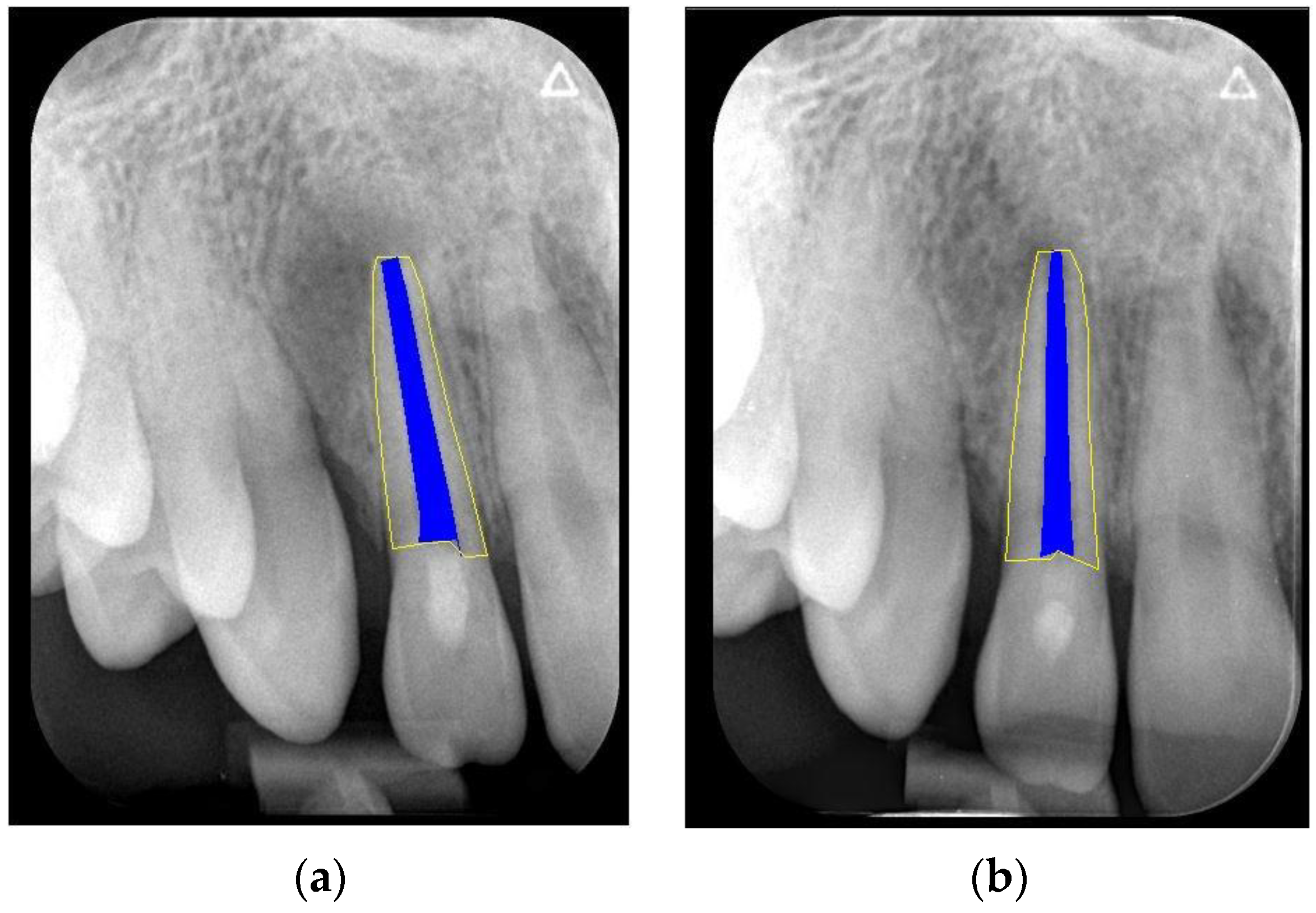
2.2. Assessment of Outcome

We clinically assessed the outcomes in the absence of any signs and symptoms and a negative cold-sensitivity test. Periapical healing, root elongation, RRA, and apical closure were assessed radiographically (Figure 1a,b). For this, we re-sized the immediate postoperative and follow-up periapical radiographs to match the size and pixels. We used the Freeware Image J software (version 1.47; National Institutes of Health, Bethesda, Rockville, MD, USA) together with the TurboReg plugin tool (Philippe Thevenaz, Biomedical Imaging Group, Swiss Federal Institute of Technology Lausanne, Lausanne, Switzerland) Rasband 1997–2018 [28] as described by Kahler et al. [29]. In brief, for radiographs obtained using different imaging systems, we used the ‘Images to Stack’ and ‘Stack to Images’ functions to equalise the number of pixels in the radiograph. Subsequently, the radiographs of each case were aligned and normalised using the TurboReg plugin tool. This tool enabled the measurement of curved roots (Figure 2a,b). Two evaluators (RY and AYK) conducted the calibration sessions using instructional videos. All cases were separately reviewed by two calibrated examiners. They discussed the case upon obtaining dissimilar results until it was resolved. The above-mentioned changes in root dimension were calculated as a percentage (Figure 3a,b). Hence, we evaluated the effect of the treatment based on the relative increase in RRA and root length.
RRA, radiographic root area; REP, regenerative endodontic procedure.
RL0 = the radiographic length at the end of REP and RL1 = the radiographic length at the final follow-up.
RRA0, the radiographic root area at the end of REP, RRA1 = the radiographic root area at the final follow-up of REP.

2.3. Data Collection

We recorded the patient age, sex, and tooth type. Furthermore, we recorded the number of months between the end of REPs and the last follow-up, periapical diagnoses, and the aetiology of pulpal necrosis. We recorded data collected at the follow-up visit, including clinical signs and symptoms. Periapical radiographs at the end of REPs and at the last follow-up were obtained from the patients’ files to assess periapical healing and changes in the RRA and root length.

2.4. Statistical Analysis

Summary statistics were computed and presented for the clinical outcomes in the cohort. We calculated the Pearson correlation coefficients and p-values of the correlation test to assess the correlation between the relative increases in root length and dentin area. The distributions were characterised using boxplots (25% quantile, median, and 75% quantile). We assessed the statistical significance of differences between the mean increases in the dentine area or root length in the anterior versus posterior roots using the t-test with unequal variance (Welch t-test). Furthermore, we determined the association between the root closure status and anterior/posterior root location using the chi-square test. We evaluated the significance of the association between the relative increases in the dentine area or root length with root closure status groups (closed/incomplete/unchanged) using the one-way analysis of variance. All analyses were performed using R software, version 3.6 (https://www.r-project.org/ (accessed on 1 September 2021)).

3. Results

A total of 50 teeth (n = 57 roots) from 46 patients who underwent REP and were followed up for 12–38 months were included in the study (Figure 4). Among the 14 posterior teeth, we could only measure 21 roots owing to superposition.
Figure 5 illustrates the RRA and length measurements obtained for all roots. Fifty-four (95%) roots exhibited increase in RRA, and 49 (86%) roots exhibited increase in length. The overall dimensions of the anterior roots were larger than those of the posterior roots. Therefore, the percentage-wise measurements are described as relative values (Figure 6). A significant overall correlation between the length and RRA changes was observed (Pearson r = 0.44, p = 0.00048). The relative average increase in the RRA of the posterior roots was 39% and that of the anterior roots was 26% (t-test p = 0.037; Table 1). The average relative increase in the length of the posterior roots was 11% and that of the anterior roots was 6% (t-test, p = 0.029; Table 1).
Table 2 shows the apical root closure status in the anterior and posterior teeth. There is a significant association between the type of tooth and apical closure status as follows: 89% (n = 17/19) of all healed posterior roots demonstrated an apical closure, whereas only 30% (10/33) of all healed anterior roots presented with apical closure. In addition, the apical diameter did not change in eight of the 33 healed anterior roots (χ2 p = 0.00018). There were no missing data.

4. Discussion

Recently, REP has gained popularity due to its potential to induce neotissue formation in the root canal space and hard tissue formation [1]. Its primary objective is to enable the healing of apical periodontitis and to help achieve other goals affecting tooth maturation [30]. Tooth maturation involves regaining normal pulpal physiological functions, which are evaluated in terms of continued root development, immune competency, and normal nociception [30]. Kim et al. [18] suggested that all teeth with immature roots (Cvek Class 1–4 (25)) that do not need intraradicular restoration could be treated with REP.
The NaOCl concentration used for disinfection irrigation contributes to the treatment success. We used 4% NaOCl in combination with 20 mL of 17% EDTA. Martin et al. [9] demonstrated that dentin conditioning with high concentrations of NaOCl exerted a profound negative effect on the survival and differentiation of stem cells of the apical papilla in vitro; however, the use of 17% EDTA following NaOCl irrigation minimised the aforementioned effect. Chrepa et al. [4] reported that 1.5% NaOCl was associated with a higher risk of failure, whereas a higher concentration of NaOCl (6%) and the complete strength of double antibiotic paste were associated with greater increase in root development.
We used a modified triple antibiotic paste (TAP) as an intracanal medication since the majority of previous studies have reported high success rates with TAP [4,31,32]. The originally suggested TAP included minocycline, which caused discoloration. Therefore, researchers suggested to discard or replace it with another type of antibiotics [31].
The scaffold is another clinical factor that contributes to REP success. Although the clinical advantages of PRF over blood clots are debatable, we used PRF as a scaffold in all cases. Moreover, the use of blood concentrates as an alternative source for blood clots may have several advantages, including the increased concentration of growth factors [33], resolution of clinical signs and symptoms of apical periodontitis, and continued root development [2,14]. Comparative clinical studies have indicated that root lengthening is significantly more frequent in REPs using PRF than in those using blood clots [34,35]. In comparison with the blood clot scaffold, PRF induced greater apical closure [3] and lesser root canal obliteration [36]. This is a better outcome since obliteration may exert an adverse effect on future endodontic treatments. Ulusoy et al. [36] demonstrated no need for prior apical bleeding for successful REP with PRF. Nonetheless, bleeding was evoked in the present study prior to PRF insertion to ensure the continuity of the PRF with apical tissues. Moreover, a recent meta-analysis by Koç and Del Fabbro [30] reported that the success rate of REP was significantly greater with blood concentrates than with blood clots.
In the present study, we observed radiographic periapical healing in 91.2% of the patients and uncertain healing in 8.8%. This result was similar to those of other studies that reported a high periapical healing rate of 90–100% [4,19,30,37,38]. However, the stratification of the anterior and posterior teeth demonstrated significantly different outcomes with respect to root development. The change in RRA was greater than that in root length, consistent with previous reports [19,36,37]. Interestingly, the average change in RRA was 26.2% for the anterior teeth and 39.3% (p = 0.037) for the posterior teeth. Similarly, the average relative increase in the root length was 6.14% in anterior teeth and 11.3% in posterior teeth (p = 0.029). The aforementioned increase in molar root dimensions was in accordance with that reported by Duggal et al. [12].
There are insufficient reports on the outcomes of REP in molars. KoÇ and Del Fabbro [30] evaluated the effect of aetiology on the REP success rate in 445 samples consisting of 432 anterior teeth and premolars and only 13 molars. However, they did not specify if ‘success’ denoted periapical healing, an increase in root dimensions, or both. The aetiology of necrosis in their sample included dental trauma (65%), broken dental cusps (29%), and dental caries (6%). There was no significant positive association between the aetiology of pulp necrosis and the REP success rate. Another meta-analysis examined the dimensional changes among anterior and posterior teeth according to the etiological factors such as caries, trauma, and dental anomalies. The majority of pronounced dimensional changes were observed in the dental anomalies group [19]. The above-mentioned studies are the only reports on the outcome of REP in molars; thus, it was difficult to draw any clear conclusions, and there is a need for further studies to determine the effect of aetiologic factors on REP outcomes. In this study, all posterior teeth presented with caries as the etiological factor, and RRA in the carious teeth was 13% greater than that in the anterior teeth, in which trauma was the etiological factor. Unlike the results of the present study, Chrepa et al. [4] demonstrated that teeth with caries were associated with a significantly lower increase in the RRA than those with trauma. This discrepancy may be attributed to the difference in the sample size as follows: the present study included 14 posterior teeth, all with caries causing pulp necrosis, whereas the San Antonio study [4] included only five teeth with caries as the aetiology with two (out of five) failures. Chrepa et al. [4] speculated that caries, as a predictor of failure, may be attributed to a long-standing biofilm structure and the inability of sufficient disinfection to eliminate it. The variations in the types of biofilms that characterised both studies may be a possible explanation for the differences in success rates between the studies. The patients referred to our clinic were young and were referred immediately following the diagnosis of pulp necrosis. The biofilm on their teeth was presumably young and susceptible to the disinfection protocol. Manoharan et al. [33] reported that the root canal microflora of traumatised teeth is diverse and differs from that of root canal infections not caused by trauma.
Our findings demonstrated no significant difference between the anterior and posterior teeth with regard to periapical healing; however, significant statistical differences regarding changes in the root dimension may contribute to the aetiological effect on the outcomes. Trauma exerts a detrimental effect on root development [39,40]. Nagata et al. [39] reported that severe trauma has the potential to cause damage to the Hertwig’s epithelial root sheath and/or apical papilla; thus, traumatised teeth may be less likely to achieve clinically meaningful continued root development than immature teeth with pulpal necrosis caused by caries. Furthermore, changes in the microenvironment in the periapical area may deplete the stem cell numbers and reduce vascularity [29,39,41,42,43]. Apical closure is an additional desired outcome following REP. Of the five types of REP outcomes described by Lv et al. [2], the first three are associated with the apical foramen. In the present study, we classified apical closure (Table 2) as complete, incomplete, and unchanged closures, which occurred in 52.9% (n = 27/52), 32.7% (n = 17/52), and 15.4% (n = eight/52) cases, respectively. These findings are consistent with those of Saoud et al. [38], who reported complete apical closure in 55% teeth at the 1-year follow-up. In a prospective study of 16 teeth, Kahler et al. [29] found complete closure in 19.4% teeth and incomplete closure in 47.2% teeth. In another study comparing platelet-rich plasma to blood clots, Bezgin et al. [43] observed complete closure in 70% (n = 7/10) teeth of the platelet-rich plasma group and in 60% (n = 6/10) teeth of the blood clot group. Apical closure was delayed in preoperatively symptomatic cases. We found a statistically significant association between the type of tooth and apical closure. In this study, the prevalence of complete or partial apical closure was considerably higher in the posterior teeth than that in the anterior teeth. The high incidence of incomplete or unchanged apical closure in the anterior teeth in this study suggests that the correlation between changes in the RRA and apical closure should be re-examined. However, no study has considered the aforementioned possibility until now. Our findings suggest that apical closure was not associated with changes in RRA.

5. Conclusions

The findings of our study contribute to the evolving field of REPs. Using a combination of blood clot and PRF as a scaffold may help enhance REP outcomes through RRA changes. Further research is needed to investigate whether the aetiology of pulpal necrosis or the tooth type (anterior/posterior) affects REP outcomes in terms of tooth maturation, including root lengthening, dentin area widening, and apical closure.

Author Contributions

Conceptualization and methodology, M.Y., A.Y.K. and N.R.; software, S.G.; formal analysis, N.R. and S.E.; investigation, M.Y., A.Y.K. and N.R.; data curation, M.Y.; writing—original draft preparation, review and editing, S.E., M.Y. and A.Y.K.; supervision, A.Y.K. All authors have read and agreed to the published version of the manuscript.

Funding

This research received no external funding.

Institutional Review Board Statement

Not applicable.

Informed Consent Statement

Informed consent was obtained from all subjects involved in the study.

Data Availability Statement

Not applicable.

Acknowledgments

The authors wish to thank Michael Kapelushnik for his contribution in mastering the evaluators to use Image software and helping in the measurements and calculations: Andrey Sivachenko, who contributed to the statistics calculations.

Conflicts of Interest

The authors declare no conflict of interest.

References

  1. Diogenes, A.; Ruparel, N.B. Regenerative endodontic procedures: Clinical outcomes. Dent. Clin. N. Am. 2017, 61, 111–125. [Google Scholar] [CrossRef] [PubMed]
  2. Lv, H.; Chen, Y.; Cai, Z.; Lei, L.; Zhang, M.; Zhou, R.; Huang, X. The efficacy of platelet-rich fibrin as a scaffold in regenerative endodontic treatment: A retrospective controlled cohort study. BMC Oral Health 2018, 18, 139. [Google Scholar] [CrossRef] [PubMed]
  3. Murray, P.E. Platelet-rich plasma and platelet-rich fibrin can induce apical closure more frequently than blood-clot revascularization for the regeneration of immature permanent teeth: A meta-analysis of clinical efficacy. Front. Bioeng. Biotechnol. 2018, 6, 139. [Google Scholar] [CrossRef] [PubMed] [Green Version]
  4. Chrepa, V.; Joon, R.; Austah, O.; Diogenes, A.; Hargreaves, K.M.; Ezeldeen, M.; Ruparel, N.B. Clinical outcomes of immature teeth treated with regenerative endodontic procedures-a San Antonio study. J. Endod. 2020, 46, 1074–1084. [Google Scholar] [CrossRef]
  5. Banchs, F.; Trope, M. Revascularization of immature permanent teeth with apical periodontitis: New treatment protocol? J. Endod. 2004, 30, 196–200. [Google Scholar] [CrossRef]
  6. Sato, I.; Ando-Kurihara, N.; Kota, K.; Iwaku, M.; Hoshino, E. Sterilization of infected root-canal dentine by topical application of a mixture of ciprofloxacin, metronidazole and minocycline in situ. Int. Endod. J. 1996, 29, 118–124. [Google Scholar] [CrossRef]
  7. Shin, S.Y.; Albert, J.S.; Mortman, R.E. One step pulp revascularization treatment of an immature permanent tooth with chronic apical abscess: A case report. Int. Endod. J. 2009, 42, 1118–1126. [Google Scholar] [CrossRef]
  8. Petrino, J.A.; Boda, K.K.; Shambarger, S.; Bowles, W.R.; McClanahan, S.B. Challenges in regenerative endodontics: A case series. J. Endod. 2010, 36, 536–541. [Google Scholar] [CrossRef]
  9. Martin, D.E.; De Almeida, J.F.; Henry, M.A.; Khaing, Z.Z.; Schmidt, C.E.; Teixeira, F.B.; Diogenes, A. Concentration-dependent effect of sodium hypochlorite on stem cells of apical papilla survival and differentiation. J. Endod. 2014, 40, 51–55. [Google Scholar] [CrossRef]
  10. American Association of Endodontists. Clinical Considerations for a Regenerative Procedure. Revised WWW Document. 2016. Available online: https://www.aae.org/uploadedfiles/publications_and_research/research/currentregenerativeendodonticconsiderations.pdf (accessed on 20 November 2020).
  11. Galler, K.M.; Krastl, G.; Simon, S.; Van Gorp, G.; Meschi, N.; Vahedi, B.; Lambrechts, P. European Society of Endodontology Position statement: Revitalization procedures. Int. Endod. J. 2016, 49, 717–723. [Google Scholar] [CrossRef]
  12. Duggal, M.; Tong, H.J.; Al-Ansary, M.; Twati, W.; Day, P.F.; Nazzal, H. Interventions for the endodontic management of non-vital traumatised immature permanent anterior teeth in children and adolescents: A systematic review of the evidence and guidelines of the European Academy of Paediatric Dentistry. Eur. Arch. Paediatr. Dent. 2017, 18, 139–151. [Google Scholar] [CrossRef]
  13. Cehreli, Z.C.; Isbitiren, B.; Sara, S.; Erbas, G. Regenerative endodontic treatment (Revascularization) of immature necrotic molars medicated with calcium hydroxide: A case series. J. Endod. 2011, 37, 1327–1330. [Google Scholar] [CrossRef] [PubMed]
  14. Tzanetakis, G.N.; Giannakoulas, D.G.; Papanakou, S.; Gizani, S.; Lygidakis, N. Regenerative endodontic therapy of immature permanent molars with pulp necrosis: A cases series and a literature review. Eur. Arch. Paediatr. Dent. 2021, 22, 515–525. [Google Scholar] [CrossRef] [PubMed]
  15. Yoshpe, M.; Kaufman, A.Y.; Lin, S.; Ashkenazi, M. Regenerative endodontics: A promising tool to promote periapical healing and root maturation of necrotic immature permanent molars with apical periodontitis using platelet-rich fibrin (PRF). Eur. Arch. Paediatr. Dent. 2021, 22, 527–534. [Google Scholar] [CrossRef] [PubMed]
  16. Keswani, D.; Pandey, R.K. Revascularization of an immature tooth with a necrotic pulp using platelet-rich fibrin: A case report. Int. Endod. J. 2013, 46, 1096–1104. [Google Scholar] [CrossRef] [PubMed]
  17. Choukroun, J.; Adda, F.; Schoeffler, C.; Vervelle, A. Une opportunite´ en paro-implantologie: Le PRF. Implantodontie 2001, 42, 55–62. [Google Scholar]
  18. He, L.; Lin, Y.; Hu, X.; Zhang, Y.; Wu, H. A comparative study of platelet-rich fibrin (PRF) and platelet-rich plasma (PRP) on the effect of proliferation and differentiation of rat osteoblasts in vitro. Oral Surg. Oral Med. Oral Pathol. Oral Radiol. Endodontology 2009, 108, 707–713. [Google Scholar] [CrossRef]
  19. Bakhtiar, H.; Esmaeili, S.; Fakhr Tabatabayi, S.; Ellini, M.R.; Nekoofar, M.H.; Dummer, P.M. Second-generation platelet concentrate (platelet-rich fibrin) as a scaffold in regenerative endodontics: A case series. J. Endod. 2017, 43, 401–408. [Google Scholar] [CrossRef]
  20. Lenzi, R.; Trope, M. Revitalization procedures in two traumatized incisors with different biological outcomes. J. Endod. 2012, 38, 411–414. [Google Scholar] [CrossRef]
  21. Nosrat, A.; Seifi, A.; Asgary, S. Regenerative endodontic treatment (Revascularization) for necrotic immature permanent molars: A review and report of two cases with a new biomaterial. J. Endod. 2011, 37, 562–567. [Google Scholar] [CrossRef]
  22. Kim, S.G.; Malek, M.; Sigurdsson, A.; Lin, L.M.; Kahler, B. Regenerative endodontics: A comprehensive review. Int. Endod. J. 2018, 51, 1367–1388. [Google Scholar] [CrossRef] [PubMed]
  23. Ong, T.K.; Lim, G.S.; Singh, M.; Fial, A.V.D. Quantitative Assessment of root development after regenerative endodontic therapy: A systematic review and meta-analysis. J. Endod. 2020, 46, 1856–1866. [Google Scholar] [CrossRef] [PubMed]
  24. Flake, N.M.; Gibbs, J.L.; Diogenes, A.; Hargreaves, K.M.; Khan, A.A. A standardized novel method to measure radiographic root changes after endodontic therapy in immature teeth. J. Endod. 2014, 40, 46–50. [Google Scholar] [CrossRef] [PubMed] [Green Version]
  25. Cvek, M. Prognosis of luxated non-vital maxillary incisors treated with calcium hydroxide and filled with gutta-percha. A retrospective clinical study. Endod. Dent. Traumatol. 1992, 8, 45–55. [Google Scholar] [CrossRef]
  26. Shao, Z.; Lyu, C.; Teng, L.; Xie, X.; Sun, J.; Zou, D.; Lu, J. Platelet-rich fibrin (PRF): A second-generation platelet concentrate. Part I: Technological concepts and evolution. Oral Surg. Oral Med. Oral Pathol. Oral Rad. Endodontology 2006, 101, E37–E44. [Google Scholar]
  27. Abuelniel, G.M.; Duggal, M.S.; Kabel, N. A comparison of MTA and Biodentine as medicaments for pulpotomy in traumatized anterior immature permanent teeth: A randomized clinical trial. Dent. Traumatol. 2020, 36, 400–410. [Google Scholar] [CrossRef]
  28. Rasband, W.S. ImageJ WWW Document, 1997–2018. Available online: https://imagej.nih.gov/ij/index.html (accessed on 28 January 2021).
  29. Kahler, B.; Mistry, S.; Moule, A.; Ringsmuth, A.K.; Case, P.; Thomson, A.; Holcombe, T. Revascularization outcomes: A prospective analysis of 16 consecutive cases. J. Endod. 2014, 40, 333–338. [Google Scholar] [CrossRef]
  30. Koç, S.; Del Fabbro, M. Does the etiology of pulp necrosis affect regenerative endodontic treatment outcomes? A systematic review and meta-analyses. J. Evid. Based Dent. Pract. 2020, 20, 101400. [Google Scholar] [CrossRef]
  31. Nagata, J.Y.; Soares, A.J.; Souza-Filho, F.J.; Zaia, A.A.; Ferraz, C.C.; Almeida, J.F.; Gomes, B.P. Microbial evaluation of traumatized teeth treated with triple antibiotic paste or calcium hydroxide with 2% chlorhexidine gel in pulp revascularization. J. Endod. 2014, 40, 778–783. [Google Scholar] [CrossRef]
  32. Ruparel, N.B.; Teixeira, F.B.; Ferraz, C.C.; Diogenes, A. Direct effect of intracanal medicaments on survival of stem cells of the apical papilla. J. Endod. 2012, 38, 1372–1375. [Google Scholar] [CrossRef]
  33. Manoharan, L.; Brundin, M.; Rakhimova, O.; Chávez de Paz, L.; Romani Vestman, N. New insights into the microbial profiles of infected root canals in traumatized teeth. J. Clin. Med. 2020, 9, 3877. [Google Scholar] [CrossRef] [PubMed]
  34. Narang, I.; Mittal, N.; Mishra, N. A Comparative evaluation of the blood clot, platelet-rich plasma, and platelet-rich fibrin in regeneration of necrotic immature permanent teeth: A clinical study. Contemp. Clin. Dent. 2015, 6, 63–68. [Google Scholar] [CrossRef] [PubMed]
  35. Alagl, A.; Bedi, S.; Hassan, K.; AlHumaid, J. Use of platelet-rich plasma for regeneration in non-vital immature permanent teeth: Clinical and cone-beam computed tomography evaluation. J. Int. Med. Res. 2017, 45, 583–593. [Google Scholar] [CrossRef] [PubMed]
  36. Ulusoy, A.T.; Turedi, I.; Cimen, M.; Cehreli, Z.C. Evaluation of blood clot, platelet-rich plasma, platelet-rich fibrin, and platelet pellet as scaffolds in regenerative endodontic treatment: A prospective randomized trial. J. Endod. 2019, 45, 560–566. [Google Scholar] [CrossRef]
  37. Nagy, M.M.; Tawfik, H.E.; Hashem, A.A.; Abu-Seida, A.M. Regenerative potential of immature permanent teeth with necrotic pulps after different regenerative protocols. J. Endod. 2014, 40, 192–198. [Google Scholar] [CrossRef]
  38. Saoud, T.M.; Zaazou, A.; Nabil, A.; Moussa, S.; Lin, L.M.; Gibbs, J.L. Clinical and radiographic outcomes of traumatized immature permanent necrotic teeth after revascularization/revitalization therapy. J. Endod. 2014, 40, 1946–1952. [Google Scholar] [CrossRef] [Green Version]
  39. Nagata, J.Y.; Gomes, B.P.; Rocha Lima, T.F.; Murakami, L.S.; de Faria, D.E.; Campos, G.R.; de Souza-Filho, F.J.; Soares, A.J. Traumatized immature teeth treated with 2 Protocols of pulp revascularization. J. Endod. 2014, 40, 606–612. [Google Scholar] [CrossRef]
  40. Shivashankar, V.Y.; Johns, D.A.; Maroli, R.K.; Sekar, M.; Chandrasekaran, R.; Karthikeyan, S.; Renganathan, S.K. Comparison of the effect of PRP, PRF and induced bleeding in the revascularization of teeth with necrotic pulp and open apex: A triple blind randomized clinical trial. J. Clin. Diagn. Res. 2017, 11, ZC34–ZC39. [Google Scholar] [CrossRef]
  41. Ricucci, D.; Siqueira, J.F., Jr.; Loghin, S.; Lin, L.M. Pulp and Apical tissue response to deep caries in immature teeth: A histologic and histobacteriologic study. J. Dent. 2017, 56, 19–32. [Google Scholar] [CrossRef]
  42. Lin, L.M.; Shimizu, E.; Gibbs, J.L.; Loghin, S.; Ricucci, D. Histologic and histobacteriologic observations of failed revascularization/revitalization therapy: A case report. J. Endod. 2014, 40, 291–295. [Google Scholar] [CrossRef]
  43. Bezgin, T.; Yilmaz, A.D.; Celik, B.N.; Kolsuz, M.E.; Sonmez, H. Efficacy of platelet-rich plasma as a scaffold in regenerative endodontic treatment. J. Endod. 2015, 41, 36–44. [Google Scholar] [CrossRef] [PubMed]
Figure 1. Measurement of RRA in an anterior tooth (a) at the end of REP, (b) at the 12-month follow-up. Uncertain healing and an unchanged apical closure are observed.
Applsci 12 06774 g001
Figure 2. The measurement of RRA in a posterior root (a) at the end of REP, (b) at the 18-month follow-up. Complete healing and apical closure are observed.
Applsci 12 06774 g002
Figure 3. The formulae used to calculate relative measurements (%) of the radiographic length (a) and root area (b).
Applsci 12 06774 g003
Figure 4. The distribution of the follow-up period.
Applsci 12 06774 g004
Figure 5. Summary of RRA (radiographic root area) and root length measurements. Solid and open circles represent the root dimensions before the procedure and at the follow-up visit, respectively. Each pair of before/after measurements for the same root is connected by an arrow. Orange and coral colours represent anterior and posterior roots, respectively.
Applsci 12 06774 g005
Figure 6. The relative increase (in percentages) in root length and RRA (radiographic root area). (A) scatterplot of increase in area vs increase in length; (B) individual distributions shown as boxplots. Coral and blue circles represent anterior and posterior roots, respectively.
Applsci 12 06774 g006
Table 1. Continued development (increase in RRA and length) of the anterior and posterior roots.
Characteristics of Continued Root DevelopmentAnteriorPosterior
MeanMedianIQRMeanMedianIQR
RRA26%25%15%39%38%30%
Length6%4%7%11%10%7%
RRA, radiographic root area; IQR, interquartile range.
Table 2. The distribution and frequency of apical closure status.
Type of TeethApical Closure Status
CompleteIncompleteUnchanged
PosteriorAnteriorPosteriorAnteriorPosteriorAnterior
Number of teeth1910216 10
Publisher’s Note: MDPI stays neutral with regard to jurisdictional claims in published maps and institutional affiliations.

