Article

Protozoa as the “Underdogs” for Microbiological Quality Evaluation of Fresh Vegetables

by
Cláudia S. Marques
1,2,3,
Susana Sousa
1,2,3,
António Castro
1,2,3,
Vânia Ferreira
4,
Paula Teixeira
4,* and
José M. Correia da Costa
1,2,3
1
Associate Laboratory for Animal and Veterinary Sciences (AL4AnimalS), Praça do Coronel Pacheco 42, 4050-083 Porto, Portugal
2
Centre for the Study in Animal Science (CECA), University of Porto, Praça do Coronel Pacheco 42, 4050-083 Porto, Portugal
3
Centre for Parasite Biology and Immunology, Department of Infectious Diseases, National Health Institute Dr. Ricardo Jorge (INSARJ), Rua de Alexandre Herculano, 321, 4000-055 Porto, Portugal
4
CBQF—Centro de Biotecnologia e Química Fina—Laboratório Associado, Escola Superior de Biotecnologia, Universidade Católica Portuguesa, Rua Diogo Botelho 1327, 4169-005 Porto, Portugal
*
Author to whom correspondence should be addressed.
Appl. Sci. 2022, 12(14), 7145; https://doi.org/10.3390/app12147145
Submission received: 1 June 2022 / Revised: 28 June 2022 / Accepted: 8 July 2022 / Published: 15 July 2022
(This article belongs to the Special Issue Multi-Contamination of Foods and Mixture Effects)

Abstract

:
The monitoring of the microbial quality of fresh products in the industrial environment has mainly focused on bacterial indicators. Protozoa, such as Giardia duodenalis, Cryptosporidium spp., Toxoplasma gondii, and Cyclospora cayetanensis, are routinely excluded from detection and surveillance systems, despite guidelines and regulations that support the need for tracking and monitoring these pathogens in fresh food products. Previous studies performed by our laboratory, within the scope of the SafeConsume project, clearly indicated that consumption of fresh produce may be a source of T. gondii, thus posing a risk for the contraction of toxoplasmosis for susceptible consumers. Therefore, preliminary work was performed in order to assess the microbiological quality of vegetables, highlighting not only bacteria (Escherichia. coli, Listeria monocytogenes, and Salmonella spp.), but also the zoonotic protozoa G. duodenalis and Cryptosporidium spp. Although all samples were found to be acceptable based on bacteriological parameters, cysts of G. duodenalis and oocysts of Cryptosporidium spp. were observed in vegetables. Moreover, it was possible to genetically characterize G. duodenalis positive samples as assemblage A, a genotype that poses risks to human health. Although these are preliminary results, they highlight the need to include protozoa in the microbiological criteria for foodstuffs, as required by EU Law No. 1441/2007, and to improve inactivation and removal procedures of (oo)cysts in fresh produce and water.

1. Introduction

Each year, nearly 600 million people become ill with foodborne diseases, leading to over 420,000 deaths [1]. Microbial contamination of food products can be caused by bacteria, viruses, or parasites, and these pathogens represent the most important cause of enteric illness [2]. During the past decade, there has been a remarkable increase in the consumption of raw or partially cooked fresh produce, due to changes in the eating habits of consumers who are more conscious of the direct and indirect health benefits of eating fruit and vegetables [3]. Hence, these types of foods have also become important vehicles for transmission of foodborne pathogens, and the number of outbreaks of foodborne diseases associated with fresh produce has increased [4,5,6]. Examples of major multi-national foodborne outbreaks include an Escherichia coli outbreak due to contaminated fenugreek seeds and an outbreak of listeriosis due to the consumption of frozen vegetables contaminated with Listeria monocytogenes [7,8]. In addition to bacteria, several protozoan parasites can also contaminate fresh produce. Giardia duodenalis, Cryptosporidium spp., Toxoplasma gondii, and Cyclospora cayetanensis have been associated with several global outbreaks of foodborne illnesses related to the consumption of contaminated fresh vegetables and fruits, and are the most important emerging parasitic protozoans that commonly infect humans and animals [9,10,11,12,13,14].
Despite guidelines and regulations that support the need for tracking and monitoring protozoa in fresh food products, their routine exclusion from detection and surveillance systems can be ascribed to the limited number of studies investigating parasite contamination of vegetables, the absence of consensual and accurate methodologies for their identification, and the wide acceptance among the expert authorities that the bacteriological aspects of the evaluation are more relevant for food safety [14]. Consequently, the monitoring of the microbial quality of fresh products in the industrial environment has mainly focused on bacterial indicators, and EU and national laws highly regulate the presence and microbiological limits of, in particular, the bacteria E. coli, L. monocytogenes, and Salmonella spp. [15,16,17,18]. In contrast, protozoan pathogens are rarely tested in fresh produce for food safety purposes. They are frequently neglected because they do not cause an immediate deleterious clinical response, but rather a chronic and long one. In addition, the associated diseases are also more common and prevalent in poor countries or communities. However, the harmful effects of these pathogens on humans in developed countries, especially on immunocompromised individuals, children, and pregnant women, and particularly their recent impacts, have increased the focus and attention of authorities [14,19,20,21,22,23].
Giardia duodenalis is the causative agent of giardiasis, resulting in acute watery diarrhea in both humans and animals. Eight major genotypes of G. duodenalis (assemblages) have been identified to date (A–H); however, assemblages A and B (and to a lesser extent assemblage E) are considered to have zoonotic interest due to their risk of infecting humans [24,25].
Cryptosporidium spp. are widespread protozoan parasites that infect humans and animals. Human cryptosporidiosis is the second commonest cause of diarrhea in children after rotavirus and can be caused either by the zoonotic Cryptosporidium parvum or anthroponotic Cryptosporidium hominis [9,26].
Toxoplasma gondii is an intracellular coccidian protozoan, for which domestic and wild felids are the only known definitive hosts responsible for oocyst dissemination in the environment. Toxoplasmosis is usually asymptomatic in immunocompetent individuals, but may cause severe infections in immunocompromised individuals, fetuses during pregnancy, and newborns [27,28].
Cyclospora cayetanensis is a human coccidia parasite that causes cyclosporiasis, a disease that causes acute diarrhea and other gastroenteritis symptoms. Humans are the only known host for C. cayetanensis oocysts, and their zoonotic role remains to be determined [29,30].
Two of the main reasons for the biological success of these parasites are their low infectious dose and their excretion in feces into the environment as resistant stages (cysts or oocysts) [31,32]. The (oo)cysts possess the ability to survive for a long period of time (weeks, months, or even years) in the environment, in extreme conditions of temperature and humidity, due to their structure, which protects them in exogenous stages. This allows the parasites to resist the common inactivation methods used for bacteria and viruses, such as heating, irradiation, and chemical disinfection [33,34,35,36,37,38].
Fresh vegetables may be contaminated at several steps along the food production chain, such as during crop production, harvesting, and processing, or directly by infected food handlers. Potential sources include water (irrigation water or water used for washing produce), wastewater discharge, soil contaminated with fecal waste from warm-blooded animals/livestock, organic fertilizers, and human handling during downstream processing and packaging steps [5,38,39,40,41].
In a recent work performed by our team [42], within the scope of the SafeConsume project (https://safeconsume.eu/, accessed on 11 July 2022), we designed a laboratory approach for the detection of T. gondii oocysts in vegetables and berry fruits based on the knowledge gained with Method 1623.1/EPA for the detection of the Cryptosporidium oocyst and Giardia cyst [43,44]. Moreover, we also described a new recombinant T. gondii oocyst wall protein 1-derived fragment, rTgOWP1-f. Strong microscopic evidence of the ability of rabbit polyclonal antibodies, anti-rTgOWP1-f, to identify environmental T. gondii oocysts, suggests rTgOWP1-f may be a potential biomarker for the detection of environmental oocysts [45]. Our studies clearly indicate that consumption of fresh produce may be a source of T. gondii infection in humans and a potential risk for consumers. However, technical refinement is still required for routine application at the industrial level or for food testing in laboratories for the detection of T. gondii oocysts.
Taking into account the experience and learning obtained from the SafeConsume work package 2 (WP2) laboratory studies, and the “One Health” approach, a parallel preliminary study was undertaken in order to assess the microbiological quality of fresh vegetables, highlighting not only bacteria (E. coli, L. monocytogenes, and Salmonella spp.) covered by the legislation [17], but also the zoonotic protozoa G. duodenalis and Cryptosporidium spp.

2. Materials and Methods

Five bulk and 11 packaged and ready-to-eat (RTE) vegetables were collected from local producers in Portugal and Spain, or provided by retail sellers, between September 2018 and June 2019 (Table 1). The fresh produce comprised lettuce (Lactuca sativa), watercress (Nasturtium officinale), coriander (Coriandrum sativum), and parsley (Petroselinum crispum). Ready-to-eat mixed salads included different varieties of lettuce, arugula (Eruca vesicaria sativa), endive (Cichorium endivia), chicory (Cichorium intybus), carrot (Daucus carota sativus), red cabbage (Brassica oleracea var. capitata f. rubra), and lamb’s lettuce (Valerianella locusta).
The concentration and recovery of G. duodenalis and Cryptosporidium spp. (oo)cysts from the vegetable samples were performed by Filtration/Immunomagnetic Separation (IMS)/Fluorescence Assay (FA) (Method 1623.1: Cryptosporidium and Giardia in water; US EPA 816-R-12-001-Jan 2012). Method 1623.1 is a validated and approved method used for surface water analysis [43]. This method was previously fully established in our laboratory, initially to gain knowledge regarding the contamination of water with Cryptosporidium spp. and Giardia spp. in the northern region of Portugal [44,46]. We also previously designed a long-term program aiming to identify the sources of surface water and environmental contamination, working with the water-supply industry, and to provide cattle and human fecal screening and molecular characterization [47,48,49].
Briefly, vegetable samples weighing between 350 and 900 g each were manually washed in large volumes of distilled water (between 10 and 80 L) (Table 1). A 1 µm Filta-Max® filter (IDEXX, Westbrook, ME, USA) applied to a peristaltic pump at a pressure of three bar was used for washing water filtration. Elution was performed in a Filta-Max® manual wash station and concentrated into 3 mL of phosphate-buffered solution (PBS) with 0.01% of Tween 20 (Merck KGaA, Darmstadt, Germany), after centrifugation at 1000× g for 10 min at room temperature. Magnetic beads conjugated with specific antibodies (Dynabeads™ GC-Combo; Thermo Fisher Scientific, Waltham, MA, USA) were added to the concentrate, and potential magnetized Cryptosporidium oocysts and Giardia cysts were recovered from the extraneous material using a magnet. Fluorescein isothiocyanate (FITC) conjugated anti-Cryptosporidium spp. and anti-Giardia spp. monoclonal antibodies were used according to the instructions of the manufacturer, Aqua-Glo™G/C (Waterborne, Inc., New Orleans, LA, USA), on slides containing the potential samples’ parasites, and observed by epifluorescence microscopy at 200× magnification. The total number of (oo)cysts per slide/sample was screened by two different microscopists to cross-check the results.
DNA was extracted from slides of positive samples containing two or more Cryptosporidium oocysts and Giardia cysts. Genomic DNA was extracted by scraping the slides to collect the (oo)cysts using the QIAamp® DNA Mini Kit (Qiagen GmbH, Hilden, Germany) according to the manufacturer’s instructions and as described previously by Almeida et al. [46]. Two-step nested PCR was performed to amplify a portion of the small subunit rRNA gene of Cryptosporidium with primers and PCR conditions described by Xiao et al. [50]. For G. duodenalis, β-giardin was the gene chosen for amplification by semi-nested PCR [51]. Positive amplification products were purified using the GRS PCR & Gel Band Purification kit (GRiSP Research Solutions, Porto, Portugal) according to the manufacturer’s instructions and sequenced using Sanger sequencing services from GATC Biotech (Eurofins Genomics, Ebersberg, Germany). Sequence comparison was made with already published sequences using the NCBI Basic Local Alignment Search Tool (BLAST).
For bacterial analysis, samples were transported to the laboratory in portable, insulated cold-bags and stored at 4 °C until analysis, normally between 1 and 2 days after collection. Twenty-five grams of each sample was added to 225 mL of sterile buffered peptone water (Biokar Diagnostics, Beauvais, France), and homogenized in a stomacher (Interscience, Saint Nom la Brèteche, France) for 2 min. Appropriate decimal dilutions were prepared in Ringer’s solution (Biokar Diagnostics) for microbial enumeration: colony counts at 30 °C on Plate Count Agar according to ISO 4833-1:2013 [52]; Enterobacteriaceae on RAPID′ Enterobacteriaceae medium (Bio-Rad, Hercules, CA, USA; ISO 16140 [53], 2016, ISO 21528-2, 2017 [54]); E. coli according to ISO 16649-2:2001 [55]; L. monocytogenes according to ISO 11290-2:2017 [56]; and yeasts and molds according to ISO 21527-1:2008 [57]. Detection of L. monocytogenes (ISO 11290-1:2017 [58]), Salmonella spp. (ISO 6579-1:2017 [59]), and E. coli was also performed. After appropriate incubation, colonies were counted and/or confirmatory tests performed and the colony forming units (CFU)/g calculated.

3. Results and Discussion

Between one and four G. duodenalis fluorescent cysts (Figure 1A) were observed in microscopic slides of four of the 16 samples (25.0%; 95% CI: 9.7 to 49.9%) (Table 2). The same percentage (25.0%) was obtained for the number of Cryptosporidium spp. (Figure 1B) oocysts (between one and eight oocysts) (Table 2); however, none of the Cryptosporidium-positive slides were confirmed by PCR. On the contrary, three of the four Giardia-positive slides were also confirmed by PCR. β-giardin fragment sequencing analysis revealed the presence of G. duodenalis assemblage A having more than 98% homology with G. duodenalis assemblage A isolates available in the Genebank database (KJ668152.1, KP687765.1, KF963547.1, EU642897.1, EU200934.1). The determination of genotypes is important for the identification of risk to human health, and potentially, the source of contamination. Assemblage A is one of the genotypes that undeniably causes human infections [24].
Globally, significant variations in the prevalence of these parasites have been observed in vegetables, ranging between 0% and 53% for G. duodenalis and between 0% and 63% for Cryptosporidium spp. (reviewed by Berrouch et al. [60]). Several factors can easily explain this discrepancy, such as different sampling procedures and detection methods having different recovery efficiencies and detection limits, distinct geographic locations having variable seasonal climatic conditions, and the degree of local development. The type of vegetable, in addition to the quality of water used for irrigation and washing, the proximity of livestock, and the presence of organic fertilizers, are also extremely relevant [60].
Unexpectedly, positive samples were only observed for the RTE type of produce. Thus, it is acceptable to discuss the management of the washing systems used in the food industry, even based on these preliminary data. Indeed, Trevisan et al. [61] noted that the use of treated wastewater for irrigation increases the chance of contamination of RTE/packaged vegetables. Other studies mention that contamination of RTE products by food handlers during packing is also a transmission route of Cryptosporidium and Giardia [61,62,63]. Therefore, it is clear that current RTE production treatment does not guarantee that fresh produce will be free from protozoa, and that alternative water treatments and increased hygienic measures for food handlers are necessary to ensure the safety of RTE vegetables.
Fresh produce has repeatedly been implicated in foodborne outbreaks and recalls, with Salmonella spp., E. coli, and L. monocytogenes the most implicated bacteria [64]. However, with the exception of a lettuce sample (sample no. 16) in which L. monocytogenes was detected, but at levels below 4 × 10 CFU/g (Table 2), all the sampled products were considered safe based on the microbiological criteria set by the European Commission Regulation (EC) No. 2073/2005 for “Pre-cut fruit and vegetables (ready-to-eat)”, i.e., the absence of Salmonella spp. and L. monocytogenes in 25 g samples and counts of E. coli not exceeding 1000 CFU/g. This is in agreement with previous studies, which demonstrated that the prevalence of these pathogens in this type of product is generally low [65,66,67]. Although the sample that tested positive for L. monocytogenes was unwashed lettuce, “Even if salad leaves and herbs are labelled with an instruction to wash before consumption, they are considered ready-to-eat. Although washing can reduce microbial contamination on the plant’s surface, it is not effective to eliminate microorganisms of concern or reduce them to an acceptable level because some pathogens can become internalised within the plant’s tissue.” [68].
The other microbiological parameters are recognized as hygiene and spoilage indicator micro-organisms in RTE foods. Most of the parameter results fell within the Satisfactory-Questionable categories according to the values recommended by the Portuguese National Health Institute [69]. Yeasts and molds were exceptions, with some products being classified as “Not Satisfactory” (yeast and mold counts exceeding, respectively, 106 CFU/g and 103 CFU/g) (Supplementary Table S1).
The knowledge gained in this field during the course of SafeConsume WP2 highlighted the need to include protozoa in the microbiological criteria for foodstuffs, as required by EU Law No. 1441/2007. This is particularly the case of T. gondii and the enteric pathogens G. duodenalis, Cryptosporidium spp., and C. cayetanensis, which are the most common protozoa associated with foodborne illness outbreaks. The absence of fecal contamination in vegetables and the guarantee of food safety for consumers can definitely no longer be defined only by the analysis of detection limits for bacteria, i.e., E. coli, L. monocytogenes, and Salmonella spp. However, microbial risk assessment evaluation and definition of strategies for the management and control of these parasites requires total collaboration between academia and industry, and investments by major companies and governments. These efforts are required to support and foster epidemiological and surveillance studies, and for the development of new technologies and “golden standard procedures” for protozoan removal (or recovery) and detection along the food chain. Monitoring of irrigation and processing water and the efficiency of wastewater treatment plants should be regularly performed. In addition, analysis of soil and manure used as fertilizer should be frequently undertaken, and animal access to crops and personal hygiene regulations applied to food handlers should be controlled. For instance, in addition to the use of sodium hypochlorite as a disinfectant, improved, safer methods, such as UV treatment, treatment with chlorine dioxide or ozone, membrane filtration, and a multi-barrier approach could be used as treatments for (oo)cyst inactivation and removal [33,37,70]. Furthermore, improvement in tools used for genotyping is necessary for the rapid and reliable detection of protozoans of concern for human health and to clarify their transmission patterns. The development of accurate methods for determining the viability and infectivity of (oo)cysts would also be invaluable in surveillance and outbreak studies, and for estimating risk to consumers. Finally, controlling foodborne (and waterborne) diseases should always be accomplished from a collaborative One Health perspective, involving a multidisciplinary team of public health and food experts, epidemiologists, veterinarians, environmental scientists, microbiologists (bacteriologists, virologists, and parasitologists), farmers, industry managers, lawmakers, and media communication specialists.

4. Conclusions

Although preliminary, the findings of the present study highlight the importance of implementing strategies for the detection, inactivation, and removal of parasite (oo)cysts in fresh produce and water, at all stages of food production, including pre- and post-harvest processes. The consumption of raw and/or undercooked vegetables, and particularly salads, has increased due to changes in the eating habits of consumers, who are switching to healthier diets. As a result, there is an urgent need to include protozoa in the microbiological criteria of EU regulations relating to foodstuffs. In addition, it is also important to make consumers aware of hygiene measures at home when washing and cooking raw vegetables.

Supplementary Materials

The following supporting information can be downloaded at: https://www.mdpi.com/article/10.3390/app12147145/s1.

Author Contributions

Conceptualization, C.S.M., S.S., A.C., V.F., P.T. and J.M.C.d.C.; Funding acquisition, P.T. and J.M.C.d.C.; Methodology, C.S.M.; Writing original draft preparation, C.S.M. and P.T.; Validation, C.S.M., S.S., A.C., V.F., P.T.; Visualization, C.S.M., S.S., A.C., V.F., P.T.; Review and editing, S.S., A.C., V.F., P.T. and J.M.C.d.C. All authors have read and agreed to the published version of the manuscript.

Funding

This research was funded by Horizon 2020 project SafeConsume, Grant Agreement No. 727580.

Institutional Review Board Statement

Not applicable.

Data Availability Statement

Not applicable.

Acknowledgments

Cristina Mena, Isabel Santos, and Luísa Carneiro (CINATE Microbiology Laboratory (Escola Superior de Biotecnologia, Universidade Católica Portuguesa) are thanked for excellent technical assistance with the bacteriological analysis. The authors would also like to thank the scientific collaboration under the Fundação para a Ciência e a Tecnologia (FCT) project UIDP/00329/2020 and fund UIDB/00211/2020.

Conflicts of Interest

The authors declare no conflict of interest.

References

  1. WHO Fact Sheet N_237, Food Safety and Foodborne Illness. Available online: https://www.who.int/news-room/fact-sheets/detail/food-safety (accessed on 25 February 2020).
  2. Prüss-Ustün, A.; Wolf, J.; Corvalán, C.; Neville, T.; Bos, R.; Neira, M. Diseases due to unhealthy environments: An updated estimate of the global burden of disease attributable to environmental determinants of health. J. Public Health 2016, 39, 464–475. [Google Scholar] [CrossRef] [PubMed]
  3. Losio, M.; Pavoni, E.; Bilei, S.; Bertasi, B.; Bove, D.; Capuano, F.; Farneti, S.; Blasi, G.; Comin, D.; Cardamone, C.; et al. Microbiological survey of raw and ready-to-eat leafy green vegetables marketed in Italy. Int. J. Food Microbiol. 2015, 210, 88–91. [Google Scholar] [CrossRef]
  4. Macieira, A.; Barbosa, J.; Teixeira, P. Food Safety in Local Farming of Fruits and Vegetables. Int. J. Environ. Res. Public Health 2021, 18, 9733. [Google Scholar] [CrossRef]
  5. Dixon, B. Transmission dynamics of foodborne parasites on fresh produce. In Foodborne Parasites in the Food Supply Web, Woodhead Publishing Series in Food Science, Technology and Nutrition; Gajadhar, A.A., Ed.; Woodhead Publishing: Ottawa, ON, Canada, 2015; Chapter 13; pp. 317–353. [Google Scholar] [CrossRef]
  6. McKerr, C.; Adak, G.K.; Nichols, G.; Gorton, R.; Chalmers, R.M.; Kafatos, G.; Cosford, P.; Charlett, A.; Reacher, M.; Pollock, K.G.; et al. An Outbreak of Cryptosporidium parvum across England & Scotland Associated with Consumption of Fresh Pre-Cut Salad Leaves, May 2012. PLoS ONE 2015, 10, e0125955. [Google Scholar] [CrossRef] [Green Version]
  7. King, L.A.; Nogareda, F.; Weill, F.-X.; Mariani-Kurkdjian, P.; Loukiadis, E.; Gault, G.; Jourdan-DaSilva, N.; Bingen, E.; Macé, M.; Thevenot, D.; et al. Outbreak of Shiga Toxin-Producing Escherichia coli O104:H4 Associated With Organic Fenugreek Sprouts, France, June 2011. Clin. Infect. Dis. 2012, 54, 1588–1594. [Google Scholar] [CrossRef] [Green Version]
  8. Willis, C.; McLauchlin, J.; Aird, H.; Amar, C.; Barker, C.; Dallman, T.; Elviss, N.; Lai, S.; Sadler-Reeves, L. Occurrence of Listeria and Escherichia coli in frozen fruit and vegetables collected from retail and catering premises in England 2018–2019. Int. J. Food Microbiol. 2020, 334, 108849. [Google Scholar] [CrossRef]
  9. Ryan, U.; Hijjawi, N.; Xiao, L. Foodborne cryptosporidiosis. Int. J. Parasitol. 2018, 48, 1–12. [Google Scholar] [CrossRef] [PubMed] [Green Version]
  10. Pinto-Ferreira, F.; Caldart, E.T.; Pasquali, A.K.S.; Mitsuka-Breganó, R.; Freire, R.L.; Navarro, I.T. Patterns of Transmission and Sources of Infection in Outbreaks of Human Toxoplasmosis. Emerg. Infect. Dis. 2019, 25, 2177–2182. [Google Scholar] [CrossRef] [Green Version]
  11. Ahlinder, J.; Svedberg, A.-L.; Nystedt, A.; Dryselius, R.; Jacobsson, K.; Hägglund, M.; Brindefalk, B.; Forsman, M.; Ottoson, J.; Troell, K. Use of metagenomic microbial source tracking to investigate the source of a foodborne outbreak of cryptosporidiosis. Food Waterborne Parasitol. 2021, 26, e00142. [Google Scholar] [CrossRef] [PubMed]
  12. Hadjilouka, A.; Tsaltas, D. Cyclospora Cayetanensis—Major Outbreaks from Ready to Eat Fresh Fruits and Vegetables. Foods 2020, 9, 1703. [Google Scholar] [CrossRef] [PubMed]
  13. Reh, L.; Muadica, A.S.; Köster, P.C.; Balasegaram, S.; Verlander, N.Q.; Chércoles, E.R.; Carmena, D. Substantial prevalence of enteroparasites Cryptosporidium spp., Giardia duodenalis and Blastocystis sp. in asymptomatic schoolchildren in Madrid, Spain, November 2017 to June 2018. Eurosurveillance 2019, 24, 1900241. [Google Scholar] [CrossRef] [PubMed]
  14. Robertson, L.J.; Lalle, M.; Paulsen, P. Why we need a European focus on foodborne parasites. Exp. Parasitol. 2020, 214, 107900. [Google Scholar] [CrossRef] [PubMed]
  15. Food Standards Australia New Zealand (FSANZ). Compendium of Microbiological Criteria for Food. Available online: https://www.foodstandards.gov.au/publications/Documents/Compedium%20of%20Microbiological%20Criteria/Compendium_revised-jan-2018.pdf (accessed on 20 May 2022).
  16. NSW Food Authority. Microbiological Quality of Fresh Cut Vegetables—A Survey to Determine the Safety of Fresh Cut Leafy Salad Vegetables Sold in NSW. Available online: https://www.foodauthority.nsw.gov.au/sites/default/files/_Documents/scienceandtechnical/microbiological_quality_fresh_cut_vegetables.pdf (accessed on 20 May 2022).
  17. COMMISSION REGULATION (EC) No. 1441/2007 of 5 December 2007 Amending Regulation (EC) No. 2073/2005 on Micro-Biological Criteria for Foodstuffs. Available online: https://eur-lex.europa.eu/legal-content/EN/TXT/PDF/?uri=CELEX:32007R1441&from=EN (accessed on 20 May 2022).
  18. European Food Safety Authority (EFSA); European Centre for Disease Prevention and Control (ECDPC). The European Union One Health 2019 Zoonoses Report. EFSA J. 2021, 19. [Google Scholar] [CrossRef]
  19. Bintsis, T. Foodborne pathogens. AIMS Microbiol. 2017, 3, 529–563. [Google Scholar] [CrossRef] [PubMed]
  20. Dorny, P.; Praet, N.; Deckers, N.; Gabriel, S. Emerging food-borne parasites. Veter. Parasitol. 2009, 163, 196–206. [Google Scholar] [CrossRef]
  21. Torgerson, P.R.; Macpherson, C.N. The socioeconomic burden of parasitic zoonoses: Global trends. Veter. Parasitol. 2011, 182, 79–95. [Google Scholar] [CrossRef]
  22. Slifko, T.R.; Smith, H.V.; Rose, J.B. Emerging parasite zoonoses associated with water and food. Int. J. Parasitol. 2000, 30, 1379–1393. [Google Scholar] [CrossRef]
  23. Dubey, J.P. Outbreaks of clinical toxoplasmosis in humans: Five decades of personal experience, perspectives and lessons learned. Parasites Vectors 2021, 14, 1–12. [Google Scholar] [CrossRef]
  24. Feng, Y.; Xiao, L. Zoonotic Potential and Molecular Epidemiology of Giardia Species and Giardiasis. Clin. Microbiol. Rev. 2011, 24, 110–140. [Google Scholar] [CrossRef] [Green Version]
  25. Dixon, B.R. Giardia duodenalis in humans and animals—Transmission and disease. Res. Veter. Sci. 2020, 135, 283–289. [Google Scholar] [CrossRef]
  26. Bouzid, M.; Kintz, E.; Hunter, P. Risk factors for Cryptosporidium infection in low and middle income countries: A systematic review and meta-analysis. PLOS Negl. Trop. Dis. 2018, 12, e0006553. [Google Scholar] [CrossRef] [PubMed]
  27. Dubey, J.P.; Murata, F.H.A.; Cerqueira-Cézar, C.K.; Kwok, O.C.H.; Villena, I. Congenital toxoplasmosis in humans: An update of worldwide rate of congenital infections. Parasitology 2021, 148, 1406–1416. [Google Scholar] [CrossRef] [PubMed]
  28. Montoya, J.G.; Liesenfeld, O. Toxoplasmosis. Lancet 2004, 363, 1965–1976. [Google Scholar] [CrossRef]
  29. Mansfield, L.S.; Gajadhar, A.A. Cyclospora cayetanensis, a food- and waterborne coccidian parasite. Veter. Parasitol. 2004, 126, 73–90. [Google Scholar] [CrossRef]
  30. Mathison, B.A.; Pritt, B.S. Cyclosporiasis—Updates on Clinical Presentation, Pathology, Clinical Diagnosis, and Treatment. Microorganisms 2021, 9, 1863. [Google Scholar] [CrossRef] [PubMed]
  31. Dawson, D. Foodborne protozoan parasites. Int. J. Food Microbiol. 2005, 103, 207–227. [Google Scholar] [CrossRef] [PubMed]
  32. Rousseau, A.; La Carbona, S.; Dumètre, A.; Robertson, L.J.; Gargala, G.; Escotte-Binet, S.; Favennec, L.; Villena, I.; Gérard, C.; Aubert, D. Assessing viability and infectivity of foodborne and waterborne stages (cysts/oocysts) of Giardia duodenalis, Cryptosporidium spp., and Toxoplasma gondii: A review of methods. Parasite 2018, 25, 14. [Google Scholar] [CrossRef] [Green Version]
  33. Mirza Alizadeh, A.; Jazaeri, S.; Shemshadi, B.; Hashempour-Baltork, F.; Sarlak, Z.; Pilevar, Z.; Hosseini, H. A review on inactivation methods of Toxoplasma gondii in foods. Pathog. Glob. Health 2018, 112, 306–319. [Google Scholar] [CrossRef]
  34. Dumètre, A.; Le Bras, C.; Baffet, M.; Meneceur, P.; Dubey, J.; Derouin, F.; Duguet, J.-P.; Joyeux, M.; Moulin, L. Effects of ozone and ultraviolet radiation treatments on the infectivity of Toxoplasma gondii oocysts. Veter. Parasitol. 2008, 153, 209–213. [Google Scholar] [CrossRef]
  35. Espinosa, M.F.; Sancho, A.N.; Mendoza, L.M.; Mota, C.R.; Verbyla, M.E. Systematic review and meta-analysis of time-temperature pathogen inactivation. Int. J. Hyg. Environ. Health 2020, 230, 113595. [Google Scholar] [CrossRef]
  36. Adeyemo, F.E.; Singh, G.; Reddy, P.; Bux, F.; Stenström, T.A. Efficiency of chlorine and UV in the inactivation of Cryptosporidium and Giardia in wastewater. PLoS ONE 2019, 14, e0216040. [Google Scholar] [CrossRef] [PubMed]
  37. Erickson, M.C.; Ortega, Y.R. Inactivation of Protozoan Parasites in Food, Water, and Environmental Systems. J. Food Prot. 2006, 69, 2786–2808. [Google Scholar] [CrossRef] [PubMed]
  38. Gajadhar, A.; Scandrett, W.B.; Forbes, L.B. Overview of food- and water-borne zoonotic parasites at the farm level. Rev. Sci. Tech. 2006, 25. [Google Scholar] [CrossRef]
  39. Tefera, T.; Tysnes, K.R.; Utaaker, K.S.; Robertson, L.J. Parasite contamination of berries: Risk, occurrence, and approaches for mitigation. Food Waterborne Parasitol. 2018, 10, 23–38. [Google Scholar] [CrossRef] [PubMed]
  40. Berger, C.N.; Sodha, S.V.; Shaw, R.K.; Griffin, P.M.; Pink, D.; Hand, P.; Frankel, G. Fresh fruit and vegetables as vehicles for the transmission of human pathogens. Environ. Microbiol. 2010, 12, 2385–2397. [Google Scholar] [CrossRef]
  41. Machado-Moreira, B.; Richards, K.; Brennan, F.; Abram, F.; Burgess, C.M. Microbial Contamination of Fresh Produce: What, Where, and How? Compr. Rev. Food Sci. Food Saf. 2019, 18, 1727–1750. [Google Scholar] [CrossRef] [Green Version]
  42. Marques, C.; Sousa, S.; Castro, A.; Da Costa, J.M.C. Detection of Toxoplasma gondii oocysts in fresh vegetables and berry fruits. Parasites Vectors 2020, 13, 180–212. [Google Scholar] [CrossRef]
  43. United States Environmental Protection Agency (EPA). Method 1623.1: Cryptosporidium and Giardia in Water by Filtra-Tion/IMS/FA. Available online: https://nepis.epa.gov/Exe/ZyPDF.cgi/P100J7G4.PDF?Dockey=P100J7G4.PDF (accessed on 20 May 2022).
  44. Almeida, A.; Moreira, M.J.; Soares, S.; Delgado, M.D.L.; Figueiredo, J.; Silva, E.; Castro, A.; Da Cosa, J.M.C. Presence of Cryptosporidium spp. and Giardia duodenalis in Drinking Water Samples in the North of Portugal. Korean J. Parasitol. 2010, 48, 43–48. [Google Scholar] [CrossRef] [Green Version]
  45. Sousa, S.; Almeida, A.; Delgado, L.; Conceição, A.; Marques, C.; Da Costa, J.M.C.; Castro, A. rTgOWP1-f, a specific biomarker for Toxoplasma gondii oocysts. Sci. Rep. 2020, 10, 1–8. [Google Scholar] [CrossRef]
  46. Almeida, A.; Moreira, M.J.; Soares, S.; Delgado, M.D.L.; Figueiredo, J.; Magalhães, E.S.; Castro, A.; Da Costa, A.V.; Da Costa, J.M.C. Biological and Genetic Characterization of Cryptosporidium spp. and Giardia duodenalis Isolates from Five Hydrographical Basins in Northern Portugal. Korean J. Parasitol. 2010, 48, 105–111. [Google Scholar] [CrossRef] [Green Version]
  47. Mendonça, C.; Almeida, A.; Castro, A.; Delgado, M.D.L.; Soares, S.; da Costa, J.M.C.; Canada, N. Molecular characterization of Cryptosporidium and Giardia isolates from cattle from Portugal. Veter. Parasitol. 2007, 147, 47–50. [Google Scholar] [CrossRef] [PubMed]
  48. Almeida, A.A.; Delgado, M.L.; Soares, S.C.; Castro, A.O.; Moreira, M.J.; Mendonça, C.M.; Canada, N.B.; Da Costa, J.M.C. Genotype Analysis of Giardia Isolated from Asymptomatic Children in Northern Portugal. J. Eukaryot. Microbiol. 2006, 53, S177–S178. [Google Scholar] [CrossRef] [PubMed]
  49. Almeida, A.A.; Delgado, M.L.; Soares, S.C.; Castro, A.O.; Moreira, M.J.; Mendonça, C.M.; Canada, N.B.; Da Costa, J.M.C.; Coelho, H.G. Genetic Characterization of Cryptosporidium Isolates from Humans in Northern Portugal. J. Eukaryot. Microbiol. 2006, 53, S26–S27. [Google Scholar] [CrossRef] [PubMed]
  50. Xiao, L.; Singh, A.; Limor, J.; Graczyk, T.K.; Gradus, S.; Lal, A. Molecular Characterization of Cryptosporidium Oocysts in Samples of Raw Surface Water and Wastewater. Appl. Environ. Microbiol. 2001, 67, 1097–1101. [Google Scholar] [CrossRef] [PubMed] [Green Version]
  51. Cacciò, S.M.; De Giacomo, M.; Pozio, E. Sequence analysis of the β-giardin gene and development of a polymerase chain reaction–restriction fragment length polymorphism assay to genotype Giardiaduodenalis cysts from human faecal samples. Int. J. Parasitol. 2002, 32, 1023–1030. [Google Scholar] [CrossRef]
  52. ISO 4833; Microbiology of the Food Chain—Horizontal Method for the Enumeration of Microorganisms—Part 1: Colony Count at 30 °C by the Pour Plate Technique. International Organization for Standardization: Geneva, Switzerland, 2013.
  53. ISO 16140-2:2016; Microbiology of the food chain—Method validation—Part 2: Protocol for the validation of alternative (proprietary) methods against a reference method. International Organization for Standardization: Geneva, Switzerland, 2016.
  54. ISO 21528-2; Microbiology of the Food Chain-Horizontal Method for the Detection and Enumeration of Enterobacteriaceae—Part 2: Colony-Count Technique. International Organization for Standardization: Geneva, Switzerland, 2017.
  55. ISO 16649-2; Microbiology of Food and Animal Feeding Stuffs—Horizontal Method for the Enumeration of Beta-Glucuronidase-Positive Escherichia coli—Part 2: Colony-Count Technique at 44 °C Using 5-bromo-4-chloro-3-indolyl beta-D-glucuronide. International Organization for Standardization: Geneva, Switzerland, 2001.
  56. ISO 11290-2; Microbiology of the Food Chain—Horizontal Method for the Detection and Enumeration of Listeria Monocytogenes and of Listeria spp.—Part 2: Enumeration Method. International Organization for Standardization: Geneva, Switzerland, 2017.
  57. ISO 21527-1; Microbiology of Food and Animal Feeding Stuffs—Horizontal Method for the Enumeration of Yeasts and Moulds—Part 1: Colony Count Technique in Products with Water Activity Greater Than 0.95. International Organization for Standardization: Geneva, Switzerland, 2008.
  58. ISO 11290-1; Microbiology of the Food Chain—Horizontal Method for the Detection and Enumeration of Listeria Monocytogenes and of Listeria spp.—Part 1: Detection Method. International Organization for Standardization: Geneva, Switzerland, 2017.
  59. ISO 6579-1; Microbiology of the Food Chain—Horizontal Method for the Detection, Enumeration and Serotyping of Salmonella—Part 1: Detection of Salmonella spp. International Organization for Standardization: Geneva, Switzerland, 2017.
  60. Berrouch, S.; Escotte-Binet, S.; Harrak, R.; Huguenin, A.; Flori, P.; Favennec, L.; Villena, I.; Hafid, J.; Salma, B.; Sandie, E.-B.; et al. Detection methods and prevalence of transmission stages of Toxoplasma gondii, Giardia duodenalis and Cryptosporidium spp. in fresh vegetables: A review. Parasitology 2020, 147, 516–532. [Google Scholar] [CrossRef]
  61. Trevisan, C.; Torgerson, P.R.; Robertson, L.J. Foodborne Parasites in Europe: Present Status and Future Trends. Trends Parasitol. 2019, 35, 695–703. [Google Scholar] [CrossRef] [Green Version]
  62. Caradonna, T.; Marangi, M.; Del Chierico, F.; Ferrari, N.; Reddel, S.; Bracaglia, G.; Normanno, G.; Putignani, L.; Giangaspero, A. Detection and prevalence of protozoan parasites in ready-to-eat packaged salads on sale in Italy. Food Microbiol. 2017, 67, 67–75. [Google Scholar] [CrossRef]
  63. Utaaker, K.S.; Skjerve, E.; Robertson, L.J. Keeping it cool: Survival of Giardia cysts and Cryptosporidium oocysts on lettuce leaves. Int. J. Food Microbiol. 2017, 255, 51–57. [Google Scholar] [CrossRef]
  64. Sheng, L.; Zhu, M. Practical in-storage interventions to control foodborne pathogens on fresh produce. Compr. Rev. Food Sci. Food Saf. 2021, 20, 4584–4611. [Google Scholar] [CrossRef]
  65. Ferreira, C.; Lopes, F.; Costa, R.; Komora, N.; Ferreira, V.; Fernandes, V.C.; Delerue-Matos, C.; Teixeira, P. Microbiological and Chemical Quality of Portuguese Lettuce—Results of a Case Study. Foods 2020, 9, 1274. [Google Scholar] [CrossRef] [PubMed]
  66. Finger, J.D.A.F.F.; Maffei, D.F.; Dias, M.; Mendes, M.A.; Pinto, U.M. Microbiological quality and safety of minimally processed parsley (Petroselinum crispum) sold in food markets, southeastern Brazil. J. Appl. Microbiol. 2020, 131, 272–280. [Google Scholar] [CrossRef] [PubMed]
  67. Zhang, H.; Yamamoto, E.; Murphy, J.; Locas, A. Microbiological safety of ready-to-eat fresh-cut fruits and vegetables sold on the Canadian retail market. Int. J. Food Microbiol. 2020, 335, 108855. [Google Scholar] [CrossRef] [PubMed]
  68. Food Safety Authority of Ireland. Guidelines for the Interpretation of Results of Microbiological Testing of Ready-to-Eat Foods Placed on the Market (Revision 4); FSAI: Dublin, Ireland, 2020; p. 50. ISBN 0-9539183-5-1. [Google Scholar]
  69. Saraiva, M.; Correia, C.B.; Cunha, I.C.; Maia, C.; Bonito, C.C.; Furtado, R.; Calhau, M.A. Interpretação dos resultados de ensaios microbiológicos em alimentos prontos para consumo e em superfícies do ambiente de produção e distribuição alimentar: Valores-guia. Repositório Científico do Instituto Nacional de Saúde Dr. Ricardo Jorge 2019. Available online: http://hdl.handle.net/10400.18/5610 (accessed on 19 May 2022).
  70. Morrison, C.M.; Hogard, S.; Pearce, R.; Gerrity, D.; von Gunten, U.; Wert, E.C. Ozone disinfection of waterborne pathogens and their surrogates: A critical review. Water Res. 2022, 214, 118206. [Google Scholar] [CrossRef]
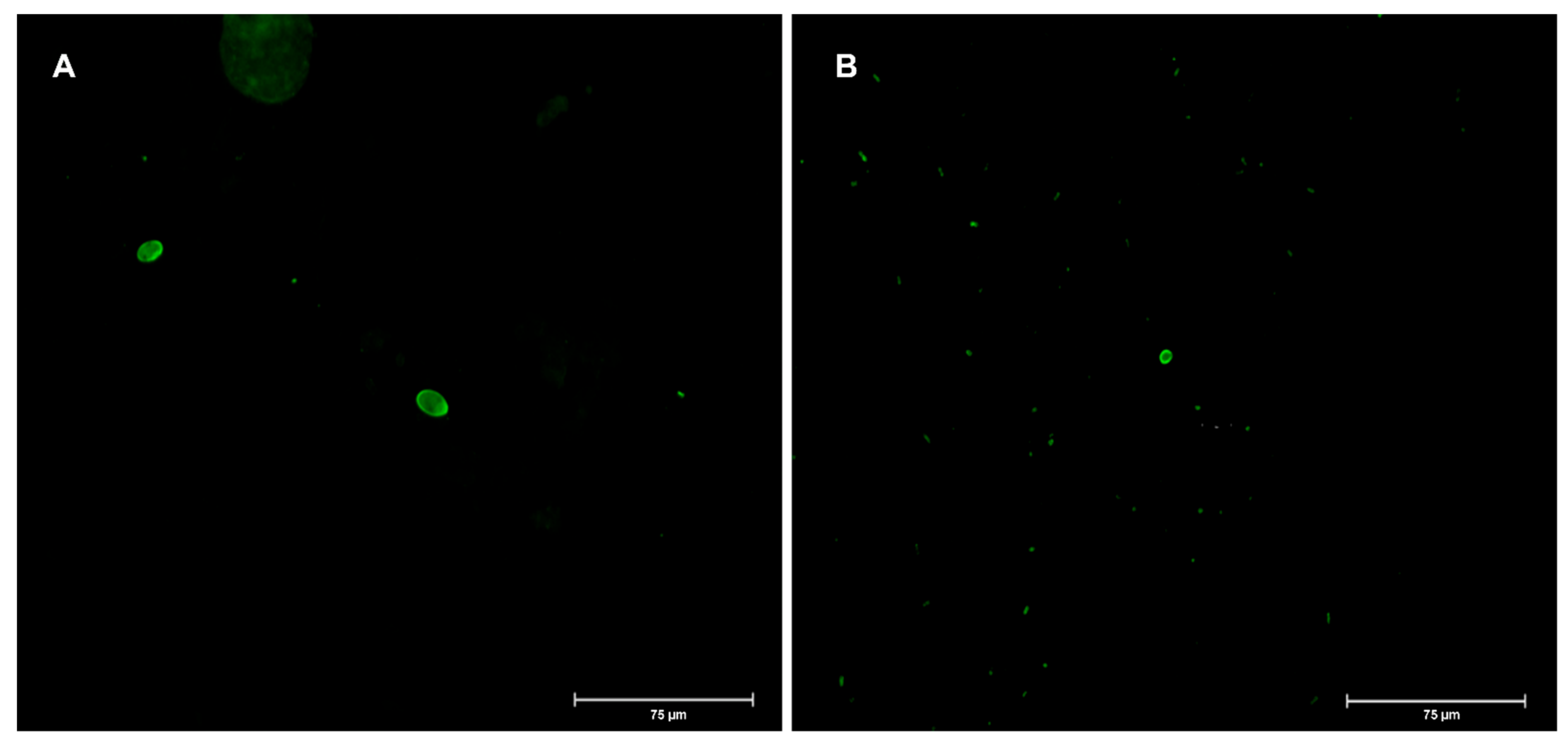
Figure 1. Epifluorescence microscopy images (400× magnification) of Giardia cysts (A) and Cryptosporidium oocysts (B) found in sample 12.
Applsci 12 07145 g001
Table 1. Origin, collection date, and product presentation of the vegetable samples.
Sample NumberProductOriginCollection DateProduct Presentation
1WatercressPortugal27 September 2018bulk
2CorianderPortugal27 September 2018bulk
3ParsleyPortugal27 September 2018bulk
4WatercressPortugal2 October 2018RTE
5Mixed saladPortugal2 October 2018RTE
6CorianderPortugal2 October 2018packaged
7ParsleyPortugal2 October 2018packaged
8WatercressPortugal12 October 2018RTE
9Mixed saladPortugal12 October 2018RTE
10CorianderPortugal12 October 2018packaged
11ParsleyPortugal12 October 2018packaged
12Mixed saladPortugal4 February 2019RTE
13Mixed saladSpain17 June 2019RTE
14Mixed saladSpain17 June 2019RTE
15ArugulaSpain17 June 2019RTE
16LettucePortugal18 June 2019bulk
RTE: ready-to-eat.
Table 2. Protozoa and bacteria positive samples.
SampleProductProduct PresentationNo. Cysts
Giardia
PCR
Giardia
No. (oo)cysts
Cryptosporidium
PCR
Cryptosporidium
Genotyping
(Assemblage)
2Corianderbulk2Negative0n.a.n.a.
8WatercressRTE1Positive0n.a.A
9Mixed saladRTE1Positive0n.a.A
12Mixed saladRTE4Positive8NegativeA
13Mixed saladRTE0n.a.1Negativen.a.
15ArugulaRTE0n.a.1Negativen.a.LMOLMO, detection
16Lettucebulk0n.a.2Negativen.a.Present < 4.0 × 101Positive in 25 g
RTE: ready-to-eat; n.a.: not applicable; LMO: Listeria monocytogenes.
Publisher’s Note: MDPI stays neutral with regard to jurisdictional claims in published maps and institutional affiliations.