Communication

Examination of Postmortem β-Hydroxybutyrate Increase in Forensic Autopsy Cases

Department of Forensic Medicine, Graduate School of Medicine, Kyoto Prefectural University of Medicine, 465 Kajiicho, Kawaramachi-dori Hirokoji-agaru, Kamigyo-ku, Kyoto 602-8566, Japan
*
Author to whom correspondence should be addressed.
Appl. Sci. 2022, 12(14), 7181; https://doi.org/10.3390/app12147181
Submission received: 31 May 2022 / Revised: 14 July 2022 / Accepted: 14 July 2022 / Published: 16 July 2022
(This article belongs to the Special Issue Forensic Medicine and Its Applications)

Abstract

:
Ketoacidosis is one of the common diseases that sometimes result in death. In forensic autopsy cases, the measuring of ketone bodies, especially β-hydroxybutyrate (BHB), is useful in diagnosing postmortem ketoacidosis as a cause of death. However, increased BHB values are not always indicative of ketoacidosis. Other causes of death, such as hypothermia, are known to increase BHB values. In addition, sometimes, we experience cases of increased BHB values that are unlikely to be associated with the pathophysiology or the cause of death resulting in an increase in BHB values. In this study, autopsies were performed to determine the relationship between blood BHB values and the postmortem interval (PMI). The patients were divided into four groups: PMI ≤ 24 h, PMI ≤ 48 h, PMI ≤ 72 h, and PMI > 72 h. The BHB values for each group were then compared. Based on the analysis, patients with a PMI ≤ 72 h and those with a PMI > 72 h had significantly higher BHB values than patients with a PMI ≤ 24 h. In conclusion, there remains a possibility that the BHB values increase after death. Forensic pathologists should consider PMI when diagnosing ketoacidosis as the cause of death.

1. Introduction

Metabolic acidosis is defined as the imbalanced condition of increased hydrogen ions and reduced bicarbonate ions in the body [1]. It has two categories, acute and chronic. According to one review, acute metabolic acidosis could result in an increased mortality rate [2]. Three pathologies are well known in the developmental mechanisms of metabolic acidosis: acid excretion decrease in the kidney, loss of bicarbonate ions, and an increase in acid generation. Usually, nonvolatile metabolic acids from daily meals are excreted in the urine from the kidney. However, in cases with decreased renal function or type 1 and 4 renal tubular acidosis, acid excretion from the kidney decreases, and it results in metabolic acidosis [1]. Loss of bicarbonate ions could happen in conditions such as severe diarrhea or type 2 renal tubular acidosis. The stool osmolality of people who have diarrhea could almost be equal to the serum osmolality. As the diarrheal stool contains electrolytes, this results in the loss of bicarbonate ions [3]. In type 2 renal tubular acidosis, the reabsorption of bicarbonate ions was decreased, which caused metabolic ketoacidosis [4]. The increase in acid generation simply means that elevated acid levels cause metabolic ketoacidosis. Although there are several causes that promote the acid generation, the above review reported that the increase in acid generation, especially lactic acidosis and ketoacidosis, accounts for up to 70% of severe metabolic acidosis [2]. Lactic acidosis is usually caused by hypoperfusion of tissues, such as sepsis, or toxin-induced such as methanol poisoning [5].
Ketoacidosis is one of the most common disease states in the field of forensic science. Ketoacidosis is known as metabolic acidosis caused by the accumulation of ketone bodies. There are several causes that could trigger ketoacidosis, such as diabetes, alcohol, or specific intoxication [6,7,8]. Diabetic ketoacidosis is the most common cause. There are multiple case reports that show death as the result of diabetic ketoacidosis [9,10,11]. Patients with type 1 diabetes are often associated with diabetic ketoacidosis. Even patients with type 2 diabetes could suffer from it. This can include severe stress, infection, and trauma [12]. Usually, serum glucose levels are maintained primarily by two hormones, glucagon, and insulin. As mechanisms of diabetic ketoacidosis, the deficiency and/or tolerance of insulin, and glucagon excess are considered [13,14]. Chronic alcoholism patients with malnutrition are at risk of suffering from alcoholic ketoacidosis. When they cannot drink alcohol due to nausea or vomiting caused by alcoholism, the catecholamine levels elevate, which results in an increase in lipolysis [15]. Therefore, fatty acid oxidation results in ketone body production.
Measuring ketone bodies, such as acetone, acetoacetate, and β-hydroxybutyrate (BHB), is necessary to diagnose diabetic or alcoholic ketoacidosis as a cause of death [16,17,18]. In addition to ketone bodies in the blood, other fluids, such as vitreous or pericardial fluid, have been investigated [19,20,21]. Samples other than blood could also be alternative tools for diagnosing ketoacidosis. BHB measurement is the most important test to establish the diagnosis of postmortem ketoacidosis [22,23]. This is because that BHB should be stable even after death for a period of 24–72 h [24]. However, patients with increased BHB values, which were unlikely to be caused by ketoacidosis or pathologies that increase BHB, such as hypothermia, can occasionally be found [25]. Although reports indicate that BHB values do not increase after death, one study demonstrated increased postmortem BHB levels [26]. Thus, a long postmortem interval (PMI) was often suspected in these patients. The current study aimed to determine the relationship between the PMI and BHB levels in autopsies.

2. Materials and Methods

2.1. Study Sample

This was a retrospective, observational study. Autopsies were performed to determine causes of death between June 2010, and March 2022. Blood was obtained from the heart during the autopsy. The reason why we used blood as samples was that it took several hours for BHB to transfer to other fluids, such as pericardial fluids [25]. BHB levels were measured using an enzymatic method by the LSI Medicine Corporation (Tokyo, Japan). The enzymatic method is one of the analyses that is performed with enzymes. It can detect samples that other methods cannot. Furthermore, it is time efficient in separating the component and its analysis [27]. One study demonstrated that the enzymatic method was suitable for the measurement of serum BHB values [28]. This study was approved by the institutional review board (ERB-C-1710-1).
Apart from diabetic and alcoholic ketoacidosis, severe hypoxia, end-stage liver disease, multiple metabolic disorders, multiple organ failure, and hypothermia also cause ketoacidosis [21,25,29]. Generally, ketoacidosis is defined as serum levels of ketone bodies >3000 µmol/L [29]. Furthermore, one study stated that cases in which BHB levels are >2500 µmol/L could result in death [30]. Exclusion criteria were set in this study to distinguish and exclude cases of increased antemortem BHB from those of increased postmortem BHB. Increased antemortem BHB cases were defined as deaths caused by increased BHB, such as in diabetic ketoacidosis, or deaths coincidentally showing increased BHB levels where that is not the cause of death, such as hypothermia. Cases involving patients <18 years old, cases wherein the above pathologies were suspected as the causes of death, and cases involving patients with BHB values > 2500 µmol/L were excluded.
The cadavers were preserved in a morgue (at 4 °C) after examination by the police until autopsy for approximately 1 d.

2.2. Characteristics of the Patients

The patients were divided into four groups based on the estimated PMI. Group 1 consisted of patients with a PMI of ≤24 h. Group 2 consisted of patients with a PMI of ≤48 h. Group 3 consisted of patients with a PMI of ≤72 h. Group 4 consisted of patients with a PMI > 72 h. In this study, PMI was defined as the duration between the estimated time of death and the autopsy.
A total of 94 men and 37 women were included in this study. The median (interquartile range [IQR]) age of men and women was 53 (42.25–70) and 60 (32–78) years, respectively. The causes of death included subarachnoid hemorrhage (n = 3), epilepsy (n = 2), malignant neoplasm (n = 1), infection (n = 8), subdural, epidural hematoma (n = 13), trauma to the brain or cervical cord (n = 6), burn death (n = 27), myocarditis (n = 3), heart failure (n = 8), renal failure (n = 1), aortic dissection (n = 2), aortic aneurysm rupture (n = 1), fatal arrhythmia (n = 5), toxicity (n = 9), heat stroke (n = 1), cerebral bleeding (n = 4), and unknown cause of death (n = 37). These causes of death were diagnosed by well-trained forensic scientists, and they concluded that these were unlikely to have been due to ketoacidosis. The PMI ranged from 12 h to 16 d. The median (IQR) BHB values were 190 (104–472.5) µmol/L in Group 1, 252 (130–777) µmol/L in Group 2, 505.5 (183.25–1185) µmol/L in Group 3, and 798 (313–1235) µmol/L in Group 4. Table 1 shows the characteristics of each group.

2.3. Statistical Analysis

Log transformation was conducted, and BHB values were analyzed using Dunnett’s test to compare each group. Steel’s test with nontransformed BHB was then performed to confirm the robustness of the results as a sensitivity analysis. In both tests, Group 1 was used as the control group.
Additionally, since Group 4 had a wide PMI range (72 h to 16 d), patients in Group 4 were categorized into two groups, PMI of ≤1 week and PMI of >1 week. Then, we compared the BHB values between these groups using Wilcoxon’s rank sum test.
Statistical analysis was performed using JMP® 16 (SAS Institute Inc., Cary, NC, USA). The significance level was set at p < 0.05.

3. Results

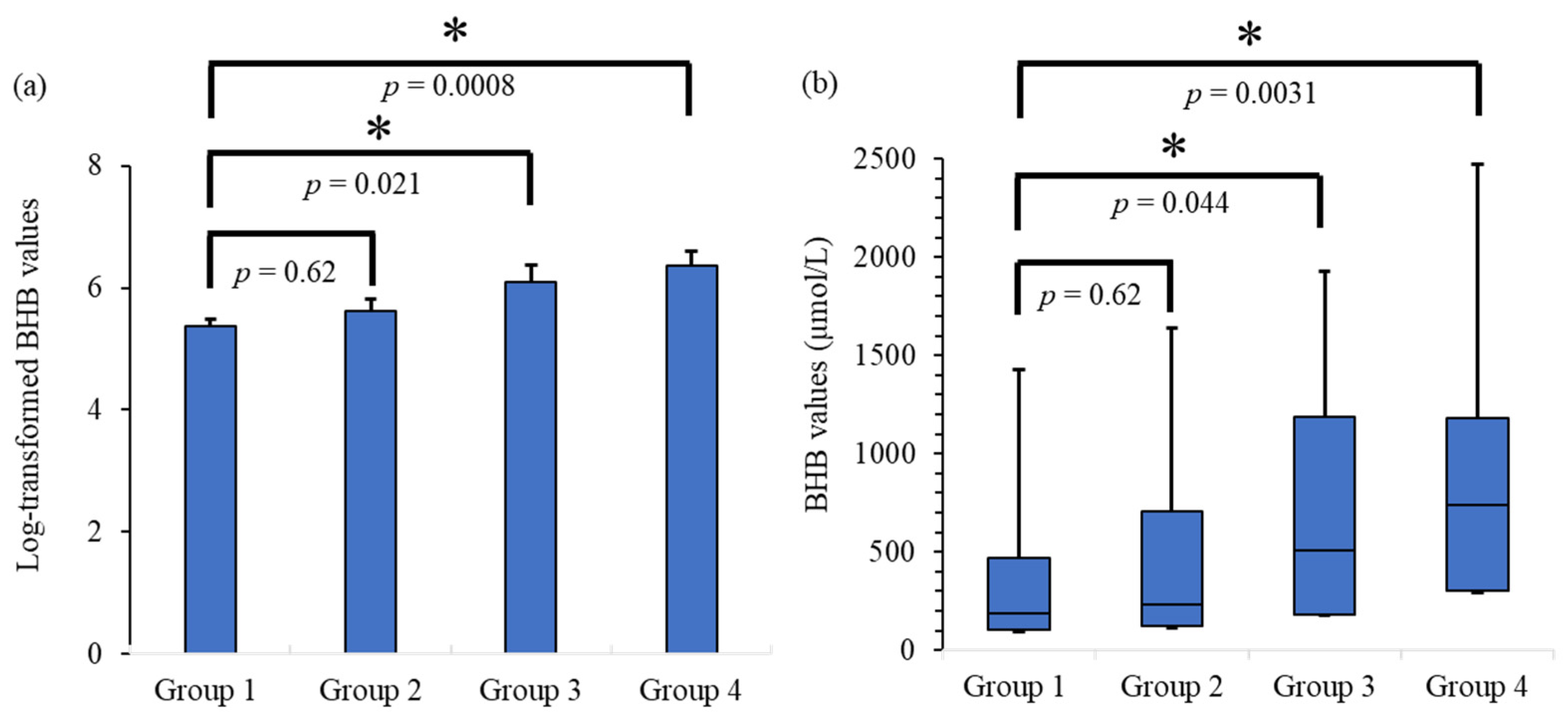
3.1. Results of Statistical Intergroup Analysis

A significant difference between Group 1 and Group 2 was not confirmed by either test (Dunnett’s test, p = 0.62; Steel’s test, p = 0.62). However, significant differences were confirmed between Groups 1 and 3 (Dunnett’s test, p = 0.021; Steel’s test, p = 0.044), as well as Groups 1 and 4 (Dunnett’s test, p = 0.0008; Steel’s test, p = 0.0031). Figure 1 shows the results of the two tests. Group 4 tended to have higher BHB levels.

3.2. Result of Wilcoxon’s Rank Sum Test

In Group 4, there were 10 males and 2 females in the PMI of ≤1-week group and 4 males in the PMI of >1-week group. The median (IQR) BHB values were 406.5 (210–862.5), and 1235 (1087.5–2162.5) in the PMI of ≤1-week and >1-week groups, respectively. Wilcoxon’s rank sum test revealed that the BHB values were significantly higher in the PMI of the >1-week group than in the PMI of the ≤1-week group (p = 0.0129). This result is shown in Figure 2.

4. Discussion

The results showed the possibility that BHB values could increase when associated with prolonged PMI. The robustness of these findings is supported by the concordant results of both the Dunnett’s and Steel’s tests. Additionally, the result of the Wilcoxon’s rank sum test of Group 4 revealed that the BHB values in the PMI of the >1-week group were higher than those in the PMI of the ≤1-week group. This corroborated with the previous findings, which showed that postmortem BHB values were associated with PMI.
On the basis of our results, postmortem production of BHB likely occurs, during the postmortem phase. It is known that ketone bodies are produced by the conversion of fatty acids mainly in the mitochondria of the liver [31,32,33]. Ketogenesis, the pathway of producing three ketone bodies, is activated in the low energic state. Carnitine palmitoyltransferase transports fatty acids to the mitochondria, and fatty acids are converted into acetyl-CoA. Thiolase synthesizes two acetyl-CoA into acetoacetyl-CoA. Then, HMG-CoA synthase converts acetoacetyl-CoA into HMG-CoA. Acetoacetate is generated from HMG-CoA by HMG-CoA lyase. In the situations in that D-β-hydroxybutyrate dehydrogenase exists, acetoacetate is converted into BHB. Conversely, if nonenzymatic decarboxylation occurs, acetoacetate converts into acetone [34]. Our bodies can use BHB and acetoacetate as alternative energies. Since they have water solubility, they can be transported to other organs even the brain. In the organs where they arrive, β-hydroxybutyrate dehydrogenase converts BHB into acetoacetate, and β-ketoacyl-CoA converts acetoacetate into acetyl-CoA, respectively. Then, acetyl-CoA is used in the citric acid cycle [34]. The citric acid cycle starts from acetyl-CoA and releases energy [35,36].
Clinically, death is defined as respiratory, cardiac, or neurologic arrest [37]. However, this does not necessarily mean the death of other organs or tissues. For example, there is a report about skeletal muscle stem cells. In the study, researchers performed a biopsy on cadavers, and they confirmed that some skeletal myogenic cells were alive and functional [38]. Furthermore, researchers succeeded in the culture of brain tissues which they obtained from autopsy cases [39]. Since dead cells cannot be cultured, the result showed a possibility that some of the brain cells were alive at the time of the autopsy. Recently, even the gene that activated after death was examined [40]. This gene keeps active for a few days after organ death. On the basis of these facts, we built a hypothesis that following the arrest of the respiratory and cardiac systems, hepatic cells become severely hypoxic or ischemic, which might result in ketone body production [29].
To the best of our knowledge, only one study has demonstrated postmortem BHB level changes [26]. That study measured BHB levels from the time of death until 49 h and showed an increase in BHB levels with the intragroup analysis. Although a significant difference was confirmed in the prolonged-PMI cases, it was not confirmed in the short-PMI cases of that study. Our results showed the same tendency. On the basis of several studies [30,41,42], it was concluded that postmortem BHB values do not increase [25]. These studies have compared the BHB values of diabetic or alcoholic ketoacidosis cases with control cases. Conversely, our analysis target was cases without ketoacidosis. This difference might result in an opposed conclusion.
This study had some limitations. Only the intergroup BHB values were examined. To demonstrate the increase in BHB values along with the PMI, intragroup examinations were required. Second, although our results showed the possibility of postmortem increases in BHB, the rate of BHB increase with PMI remains unknown. This is because we could not obtain the BHB levels at the time of death. In addition, when forensic scientists interpret the result of biochemical examination with serum samples, they should take the effect of hemolysis. In this study, we did not consider the effect of hemolysis on BHB based on the study that examined the association between serum BHB levels and hemolysis [43]. However, this study was performed with bovine blood, and we could not exclude the possibility that human BHB is affected by hemolysis. An intragroup analysis with sufficient samples is needed to demonstrate these aspects.

5. Conclusions

Our results showed the possibility that a longer PMI was associated with higher BHB values. Postmortem production of BHB may occur. Forensic pathologists should take PMI into account while diagnosing ketoacidosis as the cause of death.

Author Contributions

Conceptualization, H.K.; methodology, H.K.; software, H.K. and R.M.; validation, S.K., N.I., and R.M.; investigation, H.I. (Hiroaki Ichioka), M.K., and D.T.; data curation, R.B.; writing—original draft preparation, H.K.; writing—review and editing, H.I. (Hiroaki Ichioka); visualization, H.I. (Hiroaki Ichioka), M.K., and D.T.; supervision, H.I. (Hiroshi Ikegaya). All authors have read and agreed to the published version of the manuscript.

Funding

This research received no external funding.

Institutional Review Board Statement

This study was approved by our institution review board (ERB-C-1710-1).

Informed Consent Statement

Informed consent was obtained by opt-out.

Data Availability Statement

The datasets generated during and/or analyzed during the current study are available from the corresponding author on reasonable request due to opt-out.

Acknowledgments

We would like to thank Yoshihisa Akasaka for the help.

Conflicts of Interest

The authors declare no conflict of interest.

References

  1. UpToDate. Approach to the Adult with Metabolic Acidosis. Available online: https://www.uptodate.com/contents/approach-to-the-adult-with-metabolic-acidosis?search=metabolic%20acidosis&source=search_result&selectedTitle=1~150&usage_type=default&display_rank=1 (accessed on 4 June 2022).
  2. Jeffrey, A.K.; Nicolaos, E.M. Treatment of Acute Metabolic Acidosis: A Pathophysiologic Approach. Nat. Rev. Nephrol. 2012, 8, 589–601. [Google Scholar] [CrossRef]
  3. UpToDate. Acid-Base and Electrolyte Abnormalities with Diarrhea. Available online: https://www.uptodate.com/contents/acid-base-and-electrolyte-abnormalities-with-diarrhea?search=metabolic%20acidosis&topicRef=2291&source=see_link#H626692994 (accessed on 5 June 2022).
  4. UpToDate. Overview and Pathophysiology of Renal Tubular Acidosis and the Effect on Potassium Balance. Available online: https://www.uptodate.com/contents/overview-and-pathophysiology-of-renal-tubular-acidosis-and-the-effect-on-potassium-balance?sectionName=PROXIMAL%20(TYPE%202)%20RTA&search=metabolic%20acidosis&topicRef=2291&anchor=H42786907&source=see_link#H42786907 (accessed on 5 June 2022).
  5. UpToDate. Causes of Lactic Acidosis. Available online: https://www.uptodate.com/contents/causes-of-lactic-acidosis?search=metabolic%20acidosis&topicRef=2291&source=see_link#H7 (accessed on 5 June 2022).
  6. Benoit, S.R.; Zhang, Y.; Geiss, L.S.; Gregg, E.W.; Albright, A. Trends in Diabetic Ketoacidosis Hospitalizations and in-Hospital Mortality—United States, 2000–2014. MMWR Morb. Mortal. Wkly Rep. 2018, 67, 362–365. [Google Scholar] [CrossRef] [PubMed]
  7. McGuire, L.C.; Cruickshank, A.M.; Munro, P.T. Alcoholic Ketoacidosis. Emerg. Med. J. 2006, 23, 417–420. [Google Scholar] [CrossRef] [PubMed]
  8. Cartwright, M.M.; Hajja, W.; Al-Khatib, S.; Hazeghazam, M.; Sreedhar, D.; Li, R.N.; Wong-McKinstry, E.; Carlson, R.W. Toxigenic and Metabolic Causes of Ketosis and Ketoacidotic Syndromes. Crit. Care Clin. 2012, 28, 601–631. [Google Scholar] [CrossRef]
  9. Luna, J.; Gilliland, M.G.; Hewan-Lowe, K.O.; Tanenberg, R.J. Postmortem Diagnosis of Diabetic Ketoacidosis Presenting as the “Dead-in-Bed Syndrome”. Endocr. Pract. 2014, 20, e123–e125. [Google Scholar] [CrossRef]
  10. Irwin, J.; Cohle, S.D. Sudden Death Due to Diabetic Ketoacidosis. Am. J. Forensic Med. Pathol. 1988, 9, 119–121. [Google Scholar] [CrossRef]
  11. Kjærulff, M.L.B.G.; Astrup, B.S. Sudden Death Due to Diabetic Ketoacidosis Following Power Failure of an Insulin Pump: Au topsy and Pump Data. J. Forensic Leg. Med. 2019, 63, 34–39. [Google Scholar] [CrossRef]
  12. UpToDate. Diabetic Ketoacidosis and Hyperosmolar Hyperglycemic State in Adults: Epidemiology and Pathogenesis. Available online: https://www.uptodate.com/contents/diabetic-ketoacidosis-and-hyperosmolar-hyperglycemic-state-in-adults-epidemiology-and-pathogenesis?search=diabetic%20ketoacidosis&source=search_result&selectedTitle=5~150&usage_type=default&display_rank=5#H2 (accessed on 4 June 2022).
  13. Barnes, A.J.; Bloom, S.R.; Goerge, K.; Alberti, G.M.; Smythe, P.; Alford, F.P.; Chisholm, D.J. Ketoacidosis in Pancreatectomized Man. N. Engl. J. Med. 1977, 296, 1250–1253. [Google Scholar] [CrossRef]
  14. Miles, J.M.; Haymond, M.W.; Nissen, S.L.; Gerich, J.E. Effects of Free Fatty Acid Availability, Glucagon Excess, and Insulin Defi ciency on Ketone Body Production in Postabsorptive Man. J. Clin. Investig. 1983, 71, 1554–1561. [Google Scholar] [CrossRef]
  15. UpToDate. Fasting Ketosis and Alcoholic Ketoacidosis. Available online: https://www.uptodate.com/contents/fasting-ketosis-and-alcoholic-ketoacidosis?search=alcoholic%20ketoacidosis&source=search_result&selectedTitle=1~17&usage_type=default&display_rank=1 (accessed on 5 June 2022).
  16. Felby, S.; Nielsen, E.; Thomsen, J.L. The Postmortem Distribution of Ketone Bodies Between Blood, Vitreous Humor, Spinal Fluid, and Urine. Forensic Sci. Med. Pathol. 2008, 4, 100–107. [Google Scholar] [CrossRef]
  17. Thomsen, J.L.; Felby, S.; Theilade, P.; Nielsen, E. Alcoholic Ketoacidosis as a Cause of Death in Forensic Cases. Forensic Sci. Int. 1995, 75, 163–171. [Google Scholar] [CrossRef]
  18. Kanetake, J.; Kanawaku, Y.; Mimasaka, S.; Sakai, J.; Hashiyada, M.; Nata, M.; Funayama, M. The Relationship of a High Level of Serum Beta-Hydroxybutyrate to Cause of Death. Leg. Med. Tokyo 2005, 7, 169–174. [Google Scholar] [CrossRef]
  19. Osuna, E.; Vivero, G.; Conejero, J.; Abenza, J.M.; Martínez, P.; Luna, A.; Pérez-Cárceles, M.D. Postmortem Vitreous Humor β-Hydroxybutyrate: Its Utility for the Postmortem Interpretation of Diabetes Mellitus. Forens. Sci. Int. 2005, 153, 189–195. [Google Scholar] [CrossRef]
  20. Heninger, M. Postmortem Vitreous Beta-Hydroxybutyrate: Interpretation in a Forensic Setting. J. Forens. Sci. 2012, 57, 1234–1240. [Google Scholar] [CrossRef]
  21. Palmiere, C.; Bardy, D.; Letovanec, I.; Mangin, P.; Augsburger, M.; Ventura, F.; Iglesias, K.; Werner, D. Biochemical Markers of Fatal Hypothermia. Forens. Sci. Int. 2013, 226, 54–61. [Google Scholar] [CrossRef] [Green Version]
  22. Eriksson Hydara, Y.; Zilg, B. Postmortem Diagnosis of Ketoacidosis: Levels of Beta-Hydroxybutyrate, Acetone and Isopropanol in Different Causes of Death. Forens. Sci. Int. 2020, 314, 110418. [Google Scholar] [CrossRef]
  23. Ahlström, S.; Ahlner, J.; Jönsson, A.K.; Green, H. The Importance of BHB Testing on the Post-Mortem Diagnosis of Ketoacidosis. Biomolecules 2021, 12, 9. [Google Scholar] [CrossRef]
  24. Iliescu, D.B.; Furnica, C.; Girlescu, N.; Chistol, R.O.; Perianu, L.; Diac, M.; Timofte, A.D.; Knieling, A.; Ciureanu, I.-A. Postmortem Diagnosis of Ketoacidosis by Determining Beta-Hydroxybutyrate Levels in Three Types of Body Fluids by Two Different Methods. Appl. Sci. 2022, 12, 5541. [Google Scholar] [CrossRef]
  25. Rousseau, G.; Reynier, P.; Jousset, N.; Rougé-Maillart, C.; Palmiere, C. Updated Review of Postmortem Biochemical Exploration of Hypothermia with a Presentation of Standard Strategy of Sampling and Analyses. Clin. Chem. Lab. Med. 2018, 56, 1819–1827. [Google Scholar] [CrossRef] [Green Version]
  26. Woydt, L.; Bernhard, M.; Kirsten, H.; Burkhardt, R.; Hammer, N.; Gries, A.; Dreßler, J.; Ondruschka, B. Intra-individual Alterations of Serum Markers Routinely Used in Forensic Pathology Depending on Increasing Post-Mortem Interval. Sci. Rep. 2018, 8, 12811. [Google Scholar] [CrossRef]
  27. Pomeranz, Y.; Meloan, C.E. Enzymatic Methods. In Food Analysis; Springer: Boston, MA, USA, 1994. [Google Scholar] [CrossRef]
  28. Ren, F.Q.; Yan, S.K.; Mao, D.Y.; Li, Y.X.; Xiao, X.H.; Xu, E.M. Evaluation of Enzymatic Method for Determination of Serum Beta-Hydroxybutyrate and Its Clinical Application. Zhongguo Yi Xue Ke Xue Yuan Xue Bao 2003, 25, 702–705. (In Chinese) [Google Scholar]
  29. Palmiere, C.; Mangin, P. Postmortem Chemistry Update Part I. Int. J. Legal Med. 2012, 126, 187–198. [Google Scholar] [CrossRef] [Green Version]
  30. Iten, P.X.; Meier, M. Beta-Hydroxybutyric Acid—An Indicator for an Alcoholic Ketoacidosis as Cause of Death in Deceased Alcohol Abusers. J. Forens. Sci. 2000, 45, 624–632. [Google Scholar] [CrossRef]
  31. Jungermann, K.; Katz, N. Functional Specialization of Different Hepatocyte Populations. Physiol. Rev. 1989, 69, 708–764. [Google Scholar] [CrossRef]
  32. Watanabe, S.; Hirakawa, A.; Aoe, S.; Fukuda, K.; Muneta, T. Basic Ketone Engine and Booster Glucose Engine for Energy Production. Open J. 2016, 2, 14–23. [Google Scholar] [CrossRef]
  33. Nakamura, M.T.; Yudell, B.E.; Loor, J.J. Regulation of Energy Metabolism by Long-Chain Fatty Acids. Prog. Lipid Res. 2014, 53, 124–144. [Google Scholar] [CrossRef]
  34. Kiranjit, K.D.; Sonu, G. Biochemistry, Ketogenesis. StatPearls. Available online: https://www.ncbi.nlm.nih.gov/books/NBK493179/ (accessed on 5 June 2022).
  35. Akram, M. Citric Acid Cycle and Role of Its Intermediates in Metabolism. Cell Biochem. Biophys. 2014, 68, 475–478. [Google Scholar] [CrossRef]
  36. Kelly, D.J.; Hughes, N.J. Chapter 12. The Citric Acid Cycle and Fatty Acid Biosynthesis. In Helicobacter pylori: Physiology and Genetics; Mobley, H.L.T., Mendz, G.L., Hazell, S.L., Eds.; ASM Press: Washington, DC, USA, 2001. [Google Scholar]
  37. World Health Organization. Clinical Criteria for the Determination of Death—WHO Technical Expert Consultation; WHO Headquarters, Geneva, Switzerland, 22–23 September 2014; World Health Organization: Geneva, Switzerland, 2017; Available online: https://apps.who.int/iris/handle/10665/254737 (accessed on 30 May 2022).
  38. Latil, M.; Rocheteau, P.; Châtre, L.; Sanulli, S.; Mémet, S.; Ricchetti, M.; Tajbakhsh, S.; Chrétien, F. Skeletal Muscle Stem Cells Adopt a Dormant Cell State Postmortem and Retain Regenerative Capacity. Nat. Commun. 2012, 3, 903. [Google Scholar] [CrossRef] [Green Version]
  39. Verwer, R.W.; Hermens, W.T.; Dijkhuizen, P.; ter Brake, O.; Baker, R.E.; Salehi, A.; Sluiter, A.A.; Kok, M.J.; Muller, L.J.; Verhaagen, J.; et al. Cells in Human Postmortem Brain Tissue Slices Remain Alive for Several Weeks in Culture. FASEB J. 2002, 16, 54–60. [Google Scholar] [CrossRef]
  40. Pozhitkov, A.E.; Neme, R.; Domazet-Lošo, T.; Leroux, B.G.; Soni, S.; Tautz, D.; Noble, P.A. Tracing the Dynamics of Gene Transcripts After Organismal Death. Open Biol. 2017, 7, 160267. [Google Scholar] [CrossRef] [PubMed] [Green Version]
  41. Peter, K.; Jože, B.; Vera, F.M. Alcoholic ketoacidosis: A Cause of Sudden Death of Chronic Alcoholics. Forens. Sci. Int. 1999, 103, S53–S59. [Google Scholar]
  42. Palmiere, C.; Mangin, P.; Werner, D. Postmortem Distribution of 3-Beta-Hydroxybutyrate. J. Forens. Sci. 2014, 59, 161–166. [Google Scholar] [CrossRef] [PubMed]
  43. Stokol, T.; Nydam, D.V. Effect of Hemolysis on Nonesterified Fatty Acid and Beta-Hydroxybutyrate Concentrations in Bovine Blood. J. Vet. Diagn. Investig. 2006, 18, 466–469. [Google Scholar] [CrossRef] [PubMed] [Green Version]
Figure 1. (a) Bar graph of the log-transformed BHB values with an error bar as the result of Dunnett’s test. The error bar depicts the standard error. (b) Boxplot graph of the BHB values as the result of the Steel’s test. Both tests showed the tendency that longer PMI groups result in higher BHB values. The significant differences between Group 1 and Group 3, as well as Group 1 and Group 4, were confirmed by both tests. * means statistical significance (p < 0.05). BHB, β-hydroxybutyrate.
Applsci 12 07181 g001
Figure 2. Comparison of BHB values between the PMI of ≤1-week and >1-week groups. The median values are significantly higher in the PMI of the >1-week group than in the PMI of the ≤1-week group. * means statistical significance (p < 0.05). BHB; β-hydroxybutyrate.
Applsci 12 07181 g002
Table 1. Characteristics of each group.
Number of PatientsMedian BHB
(IQR) µmol/L
Average BHB
(SE)
(Log-Transformed)
Group 1Males: 561905.38
(PMI ≤ 24 h)Females: 18(104–472.5)(0.11)
Group 2Males: 152325.62
(PMI ≤ 48 h)Females: 10(122–705.5)(0.19)
Group 3Males: 9505.56.10
(PMI ≤ 72 h)Females: 7(183.25–1185)(0.28)
Group 4Males: 147416.37
(PMI > 72 h)Females: 2(302–1182.5)(0.24)
BHB, β-hydroxybutyrate; IQR, interquartile range; SE, standard error; PMI, post-mortem interval.
Publisher’s Note: MDPI stays neutral with regard to jurisdictional claims in published maps and institutional affiliations.

