Featured Application
The results highlighted the role of low-intensity electromagnetic fields in fish bone regeneration. Such a simple in vivo model can be used to elucidate the responsiveness of skeletal tissue to specific physical forces useful to design new therapeutic approach in human bone diseases.
Abstract
Low-Intensity electromagnetic fields (LI-PEMFs) are known to induce a trophic stimulus on bone tissue and therefore have been largely used for the treatment of several musculoskeletal disorders. High intensity (HI) PEMFs add interesting features to bio-stimulation such as electroporation, a phenomenon characterized by transient increased cell permeabilization to molecules, and diamagnetism, a water-repulsive effect based on the diamagnetic properties of water and transmembrane ions gradients. Despite the rapid evolution of technology, the biological mechanisms underlying it are still poorly understood. In order to evaluate the effectiveness of this particular stimulation, HI LF-PEMFs were used to stimulate the caudal fin rays of adult zebrafish. Actually, the zebrafish fin regeneration is a simple, well understood, and widely adopted model for studying bone regeneration. A controlled amputation fin experiment was then conducted. Regenerated bone matrix of fin rays was dyed with calcein and then analysed under fluorescence microscopy. Both the length and the area of regenerated fin’s rays treated with HI LF-PEMFs resulted significantly increased when compared with non-treated.
1. Introduction
In the last fifty years, selected low-frequency time-varying magnetic fields have been studied as therapeutic tools for several musculoskeletal diseases such as bone non-unions, failed arthrodesis, osteonecrosis, chronic refractory tendinitis. The pulsed electromagnetic fields (PEMFs), within the wide electromagnetic spectrum, resulted in clinically effective exploiting of the piezoelectrical properties of the bone [1], modulating of the membrane signal transduction processes [2], and promoting differentiation [3] and proliferation of osteoblastic cells [4]. These positive effects have been largely used for therapeutic purposes when osteogenic stimulation is requested for bone tissue repair [5,6]. Although a remarkable number of studies have been conducted on low-intensity (LI) low-frequency (LF) PEMFs, very little is known about the effects of high-Intensity (HI) LF-PEMFs on vital musculoskeletal tissues. HI LF-PEMFs, with intensity in the range of 0.3–16.4 Tesla (T) have been studied for their ability to induce electroporation (i.e., the capacity to increase transmembrane molecular transport) [7,8,9,10,11] and, with adequate levels of energy, a water repulsive and molecular diamagnetic effect [12]. This repulsive effect (diamagnetic repulsion or diamagnetism) refers to the magnetic property of some materials which, subjected to a high intensity magnetic field, receive the pushing effect as already experienced in the field of biology by the magnetic levitation [13]. At the extracellular matrix (ECM) level, diamagnetism moves water and other diamagnetic substances such us proteins and ions [12]. This occurs also at the cellular level in terms of movement of ions and the activation of transmembrane proteins, while the variability of the MF and of the magnetic field gradient influence the fundamental ion-channel on/off switching events by membrane magneto-mechanical stressing [14,15] mainly for Ca2+, Na+, K+, Li+, Mg2+ ions or other cellular activities. We, therefore, deemed interesting to ascertain the effects of HI LF-PEMF on bone regeneration. In this regard, we chose an experimental model based on the zebrafish (Danio rerio). This fish has several attractive features for research (amongst others, bone developmental and repair mechanisms very similar to those of vertebrates) [16]. Thus, zebrafish seems to be a good target for experimental studies about the effects of physical stimulation on bone tissue, especially, using caudal fin regeneration as a model [17]. Despite these interesting features, very little is known about the results of PEMFs on zebrafish. The only scientific report that can be found in medical literature is about the stimulatory effect on the pigmentation of zebrafish embryo in vivo [18]. The purpose of this experimental study is to evaluate the effects of HI LF-PEMF on caudal fin adult zebrafish regeneration.
2. Materials and Methods
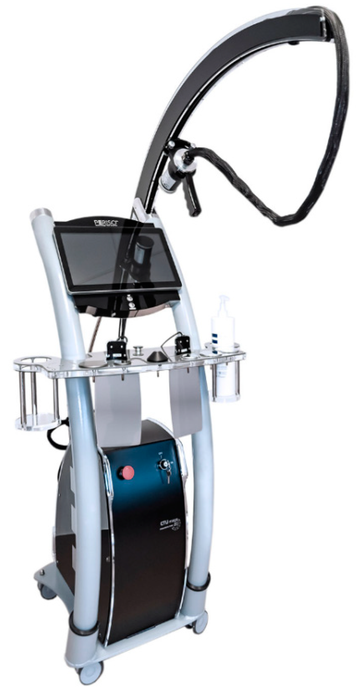
2.1. PEMF Generator
As a PEMF generator, we used a diamagnetic acceleration system able to generate at the origin a high-intensity LF-PEMF of 2 T (Figure 1).

Figure 1.
PEMF generator.
The behaviour with the distance of the magnetic field as peak amplitude at the deeper distance has been measured with a Hall sensor applying the following equation derived from the Biot-Savart law for a given type of solenoid:
(n is the number of turns, I is the peak current and l/2, z, R are the geometric parameters of coil).
The field’s gradient of the magnetic field is <400 T/s, the single pulse duration is 5 ms, the period of 1000 ms and 2.9 mT of MF at the target. Treated fish were exposed to 6 Hz of PEMFs frequency. Using carrier 1000 ms it results in a lowest main peak in frequency domain that is 1 Hz, the second highest peak, that does not result from multiple harmonic of 1 Hz, is at 200 Hz with 5 ms pulse duration. Regenerated fins images of untreated and HI LF-PEMF-treated fish were captured from day 1 to 10 after the amputation and analysed at five days after amputation. The trend of regeneration over the ten days will be described.
2.2. Animals and Treatments
Zebrafish AB strains were maintained in a ZEBTEC © bench top system (Tecniplast, Buguggiate, Italy) under standard conditions [19]. During the treatment, fish have been maintained at 28 °C in E3 medium (5 mM NaCl, 0.17 mM KCl, 0.33 mM CaCl2, 0.33 mM MgSO4. Fish have been incubated with 0.005% Alizarin Red S (ARS, Sigma Aldrich, St. Louis, MO, USA) to highlight bone mineralized matrix. At the end of the staining procedure, fish have been anaesthetized using 0.168 mg/mL tricaine methanesulfonate E3 medium solution [19] and subjected to amputation at the fin bifurcation. In a preliminary experiment (data not shown), we tested different time-protocol of treatment to identify the most effective in terms of regenerative stimulation. Time of treatment has been defined using an algorithm that considers fish morphogenetic proprieties, traducer diameter, dimensions of treatment tank and number of fish treated simultaneously. Ideal time of treatment resulted in 64 min/2 times each day. We also checked that HI LF- PEMFs treatment does not alters the temperature of the water. In every experiment, we treated 14 zebrafish. Seven zebrafish were treated with HI LF-PEMFs following the protocol cited above, whereas seven fish acted as untreated controls. We used the device preset setup specific for bone and cartilage with intensity at level 5 (maximum) and repetition rate of the pulse 5 Hz/s. The entire experiment has been repeated three times using a total amount of 42 fish. After five days of treatment, fish were incubated overnight with 0.005% calcein (Bis[N,N-bis(carboxymethyl)aminomethyl] fluorescein, Sigma Aldrich, St. Louis, MO, USA) to evidentiate the regenerated bone matrix of fin rays. Fish have been anaesthetized in 0.01% tricaine methanesulphonate (Sigma Aldrich, St. Louis, MO, USA) and fins have been analysed using a fluorescence microscope (Olympus SZX-ZB7, Tokyo, Japan) equipped with a Discovery CH30 camera (TiEsseLab, Milan, Italy). The parameters chosen for evaluating the effect of stimulation were the length of the three longest fin rays’ and the area of regenerated bone tissue. Samples were analysed with ImageJ open-source software.
2.3. Ethic Statement
This experimentation has been performed in the Zebrafish Laboratory (IRCCS R. Galeazzi, GSD Foundation, Milan, Italy) according to the Italian and European guidelines on research practice (EU Directive 2010/63/EU) and with authorization by ASL Varese With Prot. No. 2019/014/DVVS/0078143, Italy.
2.4. Statistics
We decided to analyze the effect of adhering to the intervention, conducting a t-test analysis with data collected at five days after amputation. The t-test is preceded by the D’Agostino-Pearson analysis to verify the distribution of the data. Statistical significance was determined for p-values being set at p < 0.05 *; p < 0.01 **; p < 0.001 ***. Data from the test have been analyzed by Student’s unpaired t-test analysis. We assumed a condition of homoscedasticity between the collected variables, considering also that the two groups have the same sample size and, except the treatment of study, were always maintained in the same basic conditions. We used an estimated common variance of the two groups ( pooled) for the t-test with n1+n2-2 degrees of freedom.
3. Results
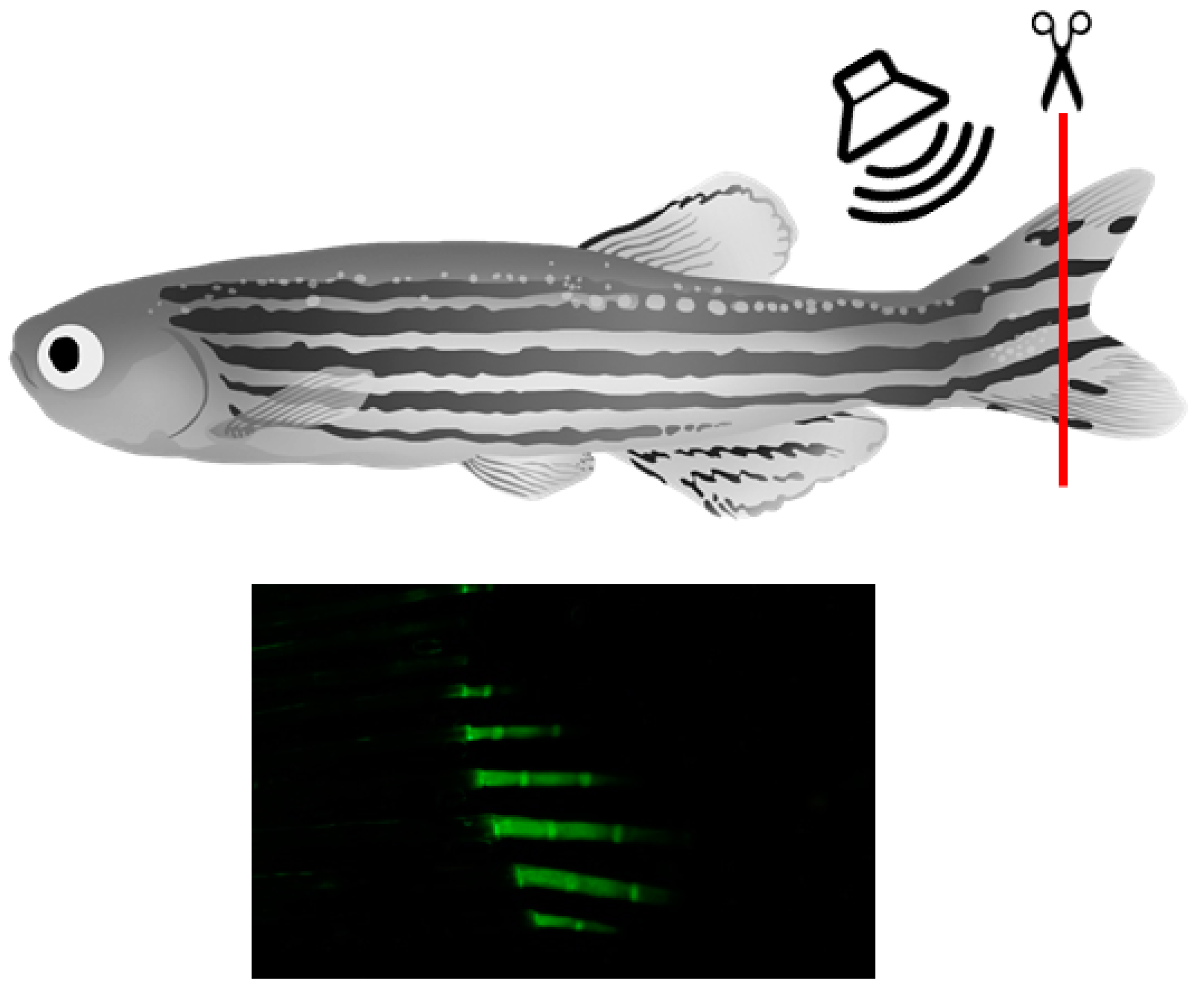
Adult zebrafish caudal fin regeneration has been used as readout model to analyze the effect of biophysical stimulation on bone regeneration by calcein live staining (Figure 2).

Figure 2.
Fin amputation and double-live staining as readout model to study the effects of biophysical stimulations of bone regeneration in vivo.
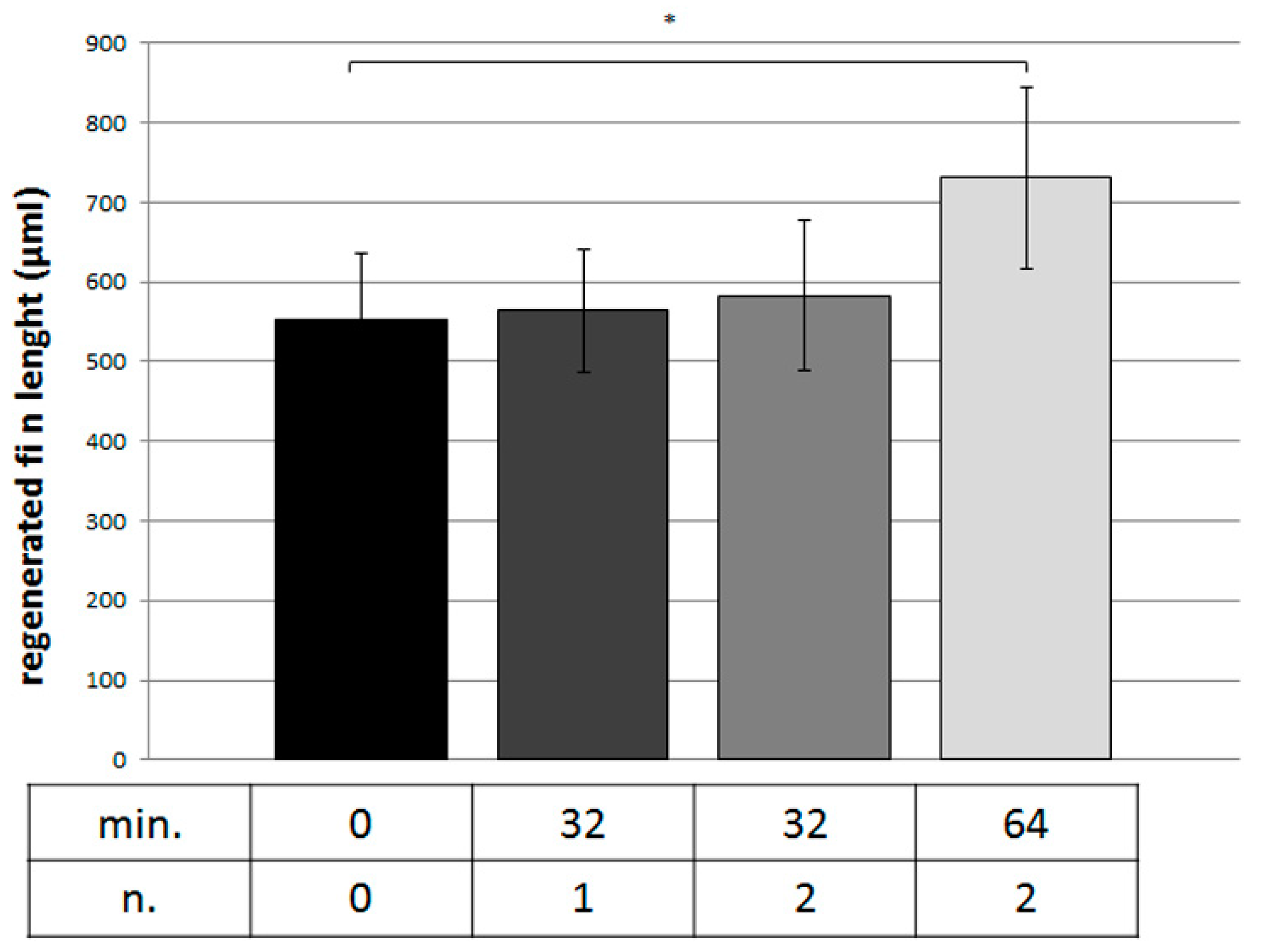
Different HI LF-PEMF treatments have been evaluated at five days after amputation in terms of duration or number of session, resulting in an effective protocol of 64 min twice a day (Figure 3).
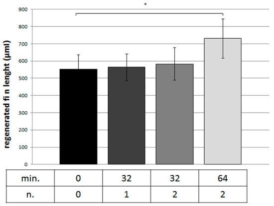
Figure 3.
Regenerated fin rays’ length measurements in different treatment with HI LF-PEMFs. Parameters were expressed as minutes of treatment (min.) and number of daily sessions (n.) (0 vs. 64, * p < 0.05).
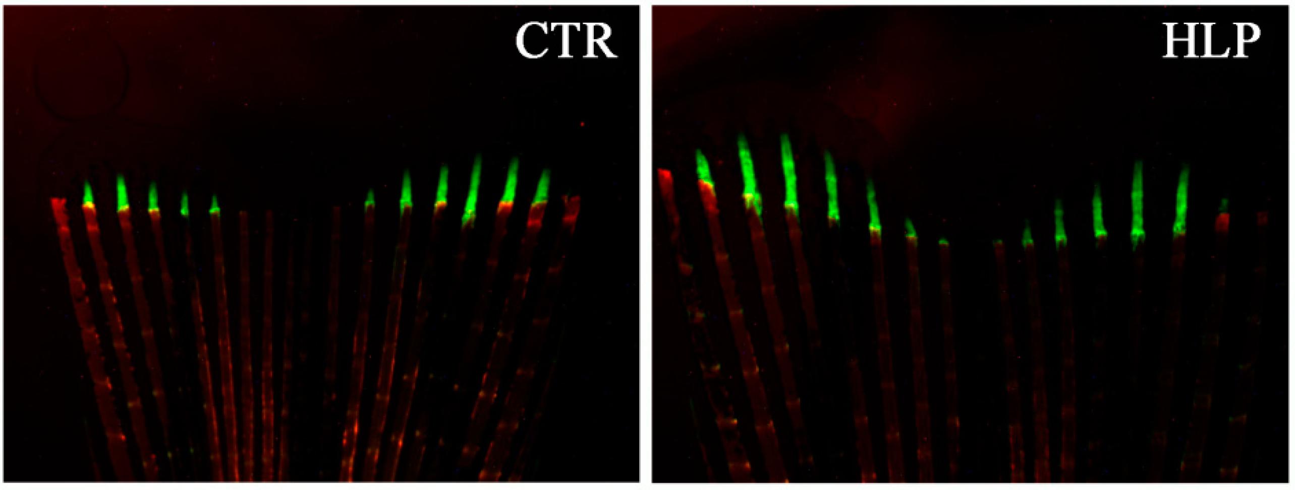
Regenerated fins images of untreated and HI LF-PEMF-treated fish were captured (Figure 4) and morphometric parameters were analyzed by imaging software.

Figure 4.
Calcein-alizarin double staining of fin rays: regenerated bone matrix in untreated (CTR) and treated with HI LFPEMFs (HLP) adult zebrafish fin (red staining highlights the old bone tissue and green staining highlights the new regenerated bone tissue).
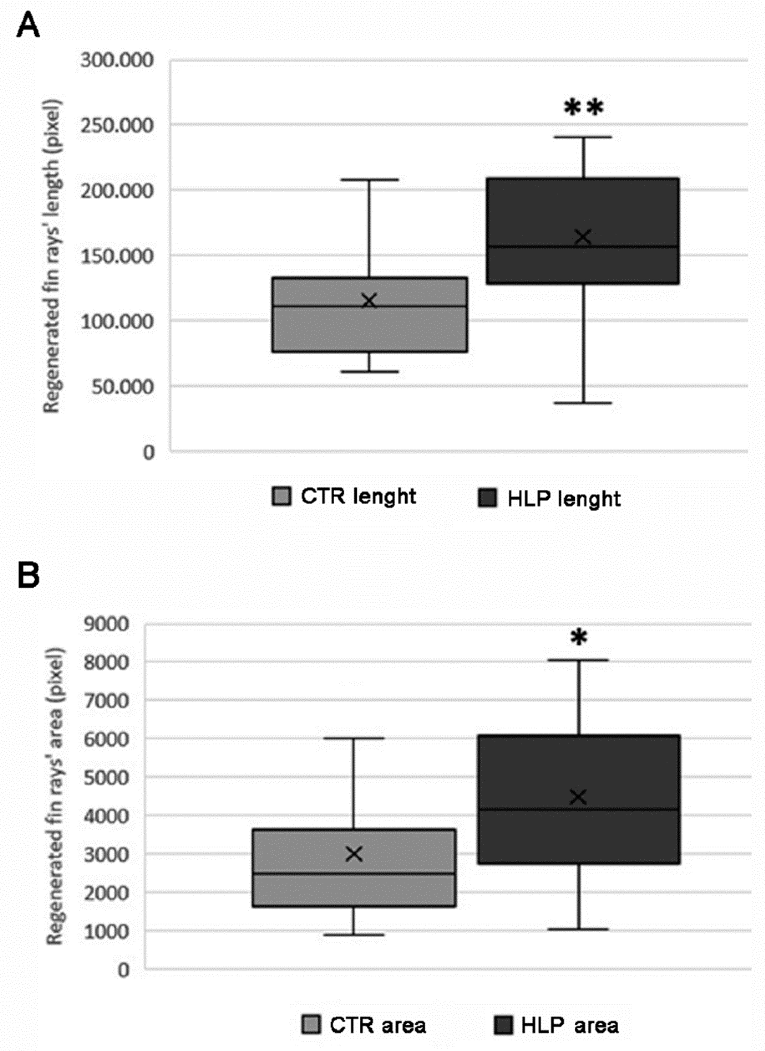
Length of regenerated fin’s rays under HI LF-PEMFs condition at five days after amputation, measured at the top of the fin, showed a statistically significant dimensional increase compared with the control group (p < 0.01, Figure 5A). Similarly, regenerated fin rays’ area of PEMFs-treated fish resulted wider compared to controls (p < 0.05, Figure 5B).

Figure 5.
Measurements in untreated (CTR, seven fish) and treated with HI LF-PEMFs (HLP, seven fish) adult zebrafish of (A) regenerated fin rays’ length (HLP vs. CTR, ** p < 0.01) and (B) regenerated fin rays’ area (HLP vs. CTR, * p < 0.05).
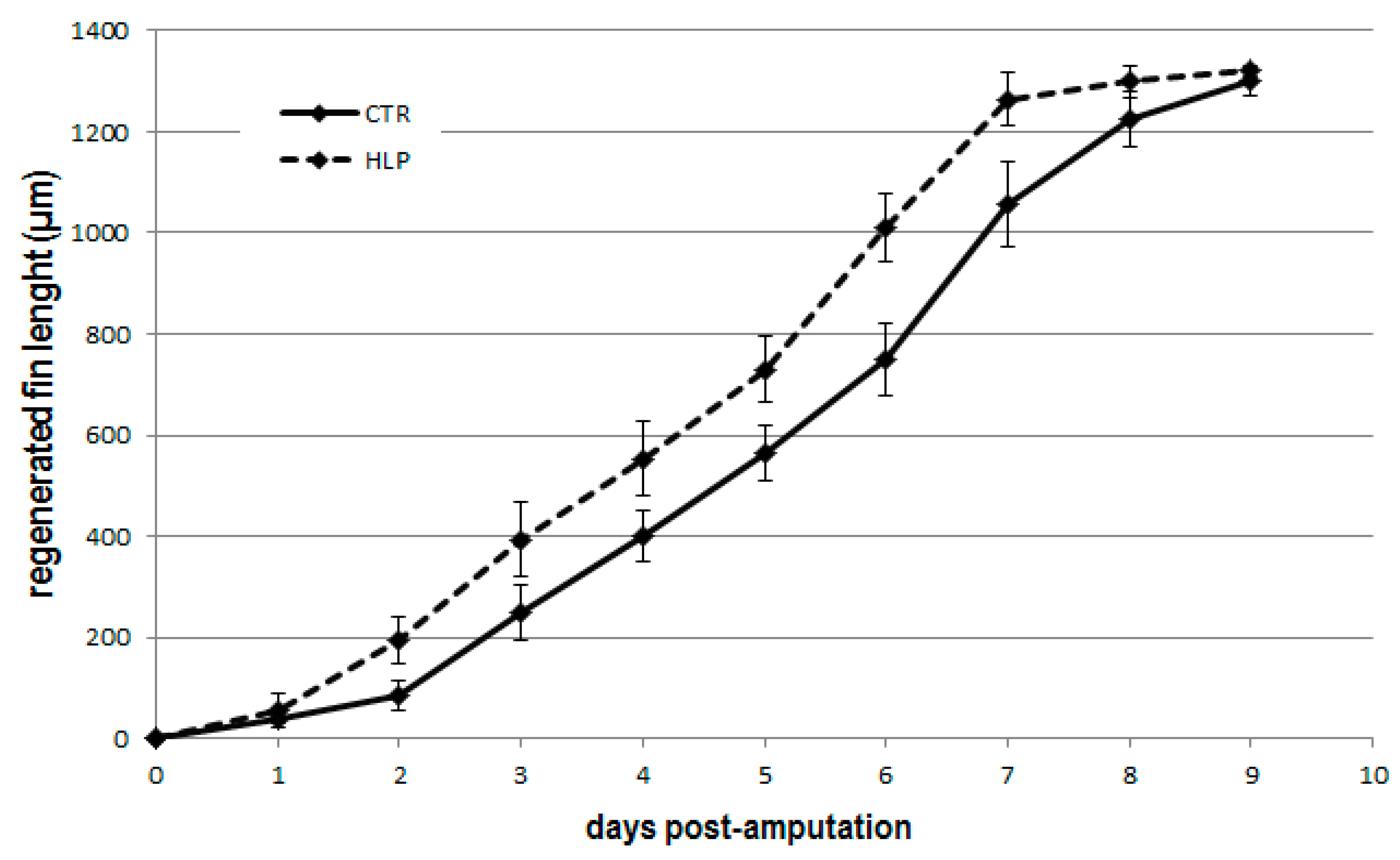
The whole growth curve, calculated until the end of the regeneration process (10 days), indicated a stimulatory effect of HI LF-PEMFs respect of untreated controls (Figure 6). In fact, the peak of regeneration is achieved before and faster in the treated group compared to the control group (8 days vs. 10 days).
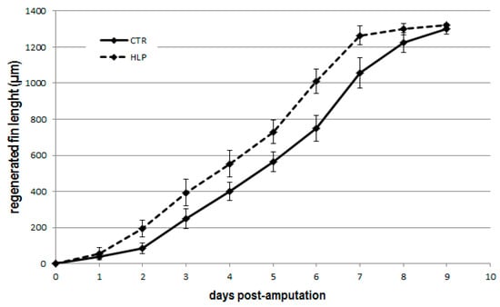
Figure 6.
Daily measurements of regenerated fin length in untreated (CTR, 7 fish) and treated with HI LF-PEMFs (HLP, 7 fish) adult zebrafish.
4. Discussion
To our knowledge, this is the first experimental study about the outcome of HI LF-PEMFs stimulation on bone tissue. On the contrary, the effects of low-intensity PEMFs stimulation on bone have been extensively studied, even though the mechanisms of action of this procedure still remain partially unknown and the quantification of the effects on bone is difficult to evaluate. Nevertheless, the positive outcome of PEMFs on osteoblast activity and differentiation have been demonstrated in vivo on animal models and in human clinical studies [6]. Owing to this, many LF-PEMFs devices for clinical use have been put on the market in recent years. However, clinical protocols for the use of these devices in the treatment of different bone conditions are still based more on anecdotal experience than on sound experimental data. Preclinical studies on simple animal models, as in this case, might contribute to screen and identify more effective protocols to be applied in the human clinical setting. Our results showed a positive, significant anabolic effect on regenerating zebrafish fin. The effect is minimal for the area but more evident in the regenerated fin length. The application of a strong magnetic field should increase the movement of intra- and extra-cellular liquids, facilitating transmembrane molecules and ions transport, enzyme-substrates interactions, and cellular metabolism as a whole. These actions are expected to promote trophic and beneficial effects in tissues. By literature, the modulation of Wnt/β-catenin, ALP, osterix, osteocalcin, and BMP signalling pathways may play a crucial role in physical stimulation of bone regeneration in zebrafish fin [20] and in other animal models [5,6]. In addition, it has been reported that signal transduction through A2A and A3 adenosine receptors stimulate the synthesis of ECM components [21]. Since these molecules and pathways are well conserved in zebrafish, we hypothesize that PEMF-mediated fin regeneration could be stimulated troughs the same mechanisms. The rationale for analyzing only the three longest regenerated fin rays’ is due to the attempt to reduce the fish-to-fish anatomic variability. Although this is a preliminary report, we can observe positive results deriving from the diamagnetic effect while interesting speculations can be made also from the technical point of view. If we do a comparison with other experimental studies on cells, our experience shows that the magnetic field values of 2.9 mT at the target, with an exposure time of 64 min/2 times each day, have allowed to reach a significative biological effect respect to other conditions that required 9 h of stimulation at 1.5 mT [22] or an exposure time of 24 h at 1.8–3 mT of Magnetic Field values [23].
The major limitation of this study resides in the relatively small sample size. Our purpose is to enlarge it during new future studies conducted on a larger fish population.
5. Conclusions
HI LF-PEMFs treatment were demonstrated to increase the regenerative re-growth potential of the adult zebrafish caudal fin, with no harmful effects. The results identify zebrafish as helpful in vivo model to study new therapeutic protocols for bone health with potential human application. Further studies with a larger number of samples, and possibly, adding gene expression and bone metabolism markers measurement, are required to confirm our data. Moreover, our results may pave the way to comparative studies on the effects of high-intensity versus low-intensity PEMFs on vital bone tissue.
Author Contributions
V.S., M.M. contributed to the ideas, M.C., N.S. contributed to data analysis, data generation and manuscript preparation. V.S. contributed to manuscript revision, project supervision and funding. P.R. and A.G. contributed to data analysis and manuscript revision. All authors have read and agreed to the published version of the manuscript.
Funding
This work was supported and funded by Italian Ministry of Health—“Ricerca Corrente” and Gruppo San Donato Foundation (grant SOYFISH). The APC was funded by IRCCS Orthopedic Institute Galeazzi.
Institutional Review Board Statement
Not applicable.
Informed Consent Statement
Not applicable.
Data Availability Statement
Not applicable.
Conflicts of Interest
The authors declare no conflict of interest.
References
- Bassett, C.A. Fundamental and practical aspects of therapeutic uses of pulsed electromagnetic fields (PEMFs). Crit. Rev. Biomed. Eng. 1989, 17, 451–529. [Google Scholar]
- Luben, R. Effects of low-energy electromagnetic fields (pulsed and DC) on membrane signal transduction processes in biological systems. Health Phys. Soc. 1991, 61, 15–28. [Google Scholar] [CrossRef]
- Ehnert, S.; Van Griensven, M.; Unger, M.; Scheffler, H.; Falldorf, K.; Fentz, A.K.; Seeliger, C.; Schröter, S.; Nussler, A.K.; Balmayor, E.R. Co-Culture with Human Osteoblasts and Exposure to Extremely Low Frequency Pulsed Electromagnetic Fields Improve Osteogenic Differentiation of Human Adipose-Derived Mesenchymal Stem Cells. Int. J. Mol. Sci. 2018, 19, 994. [Google Scholar] [CrossRef] [PubMed] [Green Version]
- Chang, W.H.; Chen, L.; Sun, J.; Lin, F. Effect of pulse-burst electromagnetic field stimulation on osteoblast cell activities. Bioelectromagnetics 2004, 25, 457–465. [Google Scholar] [CrossRef] [PubMed]
- Ehnert, S.; Schröter, S.; Aspera-Werz, R.H.; Eisler, W.; Falldorf, K.; Ronniger, M.; Nussler, A.K. Translational Insights into Extremely Low Frequency Pulsed Electromagnetic Fields (ELF-PEMFs) for Bone Regeneration after Trauma and Orthopedic Surgery. J. Clin. Med. 2019, 8, 2028. [Google Scholar] [CrossRef] [PubMed] [Green Version]
- Yuan, J.; Xin, F.; Jiang, W. Underlying Signaling Pathways and Therapeutic Applications of Pulsed Electromagnetic Fields in Bone Repair. Cell Physiol. Biochem. 2018, 46, 1581–1594. [Google Scholar] [CrossRef] [PubMed]
- Novickij, V.; Grainys, A.; Kučinskaite-Kodze, I.; Žvirbliene, A.; Novickij, J. Magneto-permeabilization of viable cell membrane using high pulsed magnetic field. IEEE Trans. Magn. 2015, 51, 5000505. [Google Scholar] [CrossRef]
- Kranjc, S.; Kranjc, M.; Scancar, J.; Jelenc, J.; Sersa, G.; Miklavcic, D. Electrochemotherapy by pulsed electromagnetic field treatment (PEMF) in mouse melanoma B16F10 in vivo. Radiol. Oncol. 2016, 50, 39–48. [Google Scholar] [CrossRef] [PubMed] [Green Version]
- Miklavcic, D.; Novickij, V.; Kranjc, M.; Polajzer, T.; Meglic, S.H.; Napotnik, T.B.; Romih, R.; Lisjak, D. Contactless electroporation induced by high intensity pulsed electromagnetic fields via distributed nanoelectrodes. Bioelectrochemistry 2020, 132, 107440. [Google Scholar] [CrossRef]
- Novickij, V.; Dermol, J.; Grainys, A.; Kranjc, M.; Miklavčič, D. Membrane permeabilization of mammalian cells using bursts of high magnetic field pulses. PeerJ 2017, 4, e3267. [Google Scholar] [CrossRef] [PubMed] [Green Version]
- Towhidi, L.; Firoozabadi, S.M.P.; Mozdarani, H.; Miklavcic, D. Lucifer Yellow uptake by CHO cells exposed to magnetic and electric pulses. Radiol. Oncol. 2012, 46, 119–125. [Google Scholar] [CrossRef] [PubMed] [Green Version]
- Premi, E.; Benussi, A.; La Gatta, A.; Visconti, S.; Costa, A.; Gilberti, N.; Cantoni, V.; Padovani, A.; Borroni, B.; Magoni, M. Modulation of long-term potentiation-like cortical plasticity in the healthy brain with low frequency-pulsed electromagnetic fields. BMC Neurosci. 2018, 19, 34. [Google Scholar] [CrossRef] [PubMed]
- Hammer, B.E.; Kidder, L.S.; Williams, P.C.; Xu, W.W. Magnetic Levitation of MC3T3 Osteoblast Cells as a Ground-Based Simulation of Microgravity. Microgravity Sci. Technol. 2009, 21, 311–318. [Google Scholar] [CrossRef] [Green Version]
- Zhadin, M.N. Combined action of static and alternating magnetic fields on ion motion in a macromolecule: Theoretical aspects. Bioelectromagnetics 1998, 19, 279–292. [Google Scholar] [CrossRef]
- Zablotskii, V.; Polyakova, T.; Lunov, O.; Dejneka, A. How a High-Gradient Magnetic Field Could Affect Cell Life. Sci. Rep. 2017, 6, 37407. [Google Scholar] [CrossRef] [PubMed]
- Renn, J.; Winkler, C.; Schartl, M.; Fischer, R.; Goerlich, R. Zebrafish and medaka as models for bone research including implications regarding space-related issues. Protoplasma 2006, 229, 209–214. [Google Scholar] [CrossRef] [PubMed]
- Uemoto, T.; Abe, G.; Tamura, K. Regrowth of zebrafish caudal fin regeneration is determined by the amputated length. Sci. Rep. 2020, 10, 649. [Google Scholar] [CrossRef] [Green Version]
- Kim, Y.M.; Lim, H.M.; Ro, H.S.; Ki, G.E.; Seo, Y.K. Pulsed Electromagnetic Fields Increase Pigmentation through the p-ERK/p-p38 Pathway in Zebrafish (Danio rerio). Int. J. Mol. Sci. 2018, 19, 3211. [Google Scholar] [CrossRef] [Green Version]
- Westerfield, M. The Zebrafish Book: A Guide for the Laboratory Use of Zebrafish (Danio rerio), 5th ed.; University of Oregon Press: Eugene, OR, USA, 2007. [Google Scholar]
- Molagoda, I.M.N.; Karunarathne, W.A.H.M.; Choi, Y.H.; Park, E.K.; Jeon, Y.J.; Lee, B.J.; Kang, C.H.; Kim, G.Y. Fermented Oyster Extract Promotes Osteoblast Differentiation by Activating the Wnt/β-Catenin Signaling Pathway, Leading to Bone Formation. Biomolecules 2019, 9, 711. [Google Scholar] [CrossRef] [Green Version]
- Cadossi, R.; Massari, L.; Racine-Avila, J.; Aaron, R.K. Pulsed Electromagnetic Field Stimulation of Bone Healing and Joint Preservation: Cellular Mechanisms of Skeletal Response. J. Am. Acad. Orthop. Surg. Glob. Res. Rev. 2020, 4, e1900155. [Google Scholar] [CrossRef]
- Lin, H.Y.; Lin, Y.J. In vitro effects of low frequency electromagnetic fields on osteoblast proliferation and maturation in an inflammatory environment. Bioelectromagnetics 2011, 32, 552–560. [Google Scholar] [CrossRef] [PubMed]
- Chang, C.H.; Loo, S.T.; Liu, H.L.; Fang, H.W.; Lin, H.Y. Can low frequency electromagnetic field help cartilage tissue engineering? J. Biomed. Mater. Res. A 2010, 92, 843–851. [Google Scholar] [CrossRef] [PubMed]
Publisher’s Note: MDPI stays neutral with regard to jurisdictional claims in published maps and institutional affiliations. |
© 2022 by the authors. Licensee MDPI, Basel, Switzerland. This article is an open access article distributed under the terms and conditions of the Creative Commons Attribution (CC BY) license (https://creativecommons.org/licenses/by/4.0/).