Article

Antiproliferative Activity of Aminobenzylnaphthols Deriving from the Betti Reaction

by
Rosanna Mallamaci
1,
Maria Annunziata M. Capozzi
2,* and
Cosimo Cardellicchio
3
1
Dipartimento di Bioscienze, Biotecnologie e Biofarmaceutica, Università di Bari, Via Orabona 4, 70125 Bari, Italy
2
Dipartimento di Chimica, Università di Bari, Via Orabona 4, 70125 Bari, Italy
3
CNR ICCOM, Dipartimento di Chimica, Università di Bari, Via Orabona 4, 70125 Bari, Italy
*
Author to whom correspondence should be addressed.
Appl. Sci. 2022, 12(15), 7779; https://doi.org/10.3390/app12157779
Submission received: 19 July 2022 / Revised: 29 July 2022 / Accepted: 30 July 2022 / Published: 2 August 2022

Abstract

:
Two aminobenzylnaphthols, which are representative items of the family of compounds synthesized with the Betti reaction, were investigated as antiproliferative agents against adenocarcinoma human colorectal (Caco-2) and human neuroblastoma (SH-SY5Y) cell lines, using cisplatin as a positive control. A better antiproliferative activity was recorded after 24 h of incubation for the first tested molecule, whereas the other one was more effective after 72 h of incubation. These results support the hypothesis that both of the tested aminobenzylnaphthols could potentially be endowed with a biological activity.

Graphical Abstract

1. Introduction

A malignant tumor, also called cancer, is among the most difficult diseases to cure [1]. Surgery, radiation, and chemotherapy are the three main treatments for cancer to date. Among these therapeutic strategies, [(cis-Diamine-dichloroplatin (II), or more commonly “cisplatin”] is one of the most used chemotherapeutic agents, since it is employed in the treatment of the testicular, ovarian, lung, head and neck, upper gastrointestinal tract, and other types of solid cancer. Cisplatin moves rapidly into the blood and has a very short retention time in the tumor [2]. Cisplatin’s mechanism of action is represented by the formation of DNA adducts blocking fundamental processes such as transcription and replication, leading to cell death [3]. However, the long-term use of the drug leads to the development of resistance, which is related to three main molecular mechanisms: DNA repair, cellular adaptations, and drug inactivation [4].
Next-generation platinum complexes have been developed to increase anticancer efficacy and minimize side effects, thus permitting higher doses to be employed [5]. In the investigations on new chemotherapeutic agents, SH-SY5Y human neuroblastoma cells have been used as a model to compare cisplatin toxicity to that of other platinum derivatives. For example, cisplatin induced apoptotic death in undifferentiated cells of SH-SY5Y human neuroblastoma [6]. SH-SY5Y cells have been employed as a model to compare cisplatin toxicity to that of other platinum derivatives, such as oxaliplatin [oxalate (trans-1,2-diamminocyclohexane) platinum (II), OHP] [7].
As far as new metal complexes tested as antiproliferative agents, cadmium complexes were tested in many cancer cell lines, e.g., MCF-7 (breast), Caco-2 (colorectal), and cisplatin-resistant lung cancer cell lines. The in vitro cytotoxicity of innovative vanadium complexes with orotic and glutamic acids were evaluated against hepatocellular carcinoma and human colorectal adenocarcinoma (Caco-2) [8].
In order to find new synthetic compounds to be employed as chemotherapeutic agents, we tested some aminobenzylnaphthols that had been synthesized with the Betti reaction [9]. This process is a multi-component reaction between 2-naphthol, aryl aldehydes, and amines that yields the cited aminobenzylnaphthols. Since its discovery in the early 20th century, the Betti reaction was seldom applied until its rediscovery due to our research, starting in 1998 [10] and continuing over the following years [9,10,11,12,13,14,15,16,17,18]. Since 1999, many researchers around the world have applied the Betti reactrion [9]. However, aminobenzylnaphthols have seldom been investigated as bioactive compounds during the last two decades (1998–2018) [18,19,20].
During the last few years (2019–2022), a new interest in the Betti reaction has emerged. A recent review [21] reported new green routes to accomplish this process, new applications of the products obtained so far, and the evaluation of their bio-activity.
To limit the investigations to only antitumor agents, Puerta et al. synthesized aminobenzylnaphthols with new amines or N-heterocycles using the Betti protocol [22]. These new intermediates were tested against some solid tumor cell lines, using cisplatin as a standard. Some of these molecules showed antiproliferative values beyond the GI50 <10 mM threshold, a value that is promising for future clinical tests. The authors speculated that this interesting activity could be due to the tryptophan mimetic of the Betti bases that is able to inhibit cell proliferation.
This hypothesis stimulated new investigations about the employment of Betti base frameworks as antitumor agents. Yellapurkar et al. synthesized and tested thiophene containing aminobenzylnaphthols [23] against four tumor cell lines (lung, prostate, breast, and liver). The in vitro results are comparable with those obtained with the standard drug doxorubicin tested against these cell lines.
Molnár et al. synthesized 2-aminomethylated estrone and estradiol derivatives with the Betti protocol [24] and tested them against gynecological tumor cell lines. They showed an antiproliferative activity that is better than or comparable with the original estrone or estradiol. However, the tested compounds had a better water solubility in comparison with the starting materials, thus emerging as a new potential clinical candidate.
Dimìc et al. prepared four new compounds starting with 4-hydroxycoumarin and using tyramine, octopamine, norepinephrine, or 3-methoxytyramine as the amine reagent in the Betti reaction [25]. In vitro cytotoxicity experiments were performed against the cell lines of adenocarcinoma, human colon and breast cancer, and, finally, against lung fibroblast lines. A significant selectivity towards colorectal carcinoma cells was observed [25].
New molecules containing both the benzothiazole and the pyrazole moieties were synthesized and evaluated for their cytotoxic activity against some human cell lines. Their action was connected to DNA binding, causing the inhibition of topoisomerase I [26].
The inhibition of YAP-TEAD complexes is a new chemotherapeutic strategy [27]. New compounds obtained with the Betti reaction were investigated to implement this strategy [28].
Finally, phthalocyanines modified with Betti bases were employed in DNA cleavage experiments as a preliminary test for future clinical applications [29], and interesting result were recorded.
A recent interest has emerged in the employment of amino acid derivatives as prodrugs [30]. The functionalization of a drug with an amino acid residue results in some positive outcomes, such as the improvement of the delivery of the drug to the target tissue, and the lowering of its toxicity. For example, boroxazolidones were reported to have modest antitumor properties; however, the L-valine derivatives showed strong cytotoxic effects [31]. Along the same lines, L-valine modified dimethyl-curcumin showed a great increase in anti-proliferative activity compared to the original drug, thus distinguishing itself as a potent anticancer agent [32].
At this stage, we chose molecules 1 [12] and 2 [14,15] represented in Figure 1. Molecule 1 is a prototypal aminobenzylnaphthol obtained with the Betti reaction; this type of molecule has not been tested as an antiproliferative agent. Moreover, molecule 2 is the simplest aminobenzylnaphthol, in which the valine residue is present, an opportunity that deserves to be investigated, considering the results obtained in valine containing prodrugs. We decided to test these compounds against the already cited Caco-2 adenocarcinoma and SH-SY5Y neuroblastoma cells chosen as representative test cell lines. Cisplatin was used as a standard in these tests.

2. Materials and Methods

2.1. Cell Cultures

Human Caco-2 adenocarcinoma and SH-SY5Y neuroblastoma lines were grown in high glucose (4.5 g L−1) Dulbecco’s Modified Eagle Medium (DMEM), reinforced with 10% fetal bovine serum (FBS, PAN Biotech, Aidenbach, Germany), 4 mM L-glutamine, penicillin, and streptomycin. Cell incubation was maintained at 37 °C (Thermo Scientific Hera Cell 240i, Waltham, MA< USA) with the atmosphere having 5% CO2 and 95% humidity.

2.2. Preparation of 1, 2, and Cisplatin Solutions

Aminobenzylnaphthol 1 and 2 were synthesized through a 2 day reaction at 60 °C without any solvent benzaldehyde, 2-naphthol, and(S)-1-phenylethylamine for compound 1 [12], or with (S)-valine methyl ester for compound 2 [14,15]. Molecule 1 was purified by crystallization [12], whereas molecule 2 was purified by chromatography followed by crystallization [14,15].
Cisplatin is commercially available.
The stock solutions were prepared by dissolving the appropriate amount of 1, 2 and cisplatin in 10 mL of DMSO under stirring for 10 min to achieve a 0.1 M concentration. The employed solutions were obtained from the stock solutions by scalar dilution with bidistilled sterile water. These solutions were maintained in the refrigerator at 4 °C.

2.3. Study Design

The Caco-2 and SH-SY5Y tumor cells lines were treated with increasing concentrations of 1 and 2 with cisplatin as a positive control. Untreated cells were considered control lines.

2.4. In Vitro Cytotoxicity Test

2.4.1. Short-Term Studies: MTT Assay and Trypan Blue Exclusion Assay

The Caco-2 and SH-SY5Y cells, after being 80% confluent, were seeded in 96-well plates at a density of 5000 cells/well in 125 mL of cell culture medium and incubated overnight.
The day after, the medium was replaced with fresh medium containing 1 and 2 solutions at final concentrations of 200, 100, 50, and 25μM for 24 and 72 h. Wells treated with only culture medium were used as a control. At the end of the exposure period, the cell viability was verified with 0.5 mg/mL MTT (3-(4,5-dimethylthiazol-2-yl)-2,5-diphenyltetrazolium bromide). The plates were incubated for 3 h in a humidified atmosphere of 95% O2 and 5% CO2. Subsequently, the MTT reagent was decanted, and the formed formazan crystals were dissolved in dimethyl sulfoxide (DMSO). The optical density (OD) of each well was spectrophotometrically quantified at 550 nm using the Bio-Rad microplate reader. Cell viability in response to treatment was calculated as a percentage of control cells treated with culture medium only. Cell Viability % = (ODtreatedcells)/(ODcontrolcells) × 100.

2.4.2. Trypan Blue Assay

A solution was prepared in PBS at a concentration of 0.4 g/mL and was then added with a 1:1 ratio to the in vitro grown mass of Caco-2 and SH-SY5Y cells for the study of membrane integrity; the mixture was kept in an incubator for 2 min at 37 °C, and cells in triplicates were evaluated in a Bürker chamber using light microscopy. The live cells remained non-stained, whereas the nuclei of dead cells appeared blue, as Trypan blue is a membrane permeable dye that enters dead cells and stains the nuclei blue. Live and dead cells were counted for each sample, with dead cells being discriminated by the incorporation of Trypan blue. The numbers of non-stained viable cells (NSt cells) and stained dead cells (St cells) were counted using a hemocytometer. Cell viability was then calculated using the following formula: Viability % = (NSt cells)/(St cells + NSt cells) × 100 [33].

2.4.3. Long-Term Studies: Clonogenic Assay

The clonogenic assay was conducted according to the literature [34], i.e., cells were seeded in twelve-well plates at a density of 200 cells/well with 1 mL volume/well. After a three-hour delay, the cells were washed with 2 mL of phosphate buffer saline (PBS) and then treated with solutions 1 and 2, achieving final concentrations of 200, 100, 50, and 25 μM over a time period required to form colonies (8 days). After this time, the medium was removed, and the colonies were fixed, stained, and counted for the evaluation of cell survival after exposure to the molecules under investigation. Control cells were treated with culture medium only. Each well is pictured. The colony-forming capacity was calculated by dividing the number of colonies that were obtained by the number of cells that were seeded. The surviving fraction (number of colonies that arose after exposure to the molecules under investigation) is the ratio of colony-forming treated cells to the colony-forming untreated controls [35].

2.5. Statistic Treatments

Data are shown as mean ± SEM. Statistical comparisons were performed by one-way ANOVA followed by multiple comparison tests (Dunnett’s test) using the statistical package in the GraphPad Prism software vers.5.01 (GraphPad Software, Inc., San Diego, CA, USA); values of p < 0.05 were considered statistically significant.

3. Results and Discussion

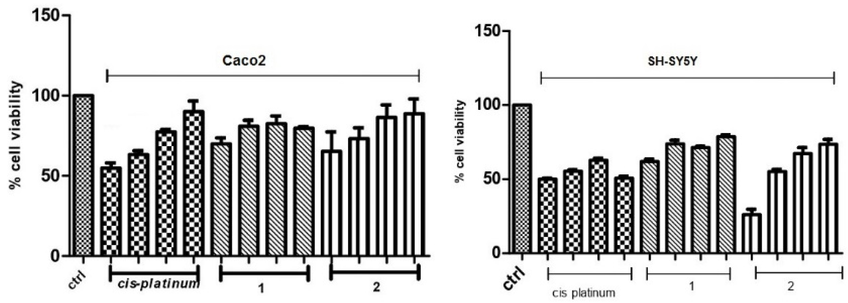
3.1. Cytotoxicity of 1 and 2 in Human Caco-2 Adenocarcinoma and SH-SY5Y Neuroblastoma Cells

Short (24 and 72 h) and prolonged (7 or 10 days) treatments of Caco-2 and SH-SY5Y cells with decreasing concentrations (200, 100, 50, and 25 μM) of 1 and 2 were used to establish the in vitro intrinsic cytotoxicity. Mitochondrial function, membrane integrity, and the capacity to form colonies were examined as final points. Untreated cells were processed and incubated in the same way in culture media and at the same time as the treated groups.

3.2. Mitochondrial Function

Mitochondrial functions were evaluated using MTT assays after a 24 and 72 h treatment with 1 and 2 at decreasing concentrations (200, 100, 50, and 25 μM). They are reported as the percentage of the viability of each untreated cell. The results are represented in Figure 2 and Figure 3.
After 24 h of exposure, 1 and 2 (200, 100, 50, and 25 μM) of Caco-2 and SH-SY5Y cells showed comparable linear tendencies of dose-dependent effects of cytotoxicity.
Concentrations of 1 from 25 to 200 μM were related with a reduction of about 80–70% and 79–62% in cell viability for Caco-2 and SH-SY5Y tumor cell lines, respectively.
On the other hand, the treatment of 2 with similar concentrations showed a reduction of about 89–65% and 73–26% in cell viability for Caco-2 and SH-SY5Y cells, respectively, thus showing a dependence on the type of employed tumor cell lines.
After a 72h treatment, the trend of cytotoxicity was analogous to the figures reported after 24 h for 1 and SH-SY5Y cells. Concentrations of molecule 1 from 25 to 200 μM were related with a reduction of about 76–15% in cell viability for Caco-2 and 80–64% for SH-SY5Y cells, respectively.
In the case of molecule 2, treatments were related with a reduction of about 84–75% and 91–37% in cell viability for Caco-2 and SH-SY5Y cells, respectively (Figure 3; p < 0.05 Dunnett’s test).
MTT assays were used to calculate cytotoxic effects of cisplatin, as a “positive control” to evaluate its antiproliferative activity on Caco-2 and on SH-SY5Y cells and to evaluate the efficacy of the compounds 1 and 2. As illustrated in Figure 2 and Figure 3, after 24 h of exposure, the cisplatin treatment was associated with a reduction of about 90–55% and 51–50%, whereas, after 72 h of exposure, a reduction of about 62–23% and 77–44% in cell viability was observed for Caco-2 and SH-SY5Y cells, respectively.
Moreover, MTT data were employed to determine EC50 (50% effective concentration) values and to evaluate the toxicity of the compounds 1 and 2 for Caco-2 and SH-SY5Y cell lines.
In summary, the results collected in Table 1 show that the antiproliferative activity towards Caco-2 cell lines at 24 h or 72 h of exposure to compound 2 is slightly better than the action of cisplatin. On the other hand, at 72 h, compound 1 increases its performance. As far as the antiproliferative activity towards SH-SY5Y cell lines is concerned, compound 1 has an antiproliferative activity at 24 h or 72 h, similar to the action of cisplatin.

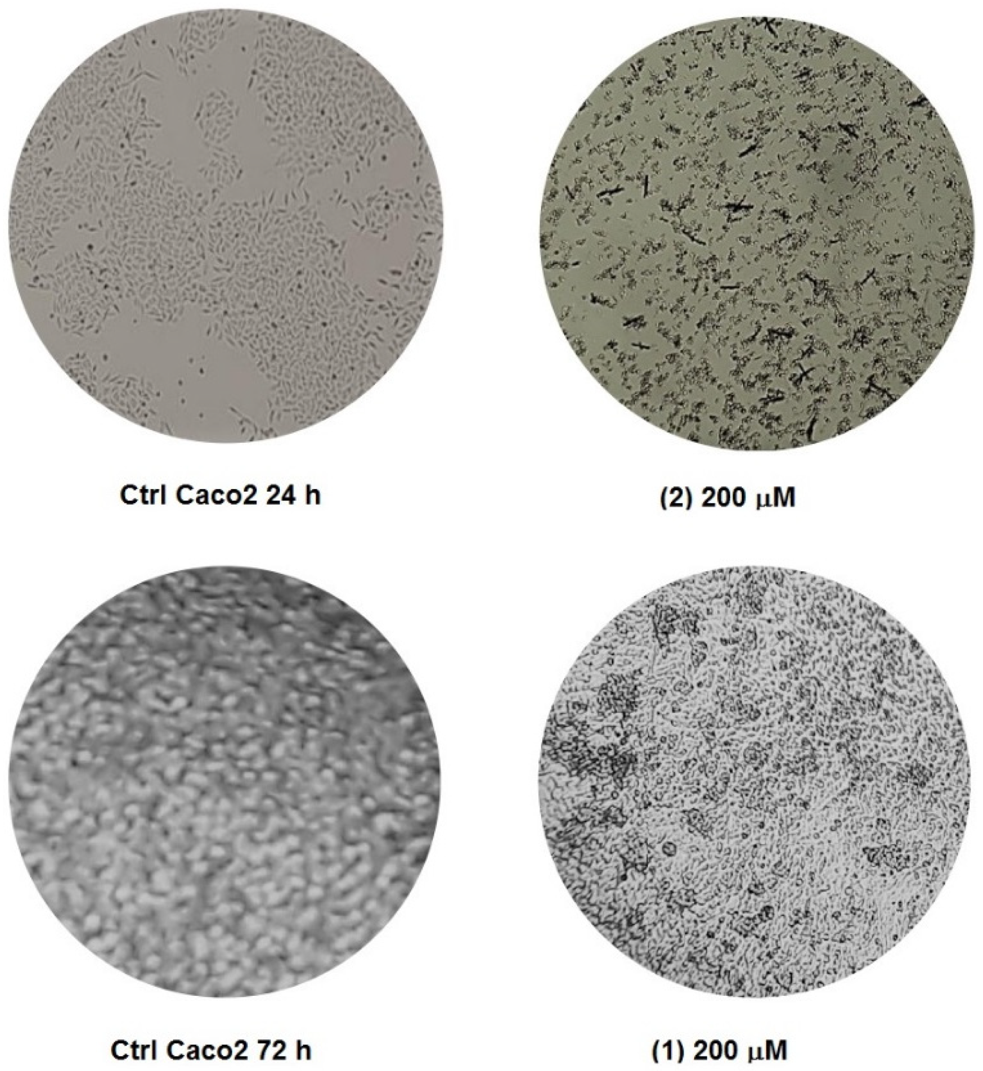
3.3. Membrane Integrity

It is interesting to highlight that the Caco-2 and SH-SY5Y cells showed a dose-dependent decrease in live cells with respect to the control after 24 h of exposure to molecule 1 and molecule 2 (200, 100, 50, and 25 μM). Linear trends of dose-dependent cytotoxic effects were observed. Concentrations of molecule 1 from 25 to 200 μM were associated with a reduction of about 80–56% and 83–64% in the number of live cells for Caco-2 and SH-SY5Y, respectively. In the case of molecule 2, a decrease in the number of live cells of about 79–52% and 77–35% for Caco-2 and SH-SY5Y, respectively, was observed (Figure 4).
After 72 h of exposure to 1 and 2 (200, 100, 50, and 25 μM) of Caco-2 and SH-SY5Y, the results obtained with the MTT assay were confirmed (Figure 5).
Concentrations of 1 ranging from 25 to 200 μM were associated with a significant reduction in the number of live cells of about 74–17% and 86–67% for Caco-2 and SH-SY5Y, respectively, while the 2 treatments were associated with a significant reduction of about 79–77% and 89–39% in number of live cells for Caco-2 and SH-SY5Y, respectively. The results are represented in Figure 6 and Figure 7.

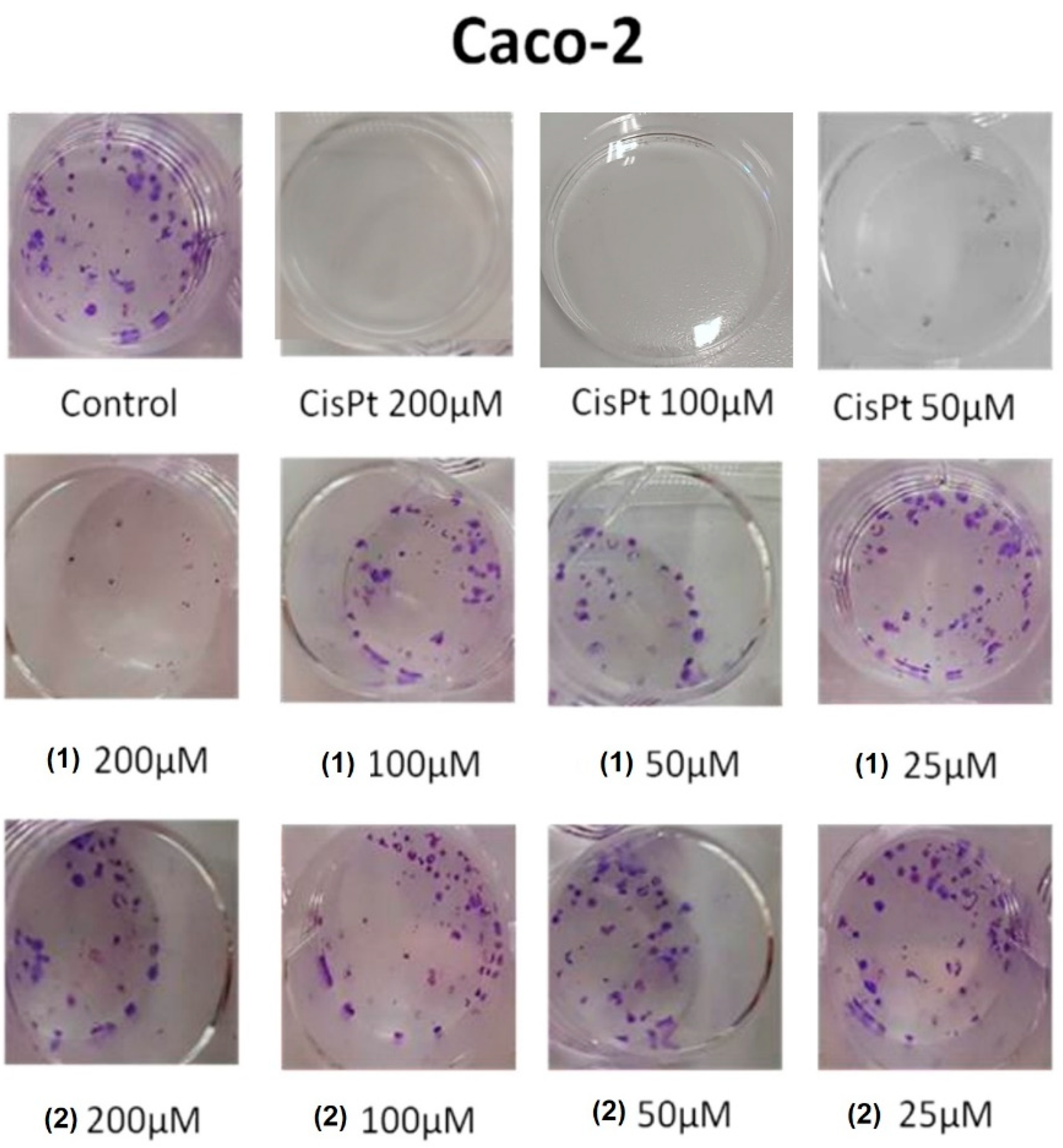
3.4. Colony-Forming Inhibition of Molecules 1 and 2 in Caco-2 Adenocarcinoma and SH-SY5Y Neuroblastoma Cells

Eventually, Caco-2 adenocarcinoma and SH-SY5Y neuroblastoma cells were seeded and monitored to evaluate if molecules 1, 2, and cisplatin might inhibit colony-forming capacity and proliferation ability (colony size). The colonies were detected by Coomassie blue staining as violet spots after 7- or 10-day treatments with the molecules under investigation.
Pictures of randomly selected microscopic fields of each cell line are represented in Figure 8 and Figure 9. Caco-2 and SH-SY5Y controls had homogeneous and similar colony patterns. Decreasing concentrations of molecules 1, 2, and cisplatin (200, 100, 50, and 25 μM) acted on colonies of Caco-2 and SH-SY5Y with a dose-dependent reduction in size and colony number (p < 0.05). In particular, the colony numbers of Caco-2 and SH-SY5Y were reduced by 30% and 47%, respectively, when compared to the control at the highest treatment dose of 200 μM of 1 (Figure 8 and Figure 9). On the other hand, the same concentration of 2 caused a reduction of 50% and 60% in the colony numbers. In this respect, the colonies of SH-SY5Y cells were more affected than those of Caco-2 cells (Figure 8 and Figure 9).

3.5. Mechanistic Considerations

The antiproliferative activity measured so far can be ascribed to the privileged molecular assembly of the aminobenzylnaphthols obtained with the Betti reaction. Even if more investigations are required to arrive at a more definitive conclusion, the comparison with other literature investigations of antiproliferative activities seems to highlight some directions that are peculiar for these structures and can be recognized as potential lines of action.
The first direction is the great ability of coordinating metal cations due to the combined action of the naphthol hydroxyl moieties with the amino residue. The coordinated cations could alter the molecular equilibrium [19], with a focus on the production of ROS (reactive oxygen species) that are connected in many chemotherapeutic actions.
On the other hand, many papers [22,25,26,29] seem to underline the ability of similar molecules to bind and alter the DNA or its action. This last mechanism is probably connected to the presence of heterocyclic moieties that are not present in our molecules.
Thus, we are inclined to think that the anti-proliferative activity that we observed is connected to the first line of action that was reported.

4. Conclusions

In summary, two Betti base derivatives were evaluated for their inhibitory activities as anticancer agents against human Caco-2 adenocarcinoma cells and SH-SY5Y neuroblastoma cells. The evaluation of anticancer activity towards the two cell lines of these aminobenzylnaphthols was accomplished through different procedures, i.e., MTT assays, membrane integrity tests, and colony-forming capacity. The main differences are bound to the different times in which these molecules (1 and 2) act; in fact, 2 is most effective towards Caco-2 cells lines for the 24 h treatment. The 8-day exposure to the tested molecules also showed a promising reduction in the formation of colonies.
The prepared compounds displayed moderate cytotoxic activity against both cancer cell lines and relatively lower inhibitory activity against the normal cell lines. In particular, compound 2 exhibited a good antiproliferative activity similar to cisplatin against Caco-2 adenocarcinoma cells, with EC50 values of 94 after 24 h and 83 after 72 h vs. cisplatin with values of 97 and 88 µM, respectively. Conversely, as far as the antiproliferative activity towards SH-SY5Y cell lines is concerned, compound 1 has an activity at 24 h or 72 h that is similar to the action of cisplatin, while compound 2 has an activity that ranges from 182 to 93 from 24 h to 72 h.
These data are a valid starting point for the evaluation of the antiproliferative activity of intermediates derived from a Betti reaction, since the investigated compounds represent the prototypal aminobenzylnaphthol (the first compound), and the aminobenzylnaphthol in which the valine residue is present (the second compound). From this perspective, the present results obtained with the simplest forms of Betti bases must be considered when more complex molecular structures should be investigated. Moreover, even if the selected tumor cell lines are well-represented, it could be envisaged that a larger panel of tumor cell lines should be tested. It must be underlined that these assays agree in showing an interesting antiproliferative activity, similar to the action of the standard cisplatin towards the same cell lines, thus proposing that they are a less toxic alternative.

Author Contributions

Synthesis of the molecules, C.C. and M.A.M.C.; biological tests, R.M.; writing—review and editing, R.M., C.C. and M.A.M.C. All authors have read and agreed to the published version of the manuscript.

Funding

This research received no external funding.

Institutional Review Board Statement

Not applicable.

Informed Consent Statement

Not applicable.

Acknowledgments

Vittoria Petrera is gratefully acknowledged for some preliminary results.

Conflicts of Interest

The authors declare no conflict of interest.

References

  1. Duffy, M.J. The war on cancer: Are we winning? Tumor Biol. 2013, 34, 1275–1284. [Google Scholar] [CrossRef] [PubMed]
  2. Chen, S.H.; Chang, J.Y. New Insights into Mechanisms of Cisplatin Resistance: From Tumor Cell to Microenvironment. Int. J. Mol. Sci. 2019, 20, 4136. [Google Scholar] [CrossRef] [PubMed] [Green Version]
  3. Fatemeh, S.J.; Mostafa, S. Different profiles of the mRNA levels of DNA repair genes in MCF-7 and SH-SY5Y cells after treatment with combination of cisplatin, 50-Hz electromagnetic field and bleomycin. Biomed. Pharmacother. 2017, 94, 564–568. [Google Scholar] [CrossRef]
  4. Rabik, C.A.; Dolan, M.E. Molecular mechanisms of resistance and toxicity associated with platinating agents. Cancer Treat. Rev. 2007, 33, 9–23. [Google Scholar] [CrossRef] [PubMed] [Green Version]
  5. Culy, C.R.; Clemett, D.; Wiseman, L.R. Oxaliplatin. A review of its pharmacological properties and clinical efficacy in metastatic colorectal cancer and its potential in other malignancies. Drugs 2000, 60, 895–924. [Google Scholar] [CrossRef] [PubMed]
  6. Sun, Y.X.; Jian, Y.; Wang, P.Y.; Li, Y.J.; Xies, S.Y.; Sun, R.P. Cisplatin regulates SH-SY5Y cell growth through downregulation of BDNF via miR-16. Oncol. Rep. 2013, 30, 2343–2349. [Google Scholar] [CrossRef] [PubMed] [Green Version]
  7. Donzelli, E.; Carfì, M.; Miloso, M.; Strada, A.; Galbiati, S.; Bayssas, M.; Griffon-Etienne, G.; Cavaletti, G.; Petruccioli, M.G.; Tredici, G. Neurotoxicity of platinum compounds: Comparison of the effects of cisplatin and oxaliplatin on the human neuroblastoma cell line SH-SY5Y. J. Neurooncol. 2004, 67, 65–73. [Google Scholar] [CrossRef]
  8. Boccato, P.F.; Aleixo, N.A.; Nogueira, A.R.F.; Massabni, A.C. In vitro Studies of Antitumor Activity of Vanadium Complexes with Orotic and Glutamic Acids. Rev. Bras. Cancerol. 2020, 66, 4649. [Google Scholar] [CrossRef]
  9. Cardellicchio, C.; Capozzi, M.A.M.; Naso, F. The Betti base: The awakening of a sleeping beauty. Tetrahedron Asymmetry 2010, 21, 507–517. [Google Scholar] [CrossRef]
  10. Cardellicchio, C.; Ciccarella, G.F.; Schingaro, E.; Scordari, F. The Betti base: Absolute configuration and routes to a family of related chiral nonracemic bases. Tetrahedron Asymmetry 1998, 9, 3667–3675. [Google Scholar] [CrossRef]
  11. Cardellicchio, C.; Ciccarella, G.; Naso, F.; Perna, F.; Tortorella, P. Use of readily available chiral compounds related to the Betti base in the enantioselective addition of diethylzinc to aryl aldehydes. Tetrahedron 1999, 55, 14685–14692. [Google Scholar] [CrossRef]
  12. Cardellicchio, C.; Capozzi, M.A.M.; Alvarez-Larena, A.; Piniella, J.F.; Capitelli, F. Investigation on the weak interactions assembling the crystal structures of Betti bases. CrystEngComm 2012, 14, 3972–3981. [Google Scholar] [CrossRef]
  13. Capozzi, M.A.M.; Terraneo, G.; Cardellicchio, C. Structural insights into methyl- or methoxy-substituted 1-(a-aminobenzyl)-2-naphthol structures: The role of C-H···p interactions. Acta Cryst. 2019, C75, 189–195. [Google Scholar] [CrossRef]
  14. Capozzi, M.A.M.; Cardellicchio, C. Stereoselection in the Betti reaction of valine methyl esters. Tetrahedron Asymmetry 2017, 28, 1792–1796. [Google Scholar] [CrossRef]
  15. Capozzi, M.A.M.; Alvarez-Larena, A.; Piniella Febrer, J.F.; Cardellicchio, C. A combined structural and computational investigation of aminobenzylnaphthol compounds derived from the Betti reaction using valine methyl ester. New J. Chem. 2021, 45, 20735–20742. [Google Scholar] [CrossRef]
  16. Capozzi, M.A.M.; Capitelli, F.; Bottoni, A.; Calvaresi, M.; Cardellicchio, C. Stacked Naphthyls and Weak Hydrogen-Bond Interactions Govern the Conformational Behavior of P-Resolved Cyclic Phosphonamides: A Combined Experimental and Computational Study. J. Org. Chem. 2014, 79, 11101–11109. [Google Scholar] [CrossRef]
  17. Capozzi, M.A.M.; Pigliacelli, C.; Terraneo, G.; Cardellicchio, C. Stacked aryl groups in P-resolved cyclic phosphonamides as a new conformational constraint. CrystEngComm 2019, 21, 7224–7232. [Google Scholar] [CrossRef]
  18. Capozzi, M.A.M.; Cardellicchio, C.; Magaletti, A.; Bevilacqua, A.; Perricone, M.; Corbo, M.R. Bioactivity of a Family of Chiral Nonracemic Aminobenzylnaphthols towards Candida albicans. Molecules 2014, 19, 5219–5230. [Google Scholar] [CrossRef] [Green Version]
  19. Georgieva, N.V.; Yaneva, Z.L.; Simova, S.D.; Nikolova, G.D. Synthesis and properties of several Betti bases as potential drugs. Bulgar. Chem. Comm. 2017, 49, 201–208. [Google Scholar]
  20. Gyémánt, N.V.; Engi, H.; Schelz, Z.; Szatmári, I.; Tóth, D.; Fülöp, F.; Molnár, J.; de Witte, P.A.M. In vitro and in vivo multidrug resistance reversal activity by a Betti-base derivative of tylosin. Brit. J. Cancer 2010, 103, 178–185. [Google Scholar] [CrossRef] [Green Version]
  21. Iftikhar, R.; Kamran, M.; Iftikhar, A.; Parveen, S.; Naeem, N.; Jamil, N. Recent Advances in the green synthesis of Betti bases and their application: A review. Mol. Divers. 2022, 1–27. [Google Scholar] [CrossRef]
  22. Puerta, A.; Galán, A.R.; Abdilla, R.; Demanuele, K.; Fernandes, M.X.; Bosica, G.; Padrón, J.M. Naphthol-derived Betti bases as a potential SLC6A14 blockers. J. Mol. Clin. Med. 2019, 2, 35–40. [Google Scholar] [CrossRef]
  23. Yellapurkar, I.; Shaikh, S.; Pavale, G.; Bhabal, S.; Ramana, M.M.V. Kaolin-catalysed one-pot synthesis of thiophene containing aminonaphthols under solvent-free condition and their in vitro anticancer and antioxidant activity. Res. Chem. Interm. 2021, 47, 4067–4082. [Google Scholar] [CrossRef]
  24. Molnár, B.; Kinyua, N.I.; Mótyán, G.; Leits, P.; Zupkó, I.; Minorics, R.; Balogh, G.T.; Frank, E. Regioselective synthesis, physicochemical properties and anticancer activity of 2-aminomethylated estrone derivatives. J. Ster. Biochem. Mol. Biol. 2022, 219, 106064. [Google Scholar] [CrossRef]
  25. Dimić, D.S.; Kaluđerović, G.N.; Avdović, E.H.; Milenković, D.A.; Živanović, M.N.; Potocnak, I.; Samol’ova, E.; Dimitrijević, M.S.; Saso, L.; Marković, Z.S.; et al. Synthesis, Crystallographic, Quantum Chemical, Antitumor, and Molecular Docking/Dynamic Studies of 4-Hydroxycoumarin-Neurotransmitter Derivatives. Int. J. Mol. Sci. 2022, 23, 1001. [Google Scholar] [CrossRef]
  26. Nagaraju, B.; Kovvuri, J.; Kumar, C.G.; Routhu, S.R.; Shareef, M.A.; Kadagathur, M.; Adiyala, P.R.; Alavala, S.; Nagesh, N.; Kamal, A. Synthesis and biological evaluation of pyrazole linked benzothiazole-β-naphthol derivatives as topoisomerase I inhibitors with DNA binding ability. Bioorgan. Med. Chem. 2019, 27, 708–720. [Google Scholar] [CrossRef]
  27. Iftikhar, R.; Zahoor, A.F.; Irfan, M.; Rasul, A.; Rao, F. Synthetic molecules targeting yes associated protein activity as chemotherapeutics against cancer. Chem. Biol. Drug Des. 2021, 98, 1025–1037. [Google Scholar] [CrossRef]
  28. Karatas, H.; Akbarzadeh, M.; Adihou, H.; Hahne, G.; Pobbati, A.V.; Ng, E.Y.; Guéret, S.M.; Sievers, S.; Pahl, A.; Metz, M.; et al. Discovery of Covalent Inhibitors Targeting the Transcriptional Enhanced Associate Domain Central Pocket. J. Med. Chem. 2020, 63, 11972–11989. [Google Scholar] [CrossRef] [PubMed]
  29. Amitha, G.S.; Vasudevan, S. DNA binding and cleavage studies of novel Betti base substituted quaternary Cu(II) and Zn(II) phthalocyanines. Polyhedron 2020, 190, 114773. [Google Scholar] [CrossRef]
  30. Vale, N.; Ferreira, A.; Matos, J.; Fresco, P.; Gouveia, M.J. Amino Acids in the Developments of Prodrugs. Molecules 2018, 23, 2318. [Google Scholar] [CrossRef] [PubMed] [Green Version]
  31. Viswanathan, A.; Sebastianelli, G.; Brown, K.; Raunio, J.; Sipilä, V.; Yli-Harja, O.; Candeias, N.R.; Kandhavelu, M. In vitro anti-glioblastoma activity of L-valine derived boroxazolidones. Eur. J. Pharmac. 2019, 854, 194–200. [Google Scholar] [CrossRef]
  32. Lee, D.-Y.; Lin, H.-Y.; Ramasamy, M.; Kuo, S.-C.; Lee, P.-C.; Hsieh, M.-T. Synthesis and Characterization of the Ethylene-Carbonate-Linked L-Valine Derivatives of 4, 4-Dimethylcurcumin with Potential Anticancer Activity. Molecules 2021, 26, 7050. [Google Scholar] [CrossRef]
  33. Tsaousis, K.T.; Kopsachilis, N.; Tsinopoulos, I.T.; Dimitrakos, S.A.; Kruse, F.E.; Welge-Luessen, U. Time-dependent morphological alterations and viability of cultured human trabecular cells after exposure to Trypan blue. Clin. Exp. Ophthalmol. 2013, 41, 484–490. [Google Scholar] [CrossRef]
  34. Herzog, E.; Casey, A.; Lyng, F.M.; Chambers, G.; Byrne, H.J.; Davoren, M. A new approach to the toxicity testing of carbon-based nanomaterials—The clonogenic assay. Toxicol. Lett. 2007, 174, 49–60. [Google Scholar] [CrossRef] [Green Version]
  35. De Simone, U.; Manzo, L.; Ferrari, C.; Bakeine, J.; Locatelli, C.; Coccini, T. Short and long-term exposure of CNS cell lines to BPA-f a radiosensitizer for Boron Neutron Capture Therapy: Safety dose evaluation by a battery of cytotoxicity tests. Neurotoxicology 2013, 35, 84–90. [Google Scholar] [CrossRef]
Figure 1. Aminobenzylnaphthols tested as anti-cancer agents.
Applsci 12 07779 g001
Figure 2. Human Caco-2 adenocarcinoma and SH-SY5Y neuroblastoma cells after 24 h exposure to cisplatin, 1, and 2 (200, 100, 50, and 25 μM).
Applsci 12 07779 g002
Figure 3. Human Caco-2 adenocarcinoma and SH-SY5Y neuroblastoma cells after 72 h exposure to cisplatin, 1, and 2 (200, 100, 50, and 25 μM).
Applsci 12 07779 g003
Figure 4. Cell viability of human Caco-2 adenocarcinoma and SH-SY5Y neuroblastoma cells after 24 h of exposure to cisplatin, 1, and 2 (200, 100, 50, and 25 μM), after exposure to Trypan blue for 2 min. The 100% control concerns the corresponding control sample.
Applsci 12 07779 g004
Figure 5. Cell viability of human Caco-2 adenocarcinoma and SH-SY5Y neuroblastoma cells after 72 h of exposure to cisplatin, 1, and 2 (200, 100, 50, and 25 μM), after exposure to Trypan blue for 2 min. The 100% control concerns the corresponding control sample.
Applsci 12 07779 g005
Figure 6. Phase-contrast micrographs showing human Caco-2 adenocarcinoma cells after 24 h and 72 h of exposure to 1 and 2 (200 μM). Gross morphological differences between cultures that were exposed to Trypan blue for 2 min are evident (magnification X100).
Applsci 12 07779 g006
Figure 7. Phase-contrast micrographs showing human SH-SY5Y neuroblastoma cells after 24 h and 72 h of exposure to 2 (200 μM). Gross morphological differences between cultures that were exposed to Trypan blue for 2 min are evident (magnification X100).
Applsci 12 07779 g007
Figure 8. Colony-forming capacity, human Caco-2 adenocarcinoma cells after 8 days of exposure to cisplatin, 1, and 2 (200, 100, 50, and 25 μM).
Applsci 12 07779 g008
Figure 9. Colony-forming capacity, SH-SY5Y neuroblastoma cells after 8 days of exposure to cisplatin, 1, and 2 (200, 100, 50, and 25 μM).
Applsci 12 07779 g009
Table 1. EC50 values for cell viability of Caco-2 and SH-SY5Y cell lines after treatments with cisplatin, molecule 1 and molecule 2 (200, 100, 50, and 25 μM).
Cell LineCisplatin12
Caco-2—24 h9712594
Caco-2—72 h888083
SH-SY5Y—24 h8880182
SH-SY5Y—72 h10511393
Statistical analysis by ANOVA of cisplatin compared to compounds 1 and 2 for Caco-2 and SH-SY5Y cell lines (p < 0.05).
Publisher’s Note: MDPI stays neutral with regard to jurisdictional claims in published maps and institutional affiliations.

