Article

Exploring the Genetic Basis of Dens Evaginatus Using Whole-Exome Sequencing

1
Department of Orthodontics, School of Dentistry, Kanagawa Dental University, 82 Inaoka-cho, Yokosuka 238-8580, Japan
2
Department of Bioinformatics and Genomics, Graduate School of Advanced Preventive Medical Sciences, Kanazawa University, 13-1 Takara-machi, Kanazawa 920-8640, Japan
3
Department of Orthodontics, School of Dentistry, Pusan National University, 2, Busandaehak-ro 63beon-gil, Geumjeong-gu, Busan 46241, Korea
*
Author to whom correspondence should be addressed.
Appl. Sci. 2022, 12(18), 8962; https://doi.org/10.3390/app12188962
Submission received: 12 August 2022 / Revised: 3 September 2022 / Accepted: 4 September 2022 / Published: 6 September 2022
(This article belongs to the Section Applied Dentistry and Oral Sciences)

Abstract

:
Dens evaginatus (DE) is a dental abnormality characterized by tubercles on the occlusal surfaces of teeth and is associated with the risk of pulpal inflammation due to fractures. The cause of DE remains unclear, as limited data are available to determine its etiology. The aim of this study was to investigate the genetic background of DE using whole-exome sequencing (WES). Saliva samples were collected from two patients of Family A and three patients of Family B, including an incident case of DE, and analyzed using WES. Rare variants were extracted from the WES data and filtered by family to extract candidate variants. Gene variants of TLR3 and MDC1 were identified as etiologic factors for DE. The variant MDC1 (c.3908C>T) was identified to be damaging, according to the scores from Polymorphism Phenotyping v2. Our findings contribute towards an understanding of the etiology of DE, which would facilitate improved treatment to prevent the risk of DE fractures and pulpal inflammation. Understanding the mechanism of DE development may also be helpful for developing regenerative medicine and gene therapy strategies.

1. Introduction

Dens evaginatus (DE) is a dental abnormality that is characterized by a conical or rod-shaped tubercle on the central portion of the occlusal surface of the premolars, especially the second mandibular premolar, and rarely on the third molar [1,2]. The same abnormalities, when present on the anterior teeth, are referred to as talon cusps and are often seen on the lingual surface [3]. DE is often found bilaterally or symmetrically [4], and the tubercles, on average, are approximately 2 mm wide and 3 mm high [5]. Moreover, the tubercles generally contain dental pulp, which is covered by enamel and dentin [6]. The presence or absence of dental pulp within the tubercles is clinically significant and distinguishes DE from other dental abnormalities, such as a cusp of Carabelli, which does not contain dental pulp [4,7].
Since patients with DE frequently experience complications, DE needs to be diagnosed early and treated appropriately. DE tubercles can wear or fracture due to occlusal and functional interference with the opposing teeth. Fractures can result in pulpal and periapical inflammation if the dental pulp is exposed [8]. DE can also cause malocclusion and loosening of the teeth [4,9]. Temporomandibular joint pain is also noted in some cases due to occlusal trauma associated with DE [8,10].
Although the etiology of DE is unclear, the general hypothesis is that DE has a genetic component that affects traits involved in tooth development at the Bell stage [11]. Abnormal proliferation and folding of the inner enamel epithelium and dental papilla are thought to be involved in DE formation [9,12].
The prevalence of DE varies widely by race and ethnicity. The incidence of DE is reported to be more common in Asians than in other races, with an incidence rate of approximately 0.5–4.3% [13]. A high incidence of DE has also been reported in Inuit, Alaskan Natives, and American Indians [14,15]. Leigh et al. [16] were the first to describe DE and its incidence in Inuit.
Racial differences are also noted in other developmental anomalies and suggest the possibility of a genetic component; for example, shovel-shaped incisors and mesiodens are common in Asians [6,17]. Hence, developmental tooth abnormalities, including cusp of Carabelli, paramolar cusp, enamel pearl, talon cusp, and shovel-shaped incisors, have been investigated in order to clarify the genetic factors that contribute to their occurrence. Shovel-shaped incisors are associated with a gene variant of EDAR (Ectodysplasin A Receptor), which influences tooth shape and size [18]. Talon cusp has been reported in a family with oculofaciocardiodental syndrome, which is considered to be caused by BCOR (BCL6 Corepressor) [19].
Although autosomal and X-linked dominant patterns have been proposed for DE [20,21], the genetic factors associated with the development of DE have not been investigated to date. Therefore, the present study investigated the genetic background of DE using whole-exome sequencing (WES).

2. Materials and Methods

2.1. Study Population and Sample Collection

In the present study, saliva specimens were collected from patients with DE and their family members receiving orthodontic treatment at Kanagawa Dental University Hospital (Kanagawa, Japan) and Pusan National University Dental Hospital (Busan, South Korea). Patients were examined for orthodontic treatment, and intraoral photographs, panoramic radiographs, and dental study models were obtained. Diagnosis of DE was made following intraoral examination by a dentist or using intraoral photographs (partially). Saliva samples were collected from five individuals: four patients with DE in the mandibular premolars and one healthy family member from each family. Patients with congenital diseases such as ectodermal dysplasia, achondroplasia, cleft lip and palate, and Down’s syndrome, with abnormalities in the morphology and number of teeth, were excluded. Informed consent was obtained from all study participants. Saliva (5 mL) was collected using the Oragene-DNA Kit (DNA Genotek Inc., Ottawa, ON, Canada) and stored at −20 °C until further use. The study was conducted according to the guidelines of the Declaration of Helsinki, and approved by the Ethics Committees of Kanagawa Dental University (protocol code 636; 2-13-2020), Pusan National University (protocol code PNUDH-2020-012; 4-28-2020), and Kanazawa University (protocol code 597-1; 11-17-2020).

2.2. Whole-Exome Sequencing (WES) Analysis

DNA was extracted from the saliva samples. Whole-exome libraries were prepared from 3 µg DNA using the SureSelect XT Target Enrichment System Kit for Illumina Multiplexed Sequencing (Agilent Technologies, Santa Clara, CA, USA). The resulting libraries were subjected to high-throughput sequencing with 150 bp paired-end reads on the Illumina HiSeq 4000 sequencer (Illumina, San Diego, CA, USA).
WES data variants were called using the workflow recommended by GATK version 3.6 (https://gatk.broadinstitute.org/hc/; accessed on 1 June 2016). The Human Gene Mutation Database (QIAGEN, Hilden, Germany) and the ANNOVAR annotation tool (https://annovar.openbioinformatics.org/; accessed on 15 April 2018) were used to extract clinically meaningful variants. We used the allele frequency spectrum of variants in 3552 Japanese individuals from the Japanese Multi Omics Reference Panel jMorp (https://jmorp.megabank.tohoku.ac.jp/201911/; accessed on 11 January 2019) and 71,702 unrelated individuals from the Genome Aggregation Database (https://gnomad.broadinstitute.org; accessed on 16 October 2019) to understand the association of DE with the identified variants.
Using combined annotation-dependent depletion v1.5 (CADD; https://cadd.gs.washington.edu/; accessed on 26 February 2019) and Polymorphism Phenotyping v2 v2.2.2 (PolyPhen2; http://genetics.bwh.harvard.edu/pph2/; accessed on 15 February 2012), we evaluated the functional effects of the variants.

3. Results

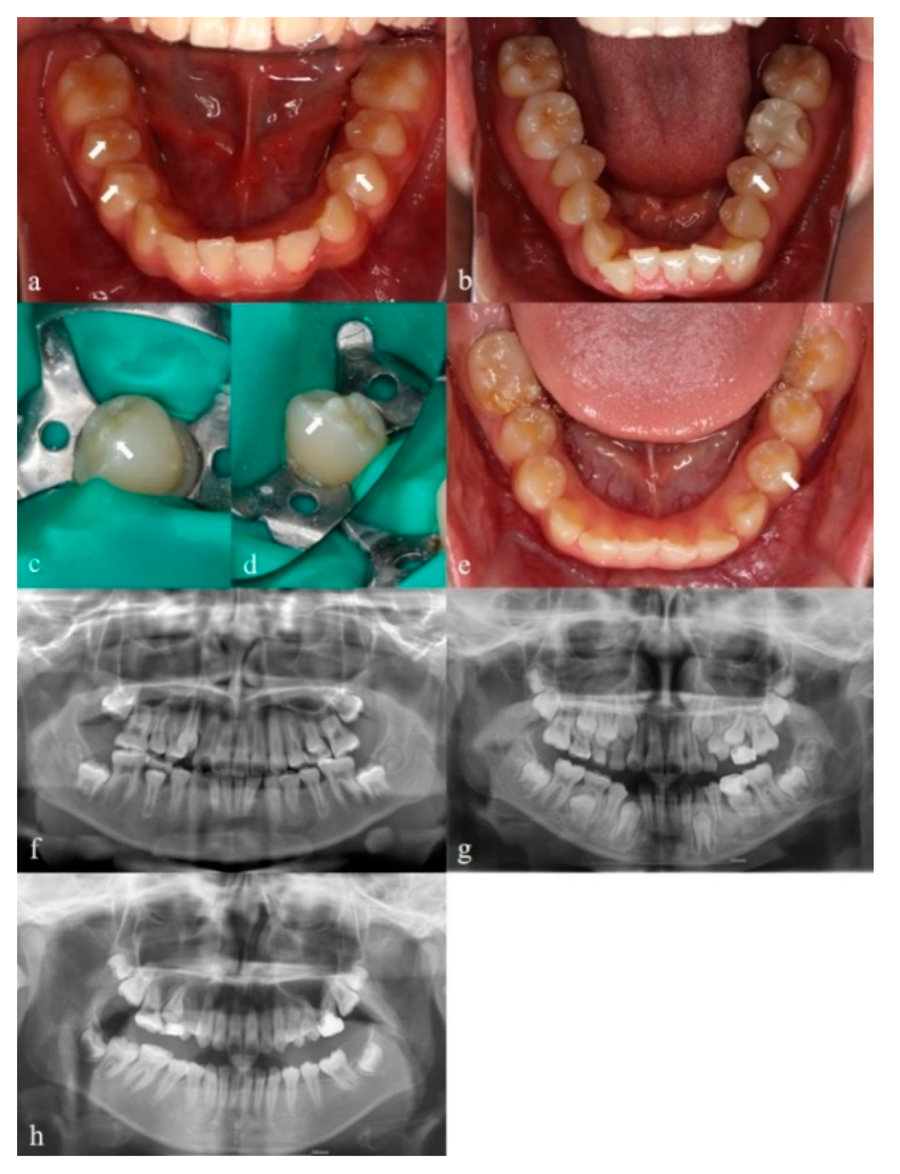
3.1. Clinical Findings

Five individuals, two from Family A and three from Family B, were included in the present study. Figure 1 shows the family trees of Families A and B. Figure 2 shows the intraoral photographs (Figure 2a–e) and panoramic radiographs (Figure 2f,h) of the patients with DE. Table 1 summarizes the clinical characteristics of the patients with DE, including sex and the position and laterality of DE.

3.2. Genetic Analysis

WES was performed for the five individuals included in the study, two from Family A and three from Family B. The WES data had a mean coverage depth of 156.9× with 96.04% of the target bases covered by at least 15 reads; the quality score (Q30) was 91.28%–93.67%. For the genome database, the frequency at which each gene variant is recognized by each allele frequency spectrum of variants was set to 1.0%, and candidate gene variants associated with DE were extracted. The variant frequency was considered to correspond to the incidence of DE.
We identified variants in the genes coding for Toll-Like Receptor 3 (TLR3) and Mediator of DNA Damage Checkpoint 1 (MDC1) as candidate gene variants that are related to the etiology of DE. Variants in TLR3 (c.1443C>G (n = 2), c.2491delA (n = 2)) and MDC1 (c.3908C>T (n = 2), c.1825G>A (n = 2)) were extracted (Table 2). Table 3 summarizes the information on the identified variants and their pathogenicity scores. The variant MDC1 (c.3908C>T) had the highest CADD score, followed by MDC1 (c.1825G>A).

4. Discussion

To the best of our knowledge, this study is the first to examine the genetic factors associated with DE using WES and identify the candidate gene variants. In total, five individuals from two families of Japanese and Korean origin were included in the present study. The results of WES analysis revealed new variants in TLR3 and MDC1, suggesting that these gene variants may be potential genetic factors associated with DE.
DE is reported more frequently in Asians, Inuit, Alaskan Natives, and American Indians [13,14,15]. The present study showed no significant differences in allele frequency, confirming racial differences in the candidate gene variants.
TLR3 encodes a protein that plays an essential role in the innate immune system and is a member of the TLR family [22]. Fawzy El-Sayed et al. [23] isolated and cultured human dental pulp stem/progenitor cells (DPSCs) from human dental pulp and confirmed the expression of TLR in DPSCs under inflamed and noninflamed conditions. Additionally, TLR3 has been reported to be expressed in odontoblasts, which are the first cells to encounter pathogens that enter the dental pulp [24,25]. Another report confirmed the expression of TLR3 in human pulp fibroblasts [26,27]. TLR3 is closely related to odontoblasts and human dental pulp. In particular, odontoblasts are associated with tooth formation, including dentin formation [28]. TLR has been reported to be associated with dental mesenchymal stromal cells (MSCs) [29]. MSCs have immunomodulatory properties and multilineage differentiation potential, and have been found in dental pulp, periodontal ligaments, dental follicles, and gingival tissues [29]. MSCs and TLR regulate each other’s activity, and can be involved in tissue regeneration and inflammatory disease progression. MSCs are interrelated with TLR and associated with dental components. Further investigation into the function of TLR may help in understanding tooth development. TLR3 is potentially related to tooth formation. The relationship between TLR3 and tooth formation needs to be further investigated.
MDC1 is a gene that encodes the nuclear protein MDC1, which is a part of the DNA damage response [30]. MDC1 is reportedly associated with Nijmegen breakage syndrome (NBS; OMIM# 251260) [31]. NBS is associated with humoral and cellular immunity [32]. Large mouth and micrognathia have also been reported as maxillofacial features of NBS [33]. Abnormal development of the tooth (hypoplasia of the enamel) was observed as an intraoral finding in a patient with NBS [34]. The genes that cause NBS and their function in tooth development and morphology need to be explored. Understanding the function of genes associated with enamel formation may help explain the mechanism of DE formation. MDC1 (C3908C>T) showed the highest CADD score and damage based on scores from PolyPhen2, which predicted the possibility of variants causing pathologic problems. There are no reports of MDC1 being associated with the oral cavity or teeth, but its functions need to be investigated through research.
The significance of the present study is that the genetic cause of DE, which remains unclear, was analyzed using WES, and candidate gene variants were identified. Identifying the genetic cause of DE is expected to be useful for early prediction, diagnosis, and treatment of DE. Early diagnosis of DE may be beneficial from the perspective of oral care because of the risk of pulpal inflammation due to fractures as a result of occlusal trauma. Understanding the mechanisms underlying the etiology of DE would lead to a better understanding of the function of genes that influence tooth morphology. This, in turn, may be helpful for developing regenerative medicine and gene therapy strategies for the treatment of DE.
This study has a few limitations. Although we identified candidate gene variants for DE, there are more gene variants that need to be identified, which require analysis in larger families and a larger sample size. Further research is required to identify the relationship between the candidate gene variants and DE, and the function of the gene variants. Additionally, the penetration rate of the DE gene variants could not be clarified in the present study and needs to be investigated in the future.

5. Conclusions

In the present study, we investigated the genetic background of DE and successfully identified candidate gene variants in TLR3 and MDC1. The candidate variants identified in this study should be investigated further to understand their role in DE. Examining the function of each variant will help us understand its relationship with DE as well as in tooth development.

Author Contributions

Conceptualization, T.Y.; methodology, T.Y.; formal analysis, H.P., K.H. and A.T.; investigation, H.P. and Y.-I.K.; writing—original draft preparation, H.P., K.H. and T.Y. All authors have read and agreed to the published version of the manuscript.

Funding

This research was funded by the Japan Society for Promotion of Science, KAKENHI program grant number [21K10173].

Institutional Review Board Statement

The study was conducted according to the guidelines of the Declaration of Helsinki, and approved by the Ethics Committees of Kanagawa Dental University (protocol code 746; [2-13-2020]), Pusan National University (protocol code PNUDH-2020-012; [4-28-2020]), and Kanazawa University (protocol code 597-1; [11-17-2020]).

Informed Consent Statement

Informed consent was obtained from all subjects involved in the study.

Data Availability Statement

Data that support the results of this study are available from the corresponding author upon reasonable request.

Conflicts of Interest

The authors declare no conflict of interest.

References

  1. Woelfel, J.B. Dental Anatomy: Its Relevance to Dentistry, 4th ed.; Lippincott Williams & Wilkins: Philadelphia, PA, USA, 1990. [Google Scholar]
  2. Manuja, N.; Chaudhary, S.; Nagpal, R.; Rallan, M. Bilateral dens evaginatus (talon cusp) in permanent maxillary lateral incisors: A rare developmental dental anomaly with great clinical significance. BMJ Case Rep. 2013, 2013, bcr2013009184. [Google Scholar] [CrossRef]
  3. Hattab, F.N.; Yassin, O.M.; al-Nimri, K.S. Talon Cusp—Clinical Significance and Management: Case Reports. Quintessence Int. 1995, 26, 115–120. [Google Scholar] [PubMed]
  4. Levitan, M.E.; Himel, V.T. Dens evaginatus: Literature review, pathophysiology, and comprehensive treatment regimen. J. Endod. 2006, 32, 1–9. [Google Scholar] [CrossRef]
  5. Hattab, F.N.; Yassin, O.M.; al-Nimri, K.S. Talon cusp in permanent dentition associated with other dental anomalies: Review of literature and reports of seven cases. ASDC J. Dent. Child. 1996, 63, 368–376. [Google Scholar] [PubMed]
  6. Yip, W.K. The prevalence of dens evaginatus. Oral Surg. Oral Med. Oral Pathol. 1974, 38, 80–87. [Google Scholar] [CrossRef]
  7. Ash, M. Wheeler’s Dental Anatomy, Physiology and Occlusion, 8th ed.; W. B. Saunders: Philadelphia, PA, USA, 2003; pp. 241–242. [Google Scholar]
  8. Echeverri, E.A.; Wang, M.M.; Chavaria, C.; Taylor, D.L. Multiple dens evaginatus: Diagnosis, management, and complications: Case report. Pediatr. Dent. 1994, 16, 314–317. [Google Scholar]
  9. Priddy, W.L.; Carter, H.G.; Auzins, J. Dens evaginatus—An anomaly of clinical significance. J. Endod. 1976, 2, 51–52. [Google Scholar] [CrossRef]
  10. Hülsmann, M. Dens invaginatus: Aetiology, classification, prevalence, diagnosis, and treatment considerations. Int. Endod. J. 1997, 30, 79–90. [Google Scholar] [CrossRef]
  11. Chen, J.W.; Huang, G.T.; Bakland, L.K. Dens evaginatus: Current treatment options. J. Am. Dent. Assoc. 2020, 151, 358–367. [Google Scholar] [CrossRef] [PubMed]
  12. Stecker, S.S.; Peterson, V.S.; Beiraghi, S.; Myers, S.L.; Bowles, W.R. Dens evaginatus: Just another cusp? Northwest Dent. 2015, 94, 31–33. [Google Scholar]
  13. Kocsis, G.; Marcsik, A.; Kokai, E.; Kocsis, K. Supernumerary occlusal cusps on permanent human teeth. Acta Biol. Szeged. 2002, 46, 71–82. [Google Scholar]
  14. Merrill, R.G. Occlusal anomalous tubercles on premolars of Alaskan eskimos and Indians. Oral Surg. Oral Med. Oral Pathol. 1964, 17, 484–496. [Google Scholar] [CrossRef]
  15. Curzon, M.E.; Curzon, J.A.; Poyton, H.G. Evaginated odontomes in the Keewatin Eskimo. Br. Dent. J. 1970, 129, 324–328. [Google Scholar] [CrossRef]
  16. Leigh, R. Dental pathology of the Eskimo. Dent. Cosmos. 1925, 67, 884–898. [Google Scholar]
  17. Geist, J.R. Dens evaginatus. Case report and review of the literature. Oral Surg. Oral Med. Oral Pathol. 1989, 67, 628–631. [Google Scholar] [CrossRef]
  18. Kimura, R.; Yamaguchi, T.; Takeda, M.; Kondo, O.; Toma, T.; Haneji, K.; Hanihara, T.; Matsukusa, H.; Kawamura, S.; Maki, K.; et al. A common variation in EDAR is a genetic determinant of shovel-shaped incisors. Am. J. Hum. Genet. 2009, 85, 528–535. [Google Scholar] [CrossRef] [PubMed]
  19. Lozić, B.; Ljubković, J.; Pandurić, D.G.; Saltvig, I.; Kutsche, K.; Krželj, V.; Zemunik, T. Oculo-facio-cardio-dental syndrome in three succeeding generations: Genotypic data and phenotypic features. Braz. J. Med. Biol. Res. 2012, 45, 1315–1319. [Google Scholar] [CrossRef] [PubMed]
  20. Stewart, R.E.; Dixon, G.H.; Graber, R.B. Dens evaginatus (tuberculated cusps): Genetic and treatment considerations. Oral Surg. Oral Med. Oral Pathol. 1978, 46, 831–836. [Google Scholar] [CrossRef]
  21. Davis, P.J.; Brook, A.H. The presentation of talon cusp: Diagnosis, clinical features, associations and possible aetiology. Br. Dent. J. 1986, 160, 84–88. [Google Scholar] [CrossRef] [PubMed]
  22. Hashimoto, C.; Hudson, K.L.; Anderson, K.V. The Toll gene of Drosophila, required for dorsal-ventral embryonic polarity, appears to encode a transmembrane protein. Cell 1988, 52, 269–279. [Google Scholar] [CrossRef]
  23. Fawzy El-Sayed, K.M.; Klingebiel, P.; Dörfer, C.E. Toll-like receptor expression profile of human dental pulp stem/progenitor cells. J. Endod. 2016, 42, 413–417. [Google Scholar] [CrossRef]
  24. Durand, S.H.; Flacher, V.; Roméas, A.; Carrouel, F.; Colomb, E.; Vincent, C.; Magloire, H.; Couble, M.L.; Bleicher, F.; Staquet, M.J.; et al. Lipoteichoic acid increases TLR and functional chemokine expression while reducing dentin formation in in vitro differentiated human odontoblasts. J. Immunol. 2006, 176, 2880–2887. [Google Scholar] [CrossRef] [PubMed]
  25. Pääkkönen, V.; Rusanen, P.; Hagström, J.; Tjäderhane, L. Mature human odontoblasts express virus-recognizing toll-like receptors. Int. Endod. J. 2014, 47, 934–941. [Google Scholar] [CrossRef]
  26. Staquet, M.J.; Carrouel, F.; Keller, J.F.; Baudouin, C.; Msika, P.; Bleicher, F.; Kufer, T.A.; Farges, J.C. Pattern-recognition receptors in pulp defense. Adv. Dent. Res. 2011, 23, 296–301. [Google Scholar] [CrossRef]
  27. Keller, J.F.; Carrouel, F.; Colomb, E.; Durand, S.H.; Baudouin, C.; Msika, P.; Bleicher, F.; Vincent, C.; Staquet, M.J.; Farges, J.C. Toll-like receptor 2 activation by lipoteichoic acid induces differential production of pro-inflammatory cytokines in human odontoblasts, dental pulp fibroblasts and immature dendritic cells. Immunobiology 2010, 215, 53–59. [Google Scholar] [CrossRef]
  28. Arana-Chavez, V.E.; Massa, L.F. Odontoblasts: The cells forming and maintaining dentine. Int. J. Biochem. Cell Biol. 2004, 36, 1367–1373. [Google Scholar] [CrossRef] [PubMed]
  29. Andrukhov, O. Toll-Like Receptors and Dental Mesenchymal Stromal Cells. Front. Oral Health. 2021, 2, 648901. [Google Scholar] [CrossRef]
  30. Zhou, B.B.; Elledge, S.J. The DNA damage response: Putting checkpoints in perspective. Nature 2000, 408, 433–439. [Google Scholar] [CrossRef]
  31. Wu, L.; Luo, K.; Lou, Z.; Chen, J. MDC1 regulates intra-S-phase checkpoint by targeting NBS1 to DNA double-strand breaks. Proc. Natl. Acad. Sci. USA 2008, 105, 11200–11205. [Google Scholar] [CrossRef]
  32. Chrzanowska, K.H.; Kleijer, W.J.; Krajewska-Walasek, M.; Białecka, M.; Gutkowska, A.; Goryluk-Kozakiewicz, B.; Michałkiewicz, J.; Stachowski, J.; Gregorek, H.; Lysón-Wojciechowska, G.; et al. Eleven Polish patients with microcephaly, immunodeficiency, and chromosomal instability: The Nijmegen breakage syndrome. Am. J. Med. Genet. 1995, 57, 462–471. [Google Scholar] [CrossRef]
  33. Maraschio, P.; Spadoni, E.; Tanzarella, C.; Antoccia, A.; Di Masi, A.; Maghnie, M.; Varon, R.; Demuth, I.; Tiepolo, L.; Danesino, C. Genetic heterogeneity for a Nijmegen breakage-like syndrome. Clin. Genet. 2003, 63, 283–290. [Google Scholar] [CrossRef]
  34. Olczak-Kowalczyk, D.; Dembowska-Bagińska, B.; Gregorek, H.; Wakulińska, A.; Pietrucha, B.; Chrzanowska, K.H. Dental Aspect of Nijmegen Breakage Syndrome. Nowa Stomatol. 2009, 3–8. [Google Scholar]
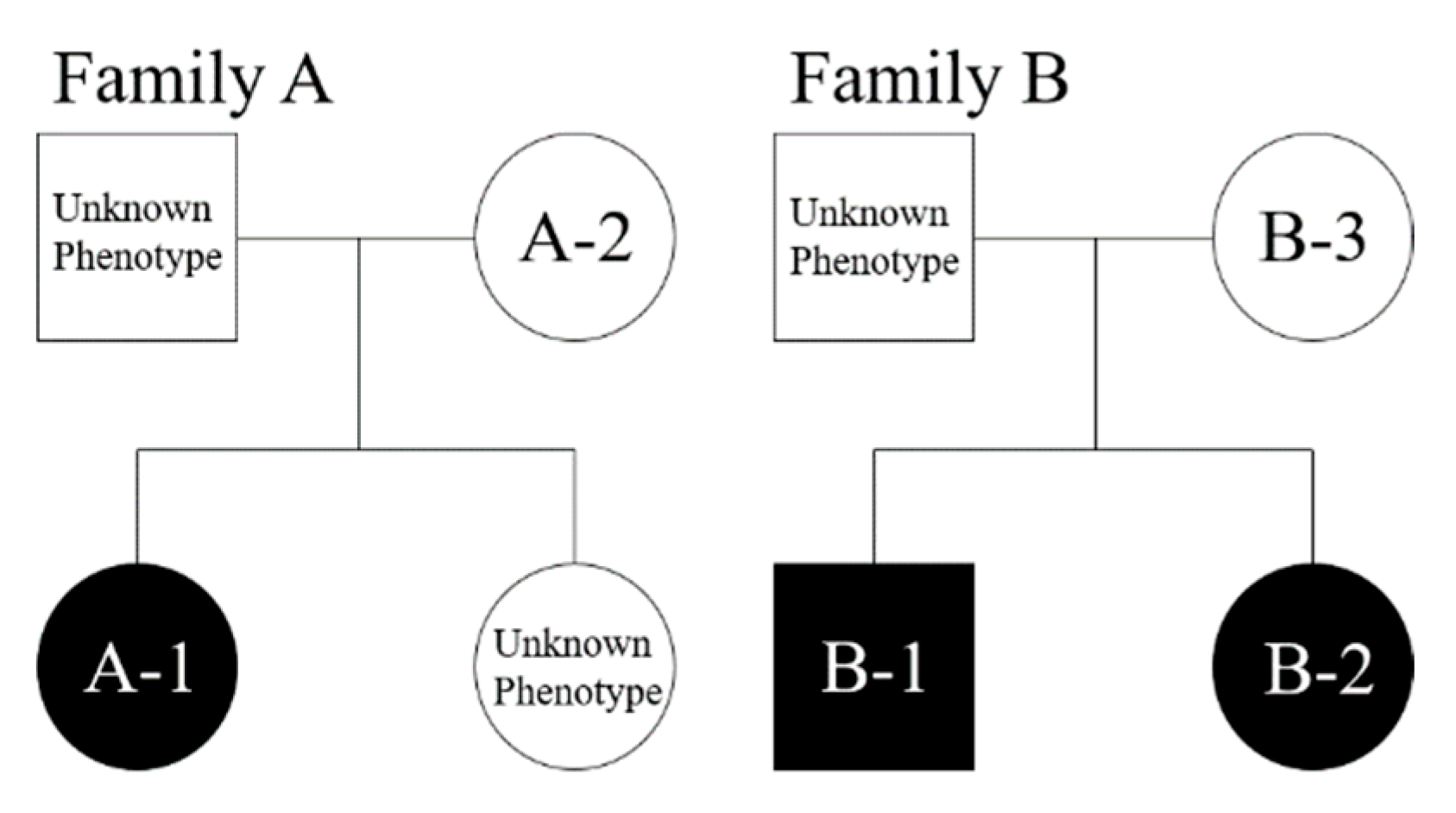
Figure 1. Family tree of Families A and B. Squares, male; circles, female; black background, individuals with dens evaginatus (DE); white background, individuals without DE.
Applsci 12 08962 g001
Figure 2. Intraoral radiographs of Patients (a) A-1, (b) A-2, (c,d) B-1, and (e) B-2. Panoramic radiographs of Patients (f) A-1, (g) B-1, and (h) B-2. White arrows denote DE.
Applsci 12 08962 g002
Table 1. Clinical characteristics of patients with dens evaginatus from Families A and B.
Subject *SexDental ArchDens EvaginatusInclusion Type
A-1FemaleLowerLeft and right first premolar, right second premolarBilateral
A-2FemaleLowerLeft second premolarMonolateral
B-1MaleLowerLeft and right first premolarBilateral
B-2FemaleLowerLeft first premolarMonolateral
B-3Female---
* Subjects are identified by family such that A-1 is Patient 1 from Family A.
Table 2. Candidate gene variants found in Families A and B.
Patient/GeneTLR3MDC1
A-1 * c.2491delAc.3908C>T
A-2 * c.2491delAc.3908C>T
B-1 *c.1443C>G c.1825G>A
B-2 *c.1443C>G c.1825G>A
B-3
* Presented with the DE phenotype.
Table 3. Identified variants and their pathogenicity scores.
GeneGenBank
Accession
Number
Nucleotide
Substitution
Amino Acid
Substitution
Variant IDMAFCADD
(Scaled)
PolyPhen2
GnomADGnomAD EAS 1000 GenomesExAC
TLR3NM_003265c.1443C>Gp.Ser481Argrs7518822990.00003229000.00000828312.35Benign
NM_003265c.2491delAp.Lys831fs-0000--
MDC1NM_014641c.3908C>Tp.Pro1303Leurs7632506510000.00000827517.49Damaging
NM_014641c.G1825Ap.Glu609Lysrs7635447940008.98 × 10−614.83Benign
Abbreviations: MAF, minor allele frequency; GnomAD, Genome Aggregation Database; ExAC, Exome Aggregation Consortium; GnomAD EAS, Genome Aggregation Database–East Asian; CADD, Combined Annotation Dependent Depletion; PolyPhen2, Polymorphism Phenotyping v2; del, deletion; fs, frame shift.
Publisher’s Note: MDPI stays neutral with regard to jurisdictional claims in published maps and institutional affiliations.

