Article

Plasticizer Di-(2-ethylhexyl) Phthalate and Its Metabolite Mono(2-ethylhexyl) Phthalate Inhibit Myogenesis in Differentiating Mouse and Human Skeletal Muscle Cell Models

1
Department of Emergency Medicine, Tri-Service General Hospital, National Defense Medical Center, Taipei 114, Taiwan
2
Department of Forensic Medicine, College of Medicine, National Taiwan University, Taipei 100, Taiwan
3
Departments of Emergency Medicine, National Taiwan University Hospital, Taipei 100, Taiwan
4
Institute of Toxicology, College of Medicine, National Taiwan University, Taipei 100, Taiwan
5
Center of Consultation, Center for Drug Evaluation, Taipei 115, Taiwan
6
Department of Geriatrics and Gerontology, College of Medicine, National Taiwan University, Taipei 100, Taiwan
7
Departments of Orthopaedics, College of Medicine, National Taiwan University and Hospital, Taipei 100, Taiwan
8
Department of Medical Research, China Medical University Hospital, China Medical University, Taichung 406, Taiwan
9
Department of Pediatrics, National Taiwan University Hospital, Taipei 100, Taiwan
*
Authors to whom correspondence should be addressed.
These authors contributed equally to this work.
Appl. Sci. 2022, 12(18), 9195; https://doi.org/10.3390/app12189195
Submission received: 31 August 2022 / Revised: 10 September 2022 / Accepted: 13 September 2022 / Published: 14 September 2022
(This article belongs to the Section Environmental Sciences)

Abstract

:
The relationship between plasticizer di(2-ethylhexyl) phthalate (DEHP) and low birth weight in neonates has been reported. Immature muscle differentiation may be involved in low birth weight. The myotoxic characteristics of chemicals have been observed in differentiating immortalized and primary muscle cells. Here, we explored the myotoxic effects of DEHP and its metabolite mono(2-ethylhexyl) phthalate (MEHP) in vitro using the immortalized mouse skeletal myoblasts C2C12 and primary human skeletal muscle progenitor cell (HSMPC) models. We found that both DEHP and MEHP at the concentrations of 10–100 μM, which were non- and low-cytotoxicity concentrations, significantly and dose-dependently inhibited the creatine kinase activity, myotube formation with multiple nuclei, and myogenin and myosin heavy chain (muscle differentiation markers) protein expression in C2C12 and HSMPCs under differentiation medium. Both DEHP and MEHP significantly decreased Akt phosphorylation in C2C12 and HSMPCs during differentiation. Taken together, DEHP and its metabolite MEHP are capable of inhibiting Akt-regulated myogenesis in myoblasts/myogenic progenitors during differentiation. These findings suggest the possibility of DEHP as an environmental risk factor affecting skeletal myogenic differentiation. Moreover, these in vitro muscle cell models may be a possible alternative method to animal myotoxicity testing.

1. Introduction

Di (2-ethylhexyl) phthalate (DEHP) is a commonly used phthalate plasticizer, which comprise ubiquitous chemicals found in many household goods, such as personal care products, food containers and packages, toys, building materials, furnishings, and medical devices [1,2]. Phthalates can be released into the environment. Humans can be exposed to phthalates through foods, indoor air, personal care products, medical procedures, and other factors [1,3,4,5,6]. After oral ingestion of DEHP, it is rapidly cleaved into monoester metabolites, such as mono(2-ethylhexyl) phthalate (MEHP), by intestinal nonspecific enzymes (e.g., esterases and lipases) [5,7]. After human exposure to DEHP, the primary metabolite MEHP and four major secondary metabolites could be significantly detected in urine [7]. DEHP has been reported to possess the ability to interfere with the reproductive and developmental systems [8,9]. Based on the analysis of urine phthalate levels in Danish children exposed to a phthalate-enriched indoor environment, DEHP was found to have the highest total daily intake [10].
A low birth weight is a marker of poor fetal development and growth [11]. An epidemiological study has shown that prenatal di-n-butyl phthalate and DEHP exposure contribute to low birth weight in Chinese newborns [12]. A relationship between prenatal exposure to phthalates and preterm delivery in Chinese women has also been reported [13]. In seriously low-birth-weight infants, a significant negative correlation between the sum of DEHP metabolites and birth weight have been demonstrated [14]. These studies indicate that DEHP exposure may contribute to low birth weight. A reduction in myogenesis has been demonstrated to be reflected in the low birth weight of intrauterine growth-restricted (IUGR) fetal piglets [15] and sheep [16]. Hansen et al. also found that myogenesis in human muscle stem cells cultured from the low-birth-weight individuals is retarded [17]. DEHP has been shown to inhibit myogenesis in C2C12 myoblasts [18]. However, it is still necessary to further explore the detailed effects and possible mechanisms of DEHP, and its metabolite MEHP, on skeletal myogenic differentiation.
The myotoxic characteristics of chemicals on differentiating immortalized and primary muscle cells have been observed in vitro [19,20,21,22,23]. Here, we aimed to study the myotoxic effects of both DEHP and MEHP on the myogenesis using immortalized mouse C2C12 skeletal myoblast and primary human skeletal muscle progenitor cell (HSMPC) models.

2. Materials and Methods

2.1. Materials

Both DEHP and MEHP, which were obtained from Sigma (St. Louis, MO, USA), were dissolved in ethanol. Antibodies for phosphorylated Akt (ab18206) were from Abcam (Cambridge, UK), and Akt (sc-8312), MHC (sc-20641), myogenin (sc-12732), and β-actin (sc-47778) were from Santa Cruz (Santa Cruz, CA, USA).

2.2. Cell Culture

Mouse C2C12 myoblasts (ATCC, Manassas, VA, USA) were incubated in cultured medium (growth medium, GM) consisting of Dulbecco’s modified Eagle’s medium (DMEM), fetal bovine serum (FBS; 10%), antibiotics (penicillin, 100 IU/mL; streptomycin, 100 μg/mL; amphotericin B, 0.25 μg/mL), and sodium bicarbonate (1.5 g/L), and placed in a humidified 5% CO2 incubator at 37 °C.

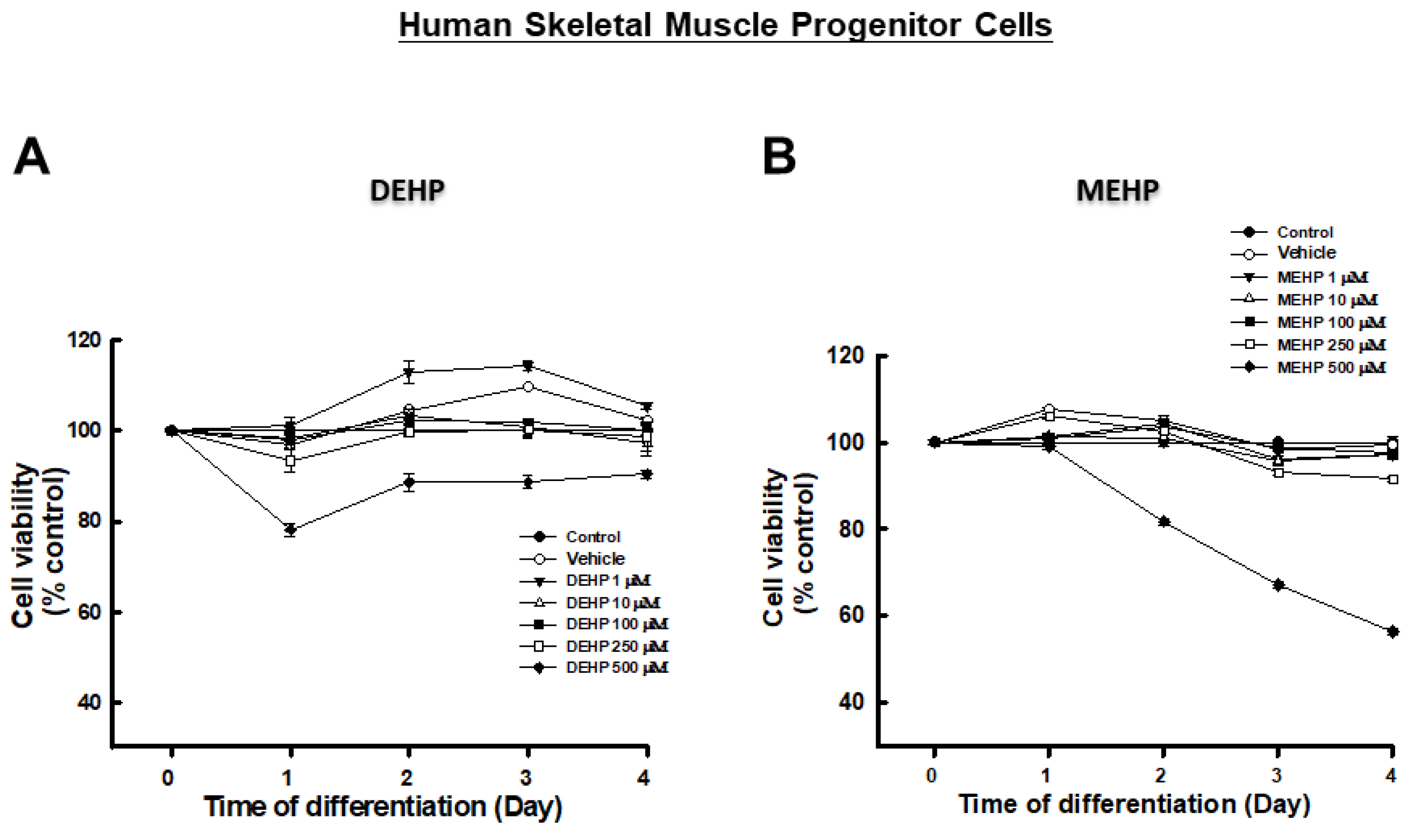
2.3. Primary HSMPC Culture

The Institutional Review Board of National Taiwan University Hospital approved the acquisition of human skeletal muscle samples. Written informed consent was obtained from all participating subjects. Human muscle sampling was performed according to the World Medical Association’s Code of Ethics (Declaration of Helsinki). As previously described [19], isolation and culture of primary HSMPCs were performed. Briefly, 0.2 g rectus muscle biopsies were obtained from the patients with orthopedic surgery. The minced muscle was reacted with serial enzymatic dissociation containing 0.5% type XI collagenase (1 h), 2.4 IU/mL dispase (45 min), and 0.2% trypsin-EDTA (15 min), and then used a 70-µm mesh filter to filter the cell suspension. Cells were seeded in a collagen-coated dish with growth medium contained FBS (20%), PSA (1%), and basic fibroblast growth factor (5 ng/mL) in Ham’s F-10. For separating fibroblasts and muscle-derived progenitor cells, the procedure of serial transfers and preplates was performed. For myogenic differentiation, cells with positive staining by desmin (a myogenic progenitor marker) were collected and cultured [24].

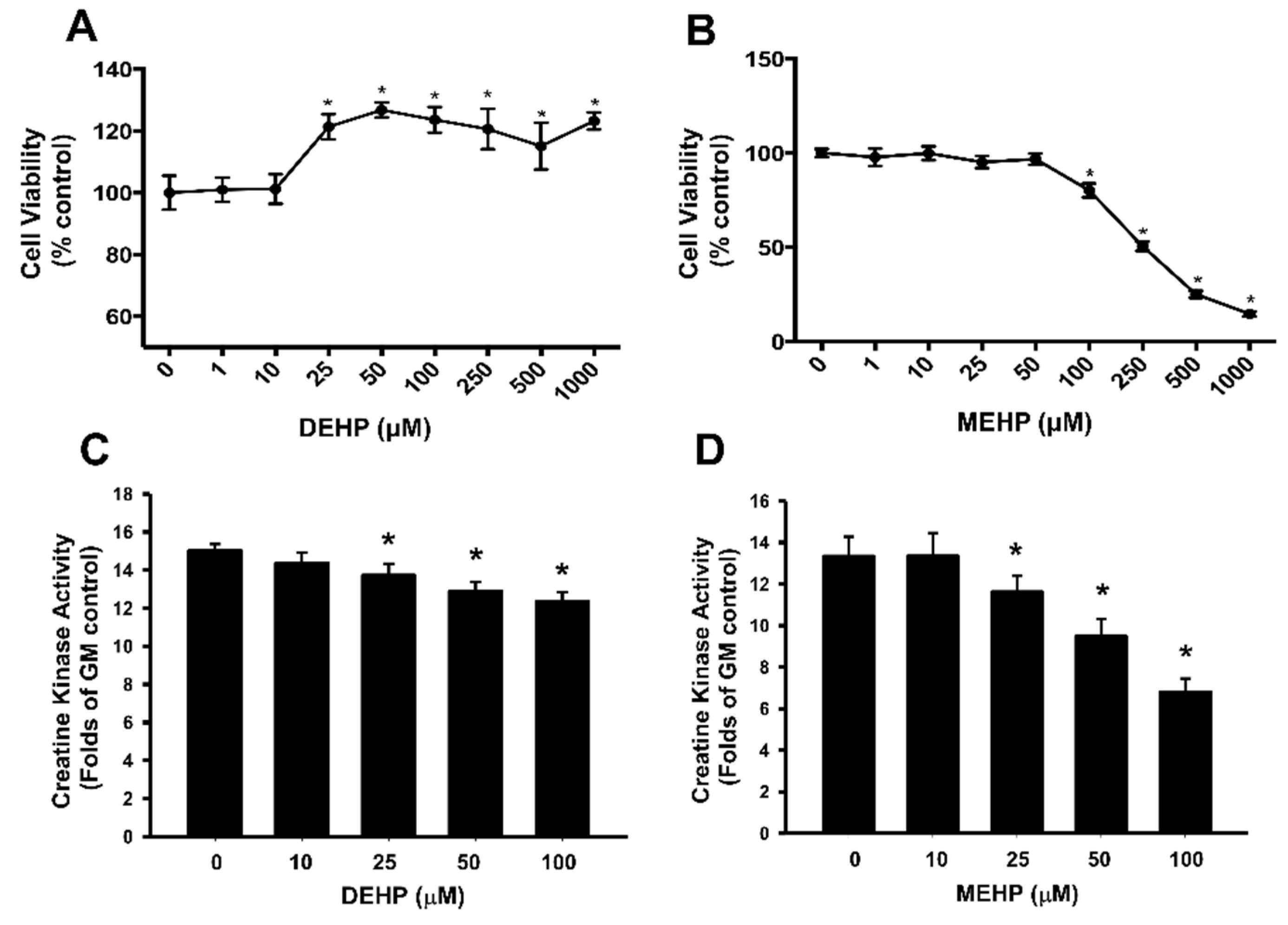
2.4. Cell Viability Measurement

Cells (104/well) were cultured in 96-well plates under growth conditions overnight, and subsequently incubated in differentiation media for 4 days. Following this, MTT (3-(4,5-dimethylthiazol-2-yl)-2,5-diphenyltetrazolium bromide (Sigma; 0.5 mg/mL)) was added and left to react for 2 h. Purple formazan crystals were formed, and then DMSO was added. Analyzed and recorded the absorbance values at 570 nm by a SpectraMax® (Shanghai, China) absorbance spectrophotometer (Molecular Devices, San Jose, CA, USA).

2.5. Creatine Kinase Activity Assay

After the cell lysates were centrifuged (13,000× g, 10 min), the supernatants were collected. A bicinchoninic acid (BCA) protein assay kit (Thermo Scientific, Rockford, IL, USA) was used to perform the protein concentration measurement. A commercial kit obtained from Teco Diagnostics (Anaheim, CA, USA) was used to determine the creatine kinase activity.

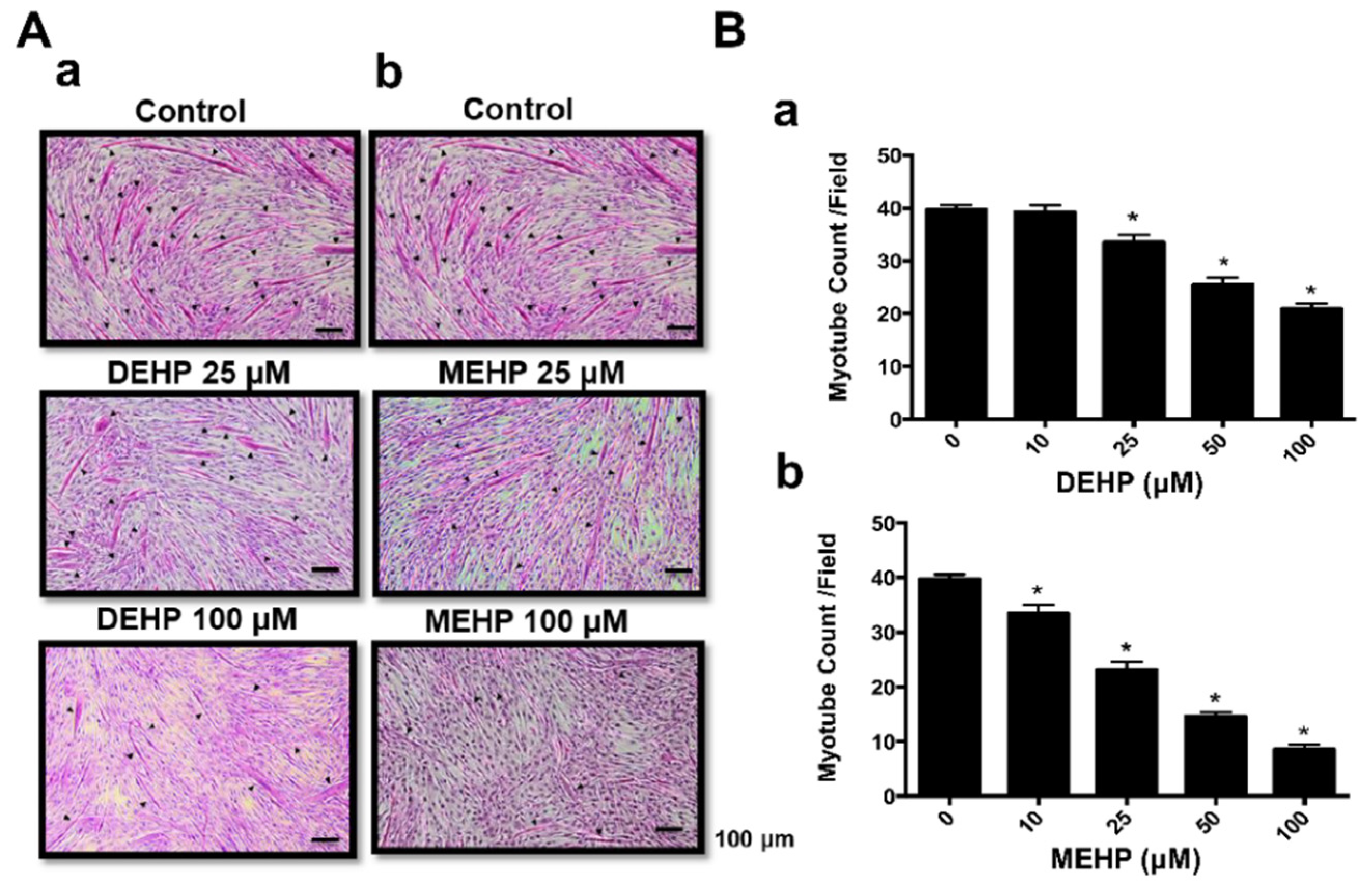
2.6. Myogenic Differentiation (Myotube Formation)
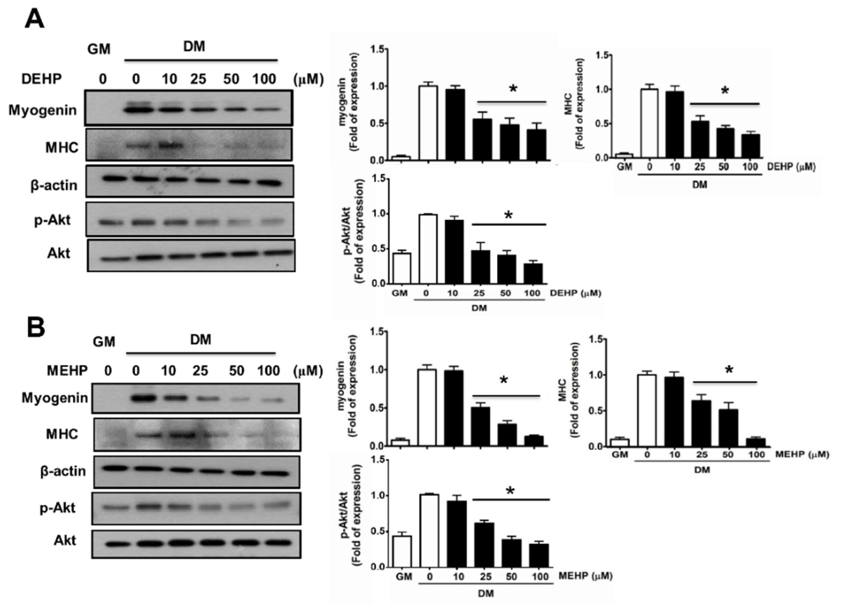
Cells were cultured in differentiation media (DM) (nutrient mixture F-12K ham medium/MCDB201 (1:1) containing 2% horse serum and antibiotics) for 4 days to induce the formation of multinucleated myotube. The morphology of myotube was analyzed by hematoxylin and eosin (H&E) staining [19].

2.7. Immunoblotting

Cells lysates were determined in RIPA buffer (containing NaCl, 150 mM; Tris, 10 mM; ethylene glycol tetraacetic acid, 1 mM; sodium dodecyl sulfate (SDS; 0.1%) supplemented with protease and phosphatase inhibitors containing NaF, sodium orthovanadate, phenylmethylsulfonyl fluoride (PMSF), aprotinin, and leupeptin at concentrations of 1 mM, 1 mM, 1 mM, 1 μg/mL, and 1 μg/mL, respectively. A BCA protein assay kit (Thermo Scientific) was used to determine the concentrations of protein. Separated proteins (20–40 μg) using the SDS-PAGE gels, and then electrotransferred onto polyvinylidene fluoride membranes, and finally blocked with 5% nonfat powdered milk. These membranes were probed with primary antibodies, and then reacted with horseradish-peroxidase-conjugated secondary anti-rabbit or anti-mouse antibodies. Finally, we developed the protein blots using an enhanced chemiluminescence reagent and these were visualized on X-ray film.

2.8. Analysis of Statistics

Representative results from at least three independent experiments in this study are shown as mean ± SEM. To check the statistical significance, one-way analysis of variance (ANOVA) and unpaired two-tailed Student’s t-tests were conducted to analyze intergroup variance. A p value less than or equal to 0.05 was considered to be significantly different.

3. Results

Exposure to DEHP at concentrations of 1–1000 μM for 96 h did not induce cytotoxicity in mouse C2C12 myoblasts under myogenic differentiation medium (Figure 1A). In fact, DEHP slightly increased cell viability at concentrations of 25 μM to 1000 μM (Figure 1A). Compared to DEHP, its metabolite MEHP at concentrations of 1–50 μM did not affect cell viability; cell viability decreased only slightly after 100 μM MEHP exposure, and displayed a greater cytotoxicity to myoblasts at concentrations of >100 μM (IC50 ≈ 250 μM; Figure 1B).
In primary HSMPCs, exposure to both DEHP and MEHP at concentrations of 1–100 μM for 96 h did not induce cytotoxicity under myogenic differentiation medium (Figure 2).
An increase in creatine kinase activity has been previously found in differentiating muscle cell cultures during myotube formation [25], which could be inhibited by environmental toxicants exposure [19,26]. Therefore, we next tested whether DEHP and MEHP affected creatine kinase activity. As shown in Figure 1C,D, both DEHP and MEHP at concentrations of 25–100 μM could significantly inhibit the creatine kinase activity in differentiating C2C12 myoblasts. MEHP exposure had greater inhibitory potency than DEHP (Figure 1D).
DEHP (10–100 μM) exposure effectively inhibited myotube formation in C2C12 myoblasts (p < 0.05), and the responses were dose-dependent (Figure 3(Aa,Ba)). MEHP (10–50 μM) could also significantly and dose-dependently decrease myotube formation (Figure 3(Ab,Bb). Moreover, the results of immunoblot analysis show that the myogenic differentiation markers MHC and myogenin protein expression in C2C12 myoblasts were also effectively inhibited by both DEHP (Figure 4A) and MEHP (Figure 4B), and the responses were dose-dependent.
Both DEHP and MEHP exposure at the concentrations of 25–100 μM also significantly decreased myotube formation (DEHP, Figure 5(Aa,Ba); MEHP, Figure 5(Ab,Bb) and protein expression of MHC and myogenin (DEHP, Figure 6A; MEHP, Figure 6B) in primary HSMPCs.
Akt signaling can positively regulate skeletal myogenesis [27]. We next tested the involvement of myogenic regulatory signal Akt in myoblasts during differentiation. As shown in Figure 3, both DEHP (Figure 4A) and MEHP (Figure 4B) effectively inhibited the phosphorylation of Akt in C2C12 myoblasts (p < 0.05), and the responses were dose-dependent. Both DEHP and MEHP did not affect total Akt protein expression in C2C12 myoblasts (Figure 4). Similarly, in the primary HSMPCs, both DEHP (Figure 6A) and MEHP (Figure 6B) could also effectively inhibit the phosphorylation of Akt (p < 0.05); there were no changes in total Akt protein expression.

4. Discussion

The present study found that DEHP and its metabolite MEHP, at non- and low-cytotoxic concentrations, inhibited myogenesis, which may further cause immature muscle fiber development, in the immortalized mouse and primary human muscle cell models. The mechanistic investigation indicated that both DEHP and MEHP markedly downregulated the Akt-regulated myogenic signaling molecules in C2C12 myoblasts and HSMPCs during differentiation.
Our data show that there was no cytotoxicity in DEHP (1–1000 μM)-treated C2C12 myoblasts under differentiation medium for 96 h. This result is consistent with the report by Chen et al., in which there was no cytotoxicity of DEHP (10–1000 μg/mL) on C2C12 cells [18]. Nevertheless, we further found that its metabolite MEHP displayed greater cytotoxicity at concentrations of >100 μM. Our data also showed that, at non-cytotoxic concentrations, DEHP (10–100 μM) and MEHP (10–50 μM) significantly suppressed myoblast differentiation and myotube formation in C2C12 myoblasts and HSMPCs. These concentrations for DEHP and MEHP seem to be relevant to human exposure in bloods, which will be discussed later. The levels of DEHP in indoor air of the Tokyo Metropolitan area have been shown to be up to 1000-fold higher than ambient levels in Japan [28]. It has been found that the concentrations of DEHP are 72 ± 13 μg/mL and 668 ± 96 μg/mL after 8 h and 24 h, respectively, in a cyclosporine solution stored in a PVC bag at room temperature [29]. In blood transfusions, the replacement blood DEHP levels have been detected to range from 10 to 650 μg/mL (about 25–1600 μM) [8]. A prospective and comparative clinical study showed that the mean maximum plasma level of DEHP was 8.3 ± 5.7 µg/mL at any time in a group of 28 consecutive term infants during an extracorporeal membrane oxygenation therapy [30]. A study of newborn infant plasticizer exposure showed that plasma DEHP levels were 3.4–1.1 μg/mL in the individual infants, and MEHP levels were 2.4–15.1 μg/mL in the corresponding samples [31]. Therefore, the test concentrations of DEHP/MEHP in the current study may be the blood concentrations to which human is likely to be exposed.
The major processes involved in skeletal myogenic differentiation are withdrawing myoblasts from the cell cycle, expressing myotube-specific genes, and the formation of multinucleated myotube [32,33]. Several factors can regulate myogenic differentiation, including transcription factors of myogenic basic helix–loop–helix family, such as MyoD, myogenin, myf5, and MRF4, and myocyte enhancer factor 2. These factors can further regulate muscle-specific gene/protein expression, including MHC and creatine kinase expression [34,35]. Moreover, the signaling of phosphatidylinositol 3-kinase (PI3K)/Akt contributes to controlling the expression of muscle genes/proteins during myogenesis [27,36]. The inhibition of the Akt-dependent signaling pathway has been demonstrated to contribute to the inhibitory effect of low-dose As2O3 on skeletal myogenic differentiation [22]. The defects in insulin activation of the PI3K/Akt signaling pathway has been shown in the skeletal muscle biopsies from young men with low birth weights [37]. Our results indicate that non-cytotoxic concentrations of both DEHP and MEHP significantly inhibit myotube formation and protein expression levels of myogenin, MHC, and phosphorylated Akt in myoblasts during differentiation, suggesting that exposure to both DEHP and MEHP can negatively impact Akt-regulated myogenic differentiation.

5. Conclusions

The current results reveal that non- and low-cytotoxic concentrations of DEHP and its metabolite MEHP were capable of inhibiting Akt-regulated myogenesis in differentiating immortalized mouse and primary human muscle cells. According to these findings, we propose that DEHP may metabolize into an active metabolite (MEHP) after DEHP intake, and both forms subsequently inhibit Akt-regulated signaling, leading to myotoxicity and myogenesis inhibition (Figure 7); this may be the cause of low birth weights in newborns reported in epidemiological studies. These findings suggest the possibility of DEHP as an environmental risk factor affecting skeletal myogenic differentiation. Moreover, these in vitro muscle cell models may be a possible alternative method to animal myotoxicity testing.

Author Contributions

K.-C.L.: Methodology, Resource, Validation, and Writing-original draft. T.-I.W.: Methodology, Resource, Validation, and Writing-original draft. C.-Y.C.: Methodology, Validation, and Writing-original draft. W.-C.C.: Investigation and Methodology. D.-C.C.: Funding and acquisition. R.-S.Y.: Conceptualization, Methodology, Resource, and Writing-Review and Editing. S.-H.L.: Conceptualization, Funding acquisition, Supervision, and Writing-Review and Editing. All authors have read and agreed to the published version of the manuscript.

Funding

This study was supported by grants from the Ministry of Science and Technology, Taiwan (MOST 102-2628-B002-030-MY3; MOST 103-2314-B-002-035).

Institutional Review Board Statement

The study was conducted in accordance with the Declaration of Helsinki, and approved by the Institutional Review Board (or Ethics Committee) of National Taiwan University Hospital (protocol code 201201044RIC and 6 December 2013).

Informed Consent Statement

Informed consent was obtained from all subjects involved in the study.

Data Availability Statement

The data presented in this study are available from the corresponding author upon reasonable request.

Conflicts of Interest

The authors declare no conflict of interest.

References

  1. Bizzari, S.; Oppenberg, B.; Iskikawa, Y. Plasticizers Chemical Economics Handbook; SRI International: Palo Alto, CA, USA, 2000. [Google Scholar]
  2. Heudorf, U.; Mersch-Sundermann, V.; Angerer, J. Phthalates: Toxicology and exposure. Int. J. Hyg. Environ. Health 2007, 210, 623–634. [Google Scholar] [CrossRef] [PubMed]
  3. Calafat, A.M.; Needham, L.L.; Silva, M.J.; Lambert, G. Exposure to di-(2-ethylhexyl) phthalate among premature neonates in a neonatal intensive care unit. Pediatrics 2004, 113, e429–e434. [Google Scholar] [CrossRef] [PubMed]
  4. Hanawa, T.; Muramatsu, E.; Asakawa, K.; Suzuki, M.; Tanaka, M.; Kawano, K.; Seki, T.; Juni, K.; Nakajima, S. Investigation of the release behavior of diethylhexyl phthalate from the polyvinyl-chloride tubing for intravenous administration. Int. J. Pharm. 2000, 210, 109–115. [Google Scholar] [CrossRef]
  5. Ito, Y.; Yokota, H.; Wang, R.; Yamanoshita, O.; Ichihara, G.; Wang, H.; Kurata, Y.; Takagi, K.; Nakajima, T. Species differences in the metabolism of di(2-ethylhexyl) phthalate (DEHP) in several organs of mice, rats, and marmosets. Arch. Toxicol. 2005, 79, 147–154. [Google Scholar] [CrossRef] [PubMed]
  6. Takehisa, H.; Naoko, E.; Masahiko, S.; Katsuhide, T.; Moriyuki, O.; Keizoh, S.; Mutsuko, T.; Kenji, K.; Shin’ichiro, N.; Toshio, O. Release behavior of diethylhexyl phthalate from the polyvinyl-chloride tubing used for intravenous administration and the plasticized PVC membrane. Int. J. Pharm. 2005, 297, 30–37. [Google Scholar] [CrossRef]
  7. Koch, H.M.; Bolt, H.M.; Preuss, R.; Angerer, J. New metabolites of di(2-ethylhexyl)phthalate (DEHP) in human urine and serum after single oral doses of deuterium-labelled DEHP. Arch. Toxicol. 2005, 79, 367–376. [Google Scholar] [CrossRef]
  8. Kavlock, R.; Boekelheide, K.; Chapin, R.; Cunningham, M.; Faustman, E.; Foster, P.; Golub, M.; Henderson, R.; Hinberg, I.; Little, R.; et al. NTP Center for the evaluation of risks to human reproduction; Phthalates expert panel report on the reproductive and developmental toxicity of di(2-ethylhexyl)phthalate. Reprod. Toxicol. 2002, 16, 529–653. [Google Scholar] [CrossRef]
  9. Latini, G.; De Felice, C.; Presta, G.; Del Vecchio, A.; Paris, I.; Ruggieri, F.; Mazzeo, P. In utero exposure to di-(2-ethylhexyl) phthalate and duration of human pregnancy. Environ. Health Perspect. 2003, 111, 1783–1785. [Google Scholar] [CrossRef]
  10. Bekö, G.; Weschler, C.J.; Langer, S.; Callesen, M.; Toftum, J.; Clausen, G. Children’s phthalate intakes and resultant cumulative exposures estimated from urine compared with estimates from dust ingestion, inhalation and dermal absorption in their homes and daycare centers. PLoS ONE 2013, 8, e62442. [Google Scholar] [CrossRef]
  11. Kensara, O.A.; Wootton, S.A.; Phillips, D.I.; Patel, M.; Jackson, A.A.; Elia, M.; Hertfordshire Study Group. Fetal programming of body composition: Relation between birth weight and body composition measured with dual-energy x-ray absorptiometry and anthropometric methods in older Englishmen. Am. J. Clin. Nutr. 2005, 82, 980–987. [Google Scholar] [CrossRef] [Green Version]
  12. Zhang, Y.; Lin, L.; Cao, Y.; Chen, B.; Zheng, L.; Ge, R.S. Phthalate levels and low birth weight: A nested case-control study of Chinese newborns. J. Pediatr. 2009, 155, 500–504. [Google Scholar] [CrossRef] [PubMed]
  13. Huang, Y.; Li, J.; Garcia, J.M.; Lin, H.; Wang, Y.; Yan, P.; Wang, L.; Tan, Y.; Luo, J.; Qiu, Z.; et al. Phthalate levels in cord blood are associated with preterm delivery and fetal growth parameters in Chinese women. PLoS ONE 2014, 9, e87430. [Google Scholar] [CrossRef]
  14. Strømmen, K.; Lyche, J.L.; Blakstad, E.W.; Moltu, S.J.; Veierød, M.B.; Almaas, A.N.; Sakhi, A.K.; Thomsen, C.; Nakstad, B.; Brække, K.; et al. Increased levels of phthalates in very low birth weight infants with septicemia and bronchopulmonary dysplasia. Environ. Int. 2016, 89–90, 228–234. [Google Scholar] [CrossRef] [PubMed]
  15. Rekiel, A.; Więcek, J.; Batorska, M.; Kulisiewicz, J. Effect of piglet birth weight on carcass muscle and fat content and pork quality—A Review. Ann. Anim. Sci. 2015, 15, 271–287. [Google Scholar] [CrossRef]
  16. Chang, E.I.; Rozance, P.J.; Wesolowski, S.R.; Nguyen, L.M.; Shaw, S.C.; Sclafani, R.A.; Bjorkman, K.K.; Peter, A.K.; Hay, W., Jr.; Brown, L.D. Rates of myogenesis and myofiber numbers are reduced in late gestation IUGR fetal sheep. J. Endocrinol. 2019, 244, 339–352. [Google Scholar] [CrossRef]
  17. Hansen, N.S.; Hjort, L.; Broholm, C.; Gillberg, L.; Schrölkamp, M.; Schultz, H.S.; Mortensen, B.; Jørgensen, S.W.; Friedrichsen, M.; Wojtaszewski, J.F.; et al. Metabolic and transcriptional changes in cultured muscle stem cells from low birth weight subjects. J. Clin. Endocrinol. Metab. 2016, 101, 2254–2264. [Google Scholar] [CrossRef]
  18. Chen, S.S.; Hung, H.T.; Chen, T.J.; Hung, H.S.; Wang, D.C. Di-(2-ethylhexyl)-phthalate reduces MyoD and myogenin expression and inhibits myogenic differentiation in C2C12 cells. J. Toxicol. Sci. 2013, 38, 783–791. [Google Scholar] [CrossRef]
  19. Chiu, C.Y.; Yen, Y.P.; Tsai, K.S.; Yang, R.S.; Liu, S.H. Low-dose benzo(a)pyrene and its epoxide metabolite inhibit myogenic differentiation in human skeletal muscle-derived progenitor cells. Toxicol. Sci. 2014, 138, 344–353. [Google Scholar] [CrossRef]
  20. Metterlein, T.; Hoffmann, P.; Späth, R.; Gruber, M.; Graf, B.M.; Zink, W. In vitro myotoxic effects of bupivacaine on rhabdomyosarcoma cells, immortalized and primary muscle cells. Cancer Cell Int. 2015, 15, 75. [Google Scholar] [CrossRef]
  21. Nouette-Gaulain, K.; Bellance, N.; Prévost, B.; Passerieux, E.; Pertuiset, C.; Galbes, O.; Smolkova, K.; Masson, F.; Miraux, S.; Delage, J.P.; et al. Erythropoietin protects against local anesthetic myotoxicity during continuous regional analgesia. Anesthesiology 2009, 110, 648–659. [Google Scholar] [CrossRef] [Green Version]
  22. Yen, Y.P.; Tsai, K.S.; Chen, Y.W.; Huang, C.F.; Yangm, R.S.; Liu, S.H. Arsenic inhibits myogenic differentiation and muscle regeneration. Environ. Health Perspect. 2010, 118, 949–956. [Google Scholar] [CrossRef] [PubMed]
  23. Yen, Y.P.; Tsai, K.S.; Chen, Y.W.; Huang, C.F.; Yang, R.S.; Liu, S.H. Arsenic induces apoptosis in myoblasts through a reactive oxygen species-induced endoplasmic reticulum stress and mitochondrial dysfunction pathway. Arch. Toxicol. 2012, 86, 923–933. [Google Scholar] [CrossRef] [PubMed]
  24. Asakura, A.; Seale, P.; Girgis-Gabardo, A.; Rudnicki, M.A. Myogenic specification of side population cells in skeletal muscle. J. Cell. Biol. 2002, 159, 123–134. [Google Scholar] [CrossRef] [PubMed]
  25. Shainberg, A.; Yagil, G.; Yaffe, D. Alterations of enzymatic activities during muscle differentiation in vitro. Dev. Biol. 1971, 25, 1–29. [Google Scholar] [CrossRef]
  26. Chen, H.J.; Wang, C.C.; Chan, D.C.; Chiu, C.Y.; Yang, R.S.; Liu, S.H. Adverse effects of acrolein, a ubiquitous environmental toxicant, on muscle regeneration and mass. J. Cachexia Sarcopenia Muscle 2019, 10, 165–176. [Google Scholar] [CrossRef]
  27. Kaliman, P.; Viñals, F.; Testar, X.; Palacín, M.; Zorzano, A. Phosphatidylinositol 3-kinase inhibitors block differentiation of skeletal muscle cells. J. Biol. Chem. 1996, 271, 19146–19151. [Google Scholar] [CrossRef]
  28. Otake, T.; Yoshinaga, J.; Yanagisawa, Y. Exposure to phthalate esters from indoor environment. J. Expo. Anal. Environ. Epidemiol. 2004, 14, 524–528. [Google Scholar] [CrossRef]
  29. Venkataramanan, R.; Burckart, G.J.; Ptachcinski, R.J.; Blaha, R.; Logue, L.W.; Bahnson, A.; Giam, C.S.; Brady, J.E. Leaching of diethylhexyl phthalate from polyvinyl chloride bags into intravenous cyclosporine solution. Am. J. Hosp. Pharm. 1986, 43, 2800–2802. [Google Scholar] [CrossRef]
  30. Karle, V.A.; Short, B.L.; Martin, G.R.; Bulas, D.I.; Getson, P.R.; Luban, N.L.; O’Brien, A.M.; Rubin, R.J. Extracorporeal membrane oxygenation exposes infants to the plasticizer, di(2-ethylhexyl) phthalate. Crit. Care Med. 1997, 25, 696–703. [Google Scholar] [CrossRef]
  31. Sjöberg, P.O.; Bondesson, U.G.; Sedin, E.G.; Gustafsson, J.P. Exposure of newborn infants to plasticizers. Plasma levels of di-(2-ethylhexyl) phthalate and mono-(2-ethylhexyl) phthalate during exchange transfusion. Transfusion 1985, 25, 424–428. [Google Scholar] [CrossRef]
  32. Weintraub, H. The MyoD family and myogenesis: Redundancy, networks, and thresholds. Cell 1993, 75, 1241–1244. [Google Scholar] [CrossRef]
  33. Weintraub, H.; Davis, R.; Tapscott, S.; Thayer, M.; Krause, M.; Benezra, R.; Blackwell, T.K.; Turner, D.; Rupp, R.; Hollenberg, S. The myoD gene family: Nodal point during specification of the muscle cell lineage. Science 1991, 251, 761–766. [Google Scholar] [CrossRef] [PubMed]
  34. Lassar, A.B.; Skapek, S.X.; Novitch, B. Regulatory mechanisms that coordinate skeletal muscle differentiation and cell cycle withdrawal. Curr. Opin. Cell Biol. 1994, 6, 788–794. [Google Scholar] [CrossRef]
  35. Olson, E.N.; Perry, M.; Schulz, R.A. Regulation of muscle differentiation by the MEF2 family of MADS box transcription factors. Dev. Biol. 1995, 172, 2–14. [Google Scholar] [CrossRef] [PubMed]
  36. Tamir, Y.; Bengal, E. Phosphoinositide 3-kinase induces the transcriptional activity of MEF2 proteins during muscle differentiation. J. Biol. Chem. 2000, 275, 34424–34432. [Google Scholar] [CrossRef]
  37. Jensen, C.B.; Martin-Gronert, M.S.; Storgaard, H.; Madsbad, S.; Vaag, A.; Ozanne, S.E. Altered PI3-kinase/Akt signalling in skeletal muscle of young men with low birth weight. PLoS ONE 2008, 3, e3738. [Google Scholar] [CrossRef] [Green Version]
Figure 1. The changes in cell viability and creatine kinase activity in myoblasts treated with both DEHP and MEHP. Under differentiation medium, C2C12 cells treated with DEHP (A,C) or MEHP (B,D) (1–1000 μM) for 24–96 h were observed. MTT assay was performed to analyze cell viability (A,B). Creatine kinase activity was assayed using a commercial kit (C,D). GM indicated as growth medium. Data are presented as means ± SEM (three independent experiments, triplicate repeats in each); p < 0.05 indicates significance compared to the control (*).
Applsci 12 09195 g001
Figure 2. The changes in cell viability in primary HSMPCs treated with both DEHP and MEHP. Under differentiation medium, HSMPCs treated with DEHP (A) or MEHP (B) (1–500 μM) for 24–96 h were observed. MTT assay was performed to analyze cell viability (A,B). Data are presented as means ± SEM (three independent experiments, triplicate repeats in each).
Applsci 12 09195 g002
Figure 3. The changes in myotube formation in C2C12 myoblasts treated with both DEHP and MEHP. Under differentiation medium, C2C12 cells treated with DEHP (Aa,Ba) or MEHP (Ab,Bb) (10–100 μM) for 96 h were observed. The cell morphology (A) was shown and myotube formation (B) was counted. Data are presented as means ± SEM (three independent experiments, triplicate repeats in each); p < 0.05 indicates significance compared to the control (*).
Applsci 12 09195 g003
Figure 4. The changes in myogenesis-related protein expression in C2C12 myoblasts treated with both DEHP and MEHP. Under differentiation medium (DM), C2C12 cells treated with DEHP (A) or MEHP (B) (10–100 μM) were observed. GM indicated as growth medium. The analysis of protein expression for myogenin, myosin heavy chain (MHC), phospho-Akt, and Akt was performed using Western blot. An internal control using β-actin was performed. The densitometric analysis of proteins via Western blot was performed. Data are presented as means ± SEM (at least three independent experiments, triplicate repeats in each); p < 0.05 indicates significance compared to the control (*).
Applsci 12 09195 g004
Figure 5. The changes in myotube formation in primary HSMPCs treated with both DEHP and MEHP. Under differentiation medium, HSMPCs treated with DEHP (Aa,Ba) or MEHP (Ab,Bb) (10–100 μM) for 96 h were observed. The cell morphology (A) was shown and the myotube formation (B) was counted (nuclei number per myotube). Data are presented as means ± SEM (three independent experiments, triplicate repeats in each); p < 0.05 indicates significance compared to the control (*).
Applsci 12 09195 g005
Figure 6. The changes in myogenesis-related protein expression in primary HSMPCs treated with both DEHP and MEHP. Under differentiation medium (DM), HSMPCs treated with DEHP (A) or MEHP (B) (10–100 μM) were observed. GM indicated as growth medium. The analysis of protein expression for myogenin, myosin heavy chain (MHC), phospho-Akt, and Akt was performed using Western blot. An internal control using β-actin was performed. The densitometric analysis of proteins via Western blot was performed. Data are presented as means ± SEM (at least three independent experiments, triplicate repeats in each); p < 0.05 indicates significance compared to the control (*).
Applsci 12 09195 g006
Figure 7. A schematic summary of the main findings for both DEHP- and MEHP-related cytotoxic effects on myogenesis in differentiating mouse and human skeletal muscle cell models.
Applsci 12 09195 g007
Publisher’s Note: MDPI stays neutral with regard to jurisdictional claims in published maps and institutional affiliations.

