Article

Removal Behavior of Heavy Metals from Aqueous Solutions via Microbially Induced Carbonate Precipitation Driven by Acclimatized Sporosarcina pasteurii

1
School of Civil and Environmental Engineering, Ningbo University, Ningbo 315211, China
2
Graduate School of Global Environment Studies, Kyoto University, Kyoto 6068501, Japan
*
Author to whom correspondence should be addressed.
Appl. Sci. 2022, 12(19), 9958; https://doi.org/10.3390/app12199958
Submission received: 30 August 2022 / Revised: 28 September 2022 / Accepted: 29 September 2022 / Published: 3 October 2022
(This article belongs to the Topic Advances in Environmental Biotechnology (AEB))

Abstract

:
Microbially induced carbonate precipitation (MICP) driven by Sporosarcina pasteurii was highly efficient for heavy metal (i.e., Cd2+, Cu2+ and Pb2+) removal in the range of 50 to 800 mg/L. Sporosarcina pasteurii bacteria were sequentially inoculated in nutrient broths amended with increased concentrations of heavy metals separately to improve the resistance to heavy metal environments. In the absence of Ca2+, the increasing urea concentration was conducive to Cd2+ removal with the best removal ratio 89.9–99.7% at a urea concentration of 2.0 mol/L, but had little positive effect on Cu2+ and Pb2+ removal, with a removal ratio of 62.6–64.4% and 71.4–97.6%, respectively, at a urea concentration of 0.5 mol/L. The heavy metal coprecipitated with Ca2+, leading to much more effective heavy metal removal, and the removal efficiency of Cd2+, Cu2+ and Pb2+ could reach 98.0–99.0, 78.1–82.1 and 98.0–100.0%, respectively. The Cu2+ deposit aggregated and cemented to form clusters, different from Cd2+ and Pb2+ deposits with comparatively dispersed microstructure. For all the three heavy metal precipitates, the predominant mineral was identified as calcite, the most thermodynamically stable polymorph of CaCO3.

1. Introduction

With the acceleration of the urbanization process in China, the environmental issues are becoming increasingly prominent, and heavy metal pollution caused by anthropogenic activities has become a focus point due to the tremendous hazard to the ecosystem [1,2]. Once enriched in the human body through the food chain [3], heavy metals will do harm to the kidney, brain and other important organs [4], and will potentially increase carcinogenic risks [5]. Research on the remediation of heavy-metal-polluted groundwater, wastewater and surface water has received worldwide attention [6,7]. The immobilization of heavy metals via microbially induced carbonate precipitation (MICP) is an environmental-friendly and energy-saving technique, and can provide a promising and alternative solution to heavy metal pollution issues, bringing new opportunity for the remediation of heavy-metal-polluted water and improvement of environmental qualities [2,8].
MICP is essentially a kind of biomineralization, which widely exists in nature. Calcium carbonate crystals with excellent cementing capacity can rapidly precipitate and accumulate when providing specific microorganisms with a wealth of calcium ions and nitrogenous nutrients [9,10,11]. Microorganisms mediate carbonate precipitation through heterotrophic pathways such as ammonification, denitrification, methane oxidation, sulfate reduction and ureolysis [12,13]. Among those microbial metabolic pathways, ureolytic bacteria, with the superiority of easy control, speedy reaction rate and efficient chemical conversion, has become the most popular bacterium in the field of MICP research [9,14]. Ureolytic bacteria can secrete urease through the metabolism process, which can catalyze the hydrolysis of urea, and the pH of the surrounding solution can correspondingly increase, resulting in the generation of ammonium and carbonate ions [15,16]. Calcium ions can be adsorbed on the cell wall surface of negatively charged bacterium. Calcium ions and carbonate ions can take the cell membrane as crystal nucleus to form calcium carbonate crystals, and also can simultaneously coprecipitate heavy metal ions to immobilize heavy metals [17,18,19,20]. The urea hydrolysis reaction process catalyzed by urease can be expressed by Equations (1)–(5). In the process of MICP, heavy metal or metalloid ions can replace Ca2+ to form coprecipitates, be adsorbed on calcite to form complexes, incorporate into the CaCO3 lattice structure, or enter into crystal interstices or defects [2,21].
CO ( NH 2 ) 2 + H 2 O urease NH 2 COOH + NH 3
NH 2 COOH + H 2 O spontaneous NH 3 + H 2 CO 3
2 NH 3 + 2 H 2 O 2 NH 4 + + 2 OH
H 2 CO 3 HCO 3 + H +
HCO 3 + H + + 2 OH CO 3 2 + 2 H 2 O
Many ureolytic bacteria have been confirmed to have the ability of immobilizing heavy metals by biomineralization. As an excellent urease producing bacterium, Sporosarcina pasteurii (S. pasteurii) is widely studied in the fields of biomaterials [11,22,23,24,25], crack sealing [26,27], sand and soil improvement [28,29,30,31,32,33,34], etc. In recent years, scholars devoted to environmental remediation have applied S. pasteurii to curb heavy metal polluted water [8,35,36]. Warren et al. [37] set up batch experimental systems and found that the solid sequestration of solution contaminants associated with this carbonate precipitation process was element-specific, the solid phase capture of strontium was highly effective and rapid, the uranyl capture was not effective and copper uptake was very poor. Fujita et al. [18] also found that strontium could coprecipitate with calcite generated by bacterial ureolysis, and the extent of the strontium incorporation appeared to be driven primarily by the overall rate of calcite precipitation. Mitchell and Ferris [21] studied the effect of temperature (i.e., 10, 15 and 20 °C) and time on the coprecipitation of strontium into calcite precipitates induced by bacterial ureolysis, and found that higher coprecipitation efficiency occurred at 20 °C. Lauchnor et al. [19] used laboratory-scale porous media flow cells to study the coprecipitation of strontium in carbonate minerals via ureolysis by S. pasteurii, and found that coprecipitation was effective (56–59%) under both pulsed and continuous flow scenarios. Li et al. [38] found that the immobilization ratio of heavy metals copper, lead, zinc, cadmium and nickel were above 88% at the initial concentration of 2 g/L. Mugwar and Harbottle [20] studied coprecipitations of a few heavy metals with calcium and found that S. pasteurii was not effective for the removal of copper (30%) in contrast with the removal efficiencies of lead, zinc and cadmium. Jiang et al. [39] found that the calcium source and initial bacteria concentration had a significant influence on the lead immobilization efficiency, and the best removal ratio could be over 95%. Jalilvand et al. [40] found that S. pasteurii could eliminate 98.71% of lead, 97.15% of cadmium and 94.83% of zinc. Duarte-Nass et al. [36] found a low removal ratio of copper (10%) in water through MICP using S. pasteurii, and attributed it to copper complexation with the ammonia generated by the urea hydrolysis. Fang et al. [35] found that Cd2+ removal efficiency could be greatly improved by the supplement of Ca2+ and nearly all the cadmium ions in the solution could by removed. Kim et al. [41] studied the removal behaviors of divalent cations when coprecipitated with calcium, and found that Pb2+ and Sr2+ had a high removal efficiency of more than 99%, while Cu2+, Zn2+ and Cd2+ demonstrated a low removal efficiency of 30–60% at an initial concentration no greater than 0.05 mM. Huang et al. [1] found that S. pasteurii could immobilize cadmium, chromium and zinc very effectively in aqueous solutions, while the immobilization efficiency for copper and nickel was very low, especially for copper. From the studies above, we can observe that significant progress has been made in removing heavy metals from aqueous solutions by S. pasteurii, and carbonate ions can promote the coprecipitaion of heavy metals in high pH environments. However, the results sometimes were very different from each other according to the removal efficiency. Moreover, the majority of the experiments were carried out at low heavy metal concentrations (0.01~1 mmol/L) due to the high toxicity of heavy metals to the bacteria [35,41,42].
Microbially induced carbonate precipitation was considered as a complex process, and both environmental conditions and biotic factors could affect the precipitation process. We can observe from the above studies that the effects of external factors such as pH, temperature, time and heavy metal concentration on the removal of heavy metals by S. pasteurii were mainly discussed, while the urea concentration of culture substrate and the calcium concentration should be taken into account simultaneously in view of the huge differences among those reported studies. Thus, in this study we designed a series of experiments to acclimatize S. pasteurii in heavy metal amended nutrient broths in order to improve the resistance of S. pasteurii to a heavy metal environment, to consider urea concentration and Ca2+ concentration in detail and to study the removal behaviors of different concentrations of heavy metals (i.e., Cd2+, Cu2+ and Pb2+) by S. pasteurii. In addition, we characterized the minerals formed by MICP through the scanning electron microscope (SEM) and X-ray diffraction spectroscopy (XRD). The results can provide a reference for the remediation of heavy-metal-contaminated groundwater and wastewater based on the MICP technique.

2. Materials and Methods

2.1. Materials

S. pasteurii can be isolated from a variety of media such as soil, water and sludge. Laboratory studies have demonstrated that this bacterium is non-pathogenic, does not easily aggregate under most growth conditions, and can produce large amounts of active ureases in cells [43]. S. pasteurii used in this study was a gram-positive and facultative anaerobic bacterium with high urease-producing ability, which was purchased from the China General Microbial Culture Collection and Management Center (CGMCC), and had a strong environmental adaptability.
The reagents such as peptone, beef extract, agar and urea are of biochemical reagents, and the reagents CdCl2 2.5H2O, CuCl2 2H2O, Pb(NO3)2 and CaCl2 are of analytical grade. The water used in the test was deionized water. All the reagents were sterilized before using.

2.2. Culture Media

The nutrient broths were prepared for incubating S. pasteurii and the following were included: peptone, 5 g; beef extract, 3 g; deionized water, 1 L.
The constituents of the solid culture media included: peptone, 5 g; beef extract, 3 g; agar, 15 g; urea, 20 g; deionized water, 1 L. The pH value of the solid culture media was adjusted to 8.0.
All those media, except for urea, were autoclaved at 121 °C for 25 min before using. The stock solution of urea was filter-sterilized using 0.22 µm filters. Then, a number of agar slant culture media were prepared and preserved in the refrigerator for later use.

2.3. Factors Affecting Growth of S. pasteurii

Different factors, i.e., incubation time (t), temperature (T), pH and urea concentration (Curea), were considered to study the growth behavior of S. pasteurii. Four groups of experiments were designed and the relevant experimental conditions are listed in Table 1. The freeze-dried bacteria S. pasteurii were activated and enriched in nutrient broth at 30 °C and for 48 h. The optical density at 600 nm wavelength incident light (OD600) of the suspension was measured by visible spectrophotometry (Unico V1800, Shanghai, China). The suspension was diluted until the OD600 = 0.4 and the bacterial concentration was approximately equivalent to 1.26 × 10⁸ cfu/mL. 1 mL of the diluted suspension was then transferred to a sterilized flask with 100 mL of nutrient broth. All the samples were prepared in this way according to Table 1, and then were incubated in a biochemical incubator. At the end of each experiment, the OD600 value was measured.

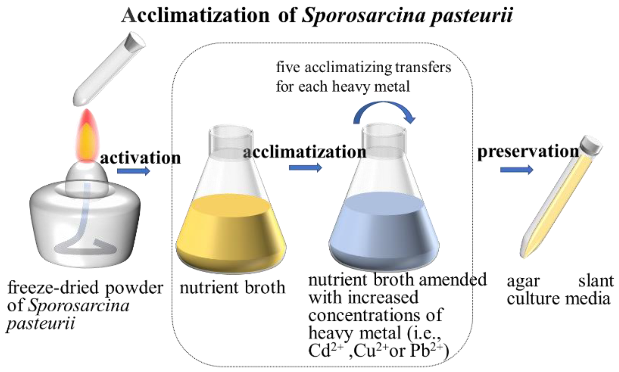
2.4. Acclimatization of S. pasteurii Using Heavy-Metal-Amended Nutrient Broths

For the sake of improving the resistance to heavy metals and the survival ability of S. pasteurii in a concentrated heavy metal environment, S. pasteurii was acclimatized in heavy-metal-amended nutrient broths. S. pasteurii was inoculated in different heavy metal (i.e., Cd2+, Cu2+ and Pb2+) amended nutrient broths separately. A series of nutrient broths with 200 mL were prepared, and in each nutrient broth, the urea concentration was 0.32 mol/L and the pH was adjusted to 8.0, which was the best culture medium condition obtained from Section 2.3. Moreover, based on the results in Section 2.3, the best incubating temperature and time was 30 °C and 48 h, respectively. Therefore, for the next experiments, the best culture conditions were applied to enrich the bacteria. A 2 mL suspension of S. pasteurii with OD600 = 0.4 was inoculated into 200 mL of the nutrient broth amended with 0.08 mol/L heavy metal, incubated at 30 °C and 180 r/min for 48 h. The acclimatizing process was repeated in the same way for 5 rounds, and the heavy metal concentration in the nutrient broth increased sequentially for each round. For the 1st, 2nd, 3rd, 4th and 5th round, the heavy metal concentration was 0.08, 0.16, 0.24, 0.32 and 0.4 mol/L, respectively. The fifth acclimatized S. pasteurii bacteria were designated as S.p. Cd-5, S.p. Cu-5 and S.p. Pb-5 separately, and were preserved in the agar slant culture media in the refrigerator at 4 °C for use. Figure 1 shows the procedure of the S. pasteurii acclimatization.

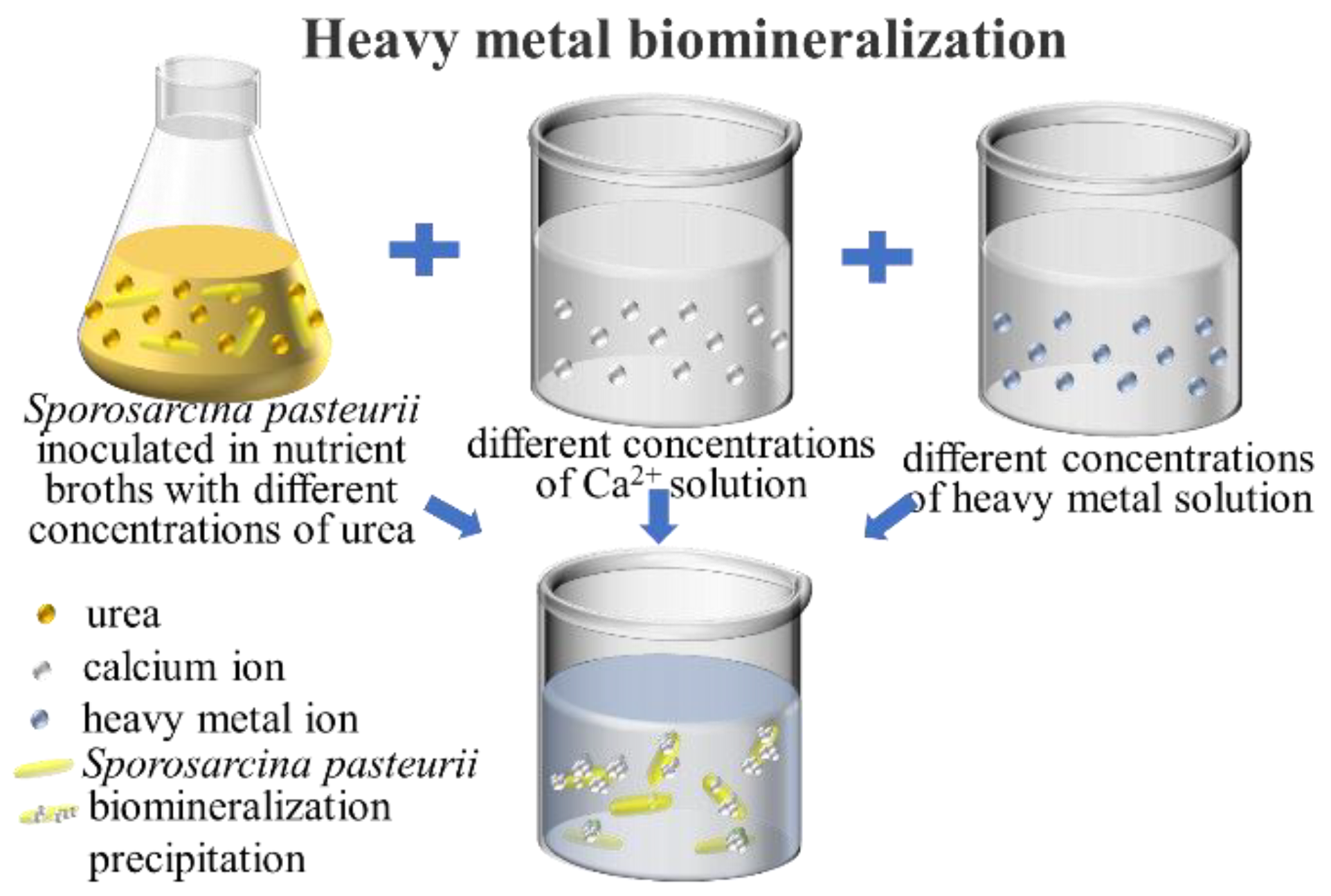
2.5. Removal of Heavy Metals from Aqueous Solutions Using Acclimatized S. pasteurii

In order to learn about the ability of acclimatized S. pasteurii mineralizing heavy metals, and how the urea and Ca2+ concentration affect the behavior of heavy metal biomineralization, a series of experiments were designed in this study. Excessive concentrations of cadmium, copper and lead have strong toxic effects on animals, plants and humans, and are easy to cause health risks and endanger the ecosystem stability. These three heavy metals are very frequently observed among heavy metal pollution issues, and are also widely studied in previous studies. Different concentrations of heavy metal (i.e., Cd2+, Cu2+ and Pb2+) solutions were prepared separately, and different weights of calcium chloride were also weighed to prepare different concentrations of Ca2+ to consider the effect of Ca2+ concentration on the heavy metal precipitation.
S.p. Cd-5, S.p. Cu-5 and S.p. Pb-5 was used for removing heavy metal Cd2+, Cu2+ and Pb2+ from aqueous solutions, respectively. These three types of acclimatized S. pasteurii were enriched in the nutrient broth at pH = 8.0 and 30 °C incubating for 48 h and diluted until the OD600 = 0.4. Then, 2 mL of the bacterial suspension was transferred into 200 mL nutrient broth with different concentrations of urea at pH = 8.0 and 30 °C, incubating for 48 h. Subsequently, 100 mL of the heavy metal solution was added into the above 200 mL nutrient broth with corresponding acclimatized S. pasteurii, and 100 mL of the Ca2+ solution was also instantly added into the bacterial solution. In the reaction systems formed by the three solutions, the concentration of heavy metals varied from 50, 100, 200, 400 to 800 mg/L, the concentration of Ca2+ varied from 0, 0.5, 1.0, 1.5 to 2.0 mol/L, and the concentration of urea varied from 0.5, 1.0, 1.5 to 2.0 mol/L. Two parallel experiments were performed for each group and all the reaction systems were placed in a thermostatic oscillator at 180 r/min, 30 °C for 48 h. The test procedure of the heavy metal biomineralization is shown in Figure 2.
At the end of the tests, the samples were taken and centrifuged at 8000 r/min for 3 min. The supernatants were collected, and heavy metal concentrations were measured by flame atomic absorption spectrophotometry (Persee TAS-990, Beijing, China). The crystal structures of the precipitates generated in the tests were detected through X-ray diffraction spectroscopy (XRD, Bruker D8 Advance Davinci, Karlsruhe, Germany), and the morphologies were also characterized using the scanning electron microscope (SEM, Hitachi SU-70, Tokyo, Japan).

3. Results and Discussion

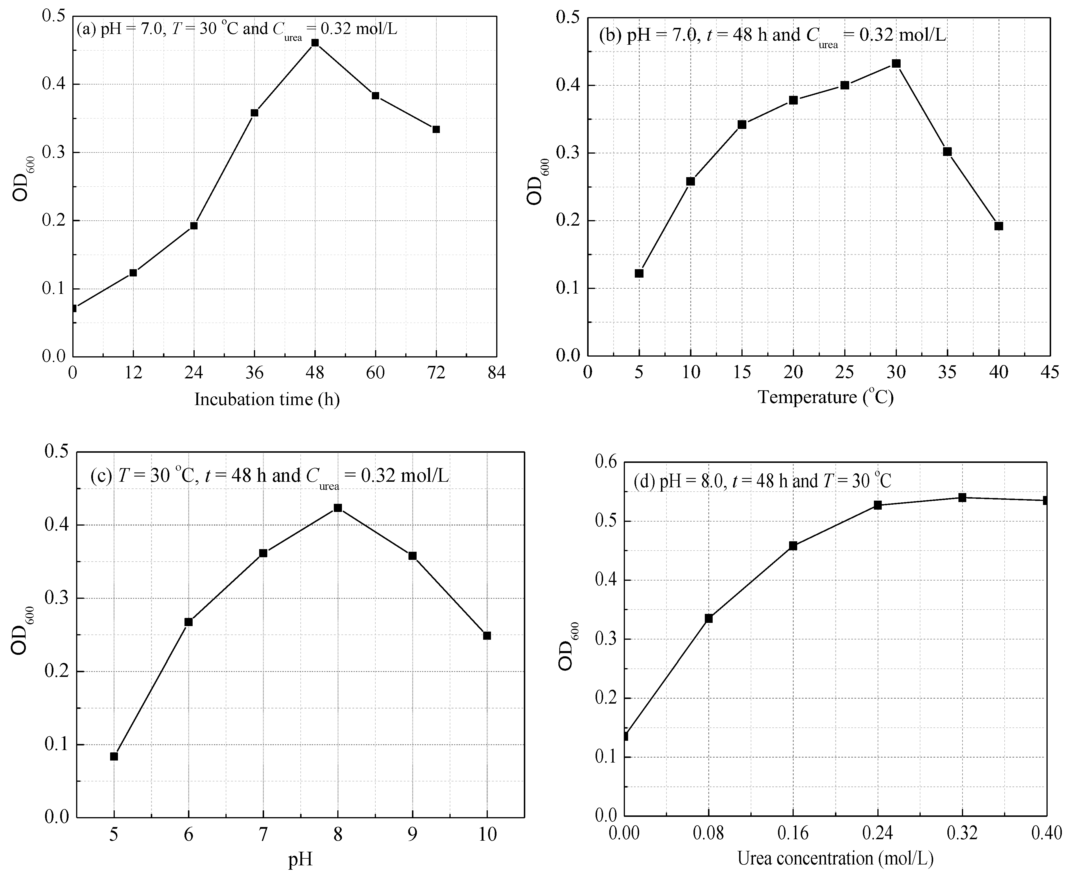
3.1. Growth Characteristic of S. pasteurii

Figure 3 shows the effect of time, temperature, pH and urea concentration on the growth characteristic of S. pasteurii. In Figure 3a, S. pasteurii exhibited a logarithmic increase at the time from 0 to 48 h, and the cell concentration reached the highest level with OD600 = 0.46. From 48 h to 72 h, the bacteria began to decay as the dissimilation of bacteria was stronger than the assimilation, leading to the death of the bacteria. As observed in Figure 3b, the OD600 value gradually increased as an increasing temperature to 30 °C, indicating that the increase in temperature could facilitate the microbial reproduction. When the temperature was above 30 °C, the OD600 value turned smaller and smaller, suggesting that the growth of microbes was inhibited, and some began to die due to bacterial inactivation. Therefore, the optimal temperature for the growth of S. pasteurii is 30 °C. The OD600 value tended to increase to the maximum at pH = 8.0 in Figure 3c, and then decrease promptly, indicating that highly acidic and alkaline environments were not suitable for the growth of S. pasteurii. In Figure 3d, the OD600 value kept gently increasing with increasing the urea concentration to 0.32 mol/L and then maintained almost constant when the urea concentration increased to 0.4 mol/L. Apart from promoting the MICP process, the urea could provide nitrogen nutrition for the growth of S. pasteurii, and the concentration of 0.32 mol/L is sufficient for the growth of S. pasteurii.

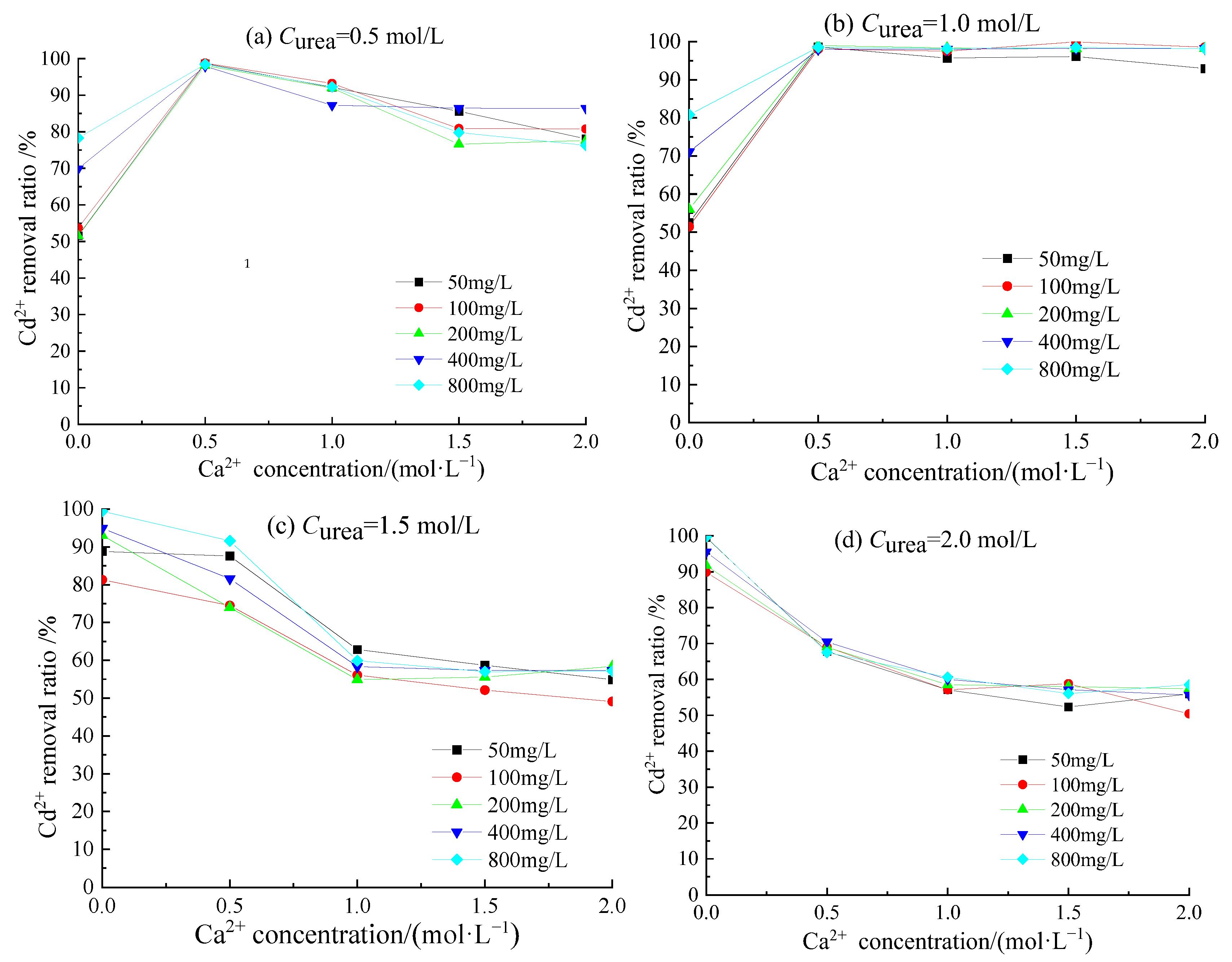
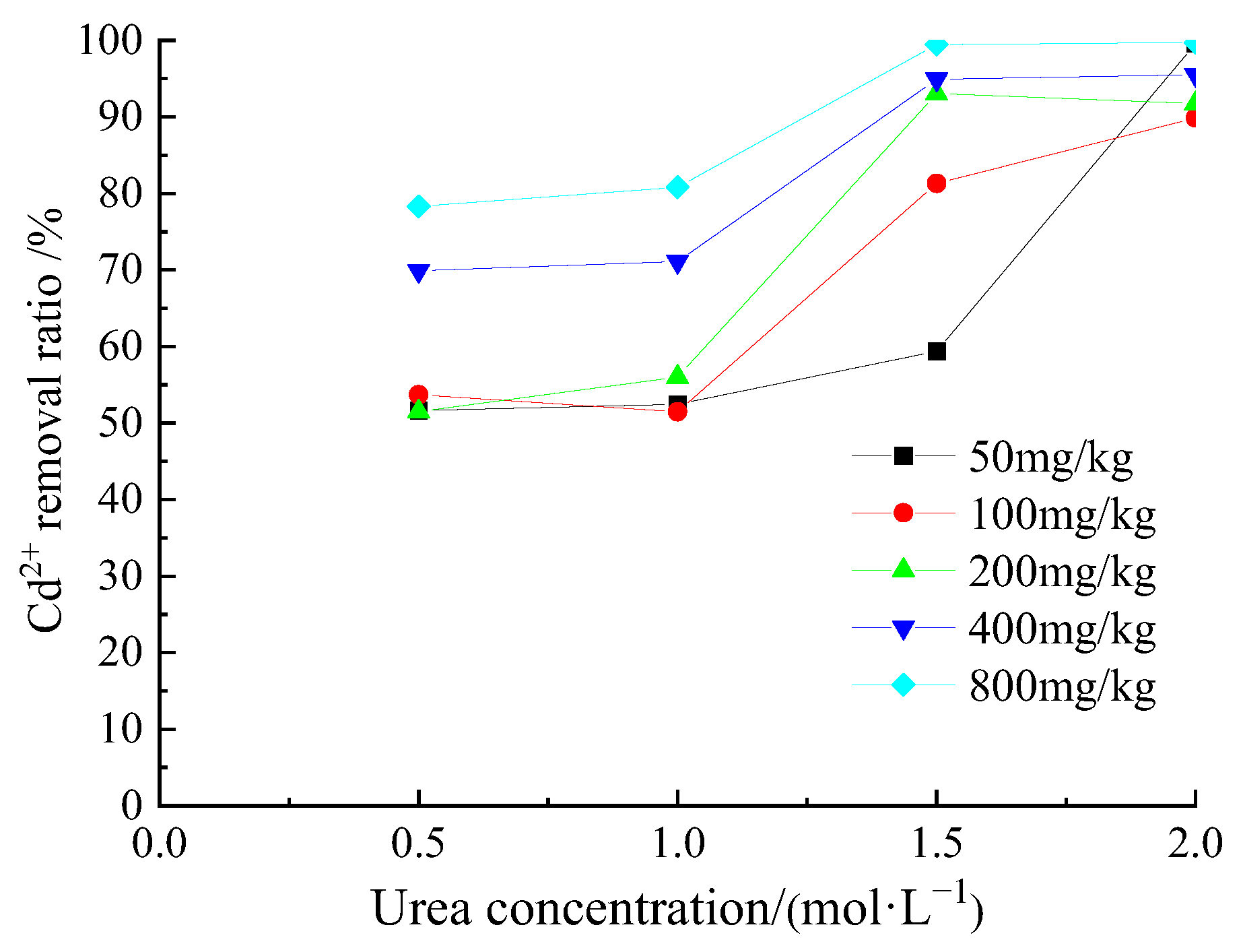
3.2. Removal Behavior of Cd2+ by Acclimatized S. pasteurii

Figure 4 shows the variation of removal ratios of Cd2+ with increasing the concentrations of Ca2+ at different urea concentrations. It can be observed from Figure 4a that when the Ca2+ concentration was 0, the removal ratio of Cd2+ was the lowest. When the Ca2+ concentration was 0.5 mol/L, the removal ratio of Cd2+ was the highest and the removal ratios of five groups with different concentrations of Cd2+ were almost the same at about 97.9–98.8%, implying that the Cd2+ ions in the solution were almost completely removed. With the increase in Ca2+ concentration, the removal ratio of Cd2+ started to decrease gradually, and the final removal ratios remained at 76.3–86.4%. The increase in Ca2+ led to the acceleration of the formation rate of CaCO3 precipitation, and the CaCO3 precipitation might adhere to the surface of bacteria, hindering the further combination of CO32− with Cd2+ and Ca2+, resulting in the decrease in MICP rate [20].
Comparing Figure 4a with Figure 4b, the removal behavior of Cd2+ had little change for the groups with 0 and 0.5 mol/L Ca2+ concentrations when raising the urea concentration from 0.5 to 1.0 mol/L. The removal ratios of Cd2+ were as high as 98.0–99.0% when the concentration of urea and Ca2+ was 1.0 and 0.5 mol/L, respectively. As observed in Figure 4b, when the Ca2+ concentration was increased from 0.5 to 1.0, 1.5 and 2.0 mol/L, the removal ratio of Cd2+ remained almost unchanged, demonstrating that a further increase in Ca2+ concentration would not be beneficial to the cadmium biomineralization process. The removal behaviors of Cd2+ in Figure 4c,d were very different from those in Figure 4a,b, and when the urea concentration was further increased to 1.5 and 2.0 mol/L, the best removal efficiency of Cd2+ happened when there was no Ca2+. After adding Ca2+ into the solutions, the Cd2+ removal efficiency began to decrease, and the removal ratio decreased with the increase in Ca2+ concentration, which seems different with the result published by Fang et al. [35], who found that the Ca2+ supplement improved the biomineralization of Cd2+ by S. pasteurii. In that study, the Ca2+ concentration was in the range of 5–250 mmol/L, and urea concentration was 20 mmol/L, both smaller than the lowest concentration of Ca2+ (0.5 mol/L) and urea (0.5 mol/L) in our study. From Figure 4a,b, we can see when the Curea ≤ 1.0 mol/L and Ca2+ ≤ 0.5 mol/L, Ca2+ supplement can facilitate Cd2+ biomineralization, and as a matter of fact, Fang et al.’s result is coincident with our results.
It can be observed from Figure 5 that when there was no Ca2+ in the solutions, the removal ratio of Cd2+ increased with the increase in urea concentration. Especially when the urea concentration increased to 1.5 mol/L, the removal ratio of Cd2+ increased significantly. When the urea concentration was raised to 2.0 mol/L, the removal ratio of Cd2+ could be in the range of 89.9 to 99.7%.
From the above results, we can infer that the increase in urea concentration was favorable for the removal of Cd2+, while the increase in Ca2+ concentration was not good for the removal of Cd2+. Both urea and Ca2+ had a significant influence on the removal behavior of Cd2+. The optimal removal efficiency of Cd2+ could be achieved when the urea and Ca2+ concentration was 1.0 and 0.5 mol/L, respectively. The acclimatized S. pasteurii presented a high affinity for a wide range concentration of cadmium ions and demonstrated an obvious advantage in removing Cd2+, in contrast to other published studies [20,41,44]. The cadmium, whose ionic radius is relatively close to the ionic radius of calcium, easily replaces the calcium in the crystal structure of CaCO3, and an octahedral carbonate mineral structure could be formed. On the other hand, a part of Cd2+ could be quickly adsorbed on the surface of the calcium carbonate minerals to form an epitaxial film [45,46].

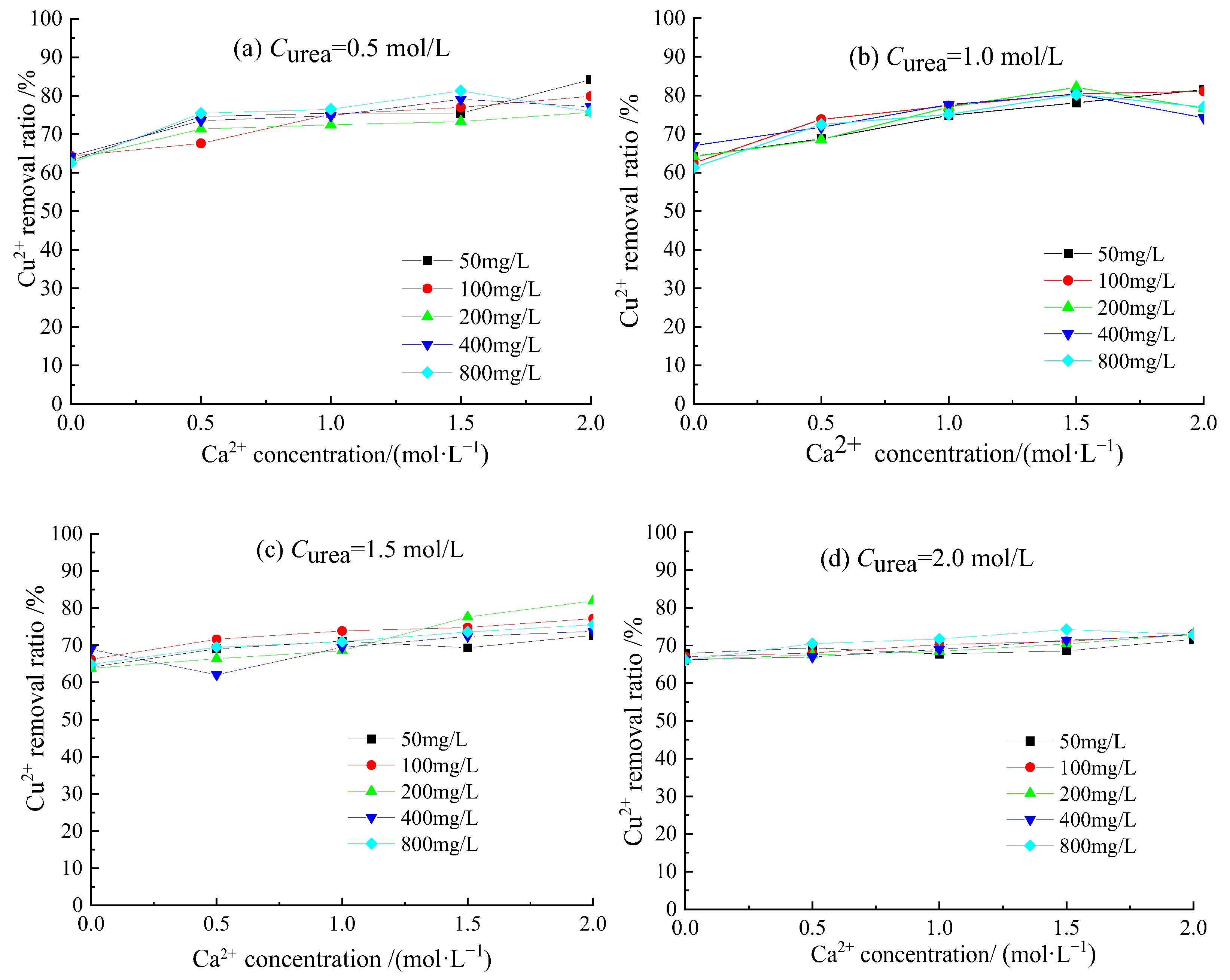
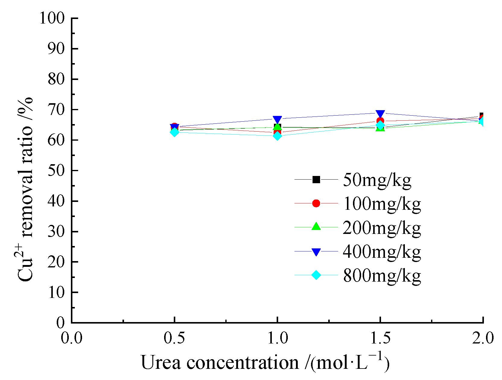
3.3. Removal Behavior of Cu2+ by Acclimatized S. pasteurii

Figure 6 shows the variation of the removal ratios of Cu2+ with increasing the concentrations of Ca2+ at different urea concentrations. As shown in Figure 6a, with the increase in Ca2+ concentration, the removal ratio of Cu2+ tended to gradually increase. When the concentration of Ca2+ increased to 2.0 mol/L, the removal ratios of Cu2+ had a slight decrease for the groups of initial concentrations of 400 and 800 mg/L, indicating that high concentrations of Ca2+ and Cu2+ inhibited the growth of bacteria, while the removal ratio increased slowly when the Cu2+ concentration was relatively small (i.e., 50, 100 and 200 mg/L). Compared with Figure 6a, the removal ratio in Figure 6b was relatively higher, because with the increase in urea concentration, the CO32− produced by urea hydrolysis continued to increase, providing more chances for precipitation. With the increase in Ca2+ concentration, the removal ratio of Cu2+ also increased, but when the Ca2+ concentration increased to 2.0 mol/L, the removal ratios of Cu2+ at concentrations of 200, 400 and 800 mg/L all decreased a little. It can be observed from Figure 6c that with further increasing the urea concentration, the corresponding Cu2+ removal ratio had a small decrease compared with that in Figure 6b. The increase in Ca2+ concentration could improve the removal efficiency of different concentrations of Cu2+, because Ca2+ concentration directly affects the formation of CaCO3 precipitation and the efficiency of coprecipitation of heavy metals [47]. The removal ratio of Cu2+ in Figure 6d was lower compared with that in Figure 6c, indicating that too much urea inhibited the biomineralization process.
Figure 7 shows the removal ratio of Cu2+ with increasing the concentration of urea from 0.5 to 2.0 mol/L without introducing a calcium ion; the removal ratio of Cu2+ was in the range of 62.6–64.4% when the urea concentration was 0.5 mol/L. In the absence of Ca2+, merely increasing the concentration of urea in the substrate of the medium did not significantly improve the removal efficiency of Cu2+, suggesting that the concentration of urea had few effects on the removal of Cu2+ when there was no calcium. It can be inferred that the mechanism of mineralization and immobilization of heavy metals by S. pasteurii is closely related to the production and quantity of CaCO3.
Under different conditions with varying the concentrations of urea and Ca2+, the removal ratio of Cu2+ could range from 62.1 to 84.2%. A higher urea concentration was not conducive to the removal of Cu2+, while the increase in Ca2+ concentration was conducive to the removal of Cu2+. When the concentration in urea was greater than 1.0 mol/L, the existence of Ca2+ could lead to a decreased removal ratio of Cu2+, as the combination of Ca2+ and CO32− was the dominant reaction. In general, when the concentrations of urea and Ca2+ were 1.0 and 1.5 mol/L, respectively, the best removal efficiency of Cu2+ could be obtained, and the removal ratio of Cu2+ could reach 78.1–82.1% when the initial concentration of Cu2+ varied from 50 to 800 mg/L. Compared with the very low removal ratio of Cu2+ from aqueous solutions by S. pasteurii in the reported studies [1,20,36,41], a much better removal efficiency of Cu2+ with the removal ratio above 80% could be obtained by elaborately adjusting the concentration of urea and calcium ion in this study, suggesting that acclimatizing the S. pasteurii bacteria by means of incubating them in a heavy-metal-amended nutrient broth is very effective.

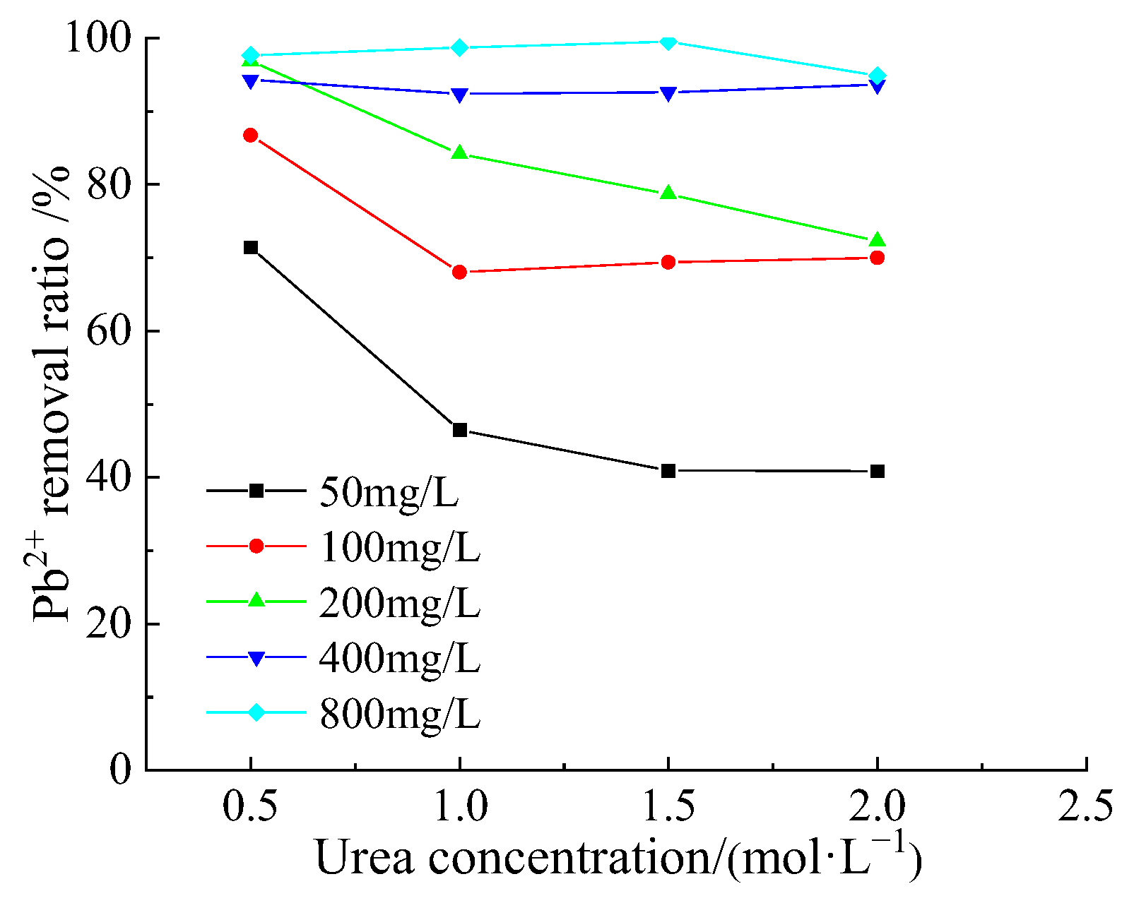
3.4. Removal Behavior of Pb2+ by Acclimatized S. pasteurii

Figure 8 shows the variation of removal ratios of Pb2+ with increasing the concentrations of Ca2+ at different urea concentrations. In Figure 8a–d, when the Ca2+ concentration was 0.5 mol/L, the removal efficiencies of Pb2+ were greatly improved in contrast with the tests with no Ca2+, and nearly all the Pb2+ ions could be removed from aqueous solutions. When both of the urea and Ca2+ concentrations were 0.5 mol/L, the removal ratio of Pb2+ could reach up to 98.0–100.0%, as shown in Figure 8a. Increasing the Ca2+ concentration from 0.5 to 1.0 mol/L had little effect on the Pb2+ removal when the urea concentrations were 0.5 and 1.0 mol/L, which can be observed from Figure 6b and Figure 8a. When increasing the urea concentration to 1.5 and 2.0 mol/L in Figure 6d and Figure 8c, the decline of the Pb2+ removal ratio was nonnegligible, and the higher the urea concentration, the more the Pb2+ removal ratio decreased.
In Figure 9, we can observed that when there was no Ca2+ in the solutions, the removal ratio of Pb2+ exhibited a decreasing trend, suggesting that the high urea concentration was unfriendly to the Pb2+ removal using S. pasteurii. The best removal ratio of Pb2+, 71.4 to 97.6%, occurred when the urea concentration was 0.5 mol/L.

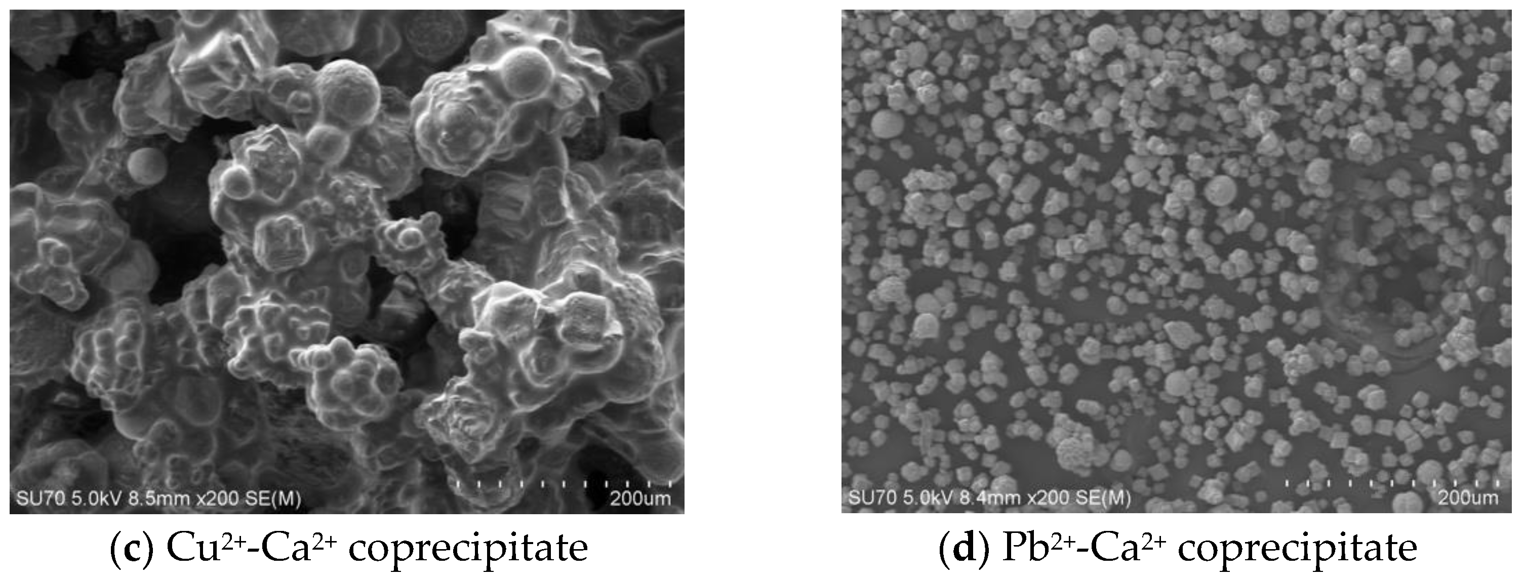
3.5. Biomineral Morphology

The removal of Cd2+, Cu2+ and Pb2+ from aqueous solutions via biomineralization driven by S. pasteurii demonstrated very good efficiency. The removal ratio of Cd2+ and Pb2+ was better than that of Cu2+, and nearly all the Cd2+ and Pb2+ ions in aqueous solutions could be removed.
Figure 10 and Figure 11 demonstrate the scanning electron microscope images and the X-ray diffraction patterns of the precipitates produced by the microbially induced carbonate precipitation. As can be observed from Figure 10a, the shape of the Ca2+ precipitate seems irregularly spherical and blocky, the particle sizes are not uniform and some particles are combined together to form agglomerates. In addition, the surfaces of some particles are smooth, and some are covered by many fine crystals; therefore, there are abundant micropores with different sizes in the entire structure. Through XRD analysis in Figure 11a, it is found that the Ca2+ precipitate is composed of two kinds of CaCO3 minerals, namely calcite and vaterite, and the proportion of calcite and vaterite is, respectively, 75.0% and 25.0%.
As shown in Figure 10b and Figure 11b, the overwhelming majority of Cd2+-Ca2+ coprecipitates are in the form of calcite minerals, accounting for as high as 98.8%, and have a blocky appearance. There is also a very small amount of another mineral called Ca0.25Cd0.75O. The particles, which have different sizes, are relatively dispersed and abundant pores can be observed in the structure.
The particle size of the Cu2+-Ca2+ coprecipitate is found to be relatively larger than the other precipitates and the color appears blue. In Figure 10c, we can observe that many particles of the Cu2+-Ca2+ coprecipitate aggregate and cement together to form clusters, and there seems to be no pore among the particles. Meanwhile, there are only large pores in this kind of structure, which is different from other precipitates. Some fine spherical particles can be observed on the surface of the clusters, and these are vaterite minerals, which can be confirmed by the XRD analysis in Figure 11c, demonstrating that the main mineral of Cu2+-Ca2+ coprecipitate is calcite (CaCO3), accounting for 95.3%.
In Figure 10d and Figure 11d, the shape of most of Pb2+-Ca2+ coprecipitates appears blocky, and the particles are much smaller and more dispersed than other precipitates. The main mineral is calcite, accounting for 96.5%, and the proportion of another mineral vaterite is only 3.5%. Although different types of crystals can be formed, vaterite is found to be in a very low percentage, similar with the reported studies [16].
As we observe from Figure 10, the shape and size of the Cu2+-Ca2+ coprecipitate are quite different from others. The clusters without any pore formed by Cu2+-Ca2+ coprecipitating enlarge the surface area of the particles, and inhibit more copper ions to enter into the structures, resulting in a much lower Cu2+ removal ratio than Cd2+ and Pb2+. As for Cd2+ and Pb2+, the sizes of the coprecipitates are relatively small, and there are many small micropores between particles. These abundant pores and great specific surface areas are conducive to the adsorption of heavy metals, leading to the very high removal ratio of Cd2+ and Pb2+.
Though the mechanism of the polymorph selection is not clearly understood now, many factors such as growth media, media concentration and calcium sources could influence the type of carbonate precipitates [48,49,50,51]. Calcite mineral is the most thermodynamically stable polymorph of CaCO3, and vaterite is a metastable and transitional phase of calcite [2,52]. In this study, we can observe from Figure 10 and Figure 11 that most coprecipitates were calcite minerals, and just a very small number of vaterite minerals were formed, indicating that the coprecipitates were relatively stable.
The heavy metals may be adsorbed on the cell surface, wrapped in calcite structure, or filled in the defect vacancy of CaCO3 crystal when coprecipitated with Ca2+; therefore, the crystal diffraction peaks of Cd2+, Cu2+ and Pb2+ in the XRD diffraction patterns cannot be easily found. However, the intensity of the diffraction peaks of calcite has been significantly enhanced. In the process of removing heavy metal ions in aqueous solutions by the MICP method, the exchangeable heavy metals will be converted into a carbonate-bonded state, thereby reducing the exchangeable heavy metal ion concentration in water. The CaCO3 crystals induced by the metabolic activity of urease producing bacteria are generally less soluble than those precipitated under abiotic environments [10,53], and the minerals formed by MICP method are very stable; therefore, the MICP technique, with a high efficiency of capturing heavy metals, can be a promising remediating method in the field of contaminated water and soil.

4. Conclusions

All the three heavy metals could be effectively removed by acclimatized S. pasteurii, and the Ca2+ and urea concentration both played critical roles in removing the heavy metals. In the absence of Ca2+, when the heavy metal concentration ranged from 50 to 800 mg/L, increasing urea concentrations in the nutrient broths was beneficial to the removal of Cd2+ with a removal ratio of 89.9–99.7% at Curea = 2.0 mol/L, but had little positive effect on the removal of Cu2+ and Pb2+ with a removal ratio of 62.6–64.4% and 71.4–97.6%, respectively at Curea = 0.5 mol/L. After introducing Ca2+ in the nutrient broths, heavy metal could coprecipitate with Ca2+, resulting in a much more effective removal of heavy metals, and the removal efficiency of Cd2+, Cu2+ and Pb2+ could reach 98.0–99.0, 78.1–82.1 and 98.0–100.0%, respectively, by adjusting the concentration of Ca2+ and urea. The overall removal efficiency for Cd2+ and Pb2+ was better than that for Cu2+ in aqueous solutions. The Cu2+ deposit aggregated and cemented to form clusters, which was different from Cd2+ and Pb2+ deposits with relatively dispersed microstructure. For all the three heavy metal precipitates, the predominant mineral was identified as calcite, the most thermodynamically stable polymorph of CaCO3. The heavy metals may be adsorbed on the cell surface, wrapped in calcite structure, or filled in the defect vacancy of CaCO3 crystal when coprecipitated with Ca2+, resulting in the not easily found XRD diffraction patterns.

Author Contributions

Conceptualization, Y.W. and X.L.; methodology, Y.W.; validation, Y.W., J.T. and X.L.; formal analysis, X.L.; investigation, K.L.; data curation, X.L. and K.L.; writing—original draft preparation, X.L.; writing—review and editing, Y.W. and J.T.; supervision, Y.W.; project administration, Y.W.; funding acquisition, Y.W. All authors have read and agreed to the published version of the manuscript.

Funding

This research was funded by the National Natural Science Foundation of China, grant number 51678311.

Institutional Review Board Statement

Not applicable.

Informed Consent Statement

Not applicable.

Data Availability Statement

Not applicable.

Acknowledgments

We would like to express our sincere gratitude for the financial support of the National Natural Science Foundation of China (grant 51678311) to this study. We also sincerely thank the reviewers for their helpful and constructive suggestions and the editors for their careful and patient work.

Conflicts of Interest

The authors declare no conflict of interest. The funders had no role in the design of the study; in the collection, analyses, or interpretation of data; in the writing of the manuscript; or in the decision to publish the results.

References

  1. Huang, X.; Zhang, R.; Cui, M.; Lai, H. Experimental Investigation on Bioremediation of Heavy Metal Contaminated Solution by Sporosarcina pasteurii under Some Complex Conditions. Water 2022, 14, 595. [Google Scholar] [CrossRef]
  2. Chen, X.; Zhang, D.; Larson, S.L.; Ballard, J.H.; Knotek-Smith, H.M.; Nie, J.; Hu, N.; Ding, D.; Han, F.X. Microbially Induced Carbonate Precipitation Techniques for the Remediation of Heavy Metal and Trace Element–Polluted Soils and Water. Water Air Soil Pollut. 2021, 232, 1–15. [Google Scholar] [CrossRef]
  3. Rai, P.K.; Lee, S.S.; Zhang, M.; Tsang, Y.F.; Kim, K.-H. Heavy metals in food crops: Health risks, fate, mechanisms, and management. Environ. Int. 2019, 125, 365–385. [Google Scholar] [CrossRef] [PubMed]
  4. Hu, B.; Wang, J.; Jin, B.; Li, Y.; Shi, Z. Assessment of the potential health risks of heavy metals in soils in a coastal industrial region of the Yangtze River Delta. Environ. Sci. Pollut. Res. 2017, 24, 19816–19826. [Google Scholar] [CrossRef]
  5. Hu, B.; Shao, S.; Ni, H.; Fu, Z.; Hu, L.; Zhou, Y.; Min, X.; She, S.; Chen, S.; Huang, M.; et al. Current status, spatial features, health risks, and potential driving factors of soil heavy metal pollution in China at province level. Environ. Pollut. 2020, 266, 114961. [Google Scholar] [CrossRef]
  6. Iqbal, A.; Jalees, M.I.; Farooq, M.U.; Cevik, E.; Bozkurt, A. Superfast adsorption and high-performance tailored membrane filtration by engineered Fe-Ni-Co nanocomposite for simultaneous removal of surface water pollutants. Colloids Surfaces A Physicochem. Eng. Asp. 2022, 652, 129751. [Google Scholar] [CrossRef]
  7. Iqbal, A.; Cevik, E.; Bozkurt, A.; Asiri, S.M.M.; Alagha, O.; Qahtan, T.F.; Jalees, M.I.; Farooq, M.U. Ultrahigh adsorption by regenerable iron-cobalt core-shell nanospheres and their synergetic effect on nanohybrid membranes for removal of malachite green dye. J. Environ. Chem. Eng. 2022, 10, 107968. [Google Scholar] [CrossRef]
  8. Wu, Y.; Li, H.; Li, Y. Biomineralization Induced by Cells of Sporosarcina pasteurii: Mechanisms, Applications and Challenges. Microorganisms 2021, 9, 2396. [Google Scholar] [CrossRef]
  9. Song, M.; Ju, T.; Meng, Y.; Han, S.; Lin, L.; Jiang, J. A review on the applications of microbially induced calcium carbonate precipitation in solid waste treatment and soil remediation. Chemosphere 2021, 290, 133229. [Google Scholar] [CrossRef]
  10. Arias, D.; Cisternas, L.A.; Rivas, M. Biomineralization Mediated by Ureolytic Bacteria Applied to Water Treatment: A Review. Crystals 2017, 7, 345. [Google Scholar] [CrossRef]
  11. Chuo, S.C.; Mohamed, S.F.; Mohd Setapar, S.H.; Ahmad, A.; Jawaid, M.; Wani, W.A.; Yaqoob, A.A.; Ibrahim, M.N.M. Insights into the Current Trends in the Utilization of Bacteria for Microbially Induced Calcium Carbonate Precipitation. Materials 2020, 13, 4993. [Google Scholar] [CrossRef] [PubMed]
  12. Zhu, T.; Edittrich, M. Carbonate Precipitation through Microbial Activities in Natural Environment, and Their Potential in Biotechnology: A Review. Front. Bioeng. Biotechnol. 2016, 4, 4. [Google Scholar] [CrossRef] [PubMed] [Green Version]
  13. Görgen, S.; Benzerara, K.; Skouri-Panet, F.; Gugger, M.; Chauvat, F.; Cassier-Chauvat, C. The diversity of molecular mechanisms of carbonate biomineralization by bacteria. Discov. Mater. 2020, 1, 1–20. [Google Scholar] [CrossRef]
  14. Yin, T.; Lin, H.; Dong, Y.; Li, B.; He, Y.; Liu, C.; Chen, X. A novel constructed carbonate-mineralized functional bacterial consortium for high-efficiency cadmium biomineralization. J. Hazard. Mater. 2020, 401, 123269. [Google Scholar] [CrossRef] [PubMed]
  15. Rajasekar, A.; Wilkinson, S.; Moy, C.K. MICP as a potential sustainable technique to treat or entrap contaminants in the natural environment: A review. Environ. Sci. Ecotechnol. 2021, 6, 100096. [Google Scholar] [CrossRef]
  16. Phillips, A.J.; Gerlach, R.; Lauchnor, E.; Mitchell, A.C.; Cunningham, A.B.; Spangler, L. Engineered applications of ureolytic biomineralization: A review. Biofouling 2013, 29, 715–733. [Google Scholar] [CrossRef] [Green Version]
  17. Silva-Castro, G.A.; Uad, I.; Rivadeneyra, A.; Vilchez, J.I.; Martin-Ramos, D.; González-López, J.; Rivadeneyra, M.A. Carbonate Precipitation of Bacterial Strains Isolated from Sediments and Seawater: Formation Mechanisms. Geomicrobiol. J. 2013, 30, 840–850. [Google Scholar] [CrossRef]
  18. Fujita, Y.; Redden, G.D.; Ingram, J.C.; Cortez, M.M.; Ferris, F.; Smith, R.W. Strontium incorporation into calcite generated by bacterial ureolysis. Geochim. Et Cosmochim. Acta 2004, 68, 3261–3270. [Google Scholar] [CrossRef] [Green Version]
  19. Lauchnor, E.G.; Schultz, L.N.; Bugni, S.; Mitchell, A.C.; Cunningham, A.B.; Gerlach, R. Bacterially Induced Calcium Carbonate Precipitation and Strontium Coprecipitation in a Porous Media Flow System. Environ. Sci. Technol. 2013, 47, 1557–1564. [Google Scholar] [CrossRef] [Green Version]
  20. Mugwar, A.J.; Harbottle, M.J. Toxicity effects on metal sequestration by microbially-induced carbonate precipitation. J. Hazard. Mater. 2016, 314, 237–248. [Google Scholar] [CrossRef]
  21. Mitchell, A.C.; Ferris, F.G. The coprecipitation of Sr into calcite precipitates induced by bacterial ureolysis in artificial groundwater: Temperature and kinetic dependence. Geochim. Et Cosmochim. Acta 2005, 69, 4199–4210. [Google Scholar] [CrossRef]
  22. Gebru, K.A.; Kidanemariam, T.G.; Gebretinsae, H.K. Bio-cement production using microbially induced calcite precipitation (MICP) method: A review. Chem. Eng. Sci. 2021, 238, 116610. [Google Scholar] [CrossRef]
  23. Zhan, M.; Pan, G.; Wang, Y.; Fu, M.; Lu, X. Recycled aggregate mortar enhanced by microbial calcite precipitation. Mag. Concr. Res. 2020, 72, 622–633. [Google Scholar] [CrossRef]
  24. Dhami, N.K.; Ereddy, M.S.; Mukherjee, A. Biomineralization of calcium carbonates and their engineered applications: A review. Front. Microbiol. 2013, 4, 314. [Google Scholar] [CrossRef] [PubMed] [Green Version]
  25. Balam, N.H.; Mostofinejad, D.; Eftekhar, M. Effects of bacterial remediation on compressive strength, water absorption, and chloride permeability of lightweight aggregate concrete. Constr. Build. Mater. 2017, 145, 107–116. [Google Scholar] [CrossRef]
  26. Vijay, K.; Murmu, M.; Deo, S.V. Bacteria based self healing concrete–A review. Constr. Build. Mater. 2017, 152, 1008–1014. [Google Scholar] [CrossRef]
  27. Al-Salloum, Y.; Hadi, S.; Abbas, H.; Almusallam, T.; Moslem, M. Bio-induction and bioremediation of cementitious composites using microbial mineral precipitation–A review. Constr. Build. Mater. 2017, 154, 857–876. [Google Scholar] [CrossRef]
  28. De Jong, J.T.; Soga, K.; Kavazanjian, E.; Burns, S.; van Paassen, L.A.; Al Qabany, A.; Aydilek, A.; Bang, S.S.; Burbank, M.; Caslake, L.F.; et al. Biogeochemical processes and geotechnical applications: Progress, opportunities and challenges. Géotechnique 2013, 63, 287–301. [Google Scholar] [CrossRef] [Green Version]
  29. Chu, J.; Ivanov, V.; Stabnikov, V.; Li, B. Microbial method for construction of an aquaculture pond in sand. Géotechnique 2013, 63, 871–875. [Google Scholar] [CrossRef]
  30. Salifu, E.; MacLachlan, E.; Iyer, K.R.; Knapp, C.W.; Tarantino, A. Application of microbially induced calcite precipitation in erosion mitigation and stabilisation of sandy soil foreshore slopes: A preliminary investigation. Eng. Geol. 2016, 201, 96–105. [Google Scholar] [CrossRef]
  31. Xiao, P.; Liu, H.; Xiao, Y.; Stuedlein, A.; Evans, T.M. Liquefaction resistance of bio-cemented calcareous sand. Soil Dyn. Earthq. Eng. 2018, 107, 9–19. [Google Scholar] [CrossRef]
  32. Duo, L.; Kan-Liang, T.; Hui-Li, Z.; Yu-Yao, W.; Kang-Yi, N.; Shi-Can, Z. Experimental investigation of solidifying desert aeolian sand using microbially induced calcite precipitation. Constr. Build. Mater. 2018, 172, 251–262. [Google Scholar] [CrossRef]
  33. Sharaky, A.M.; Mohamed, N.; Elmashad, M.E.; Shredah, N.M. Application of microbial biocementation to improve the physico-mechanical properties of sandy soil. Constr. Build. Mater. 2018, 190, 861–869. [Google Scholar] [CrossRef]
  34. Sasaki, T.; Kuwano, R. Undrained cyclic triaxial testing on sand with non-plastic fines content cemented with microbially induced CaCO3. Soils Found. 2016, 56, 485–495. [Google Scholar] [CrossRef]
  35. Fang, L.; Niu, Q.; Cheng, L.; Jiang, J.; Yu, Y.-Y.; Chu, J.; Achal, V.; You, T. Ca-mediated alleviation of Cd2+ induced toxicity and improved Cd2+ biomineralization by Sporosarcina pasteurii. Sci. Total Environ. 2021, 787, 147627. [Google Scholar] [CrossRef]
  36. Duarte-Nass, C.; Rebolledo, K.; Valenzuela, T.; Kopp, M.; Jeison, D.; Rivas, M.; Azócar, L.; Torres-Aravena, Á.; Ciudad, G. Application of microbe-induced carbonate precipitation for copper removal from copper-enriched waters: Challenges to future industrial application. J. Environ. Manag. 2019, 256, 109938. [Google Scholar] [CrossRef]
  37. Warren, P.A.M.L.A. Microbially Mediated Calcium Carbonate Precipitation: Implications for Interpreting Calcite Precipitation and for Solid-Phase Capture of Inorganic Contaminants. Geomicrobiol. J. 2001, 18, 93–115. [Google Scholar] [CrossRef]
  38. Li, M.; Cheng, X.; Guo, H. Heavy metal removal by biomineralization of urease producing bacteria isolated from soil. Int. Biodeterior. Biodegrad. 2013, 76, 81–85. [Google Scholar] [CrossRef]
  39. Jiang, N.-J.; Liu, R.; Du, Y.-J.; Bi, Y.-Z. Microbial induced carbonate precipitation for immobilizing Pb contaminants: Toxic effects on bacterial activity and immobilization efficiency. Sci. Total Environ. 2019, 672, 722–731. [Google Scholar] [CrossRef]
  40. Jalilvand, N.; Akhgar, A.; Alikhani, H.A.; Rahmani, H.A.; Rejali, F. Removal of Heavy Metals Zinc, Lead, and Cadmium by Biomineralization of Urease-Producing Bacteria Isolated from Iranian Mine Calcareous Soils. J. Soil Sci. Plant Nutr. 2019, 20, 206–219. [Google Scholar] [CrossRef]
  41. Kim, Y.; Kwon, S.; Roh, Y. Effect of Divalent Cations (Cu, Zn, Pb, Cd, and Sr) on Microbially Induced Calcium Carbonate Precipitation and Mineralogical Properties. Front. Microbiol. 2021, 12, 646748. [Google Scholar] [CrossRef] [PubMed]
  42. Dhami, N.K.; Quirin, M.E.C.; Mukherjee, A. Carbonate biomineralization and heavy metal remediation by calcifying fungi isolated from karstic caves. Ecol. Eng. 2017, 103, 106–117. [Google Scholar] [CrossRef]
  43. Lapierre, F.M.; Schmid, J.; Ederer, B.; Ihling, N.; Huber, R. Revealing nutritional requirements of MICP-relevant Sporosarcina pasteurii DSM33 for growth improvement in chemically defined and complex media. Sci. Rep. 2020, 10, 22448. [Google Scholar] [CrossRef] [PubMed]
  44. Kang, C.-H.; Han, S.-H.; Shin, Y.; Oh, S.J.; So, J.-S. Bioremediation of Cd by Microbially Induced Calcite Precipitation. Appl. Biochem. Biotechnol. 2013, 172, 1929–1937. [Google Scholar] [CrossRef]
  45. Chada, V.G.R.; Hausner, D.B.; Strongin, D.R.; Rouff, A.A.; Reeder, R.J. Divalent Cd and Pb uptake on calcite {1014} cleavage faces: An XPS and AFM study. J. Colloid Interface Sci. 2005, 288, 350–360. [Google Scholar] [CrossRef] [PubMed]
  46. Du, Y.; Lian, F.; Zhu, L. Biosorption of divalent Pb, Cd and Zn on aragonite and calcite mollusk shells. Environ. Pollut. 2011, 159, 1763–1768. [Google Scholar] [CrossRef]
  47. Kumari, D.; Qian, X.-Y.; Pan, X.; Achal, V.; Li, Q.; Gadd, G.M. Microbially-induced Carbonate Precipitation for Immobilization of Toxic Metals. Adv. Appl. Microbio. 2016, 94, 79–108. [Google Scholar] [CrossRef] [Green Version]
  48. Li, Q.; Zhang, B.; Ge, Q.; Yang, X. Calcium carbonate precipitation induced by calcifying bacteria in culture experiments: Influence of the medium on morphology and mineralogy. Int. Biodeterior. Biodegrad. 2018, 134, 83–92. [Google Scholar] [CrossRef]
  49. Rodriguez-Navarro, C.; Jroundi, F.; Schiro, M.; Ruiz-Agudo, E.; González-Muñoz, M.T. Influence of Substrate Mineralogy on Bacterial Mineralization of Calcium Carbonate: Implications for Stone Conservation. Appl. Environ. Microbiol. 2012, 78, 4017–4029. [Google Scholar] [CrossRef] [Green Version]
  50. Omoregie, A.I.; Ngu, L.H.; Ong, D.E.L.; Nissom, P.M. Low-cost cultivation of Sporosarcina pasteurii strain in food-grade yeast extract medium for microbially induced carbonate precipitation (MICP) application. Biocatal. Agric. Biotechnol. 2018, 17, 247–255. [Google Scholar] [CrossRef]
  51. Xu, J.; Du, Y.; Jiang, Z.; She, A. Effects of Calcium Source on Biochemical Properties of Microbial CaCO3 Precipitation. Front. Microbiol. 2015, 6, 1366. [Google Scholar] [CrossRef] [PubMed] [Green Version]
  52. Boulos, R.A.; Zhang, F.; Tjandra, E.S.; Martin, A.; Spagnoli, D.; Raston, C.L. Spinning up the polymorphs of calcium carbonate. Sci. Rep. 2014, 4, 3616. [Google Scholar] [CrossRef] [PubMed] [Green Version]
  53. Mitchell, A.C.; Ferris, F.G. The Influence of Bacillus pasteurii on the Nucleation and Growth of Calcium Carbonate. Geomicrobiol. J. 2006, 23, 213–226. [Google Scholar] [CrossRef]
Figure 1. Procedure of S. pasteurii acclimatization.
Applsci 12 09958 g001
Figure 2. Test procedure of heavy metal biomineralization by acclimatized S. pasteurii.
Applsci 12 09958 g002
Figure 3. Growth characteristics of S. pasteurii under different conditions ((a) pH = 7.0, T = 30 °C and Curea = 0.32 mol/L; (b) pH = 7.0, t = 48 h and Curea = 0.32 mol/L; (c) T = 30 °C, t = 48 h and Curea = 0.32 mol/L; (d) pH = 8.0, t = 48 h and T = 30 °C).
Applsci 12 09958 g003
Figure 4. Removal ratio of Cd2+ with different urea concentrations ((a) Curea = 0.5 mol/L; (b) Curea = 1.0 mol/L; (c) Curea = 1.5 mol/L; (d) Curea = 2.0 mol/L).
Applsci 12 09958 g004
Figure 5. Removal ratio of Cd2+ without Ca2+.
Applsci 12 09958 g005
Figure 6. Removal ratio of Cu2+ with different urea concentrations ((a) Curea = 0.5 mol/L; (b) Curea = 1.0 mol/L; (c) Curea = 1.5 mol/L; (d) Curea = 2.0 mol/L).
Applsci 12 09958 g006
Figure 7. Removal ratio of Cu2+ without Ca2+.
Applsci 12 09958 g007
Figure 8. Removal ratio of Pb2+ with different urea concentrations ((a) Curea = 0.5 mol/L; (b) Curea = 1.0 mol/L; (c) Curea = 1.5 mol/L; (d) Curea = 2.0 mol/L).
Applsci 12 09958 g008
Figure 9. Removal ratio of Pb2+ without Ca2+.
Applsci 12 09958 g009
Figure 10. SEM images of precipitates.
Applsci 12 09958 g010aApplsci 12 09958 g010b
Figure 11. XRD spectra of precipitates ((a) Ca2+ precipitate; (b) Cd2+-Ca2+ coprecipitate; (c) Cu2+-Ca2+ coprecipitate; (d) Pb2+-Ca2+ coprecipitate).
Applsci 12 09958 g011
Table 1. Growing conditions of S. pasteurii.
GrouppHT (°C)t (h)Curea (mol/L)
17.0300, 12, 24, 36, 48, 60, 720.32
27.010, 15, 20, 25, 30, 35, 40, 45480.32
35.0, 6.0, 7.0, 8.0, 9.0, 10.030480.32
48.030480, 0.08, 0.16, 0.32, 0.4
Publisher’s Note: MDPI stays neutral with regard to jurisdictional claims in published maps and institutional affiliations.

