Article

Distribution of Diatom Resting Stages in Sediment near Artificial Reefs Deployed in the Dysphotic Zone: Exploration of New Artificial Reef Function

1
Civil Engineering Research Institute for Cold Region, Public Works Research Institute, Sapporo 062-8602, Japan
2
Seed Bank Co., Ltd., Kyoto 606-8267, Japan
*
Authors to whom correspondence should be addressed.
Appl. Sci. 2022, 12(19), 9972; https://doi.org/10.3390/app12199972
Submission received: 16 September 2022 / Revised: 30 September 2022 / Accepted: 1 October 2022 / Published: 4 October 2022

Abstract

:

Featured Application

Our findings provide a crucial perspective on a novel accumulation effect of diatom resting stages by the deployment of artificial reefs, which may enhance the primary production of the overlying water column, subsequently contributing to the augmentation of fishery productivity through food chains.

Abstract

Artificial reefs (ARs) have multiple beneficial effects involved in increased fishery production. The present study investigated diatom resting stages in the sediments near ARs at a depth of about 90 m by the combined use of direct microscopic observation and the MPN method, seeking a novel function of AR deployment that potentially increases biological production. Although the concentration of diatom resting stages at A2 (north end station of the AR area), composed mainly of Chaetoceros, was ten times the concentration at A5 (non-reef station), the concentration at A1 (south end station of the AR area) was the same level as at A5. These results suggest that the AR contributes to the accumulation of diatom resting stages while the degree of accumulation is attributed to the arrangement of ARs and their physical environment such as flow directions. Interestingly, live pennate diatoms possessing photosynthetic pigments were constantly observed in all samples despite the low light intensity (≤1.2 µmol photons m−2 s−1), which provides new ecological insight into their biological and physiological characteristics. In this study, the distribution of viable diatom resting stages and live pennate diatoms in the sediment around ARs in a dysphotic zone was revealed for the first time, casting a light on the novel function of ARs potentially enhancing primary production around them.

1. Introduction

Artificial reefs (ARs), which are already used worldwide for multiple purposes, mainly involving fishery enhancement [1,2,3], are defined as “submerged structures deliberately constructed or placed on the seabed to emulate some functions of a natural reef such as protecting, regenerating, concentrating, and/or enhancing populations of living marine resources” [4], and the deployment of ARs has been proposed as an effective tool to enhance fishery resources in the ocean [5]. Although it is a well-known fact that fish aggregate densely around ARs, it is still controversial whether the fish aggregation is attributed to attraction (ARs merely attract fish from surrounding areas) or production (ARs contribute to fish reproduction), and Simon et al. [6] concluded that both attraction and production may occur around ARs by comparing ARs with natural reefs. An ecosystem perspective on the function of ARs, such as enhanced primary productivity, will surely provide new insight into the unresolved “attraction versus production” debate [7,8]. Actually, an increase in phytoplankton in a water column was pointed out by previous studies after the deployment of ARs [9,10]. In this way, AR deployment may play a role in enhancing the primary production of the overlying water column, subsequently contributing to the augmentation of fishery productivity through food chains.
Among the primary producers in the ocean, diatoms (Bacillariophyceae) are the main organisms and are estimated to contribute as much as 20% to the total primary production [11,12]. Many diatom species have been shown to form dormant cells called resting stages (resting spores and/or resting cells) to survive under unfavorable environmental conditions [13,14]. The formation of resting stages is induced when they are exposed to adverse conditions, such as nutrient depletion [15]. Resting stages settle down on the seafloor and will germinate/rejuvenate when the environmental conditions are favorable for their growth by being resuspended in a water column by a disturbing event such as vertical mixing and/or upwelling [16,17]. Therefore, diatom resting stages can serve as a seed population for a future diatom bloom [18]. If AR deployment increases pelagic diatom populations in the water column, the subsequent formation of diatom resting stages around AR is likely to be enhanced simultaneously.
One of the beneficial physical characteristics of ARs is a wake region generated behind an AR on the downstream side of the prevailing flow, where counterflows and eddies occur [19]. The flows and eddies allow phytoplankton and biodeposits in the water column to remain there and accumulate around the structure [19,20]. It is thus likely that an artificial reef also traps settling diatom resting stages and accumulates them in the vicinity. Through the above-mentioned processes, deployment of an AR may not only enhance primary production in the sympatric water column but also potentially boost primary production by supplying diatom resting stages. To our best knowledge, however, no studies have ever investigated the distribution of diatom resting stages around ARs.
In the present study, we investigated the concentration and composition of resting stages and vegetative cells of diatoms by direct microscopic observation and a culturing method (MPN method) of the sediment around ARs and a non-reef station at a depth of about 90 m in the dysphotic zone to reveal the accumulation effect of ARs.

2. Materials and Methods

2.1. Study Area

The study area was around an artificial reef system established by the Hokkaido prefectural government in 2017, located 10 km off the south of Rishiri Island (45°10′ N, 141°13′ N), Hokkaido, in the northernmost part of Japan (Figure 1a). The AR system comprised 3 artificial tower reefs made of steel (10 m by 10 m, 20 m tall) in the center and 156 cube reefs made of concrete (3 × 3 × 3 m) sporadically deployed in the outer circular zone (Figure 1b,c). Samplings were conducted at three stations (A1, A2, and A5) in the study area (Figure 1d). A1 was located at the innermost side of the circular zone; in contrast, A2 was located at the outermost side, while A5 was a non-reef station 150 m off the outer edge of the circular zone. Detailed information on the position and bottom depth of each station is summarized in Table 1.

2.2. Hydrographic Observation

The water temperature and light intensity at A2 and A5 were continuously observed with a water temperature logger (HOBO TidbiT v2 UTBI-001, Onset, Bourne, MA, USA) installed right above the bottom and a light quantum meter (COMPACT-LW, JFE Advantech, Hyogo, Japan) deployed 3.0 m above the seafloor. To observe the flow speed and direction near the seafloor, an electromagnetic flowmeter (INFINITY-EM, JFE Advantech, Hyogo, Japan) was also installed at 3.0 m above the seafloor of A5. All the observation devices were moored to an anchored rope at each station. The observation period was from 25 July 2018 to 7 August 2018. Vertical profiles of environmental variables (temperature, salinity, density, and chlorophyll fluorescence) were also measured with CTD (RINKO-Profiler ASTD102, JFE Advantech, Hyogo, Japan) at all the stations on 29 July 2018.

2.3. Particle Size Distribution and Organic Matter Measurements

Bottom sediments were collected with a Smith–McIntyre grab (0.05 m2) (RIGO, Tokyo, Japan) at each station. The particle size distribution in part of each sample was determined according to the Japanese Industrial Standards A 1204 (JIS A 1204). Sediment samples for loss on ignition (LOI) measurement were weighed and then heated to 600 °C for 2 h in a muffle furnace (FO510, Yamato Scientific, Tokyo, Japan) after drying at 105 °C until a constant weight was obtained. The total organic carbon (TOC) and total organic nitrogen (TON) concentrations of each sediment were also determined by dry combustion using a Sumigraph NC-220F (Sumika Chemical Analysis Service, Osaka, Japan).

2.4. Diatom Community and Population in Sediment

2.4.1. Sampling

Bottom sediments for diatom analysis were collected with the Smith–McIntyre grab at A5, while the samples at A1 and A2 were collected with an Ekman–Birge grab (0.02 m2) (RIGO, Tokyo, Japan) attached to a remotely operated vehicle (ROV) (Mitsui RTV. N-Hyper300EXY, Mitsui E&S Holdings, Tokyo, Japan) from the vicinity of a cube reef after visual confirmation through the ROV. The sediment samples were rapidly transported under cool-dark conditions to a laboratory and stored for two months at 4 °C under dark conditions. In the present study, compositions and concentrations of diatom resting stages were determined using the two methods below.

2.4.2. Direct Microscopic Observation

Resting stages of diatoms in the sediment samples were counted directly as follows, according to the method described by Ishii et al. [21]. One gram in wet weight of the sediment sample was suspended in filtered autoclaved seawater and sieved with 100, 20, and 10 µm nylon mesh in succession. The respective fractions captured on the 20 and 10 µm nylon mesh were each suspended in 10 mL of filtered autoclaved seawater. An aliquot of the suspensions was then placed on a glass slide and examined with inverted fluorescence microscope (IX-70, OLYMPUS, Tokyo, Japan) to identify and enumerate vegetative cells and resting stages of diatoms. The presence or absence of chloroplasts was also determined using the inverted fluorescence microscope. Identification of diatom resting stages was conducted in reference to Rines and Hargraves [22], Hasle and Syvertsen [23], Jensen and Moestrup [24], and Ishii et al. [25].

2.4.3. MPN Method

The number of viable diatom resting stages in the sediments was estimated by the most probable number (MPN) method [26,27]. The sediment sample (1.0 g wet weight) was suspended in autoclaved culture medium (Daigo’s IMK medium (Shiotani MS, Hyogo, Japan) + silicate) at a concentration of 0.1 g mL−1. The suspension was successively diluted with the medium (10−2, 10−3, 10−4, and 10−5), and 1 mL aliquots of each dilution were inoculated into five wells each on a microplate (48 wells). Incubation was carried out at 20 °C under a light intensity of about 100 µmol photons m−2 s−1 with a 14:10 h light–dark photocycle for 7 days, and the appearance of diatom vegetative cells in the five wells of each dilution series was examined with the inverted fluorescence microscope to calculate MPN, according to the statistical table of Throndsen [28].

3. Results

3.1. Environmental Conditions

The vertical profiles of the environmental variables closely agreed between the stations (Figure 2). Thermal and salinity stratification was observed at depths greater than about 55 m and chlorophyll fluorescence also peaked at around 55 m depth at all the stations. The vertical profile of water temperature was consistent among the stations, ranging from 7.5 to 20 °C. In particular, the influence of artificial reefs on hydrography was not seen. The temporal changes in water temperature near the seabed at A2 and A5 also exhibited a similar trend, ranging from 7.0 to 7.9 °C and from 7.1 to 8.0 °C, respectively. The light intensity near the seafloor was generally consistent at the two stations, ranging between 0.10 and 1.2 µmol photons m−2 s−1 at A2, and between 0.10 and 0.89 µmol photons m−2 s−1 at A5, respectively. The measured average flow speed near the seafloor at A5 was 0.06 m s−1, and the maximum flow speed was 0.16 m s−1 during the study period, and north-northwestward flows were dominant (31.4%) (Figure 3). The particle size in the bottom sediment was the coarsest at A1 among the stations; the fraction of 4.75–19 mm contributed 44.4% and the median diameter (D50) was 3.70 mm (Table 2). The particle size distribution at A2 showed a higher proportion of particles smaller than 0.85 mm than at A1 (A1: 35.8%, A2: 49.7%), although the fraction of particles of 4.75–19 mm was also relatively high (29.5%), while the D50 showed a middle value (0.89 mm) among the sites. The particle size distribution of A5 was dominated by the fraction of <0.005 mm (28.3%) and the D50 was the smallest among the three stations (0.12 mm). Overall, the particle sizes of the sediment at the AR stations (A1, A2) were larger than at the non-reef station (A5). The LOI of the sediment sample was 4.1% at A2 and A5 and was relatively low at A1 (3.5%). The TOC concentration in the sediment of A1, A2, and A5 was 5.2, 5.9, and 5.4 mg g−1, respectively, while the content of TON was under the limit of quantification (<1.0 mg g−1) at all the stations. The LOI and TOC concentrations at A2 showed the highest values among the three stations. In contrast, those at A1 located in the AR area also showed the lowest values among the stations.

3.2. Diatom Resting Stages

3.2.1. Abundances and Composition Based on Direct Microscopic Observation

The concentration of viable cells at A1, A2, and A5 was 560, 8820, and 840 cells g−1 wet sediment, respectively, and the proportion of Chaetoceros was more than 50% at all the stations, in particular, accounting for 93.4% at A2 (Figure 4a). The concentration and composition of the dominant Chaetoceros resting stages are displayed in Figure 4b, and seven different representative resting stage types morphologically identified as Chaetoceros in the present study are also shown in Figure 4c. However, the resting stages of C. affinis and C. vanheurckii were counted as “Chaetoceros affinis or vanheurckii”, owing to inadequate information on morphological characteristics to distinguish between the two species. There were also multiple resting stages of Chaetoceros that could not be identified at the species level in this study. The species composition of Chaetoceros exhibited a general resemblance among the stations, in which C. contortus and C. radicans were consistently dominant. Putative resting stages of Actinocyclus sp., Actinoptychus senarius, and Odontella aurita, and Thalassiosira sp., together with resting spores of Leptocylindrus danicus, were also observed (Figure 5). It is noteworthy that several motile pennate diatoms with chloroplasts were constantly observed in all the sediment samples, reaching the highest concentration (260 cells g−1 wet sediment) at A2 (Figure 6 and Table 3). The concentration of diatom resting stages at A2 located on the northernmost side of the AR area was about 10-fold that at A5 (non-reef station), although the concentration at A1, the south end station of the AR area, was lower than at A5.

3.2.2. Abundance of Viable Cells Estimated by MPN

The diatom species confirmed by MPN belonged to four genera (Chaetoceros spp., Odontella spp., Skeletonema spp., and Thalassiosira spp.) which were fewer than those with direct observation. However, the MPNs of viable cells at A1, A2, and A5 were 1735, 9920, and 741 cells g−1 wet sediment, respectively (Figure 7), and the highest value calculated by MPN at A2 showed close agreement with the direct observation. On the other hand, the contribution of Chaetoceros spp. was relatively low in the species composition of the MPN, unlike the result of the direct observation. The pennate diatoms were generally dominant in the composition of each station, accounting for 74.9%, 23.2%, and 72.9% at A1, A2, and A5, respectively. In the species composition of A2, however, unidentified spherical cells of a size of about 10 µm were abundantly observed (49.4%). The MPN value of viable diatom cells at A2 was more than 13 times that at the non-reef station (A5).

4. Discussion

The previous studies reported that the deployment of ARs resulted in increases in primary production in the surrounding environment, enhancing the feeding environment for marine organisms induced by the promotion of settling suspended particles, thus contributing to the augmentation of fishery productivity [10,29,30]. The primary productivity increment is often explained as the upwelling generated by ARs bringing nutrient-rich water, which enhances primary production in the overlying water [9,10]. Diatoms, the major marine primary producer [31], are classified into two types, namely centric and pennate [23], and the key ecophysiological characteristic of the most centric planktonic diatom species is their dormant stages [13]. These dormant stages can be of two types: resting cells [32] or resting spores [33]. Once produced, they settle down to the bottom, serving as seed populations for a future diatom bloom [18]. Thus, if enhancement of primary production by ARs is contemplated, it is essential to also integrate information on the distribution of diatom dormant cells and the physical effect of ARs such as the wake region, known to accumulate biodeposits [19,20]. The present study has demonstrated that the concentration of the diatom resting stages at A2 (north end of the cube reef area) reached 8920 cells g−1 wet sediment with the direct microscopic observation method, mainly composed of Chaetoceros, the dominant phytoplankton group distributed worldwide [34], and the value was more than ten times the concentration at the non-reef station (A5). On the other hand, interestingly, the cell concentration at A1, also a station near ARs but located at the south end, was the same level as that at the non-reef station. From the point of view of the environmental variables observed in this study, there were no distinct differences in the vertical environmental profiles (Figure 2) and temporal changes in water temperature at the seafloor. On the west coast of Hokkaido, where the study area is located, the Tsushima Warm Current flows northward in the surface layer, and northward flows were also observed at a depth of 100 m [35]. The dominant flow direction observed near the seafloor at A5 during the study period was also northerly (Figure 3). A2 was located approximately 55 m north of A1, and multiple cubic reefs were densely distributed to the south of A2, while only tower reefs were distantly located to the south of A1 (Figure 1c,d). In general, TOC concentration is inversely proportional to the particle size of the sediment; however, both the TOC concentration and LOI at A2 showed the highest values, even though the particle size at A2 was much larger than at A5 (Table 2). In general, soft sediments such as clay and silt are not suitable substrates for AR placement because ARs may sink into these sediments; thus, hard substrates are recommended as a deployment site for ARs [36]. The original substratum of the AR system in this study had mainly been composed of gravel before its deployment. However, although the proportion of gravel (>2.0 mm) was still high, accounting for 58.4% and 44.4% at A1 and A2, respectively, the proportion of clay and silt smaller than 0.075 mm was also relatively high (A1: 20.4%, A2: 28.5%), and the particle size at A2 was also smaller than at A1. It is therefore assumed that A2, which is positioned on the downstream side of A1, was within the range of the wake region that spreads out northward. Assuming the same amount of supply, the highest concentration of diatom resting stages at A2 can reasonably be explained by the positional relationship between the stations and the dominant flow direction. In other words, our results suggest that ARs do not necessarily accumulate diatom resting stages in their vicinity, but the wake region generated by ARs traps settling diatom resting stages and accumulates in their vicinity.
In this study, similar values to the direct observation values were also obtained by the MPN method, indicating that diatom cells in the sediment collected at a depth of about 90 m were mostly viable (Figure 4a and Figure 7). However, the species composition obtained with the MPN method showed a much lower ratio of Chaetoceros compared to that with direct observation (Figure 7). In contrast, the ratio of pennate diatoms was confirmed to be high at all stations with the MPN method. For the direct microscopic observation, the sediment subsamples were consecutively sieved through 100, 20, and 10 µm mesh and the remaining diatoms on the 20 and 10 µm mesh were observed, while the sediment samples only diluted with the medium were used for the MPN method. The above-mentioned differences may be attributed to the compositions derived from different methods, notably the pennate diatoms smaller than 10 µm and unidentified spherical cells (10 µm in diameter) by the MPN method. Shinada et al. [37] calculated the concentration of diatom resting stages in sediment at a depth of 90 m in Funka Bay on the Pacific coast of Hokkaido, reporting that the concentration of Chaetoceros (C. sociale, C. compressus, and Chaetoceros spp.) was 18,000 cells g−1 wet sediment in July 1995, corresponding to the timing of the present study (late July 2018). Although the concentration of Chaetoceros at A2 in the present study was 8240 cells g−1 wet sediment determined with direct microscopic observation, which was less than half of the value in Funka bay, it was more than fifteen times the Chaetoceros concentration in Funka Bay in August 1995 (523 cells g−1 wet sediment) [37]. However, diatom resting stages are generally more abundant in coastal than offshore regions [15] and the Sea of Japan off Hokkaido, where the present study was conducted, is also characterized as oligotrophic compared to other regions of Hokkaido [38]. Considering all the circumstances, the concentration of diatom resting stages at A2 was sufficiently high, subject to the effect of ARs. The resting stages of Chaetoceros accounted for a large proportion (>50%) of the species composition at all of the stations. In the coastal waters of the Japan Sea and the Okhotsk Sea off Hokkaido, Chaetoceros species are frequently observed and are dominant from April/May to November [39,40]. This is consistent with the results that Chaetoceros are the most dominant in the sediment collected in the present study. Light is considered a critical factor for germination of diatom resting stages [13,15,41], and the minimum light intensity required for germination is reported to be 1.3 µmol photons m−2 s−1 for resting spores of Chaetoceros spp. [41]. Although the light intensity measured during the present study was mostly below 1.0 µmol photons m−2 s−1, the light intensity monitored at the same location in September 2019 was often confirmed to be over 1.3 µmol photons m−2 s−1 during the daytime, indicating that the minimum light intensity for germination reached the sampling stations (ca. 90 m) although not always. The germinated cells may not effectively enhance primary production on a regular basis if the cells are not carried to the euphotic zone by an upward flow such as upwelling and/or vertical mixing [16,17]. According to previous studies, the resting stages of Chaetoceros spp. usually keep their viability for approximately 500 to 700 days [15,18,41], while a recent study succeeded with the germination of several millennia-old ancient Chaetoceros resting spores from the Baltic Sea [42]. The advantageous conditions for the survival of diatom resting stages are cool temperatures and dark conditions [15], and diatom resting stages were accordingly confirmed to be in a good state of preservation in this study (Figure 4c and Figure 5), indicating that the environmental conditions at A2 (north end of the cube reef area) were favorable for the survival of resting stages, potentially serving as a “seed bank” for many years.
Interestingly, viable pennate diatoms with chloroplasts were constantly observed in the bottom sediments at a depth of about 90 m both with direct observation and the MPN method (Figure 6 and Figure 7). Live benthic pennate diatoms have been found in sediments at a depth of 191 m in Onslow Bay, North Carolina [43], as well as in sediments at a depth of more than 100 m in the Beaufort Sea according to several studies using the MPN method [44,45], implying that the pennate diatoms adapted chromatically [43,46] and grew autotrophically under low light or hetero-/mixotrophic living conditions [47]. On the other hand, since some pennate diatoms are known to form resting stages [14,48,49], they may be in a dormant period on the seafloor, even though the formation of resting stages is not common for pennate diatoms [13,23,50]. However, one pennate diatom species exhibited growth at light intensity controlled by about 1 µmol photon m−2 s−1, which was isolated from the bottom sediment of the study area [51]; thus, it is likely that these pennate diatoms were living as vegetative cells on the seafloor of the study area, suggesting novel benthic food webs around ARs deployed at a dysphotic seafloor starting from these diatoms. Few studies have investigated such benthic microalgal production below a depth of 60 m [43,52], and further investigation of the ecology and physiology of pennate diatoms in the dysphotic zone is required to understand their ecological role in an offshore AR ecosystem.

5. Conclusions

This study investigated the distribution of diatom resting stages in sediments around ARs at a depth of about 90 m for the first time, suggesting that the AR has a novel function of accumulating diatom resting stages in its vicinity. Diatom resting stages can survive for years in sediment and serve as a seed population for a future diatom bloom. Thus, the ARs may function as a reservoir of diatom resting stages over the years, contributing to enhancing the primary production around it. However, further investigations are needed to test the universality of the accumulation effect of diatom resting stages by ARs. Moreover, motile pennate diatoms with chloroplasts were constantly observed in the bottom sediments of the study area, suggesting that the pennate diatoms are alive as vegetative cells, serving as a base for novel food webs around ARs in a dysphotic zone. However, their ecology and physiology remain largely unknown. We hope that potential primary productions of diatom resting stages and those benthic diatoms are taken into account for AR researchesin the future.

Author Contributions

Conceptualization, K.S. and N.I.; methodology and formal analysis, K.-I.I. and A.I.; investigation, K.S.; writing―original draft preparation, T.M. and N.I.; visualization, T.M.; writing―review and editing, T.M., K.S., K.-I.I., A.I., and N.I. All authors have read and agreed to the published version of the manuscript.

Funding

This research received no external funding.

Institutional Review Board Statement

Not applicable.

Data Availability Statement

Not applicable.

Acknowledgments

We express our sincere gratitude to the members of the Fisheries Engineering Research Team, Civil Engineering Research Institute for Cold Region (CERI), Japan. We would like to sincerely thank Kazumi Matsuoka from Nagasaki University for his helpful comments on the manuscript.

Conflicts of Interest

Authors Ken-Ichiro Ishii and Ayako Imura were employed by the company Seed Bank Co., Ltd. The remaining authors declare that the research was conducted in the absence of any commercial or financial relationships that could be construed as a potential conflict of interest.

References

  1. Baine, M. Artificial reefs: A review of their design, application, management and performance. Ocean Coast. Manag. 2001, 44, 241–259. [Google Scholar] [CrossRef]
  2. Becker, A.; Taylor, M.D.; Folpp, H.; Lowry, M.B. Managing the development of artificial reef systems: The need for quantitative goals. Fish Fish. 2018, 19, 740–752. [Google Scholar] [CrossRef]
  3. Lee, M.O.; Otake, S.; Kim, J.K. Transition of artificial reefs (ARs) research and its prospects. Ocean Coast. Manag. 2018, 154, 55–65. [Google Scholar] [CrossRef]
  4. London Convention and Protocol/UNEP. London Convention and Protocol/UNEP Guidelines for the Placement of Artificial Reefs; IMO (The International Marine Organization): London, UK, 2009; pp. 1–100. [Google Scholar]
  5. Paxton, A.B.; Shertzer, K.W.; Bacheler, N.M.; Kellison, G.T.; Riley, K.L.; Taylor, J.C. Meta-analysis reveals artificial reefs can be effective tools for fish community enhancement but are not one-size-fits-all. Front. Mar. Sci. 2020, 7, 282. [Google Scholar] [CrossRef]
  6. Simon, T.; Pinheiro, H.T.; Joyeux, J. Target fishes on artificial reefs: Evidences of impacts over nearby natural environments. Sci. Total Environ. 2011, 409, 4579–4584. [Google Scholar] [CrossRef]
  7. Layman, C.A.; Allgeier, J.E. An ecosystem ecology perspective on artificial reef production. J. Appl. Ecol. 2020, 57, 2139–2148. [Google Scholar] [CrossRef]
  8. Layman, C.A.; Allgeier, J.E.; Montaña, C.G. Mechanistic evidence of enhanced production on artificial reefs: A case study in a Bahamian seagrass ecosystem. Ecol. Eng. 2016, 95, 574–579. [Google Scholar] [CrossRef] [Green Version]
  9. Jeong, Y.K.; Lee, H.N.; Park, C.; Kim, D.S.; Kim, M.C. Variation of phytoplankton and zooplankton communities in a sea area, with the building of an artificial upwelling structure. Anim. Cells Syst. 2013, 17, 63–72. [Google Scholar] [CrossRef]
  10. Yu, J.; Chen, P.; Tang, D.; Qin, C. Ecological effects of artificial reefs in Daya Bay of China observed from satellite and in situ measurements. Adv. Space Res. 2015, 55, 2315–2324. [Google Scholar] [CrossRef]
  11. Field, C.B.; Behrenfeld, M.J.; Randerson, J.T.; Falkowski, P. Primary production of the biosphere: Integrating terrestrial and oceanic components. Science 1998, 281, 237–240. [Google Scholar] [CrossRef] [PubMed]
  12. Mann, D.G. The species concept in diatoms. Phycologia 1999, 38, 437–495. [Google Scholar] [CrossRef] [Green Version]
  13. McQuoid, M.R.; Hobson, L.A. Diatom resting stages. J. Phycol. 1996, 32, 889–902. [Google Scholar] [CrossRef]
  14. Belmonte, G.; Rubino, F. Resting cysts from coastal marine plankton. In Oceanography and Marine Biology: An Annual Review; Hawkins, S.J., Allcock, A.L., Bates, A.E., Firth, L.B., Smith, I.P., Swearer, S.E., Todd, P.A., Eds.; CRC Press: Boca Raton, FL, USA, 2019; Volume 57, pp. 1–88. [Google Scholar]
  15. Hargraves, P.E.; French, F.W. Diatom resting spores: Significance and strategies. In Survival Strategies of the Algae; Fryxell, G.A., Ed.; Cambridge University Press: Cambridge, UK, 1983; pp. 49–67. [Google Scholar]
  16. Pitcher, G.C. Phytoplankton seed populations of the Cape Peninsula upwelling plume, with particular reference to resting spores of Chaetoceros (Bacillariophyceae) and their role in seeding upwelling waters. Estuar. Coast. Shelf Sci. 1990, 31, 283–301. [Google Scholar] [CrossRef]
  17. Ishikawa, A.; Furuya, K. The role of diatom resting stages in the onset of the spring bloom in the East China Sea. Mar. Biol. 2004, 145, 633–639. [Google Scholar] [CrossRef]
  18. Itakura, S.; Imai, I.; Itoh, K. “Seed bank” of coastal planktonic diatoms in bottom sediments of Hiroshima Bay, Seto Inland Sea, Japan. Mar. Biol. 1997, 128, 497–508. [Google Scholar] [CrossRef]
  19. Oh, T.G.; Otake, S.; Lee, M.O. Estimating the effective wake region (current shadow) of artificial reefs. In Artificial Reefs in Fisheries Management; Bortone, S.A., Brandini, F.P., Fabi, G., Otake, S., Eds.; CRC Press LLC: Boca Raton, FL, USA, 2011; pp. 279–295. [Google Scholar]
  20. Sheng, P.Y. Physical characteristics and engineering at reef sites. In Artificial Reef Evaluation: With Application to Natural Marine Habitats; Seaman, W.J., Ed.; CRC Press LLC: Boca Raton, FL, USA, 2000; pp. 51–94. [Google Scholar]
  21. Ishii, K.-I.; Imai, I.; Natsuike, M.; Sawayama, S.; Ishino, R.; Liu, W.; Fukusaki, K.; Ishikawa, A. A simple technique for establishing axenic cultures of centric diatoms from resting stage cells in bottom sediments. Phycologia 2018, 57, 674–679. [Google Scholar] [CrossRef]
  22. Rines, J.E.B.; Hargraves, P.E. The Chaetoceros Ehrenberg (Bacillariophyceae) Flora of Narragansett Bay, Rhode Island, U.S.A.; Lubrecht & Cramer: New York, NY, USA, 1988; pp. 1–196. [Google Scholar]
  23. Hasle, G.R.; Syvertsen, E.E. Marine Diatoms. In Identifying Marine Phytoplankton; Tomas, C.R., Ed.; Academic Press: London, UK, 1997; pp. 5–385. [Google Scholar]
  24. Jensen, K.G.; Moestrup, Ø. The Genus Chaetoceros (Bacillariophyceae) in inner Danish coastal waters. Opera Bot. 1998, 133, 5–68. [Google Scholar] [CrossRef]
  25. Ishii, K.-I.; Iwataki, M.; Matsuoka, K.; Imai, I. Proposal of identification criteria for resting spores of Chaetoceros species (Bacillariophyceae) from a temperate coastal sea. Phycologia 2011, 50, 351–362. [Google Scholar] [CrossRef]
  26. Imai, I.; Itoh, K.; Anraku, M. Extinction dilution method for enumeration of dormant cells of res tide organisms in marine sediments. Bull. Plankton Soc. Jpn. 1984, 31, 123–124. [Google Scholar]
  27. Anderson, D.M.; Fukuyo, Y.; Matsuoka, K. Cyst methodologies. In Manual on Harmful Marine Microalgae; Hallegraeff, G.M., Anderson, D.M., Cembella, A.D., Eds.; IOC-UNESCO: Paris, France, 1995; pp. 229–249. [Google Scholar]
  28. Throndsen, J. The dilution-culture method. In Phytoplankton Manual; Sournia, A., Ed.; UNESCO: Paris, France, 1978; pp. 218–224. [Google Scholar]
  29. Okano, T.; Takeda, M.; Nakagawa, Y.; Hirata, K.; Mitsuhashi, K.; Kawaguchi, S.; Ito, J. Artificial reefs to induce upwelling to increase fishery resources. In Artificial Reefs in Fisheries Management; Bortone, S.A., Brandini, F.P., Fabi, G., Otake, S., Eds.; CRC Press LLC: Boca Raton, FL, USA, 2011; pp. 265–278. [Google Scholar]
  30. Leitão, F. Artificial reefs: From ecological processes to fishing enhancement tools. Braz. J. Oceanogr. 2013, 61, 77–81. [Google Scholar] [CrossRef]
  31. Armbrust, E.V. The life of diatoms in the world’s oceans. Nature 2009, 459, 185–192. [Google Scholar] [CrossRef]
  32. Jewson, D.H.; Lowry, S.F.; Bowen, R. Co-existence and survival of diatoms on sand grains. Eur. J. Phycol. 2006, 41, 131–146. [Google Scholar] [CrossRef] [Green Version]
  33. Sugie, K.; Kuma, K. Resting spore formation in the marine diatom Thalassiosira nordenskioeldii under iron- and nitrogen-limited conditions. J. Plankton Res. 2008, 30, 1245–1255. [Google Scholar] [CrossRef] [Green Version]
  34. Lebranc, K.; Arístegui, J.; Armand, L.; Assmy, P.; Beker, B.; Bode, A.; Breton, E.; Cornet, V.; Gibson, J.; Gosselin, M.-P.; et al. A global diatom database—Abundance, biovolume and biomass in the world ocean. Earth Syst. Sci. Data 2012, 4, 149–165. [Google Scholar] [CrossRef] [Green Version]
  35. Higaki, N.; Isoda, Y.; Honda, S. Mode waters observed around Musashi Rise west of Hokkaido. Oceanogr. Jpn. 2009, 18, 335–350. [Google Scholar]
  36. United States Department of Commerce, National Oceanic and Atmospheric Administration. National Artificial Reef Plan (As Amended): Guidelines for Siting, Construction, Development, and Assessment of Artificial Reefs; NOAA: Silver Spring, MD, USA, 2007; pp. 1–54.
  37. Shinada, A.; Shiga, N.; Ban, S. Origin of Thalassiosira diatoms that cause the spring phytoplankton bloom in Funka Bay, southwestern Hokkaido, Japan. Plankton Biol. Ecol. 1999, 46, 89–93. [Google Scholar]
  38. Kuribayashi, T.; Abe, T.; Montani, S. Historical δ15N records of Saccharina specimens from oligotrophic waters of Japan Sea (Hokkaido). PLoS ONE 2017, 12, e0180760. [Google Scholar] [CrossRef] [Green Version]
  39. Fukui, D.; Kitatsuji, S.; Ikeda, T.; Shiga, N.; Yamaguchi, A. Long-term changes in the abundance and community structure of net-phytoplankton in Oshoro Bay, Hokkaido. Bull. Plankton. Soc. Jpn. 2010, 57, 30–40. [Google Scholar]
  40. Matsumoto, T.; Matsuno, K.; Katakura, S.; Kasai, H.; Yamaguchi, A. Seasonal variability of the protist community and production in the southern Okhotsk Sea revealed by weekly monitoring. Reg. Stud. Mar. Sci. 2021, 43, 101683. [Google Scholar] [CrossRef]
  41. Hollibaugh, J.T.; Seibert, D.L.R.; Thomas, W.H. Observations on the survival and germination of resting spores of three Chaetoceros (Bacillariophyceae) species. J. Phycol. 1981, 17, 1–9. [Google Scholar] [CrossRef]
  42. Sanyal, A.; Larsson, J.; van Widum, F.; Andrén, T.; Moros, M.; Lönn, M.; Andrén, F. Not dead yet: Diatom resting spores can survive in nature for several millennia. Am. J. Bot. 2022, 109, 67–82. [Google Scholar] [CrossRef] [PubMed]
  43. McGee, D.; Laws, R.A.; Cahoon, L.B. Live benthic diatoms from the upper continental slope: Extending the limits of marine primary production. Mar. Ecol. Prog. Ser. 2008, 356, 103–112. [Google Scholar] [CrossRef] [Green Version]
  44. Tsukazaki, C.; Ishii, K.-I.; Saito, R.; Matsuno, K.; Yamaguchi, A.; Imai, I. Distribution of viable diatom resting stage cells in bottom sediments of the eastern Bering Sea shelf. Deep-Sea Res. II 2013, 94, 22–30. [Google Scholar] [CrossRef]
  45. Fukai, Y.; Matsuno, K.; Fujiwara, A.; Suzuki, K.; Richlen, M.L.; Fachon, E.; Anderson, D.M. Impact of sea-ice dynamics on the spatial distribution of diatom resting stages in sediments of the Pacific Arctic Region. J. Geophys. Res. Oceans 2021, 126, e2021JC017223. [Google Scholar] [CrossRef]
  46. Cahoon, L.B.; Laws, R.A.; Savidge, T.W. Characteristics of benthic microalgae from the North Carolina outer continental shelf and slope: Preliminary results. In Diving for Science. Proceedings of the 12th Annual Scientific Diving Symposium, Wilmington, NC, USA, 24–27 September 1992; Cahoon, L.B., Ed.; American Academy of Underwater Sciences: Costa Mesa, CA, USA, 1992; pp. 61–68. [Google Scholar]
  47. Marella, T.K.; Bhattacharjya, R.; Tiwari, A. Impact of organic carbon acquisition on diatom regrowth and functional biomolecule production in diatoms. Microb. Cell Fact. 2021, 20, 135. [Google Scholar] [CrossRef]
  48. Anderson, R.O. Respiration and photosynthesis during resting cell formation in Amphora coffeaeformis (Ag.) Kütz. Limnol. Oceanogr. 1976, 21, 452–456. [Google Scholar] [CrossRef]
  49. von Stosch, H.A.; Fecher, K. “Internal thecae” of Eunotia soleirolii (Bacillariophyceae): Development, structure, and function as resting spores. J. Phycol. 1979, 15, 233–243. [Google Scholar] [CrossRef]
  50. Ishii, K.-I.; Matsuoka, K.; Imai, I.; Ishikawa, A. Life cycle strategies of the centric diatoms in a shallow embayment revealed by the plankton emergence trap/chamber (PET chamber) experiments. Front. Mar. Sci. 2022, 9, 889633. [Google Scholar] [CrossRef]
  51. Sudo, K.; Inaba, N.; Matsumoto, T.; Matono, H.; Ishii, K. Growth characteristics of diatom resting stage cells in sediments around offshore artificial reefs. In Abstracts for the 2021 Joint Meeting of The Japanese Association of Benthology and The Plankton Society of Japan; Joint Meeting of The Japanese Association of Benthology and The Plankton Society of Japan: Kagoshima, Japan, 2021; p. 91. [Google Scholar]
  52. Cahoon, L.B. The role of benthic microalgae in neritic ecosystems. Oceanogr. Mar. Biol. Annu. Rev. 1999, 37, 47–86. [Google Scholar]
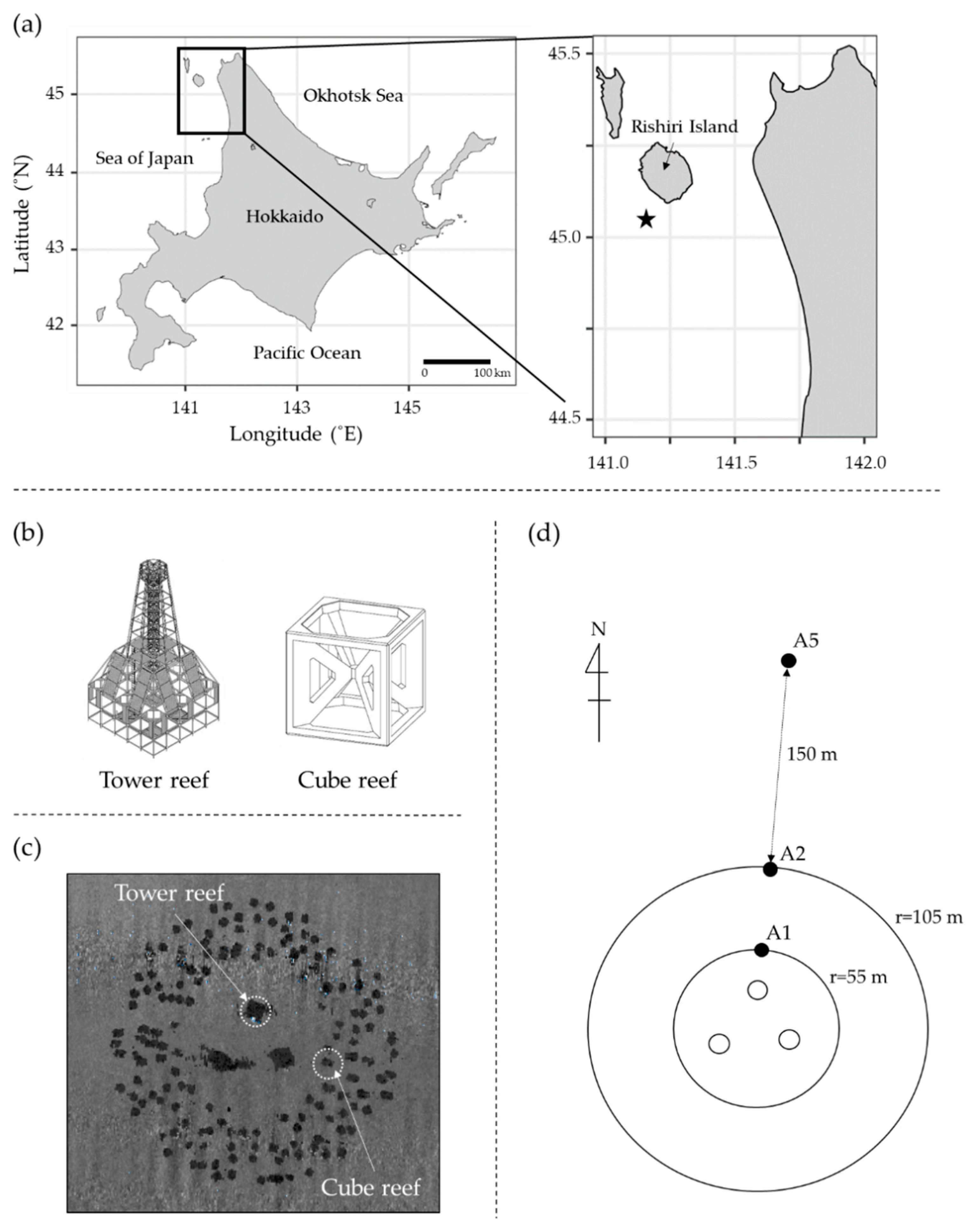
Figure 1. Location of the study area and details of the artificial reef (AR) system: (a) location of the AR system (black star), (b) detailed structure of a tower reef (20 m high), and a cube reef (3 m high) deployed in the AR system, (c) the distribution of tower reefs and cube reefs in the AR system detected by side-scan sonar, and (d) location of the sampling stations (A1, A2, and A5); an open circle represents a tower reef.
Applsci 12 09972 g001
Figure 2. Vertical profiles of the environmental variables (water temperature, salinity, density (σT), and chlorophyll fluorescence) at A1, A2, and A5.
Applsci 12 09972 g002
Figure 3. Frequency distribution of flow speed and direction at 3 m above the bottom of A5 (non-reef station) from 25 July to 7 August 2018. Flow speeds and directions were measured every one second for two minutes at an interval of twenty minutes through the period.
Applsci 12 09972 g003
Figure 4. Cell concentrations and species compositions of (a) diatoms and (b) Chaetoceros species in the bottom sediment at A1, A2, and A5, determined by direct microscopic observation (an open circle represents cell concentration), and (c) photomicrographs of the resting spores/cells of Chaetoceros spp.; (I) C. contortus, (II) C. debilis, (III) C. radicans, (IV) C. affinis or vanheurckii, (V) C. didymus, (VI) C. coronatus, (VII) C. diadema. Scale bars are 20 µm.
Applsci 12 09972 g004
Figure 5. Photomicrographs of the resting stages of diatoms other than Chaetoceros spp. by direct microscopic observation; (I) Actinocyclus sp., (II) Actinoptychus senarius, (III) Thalassiosira sp., (IV) Odontella aurita, and (V) Leptocylindrus danicus. Scale bars are 20 µm.
Applsci 12 09972 g005
Figure 6. Photomicrographs of representative pennate diatoms by direct microscopic observation; (I) Naviculoid type diatom, (II) Haslea sp., (III) Nitzschia sp., (IV) Surirella sp., (V) Fragilariopsis sp., (VI) unidentified pennate diatoms. Scale bars are 20 µm.
Applsci 12 09972 g006
Figure 7. MPN of viable cells and species composition of diatoms in the bottom sediment at A1, A2, and A5; an open circle represents cell concentration.
Applsci 12 09972 g007
Table 1. The position and bottom depth of the sampling stations (A1, A2, and A5).
StationLatitude (N)Longitude (E)Water Depth (m)
A145°02′32.600″141°07′54.368″90.9
A245°02′34.220″141°07′55.028″91.7
A545°02′39.079″141°07′54.848″93.7
Table 2. The particle size distribution and D50 (median diameter) of the bottom sediment at A1, A2, and A5.
StationParticle Size Distribution (%)D50
(mm)
ClaySiltSandGravel
FineMediumCoarseFineMediumCoarse
<0.005
mm
0.005–0.075
mm
0.075–0.25
mm
0.25–0.85
mm
0.85–2.0
mm
2.0–4.75
mm
4.75–19
mm
19–75
mm
A110.59.97.18.35.814.044.40.03.70
A216.611.98.812.45.914.929.50.00.89
A528.318.113.820.93.85.79.40.00.12
Table 3. Taxa and concentrations of the diatom resting stages in the bottom sediment at A1, A2, and A5, determined by direct microscopic observation. Cells of species other than pennate diatoms and unidentified diatoms were identified as resting stages in the present study.
SpeciesCell Concentration (Cells g−1 Wet Sediment)
A1A2A5
Actinocyclus spp.0040
Actinoptychus senarius608020
Chaetoceros affinis or vanheurckii2084020
Chaetoceros contortus902560150
Chaetoceros coronatus0200
Chaetoceros debilis10100010
Chaetoceros diadema6036080
Chaetoceros didymus1018010
Chaetoceros radicans802480190
Coscinodiscus spp.0200
Leptocylindrus danicus0400
Odontella aurita3000
Thalassiosira spp.6018030
Pennate diatoms50260130
Unidentified Chaetoceros spp.3080020
Unidentified diatoms600140
Publisher’s Note: MDPI stays neutral with regard to jurisdictional claims in published maps and institutional affiliations.