Review

Nanotechnology in Plant Metabolite Improvement and in Animal Welfare

1
Department of Biology, Institut Teknologi Sepuluh Nopember, Surabaya 60111, Indonesia
2
Department of Development Studies, Institut Teknologi Sepuluh Nopember, Surabaya 60111, Indonesia
3
Department of Biology and Biotechnologies “Charles Darwin”, Università di Roma La Sapienza, Piazzale Aldo Moro 5, 00185 Rome, Italy
*
Authors to whom correspondence should be addressed.
Appl. Sci. 2022, 12(2), 838; https://doi.org/10.3390/app12020838
Submission received: 6 December 2021 / Revised: 10 January 2022 / Accepted: 11 January 2022 / Published: 14 January 2022
(This article belongs to the Section Nanotechnology and Applied Nanosciences)

Abstract

:
Plant tissue culture plays an important role in plant biotechnology due to its potential for massive production of improved crop varieties and high yield of important secondary metabolites. Several efforts have been made to ameliorate the effectiveness and production of plant tissue culture, using biotic and abiotic factors. Nowadays, the addition of nanoparticles as elicitors has, for instance, gained worldwide interest because of its success in microbial decontamination and enhancement of secondary metabolites. Nanoparticles are entities in the nanometric dimension range: they possess unique physicochemical properties. Among all nanoparticles, silver-nanoparticles (AgNPs) are well-known for their antimicrobial and hormetic effects, which in appropriate doses, led to the improvement of plant biomass as well as secondary metabolite accumulation. This review is focused on the evaluation of the integration of nanotechnology with plant tissue culture. The highlight is especially conveyed on secondary metabolite enhancement, effects on plant growth and biomass accumulation as well as their possible mechanism of action. In addition, some perspectives of the use of nanomaterials as potential therapeutic agents are also discussed. Thus, the information provided will be a good tool for future research in plant improvement and the large-scale production of important secondary metabolites. Elicitation of silver-nanoparticles, as well as nanomaterials, function as therapeutic agents for animal well-being is expected to play a major role in the process. However, nanosized supramolecular aggregates have received an increased resonance also in other fields of application such as animal welfare. Therefore, the concluding section of this contribution is dedicated to the description and possible potential and usage of different nanoparticles that have been the object of work and expertise also in our laboratories.

1. Introduction

Plant in vitro culture is considered a biotechnological process that involved growing plant cells, tissues, or organs in an artificial nutrition media in a controlled environment and aseptic condition. Application of plant in vitro culture for plant propagation and crop improvement has been discussed [1,2,3,4,5]. In addition, it is also utilized for pharmacological purposes [5,6]. As far as agriculture is concerned, cell propagation in vitro, has been widely used for massive production of low-proliferating crops, for improvement of crop varieties, and preparation of pathogen-free plants, as well as for maintaining stocks of endangered species as an ex-situ conservation approach [5,6,7,8,9,10]. Plant cell culture can be applied to a wide range of botanical species through different methods and procedures and this assumes strategic importance considering the present concern about environmental issues [10]. Plant tissue culture has also been used as a major platform for the production of secondary metabolites, which are widely used for pharmacological as well as industrial purposes [10,11,12]. Plant secondary metabolites contained in medicinal plants are the major reason why they have been used traditionally by society worldwide [13]. Many bioassays and phytochemical studies have been carried out to scientifically prove the pharmacological effect of the plant’s natural constituents [14,15].
The culture of plants in vitro is directed towards the aseptic growth of parts or whole plants under specific nutritional and environmental conditions [1,12]. This technique depends on the concept of totipotency, which refers to the ability of plant cells to express a full genome through cell division [1,2]. Several factors affect the success of in vitro culture, such as the genotype, physiological status, and type of explants, disinfection method employed in the preparation of the sample, culture medium, plant growth regulators (PGRs), light intensity, photoperiod, and temperature. Generally, medium composition strongly influences morphogenesis in explants [16,17,18]. Media are typically composed of macro- and micronutrients, amino acid organic supplements, vitamins, carbon sources, and PGRs, as well as solidifying agents [4,7,12]. Elicitors can be defined as molecules or agents known for their ability to induce a response in the organisms they are administered to. They are also added for some special purposes, such as enhancing phytochemical production in plants [19,20]. These molecules are of biological or non-biological origin and can recognize cytoplasmic membrane receptors on the plant cells. Recognition and binding led to signal elicitation, which stimulates the expression of genes related to secondary metabolite production [20,21,22]. The elicitors work as plant stressors in a natural environment: they can be represented by fungi, bacteria, activated enzymes, and other biological compounds [22,23]. Other forms of elicitors are constituted by physical agents such as temperature, salinity, heavy metals, and mineral stress [22,23,24]. With respect to this, nanoparticles/heavy metals conjugates show high potential as in vitro culture elicitors for their physiochemical properties and fall within the field of nanotechnology [22,25].
Nanotechnology has rapidly become prominent in various scientific fields of application [26]. Nanobiotechnology has been widely utilized for drug and gene delivery, bio-detection of pathogens and proteins, disease control, and fluorescent biological labeling. Also, in plant tissue culture it has found use in seed germination, yield, and bioactive compound improvement, as well as a plant protection [12,26,27]. Nanomaterials exhibit unique properties such as low molecular, large surface-area-to-volume ratio, ability to engineer electron exchanges, special electronic and optical attributes, and surface reactive capability [26,28]. Previous studies have shown that metallic (Ag-, Au-, and Fe-nanoparticles) and metal oxide nanoparticles (nano-ZnO2, -TiO2, and -CuO2) positively impact plant tissue culture by supporting morphological potential and propagation, as well as improving plant resistance to stress [12,26,28].
Silver-nanoparticles (AgNPs) have raised great interest due to their strong biological activity, resulting in outstanding anti-microbial performance, and their hormetic effects, which, in appropriate doses, lead to the improvement of plant biomass as well as secondary metabolite accumulation. However, the molecules inhibit plant growth in high doses [28,29]. These AgNPs nanoparticles affect plants at many different levels, resulting in stimulation of plant germination and growth, biomass accumulation, improved shoot induction and proliferation, and enhancement of pigment content [28]. Numerous positive results have been gained from nanotechnology in plant tissue culture, where the involvement of nanoparticles has become a promising method of increasing effective plant propagation and production. A recent report [8] showed the effective propagation and secondary metabolite enhancement of the endangered plant species Caralluma tuberculata via AgNP elicited culture. It has been demonstrated that a combination of AgNPs and plant growth regulators increased callus biomass as well as total phenolics and flavonoids. This review mainly focuses on the most recent progress and achievement in AgNP elicitation in plant tissue culture for the purpose of secondary metabolite enhancement. Their effect on plant growth and biomass accumulation as well as the possible mechanism of action and some perspectives of the use of nanomaterials as potential therapeutic agents are also reviewed and presented. The final section of this contribution is dedicated to the application of nanotechnological to fields different from plants but rather to animal model systems. This is actually a major field of expertise in the laboratory of one of the authors (GR). Due to the very high versatility of the carbon-based nanoparticles, the reader is addressed to Section 5 of this work for a comprehensive illustration and discussion of the most common entities. In particular, their potential delivery action into recipient cells and/or whole organisms is examined. With respect to his specific usage of cargo nanoparticles loaded with bioactive principles of botanical origin, scientific literature exists. Natural raw materials from plants are since the dawn of ages used as therapeutic drugs well as other diverse tools in herbal treatments, food, and cosmetics. The phyto-molecules isolated from the medicinal and aromatic plants have become a commodity of primary demand within the pharma industry. However, some drawbacks exist since phyto-molecules may show limited absorption, not well-defined bioavailability, and at times disputable efficacy: see for instance the case of neem oil and its derivates [30].
Phytomolecules or compounds of essential oils from medicinal and aromatic plants like camptothecin, curcumin, thymol, and eugenol, for instance, may show reduced solubility in pure form which hinders their bioavailability and efficacious uptake by the target cell/organ. This represents a serious limitation in their biomedical application. These limitations may be overcome by using nanotechnological tools. This was discussed in a relatively recent review article. Specifically, improvement of the biological activity and delivery was observed after phyto-molecules encapsulation in the nanocarriers. In conclusion, the combinatorial application of plant products with medicinal properties and nanotechnology would be advantageous in the healthcare area [31]. Regarding this point, it has been extensively discussed that the “conventional” antitumor therapy may show a significant improvement using a combination of drugs incorporated into nano-delivery drug systems. The application of nanotechnology in the codelivery of plants and the chemotherapeutic agent has been adopted in tumor-targeted therapy because of their efficacy in delivering antitumor drugs. The in vivo uptake of tumor-targeted nano-delivered active substances consider several physiological aspects such as blood circulation, tumor nature, composite internalization, and drug release as well as its pharmacodynamics. Studies have been performed to find alternative strategies to overcome these bottlenecks. However, the cell membrane remains the first, and indeed most complex, the barrier that the nanocomposite must traverse in order to reach the target site within the cell. This particular aspect has been deeply studied also in our laboratory (see for instance [32,33,34,35,36] and references therein). Finally, the role and nature of “hybrid” supramolecular aggregates and their chemical, biological, and usage in the biomedical field [37].

2. Silver-Nanoparticles in Crop Improvement

Plant tissue culture plays important roles in agrotechnology [2,10,38]. Plant micropropagation is one of the best alternatives for crop improvement and shows a special significance in multiplication through a vegetative approach. This minimizes the risk of pathogen accumulation and the slow proliferation rate in generative multiplication, which in turn, causes a loss in both crop quantity and quality [39,40,41,42]. Several studies have shown the positive impacts of AgNP augmentation for callus induction, shoot generation, and growth (Table 1). Calli obtained from in vitro culture of Caralluma tuberculata were compact and greenish in color. The water content, which is an important parameter for metabolic and physiological status, increased along with the callus biomass values [8,37]. However, administration of AgNPs above the estimated optimum level (60 µg/L) caused a significant decline in callus biomass [8].
In the sugarcane (Saccharum officinarum) [43], the addition of AgNPs resulted in significantly higher shoot multiplication and elongation at high concentrations of these nanoparticles while showing no effect on growth. This study exhibited the hormetic effect of AgNPs in plant tissue culture, as it was clearly shown that the application of AgNPs in the medium resulted in inhibition of shoot multiplication and elongation. This study also revealed that the accumulation of important nutrients in the medium, such as N, Mg, and Fe was also increased. Therefore, the increased levels of nutrients related to chlorophyll biosynthesis, may lead to higher photosynthetic activity, conversely, higher concentrations of AgNPs may lead to phytotoxicity. These results were similar to those of previous studies, such as those on Cucurbita pepo [44] and Raphanus sativus [45]. The addition of the same size of AgNPs to the MS culture medium of Stevia rebaudiana B. Castro-González et al. [29] exhibited an increase in shoot number and length in all AgNP treatments compared to those in the control. However, the highest concentration of AgNPs showed a lower amount of shoot multiplication and elongation.
The in vitro culture of Prunella vulgaris L., which is known as self-healing, using a medium containing Naphthaleneacetic acid (NAA) + AgNPs alone or augmented with Gold-Nanoparticles (AuNPs) showed a very significant increase in callus proliferation as compared to that in the control [46]. In Lavandula angustifolia shoot propagation, media enrichment with AgNPs and AuNPs showed significantly improved plant development compared to that in the control. Regardless of the components and concentration, the addition of those nanoparticles (NPs) was reported to enhance the formation of lateral shoots. Generally, the addition of AgNPs even at the highest level did not exhibit any visible toxic effects such as necrosis; however, higher levels of AuNPs exhibited a slight color change in the leaf blades to yellowish.
The effect of AgNPs’ was also investigated in callus induction of Oryza sativa cv. IR64; where the highest callus induction was observed at a relatively low concentration of AgNPs [47]. The callus induction increased significantly as compared to that in the control medium. A study on the optimum concentration of AgNPs for three possible propagation methods of Chrysanthemum morifolium was also investigated [48] in a solid and liquid medium, as well as microphonics. The addition of AgNPs to the culture medium improved dramatically the number of shoots as well as the plant height and biomass. It is also worth noting that callus cultures, as well as cell suspension culture (CSC), are used to enhance the accumulation of plant secondary metabolites. Callus which is derived from plant organs cultured in vitro can be defined as unorganized or undifferentiated cell mass that can serve as a potential plant biotechnological platform to manufacture significant bioactive compounds of pharmaceutical interests [10]. In addition, callus culture coupled with nano-elicitation has been reported as a promising strategy for enhancing alkaloid [49], flavonoid, and phenolic compounds [50]. For instance, AgNPs have been reported to boost the concentration of different flavonoids including flavonols, hydroxybenzoic and hydroxycinnamic acids in the CSC of bitter gourd [21]. An increase of total phenolic and flavonoid content had also been demonstrated in callus culture of Caralluma tuberculata and Prunella vulgaris after being elicited by AgNPS and AgAuNP, respectively [8,41].
Along with their effects on growth improvement, AgNPs also possess anti-microbial properties since it was reported that the addition of AgNPs in culture media postponed the appearance of internal contamination of Araucaria excelsa [27]. According to this study administration of nanoparticles reduced bacterial contamination and, in addition, application of AgNPs on the medium was also reported to control pathogenic fungal proliferation [51].
Although several studies have shown the positive effects of AgNPs on plant propagation, its exact mechanism has not been understood in detail. From the data presented, the effect of AgNPs on plant growth in vitro and media decontamination was dependent on various factors, such as plant species, nature, explant type, and age, as well as the AgNP size and concentration [8,27,52]. Thus, the results of the examinations suggested that the effect of AgNPs is dose-dependent, in which application of high concentrations of these nanomaterials leads to growth inhibition and necrosis. It was suggested that the increase in callus biomass was due to AgNPs modifying the plant cell wall structure, possibly allowing the plant cells to uptake more water and nutrients from the medium [8]. This suggestion was based on the structural impact of the cell wall in plant cells, compared to that in animal cells [53].
In Section 5 of this review article, we discuss the potential use of different nanoparticles. However, the reader should consider that the sophistication of molecular carriers or cargo-nanoparticles, as they are often also defined, for the delivery of exogenous material within a living cell is highly diversified. Also, they have different purposes and become ever more numerous with different chemical features often designed to reach specific bio-targets. In this work, for the sake of conciseness, we briefly overview the nature and chemical-physical properties of cationic liposomes, cat-anionic vesicles, and graphene-based carbon nanotubes. With respect to this, drug absorption into a cell, organ, or whole organism, depends also on the chemical structure of the compound and on the hydrophilic/lipophilic balance of the tissue. This constitutes a major challenge. The goal in developing the features of the cargo supramolecular carriers is, in fact, endowing them with the possibility of reaching the appropriate site in a living organism. This means, in other words, that they should identify the appropriate receptor on the cell surface, alternatively, these supramolecular aggregates should be flexible enough to be assumed actively by the target cell or to freely assumed by passive diffusion. In any case, the ideal properties of a molecular cargo in drug delivery should assure the uptake of an efficacious amount of drug within the target tissue. This latter can have different chemical characteristics, such as polarity of the membrane or of the internal cytoplasmic matrix. These parameters play a crucial role in the optimal solution of the drug within an aqueous medium: with respect to this, molecules may be transformed into acid salts when target tissues are strongly polar, or they may form complexes with crown ethers. Sophisticated methods exist for the transport of an exogenous molecule such as gels and dendrimers carbon nanotubes. These latter are raising an enormous interest for their potential usage as cargo molecules both in cultured cells and in whole organisms but also in the extremely variegated field of applications. For a comprehensive discussion of these aspects, the reader is addressed to Section 5 of this review article and to citation [54] and references therein.

3. Silver-Nanoparticles in Plant Secondary Metabolite Enhancement

Plants are rich in secondary metabolites helping them to survive under challenging environmental conditions. Furthermore, plant secondary metabolites are also well known as an important source of pharmacological or industrial means. In vitro plant cell and organ, culture has been proven to be advantageous for plant secondary metabolite production [12]. The application of elicitors, such as AgNPs, also has a positive effect on secondary metabolite enhancement in several studies [43,46,55,56,64]. In Caralluma tuberculata, application of AgNPs in MS medium showed the highest result for total phenolic compounds (total flavonoid content), phenylalanine ammonia-lyase, and DPPH free radical activity. Even though no significant increase was observed from the augmentation of AgNPs and PGRs, data showed that application of AgNPs with PGR resulted in the second-highest secondary metabolite production in C. tuberculata culture [51]. This evidence suggests that the addition of AgNPs per se in the medium was more supportive for secondary metabolite enhancement.
An increase in total phenolic compounds on sugarcane after media elicitation with AgNPs was also demonstrated [43]. This study also reported that the optimum concentration for phenolic compound enhancement in sugarcane occurred at medium concentration; higher concentrations caused a decrease of phenolic compound and antioxidant production while the production of reactive oxygen species (ROS) continued to rise. Administration of differently sized nanoparticles at various concentrations to Arabidopsis thaliana exhibited a significant change in camalexin content as compared to that in the control where the best result for camalexin accumulation was observed. Pathway network analysis on the AgNPs in A. thaliana revealed increases in the TCA-cycle, sugar metabolism, and shikimate phenylpropanoid metabolism, which are also associated with plant resistance to abiotic stress. Therefore, the ability of AgNPs to induce the production of phytoalexins and other secondary metabolites is promising for the enhancement of plant immunity against pathogens [57].
Capsaicin enhancement through Cell Suspension Culture (CSC) of Capsicum spp. where higher than those in Capsicum fruit was observed: capsaicin level raised up to two-fold after three to six days of culture [55]. It should be pointed out that the CSC approach was also adopted for the evaluation of important secondary metabolite production in bitter gourd. Total phenolic and flavonoid compounds were examined, and an increase of total phenolic and flavonoid contents was monitored. Ultra-HPLC assessment showed 20 different individual phenolic compounds among which hydroxybenzoic acid, hydroxycinnamic acid, and other flavanols [21].
Elicitation by AgNPs affected the pharmacological activity of the bitter gourd extracts since CSC, extracts thus obtained, showed a significantly higher inhibitory effect towards α-amylase as compared to that of the non-elicited ones. This may represent a potentially improved therapeutic tool as bitter gourd extracts are used for postprandial hyperglycemia treatment in diabetes patients [21,68]. These elicited extracts also showed higher anti-microbial and inhibitory effects on MCF-7 and HT-29 cell propagation. Studies performed on stabilized Prunella vulgaris CSCs’ yielded analogous results [46,56]. The TPC and TFC of the suspended cell showed significantly higher results in the NP-elicited-CSC, with the highest result in the combined AgAuNP treatment; similarly, administration of nanoparticles to E. purpurea cultures resulted in a significant increase in cichoric acid, an important specific secondary metabolite. Nanoparticle suspensions at different concentrations were added to cells in logarithmic growth: results showed that maximum level of cichoric acid was obtained from elicitation with 2 mg/L AgNPs at 72 and 48 h of exposure respectively, for leaf and root-derived callus. The biomolecular mechanisms whereby AgNPs affect the biosynthesis of this secondary metabolite is not yet fully understood, but it has been suggested that these nanoparticles stimulate the stress response to metals which would lead to the production of phenolic compounds [66].
In the hairy root culture of Curcumis anguria obtained from Agrobacterium-mediated transformation, medium elicitation with AgNPs was reported to increase significantly the total phenolic and flavonoid content and the direct uptake of the silver moiety in the root surface [64].

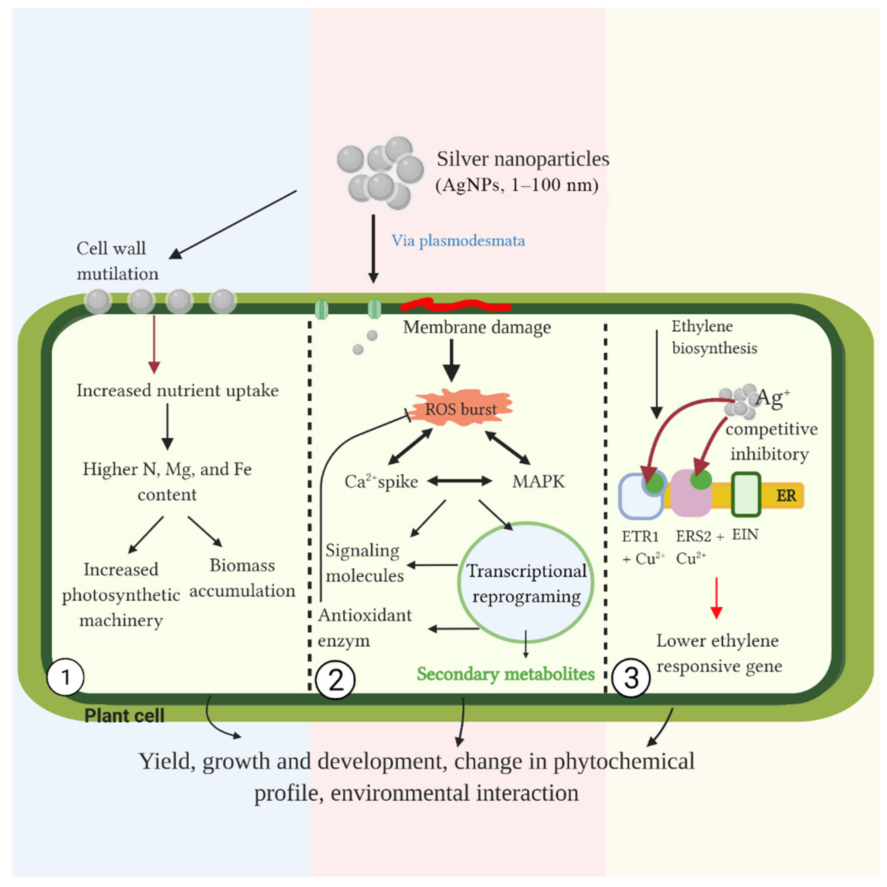
4. Possible Mechanism of AgNPs in Plant Cells

Literature addresses the possible mechanisms of growth improvement and biomass accumulation due to AgNP administration. The significant increase in some macronutrient contents and the accumulation of larger amounts of N, Mg, and Fe in the shoots of sugarcane exposed to AgNPs may contribute to the increased shoot number and length [29,43]. Nitrogen is a constituent of chlorophyll, proteins, nucleic acid, and hormones. The photosynthetic activity leads to the formation of biomass in plants therefore, it is possible that this mechanism helps improve growth in plants exposed to AgNPs. However, information on the mode of AgNPs action on nutrient absorption is lagging behind; but, in any case, it may be speculated that these particles may enhance the plant uptake inflicting damage to the cell wall. Thus, cell wall malfunction may lead to higher uptake of nutrients and water; incidentally, the higher penetration of drugs consequent to cell membrane permeabilization has been observed in both plant and animal cells [8].
Another possible mechanism of growth stimulation mediated by AgNPs, can be ascribed to PGR-responsive gene expressions. As a matter of fact, investigations demonstrated that these nanoparticles decreased the expression of ethylene- and ABA-responsive genes, resulting in callus regeneration rate [47]. Under closed container culture conditions, plant development will be highly influenced by self-produced gases, such as ethylene, which possibly regulate senescence. The introduction of AgNPs in olive cells showed positive impacts on cell growth, reducing necrosis and improving growth vigor [69]. The rapid growth effects on plant cultures might be due to the inhibitory effect of silver ions on ethylene production. Silver ions from AgNPs act as ethylene perception inhibitors, by replacing the copper cofactor, leading to the downregulation of genes involved in ethylene production [47,70]. It has been also reported that the application of AgNPs decreased the ACS transcript level on the AgNPs-elicited culture of T. undulata [59]. The expression of ACS had been widely studied to understand growth and stress response, as it catalyzes the conversion of S-adenosylmethionine to 1-aminocyclopropane-1-carboxylic acid, the precursor of ethylene. This low expression of ACS may explain how AgNP-treated culture showed a decreased concentration of ethylene. It was suggested that the action of AgNPs consisted of replacing ethylene in the receptors located at the level of the endoplasmic reticulum thus leading to the saturation of ethylene production. A significant decrease of ethylene content in AgNPs-elicited culture was also reported and another study showed that the particles affect root growth improvement through the upregulation of auxin [60].
Dynamic biosynthesis and signaling of PGRs are also influenced by the application of nanoparticles. Some of them include auxin, abscisic acid (ABA), and ethylene [71]. However, another study also demonstrated that nanoparticles are involved in the determination of plant growth and development [72]. Vankova et al. [72] showed that the hormonal pool is affected by the application of nanoparticles. It has been suggested that different plant growth and physiological responses in various plant species might result from different pools of PGRs. A high concentration of nanoparticles suppressed the biosynthesis of cytokinins and auxins in the shoot apical meristem (SAM). Similar to that, jasmonic acid was also decreased during nanoparticle treatment. Meanwhile, accumulation of abscisic acid, cis-zeatin, and salicylic acid was observed in plant roots upon nanoparticle exposure. This indicates that nanoparticle serves as a source of plant stressors [72].
Even though the direct biophysical and biochemical interactions between nanoparticles and the biological system have not yet been explained, it has been strongly suggested that the initial response of plants exposed to NPs might include increased levels of ROS, cytoplasmic Ca2+, and mitogen-activated protein kinase (MAPK) cascades; these responses were the same as those in the biotic and abiotic stress defense mechanisms [8,26,56,73]. AgNPs were reported to adhere to the surface of the primary root in the early stage of exposure. Transport of the particles in plants occurs by two mechanisms, i.e., via the intercellular transport or vascular tissue. The initial penetration of nanoparticles is at the level of the epidermal layer of the root, followed by a series of events ending in the entry of the plant vascular tissue. This mechanism enables the translocation of AgNPs in whole plants or explants [74]. AgNPs are reported to remain in nanoparticle form after intake into the plant cells, causing less severe effects than one generated by free Ag+ derived from silver nitrate [75]. AgNPs could enter cells through plasmodesmata, after direct contact with the cell membrane. In this adverse condition, plants produce ROS that causes damage to the biological membrane system and to macromolecules [73,74]. As reported in A. thaliana, the initial recognition of AgNPs by membrane-bound receptors triggers Ca2+ burst and ROS induction [69].
Nanoparticles and silver ions were reported to stimulate changes in the secondary metabolite profile, which suggested that AgNPs affect plant metabolism via ions release in the medium. In stevia (Stevia rebaudiana), high doses of AgNPs were reported to accumulate in plant cells and tissues [57]. These molecules were internalized in the plant through the vascular system and translocated to neighborhood cells via apoplasts. The transfer mechanism via apoplasts requires NPs to cross a membrane; this indicates that NP uptake must be size-specific. Nanomaterials could also be transferred via plasmodesmata; this channel is approximately 40 nm in diameter, so it is possible for 40 nm-sized AgNPs to be transferred via plasmodesmata. Interestingly, nanoparticles are also reported to possibly create a pore for bigger size NPs, via NP-induced conductance mechanism [74,76,77,78]. Another study demonstrated that the morphology and size of the AgNPs affected the plant response during exposure [79]. The study exhibited that smaller size AgNPs showed a more efficacious anti-microbial activity and increased response to oxidative stress.
Nanoparticles also induced a ROS-mediated MAPK cascade and a calcium spike, which possibly enhanced secondary metabolite production to counteract the oxidative stress, resulting in a higher quality and quantity of secondary metabolites during AgNPs exposure [56] (Figure 1). MAPK has been known to play an important function in plant responses, triggered by ROS, through dynamic gene expression and regulations. It has been reported to be crucial for flavonoid [80] and alkaloid [81] biosynthesis when plants are subjected to stress conditions. Paezi et al. [82] reported up-regulation of several genes involved in the biosynthesis of alkaloids, coupled with high expression of Mitogen-Activated Protein Kinase 3 (CrMPK3) gene on Catharanthus roseus after silver exposure. Sosan et al. [79] reported Ca2+ burst and ROS induction on the cell membrane after AgNPs exposure. Nanoparticles induced single channel-like conductance that allows Ca2+ entry, resulting in ROS generation because of the cytosolic Ca2+. This condition leads to the expression of MAPK genes that affects transcriptional reprogramming of secondary metabolites-related genes. Interestingly, a recent study conducted by Fouad et al. [49] has reported that AgNPs triggered the accumulation of alkaloids in C. roseus via ROS-mediated MAPK cascade through the up-regulation of the MAK3 gene. It leads finally to the overexpression of alkaloid biosynthetic genes.

5. Nanomaterials: The Future Goal in Many Multi-Faceted Fields of Animal Welfare

The final part of this review focuses on the supramolecular structure of some nanoparticles that could be potentially used in a number of different fields: ranging from agriculture to human advanced biomedicine [26]. The main body of this work has been concentrated on the elicitation by silver nanoparticles in plant cells. However, the relevance of these nanosized supramolecular aggregates also in other fields of application cannot be disregarded. Therefore, the concluding section of this contribution is dedicated to the description and possible potential and usage of different nanoparticles that have been the object of work and expertise also in our laboratories. With respect to this, this contribution represents a joint effort between the Institut Teknologi Sepuluh Nopember (ITS; Surabaya-Indonesia) and the University of Rome “La Sapienza”. This work was accomplished also thanks to the grant by the European Union to one of us (G.R.) within the framework of the ERASMUS Mundus exchange program.
Figure 2 reports the main structural features of the supramolecular aggregates discussed hereafter.
Liposomes are among the most studied drug delivery systems; they show high biocompatibility, target selectivity, and advantageous cost/production ratio. This may play an important role in their selection as therapeutic agents in the emerging of disadvantaged situations. The validity of liposomes as cargo particles is mainly due to their flexibility since they may function as vehicles for DNA, RNA, and proteins as well as small molecules like hormones, natural compounds, and/or drugs in general, also of botanical origin. Liposomes based on cationic lipids are not found in nature but are synthesized in the chemistry laboratory but possessing a net positive charge they may promptly interact with the negatively charged cell membrane and nucleic acids this has evidenced their ability to act as vaccine carrier/adjuvants [32,33]. This is an added value in the present pandemics contingency due to the COVID-19 virus. The possibility of using liposomes also exists in cancer therapy since cationic liposomes are able to deliver specifically their payload to embryologically different tissues. Finally, the antibacterial action of cationic liposomes has been also examined in antibiotic-resistant pathogenic microorganisms. This action may derive from the ability of liposomes to increase the bacterial membrane permeability, with consequent higher susceptibility to drug uptake [34,35,36,83].
Cat-anionic vesicles are fabricated by mixing in non-stoichiometric ratios cat-ionic and anionic surfactant species. Surfactants of opposite charge aggregate in aqueous polar solvents [84]. Vesicles, analogously to liposomes, interact with nucleic acids and other biopolymers in general. The complexes vesicle/macromolecule, are also known as lipoplexes and are of crucial importance in biotechnology and biomedicine: for instance, they can deliver exogenous material across the cell membrane but do not cause permanent or relevant damages [85,86]. In any case, vesicles may show a time/dose cytotoxic effect, which is also due to the composing cat-anionic moieties. With respect to this, results from our laboratories show that the transfected cat-anionic/RNA lipoplex is efficiently translated into protein as shown in our laboratory [87,88,89,90,91,92,93].
Graphene sheets organized in cylindrical structures with single or multi-walled tube-like structures are by definition carbon nanotubes. They diversified applications in biochemistry, nanomedicine, pharmacology, and industry such as avionics as well as space engineering, see, for instance: [92,93,94,95,96,97]. The biocompatibility of carbon nanotubes must be evaluated in terms of toxicity and immuno-tolerance to guarantee, the overall impact of these bioactive entities. The cell membrane represents the first barrier for the nanotube prior to cell entry. With the poor dispersibility of nanotubes in aqueous environments, van der Waals interactions are primarily established. However, non-covalent carbon nanotubes/BSA complexes did not alter the cell viability in murine fibroblasts, human embryonic kidney cells, and murine macrophages [96,97]. Also, nanotubes crossing of the plasma membrane, cause no alterations of its dielectric parameters. This strongly suggests that nanoparticle/cell membrane impact does is not detrimental to the overall membrane structure, function, and permeability [98,99,100].
Graphene which is the basic component of the nanotube technology consists of a one-atom-thick planar sheet of carbon atoms. The final structure consists of a densely packed honeycomb crystal lattice. These structures’ chemical structure is derived from the basic chemical-physical features of carbon allotropes. Graphene-based nanotechnology represents an ever-expanding area of basic research and industrial applications ranging from material sciences, electronics, photonics, composite materials, energy generation and storage, sensors as well as biological applications [34,99,100,101]. A side problem, however, may be represented by their release into the environment. Data exist that these nanoparticles are essentially non-toxic in diverse protozoan and animal models, in amphibians, for instance, it was demonstrated that the life span was affected though their growth rate was slower- This was possibly due to digestive and respiratory problems [101,102,103,104,105].
The central goal of this interdisciplinary review is to evidence the multi-task potentials of nanoparticles. It is evident from this contribution that the flexibility and versatility of nanoparticles, render them an ideal tool in fields as diverse as agriculture, human and veterinary advanced therapies, and, in general, for the better general welfare. The authors hope that this goal has been reached.

6. Conclusions and Perspectives

The use of AgNPs on plant tissue culture may have positive impacts on plant growth and secondary metabolite enhancement. Various sizes and concentrations of AgNPs were employed in the presented review. It ranged from 1–40 nm in size and 30 µg/L to 200 mg/L in concentrations. This application affects plant growth responses, including callus proliferation, shoot multiplication, and elongation, as well as root formation. In addition, AgNPs induce the accumulation of plant secondary metabolites. These responses result from complex physiological mechanisms altering antioxidant enzyme activities, genes expression, plant hormone signaling and regulations, and ROS production. However, different responses and hormetic effects were observed depending on the plant species and explant type. Thus, optimization of AgNP properties is required for each plant species to improve secondary metabolite enhancement. However, AgNPs show high potential to be employed for those improvement purposes in plants. Large-scale approaches, such as nano-integrated suspension culture, are a promising method of secondary metabolite production in the future. The findings on the growth improvement effects of AgNPs could also provide basic knowledge to employ this nanomaterial for plant propagation, which could possibly support crop improvement efforts as well as plant conservation. Further assessment and evaluation are required to understand the mechanism behind the effects of AgNPs in plant culture.
In any case, the potential activity of silver nanoparticles in human therapy should be also taken into account. Silver nanoparticles have shown an effective antiviral action also towards viral agents involved in the etiology of respiratory diseases. Regarding the potential role of silver nanoparticles, we would like to point out that recent works highlighted how they could play role in the control of different and severe acute respiratory syndromes such as the acute respiratory syndrome (SARS-CoV) and the Middle East respiratory syndrome (MERS-CoV). In particular human coronaviruses are known for their ability to spread within the healthy population in an extremely rapid and explosive manner [106].
The outbreak of the pandemics is dated at the end of 1919, and the characterizing pathology is characterized by a very serious, often lethal symptomatology: it has been estimated that, as of January 2022, about 5.5 million people have been the victim of the viral infection all over the world. But yet this is possible and underestimation due to the continuous emergence of viral variants. Only three states in the world have not yet recorded any death: Vatican City, Republic of Palau, and the Federated States of Micronesia [107]. However, in the opinion of the Authors, although treatment of the disease with AgNPs to combat SARS-CoV-2 could be viable, the technology and costs of this treatment could be prohibitive in some disadvantaged areas of the world.
One final consideration concerns the possible toxic of AgNPs which might appear in human health and in the pharmaceutical industry, without disregarding environmental and industrial applications. Literature exists on this specific subject (see for instance [108]). This review is mainly focused on the elicitation of plant tissue culture medium with AgNPs; however, it should be borne in mind that a large number of studies exist on the relevance of nanotechnology in an extreme variety of biological fields. For instance, the possibility of delivering exogenous macromolecules has been successfully examined in our laboratories and the delivery of natural products, with potentially therapeutic action, has been amply reviewed and discussed. Therefore, due to the interdisciplinary and comprehensive character of this contribution the application of nanotechnological strategies applied to the animal kingdom cannot be ignored. In conclusion, the field of nanotechnology is in an ever-growing status, and in the very close future, the hypotheses earlier formulated by P. Ehrlich and J.H. Muller may come true [109].

Author Contributions

Conceptualization, N.J.; data curation, M.R.; writing—review and editing, N.J., G.R. and M.R.; supervision, N.J., C.M. and G.R.; funding acquisition, N.J. and C.M. All authors have read and agreed to the published version of the manuscript.

Funding

This research was funded by the Ministry of Education, Culture, Research and Technology of the Republic of Indonesia, grant number 894/PKS/ITS/2021. This research is also part of the project scheme of the publication writing and IPR incentive program (PPHKI) supported by Institut Teknologi Sepuluh Nopember (ITS), Surabaya-Indonesia.

Institutional Review Board Statement

Not applicable.

Informed Consent Statement

Not applicable.

Acknowledgments

Authors are grateful to The Centre of Public Policy, Business, and Industry (PKKPBI) of Institut Teknologi Sepuluh Nopember (ITS), Surabaya-Indonesia for all supports given during this study. This work is based on several years of research work of N.J., G.R. was the recipient of a traveling/teaching grant within the framework of the ERASMUS Mundus exchange program funded by the European Union. Part of the work reported in this contribution was developed during his stage at the ITS; the collaboration between G.R. and the scientists at this institution is ongoing.

Conflicts of Interest

The authors declare no conflict of interest.

References

  1. Ali, A.M.A.; El-Nour, M.E.A.M.; Yagi, S.M. Callus Induction, Direct and Indirect Organogenesis of Ginger (Zingiber Officinale Rosc). Afr. J. Biotechnol. 2016, 15, 2106–2114. [Google Scholar] [CrossRef] [Green Version]
  2. Bidabadi, S.S.; Jain, S.M. Cellular, Molecular, and Physiological Aspects of In Vitro Plant Regeneration. Plants 2020, 9, 702. [Google Scholar] [CrossRef] [PubMed]
  3. Kasilingam, T.; Raman, G.; Sundramoorthy, N.; Supramaniam, G.; Mohtar, S.; Avin, F. A Review on In Vitro Regeneration of Ginger: Tips and Highlights. Eur. J. Med. Plants 2018, 23, 1–8. [Google Scholar] [CrossRef]
  4. Singh, C.R. Review on Problems and Its Remedy in Plant Tissue Culture. Asian J. Biol. Sci. 2018, 11, 165–172. [Google Scholar] [CrossRef] [Green Version]
  5. Álvarez, S.P.; Tapia, M.A.M.; Vega, M.E.G.; Ardisana, E.F.H.; Medina, J.A.C.; Zamora, G.L.F.; Bustamante, D.V. Nanotechnology and Plant Tissue Culture. In Plant Nanobionics; Nanotechnology in the Life Sciences; Prasad, R., Ed.; Springer International Publishing: Cham, Switzerland, 2019; pp. 333–370. ISBN 978-3-030-12495-3. [Google Scholar]
  6. Acemi, R.K.; Acemi, A.; Cakir, M.; Polat, E.G.; Ozen, F. Preliminary screening the antioxidant potential of in vitro-propagated Amsonia orientalis: An example to sustainable use of rare medicinal plants in pharmaceutical studies. Sustain. Chem. Pharm. 2020, 17, 100302. [Google Scholar] [CrossRef]
  7. Oseni, O.M.; Pande, V.; Nailwal, T.K. A Review on Plant Tissue Culture, A Technique for Propagation and Conservation of Endangered Plant Species. Int. J. Curr. Microbiol. App. Sci. 2018, 7, 3778–3786. [Google Scholar] [CrossRef]
  8. Ali, A.; Mohammad, S.; Khan, M.A.; Raja, N.I.; Arif, M.; Kamil, A.; Mashwani, Z.-R. Silver Nanoparticles Elicited in Vitro Callus Cultures for Accumulation of Biomass and Secondary Metabolites in Caralluma tuberculata. Artif. Cells Nanomed. Biotechnol. 2019, 47, 715–724. [Google Scholar] [CrossRef] [Green Version]
  9. Abbas, M.S.; Taha, H.S.; Aly, U.I.; El-Shabrawi, H.M.; Gaber, E.-S.I. In Vitro Propagation of Ginger (Zingiber Officinale Rosco). J. Genet. Eng. Biotechnol. 2011, 9, 165–172. [Google Scholar] [CrossRef] [Green Version]
  10. Chandran, H.; Meena, M.; Barupal, T.; Sharma, K. Plant Tissue Culture as a Perpetual Source for Production of Industrially Important Bioactive Compounds. Biotechnol. Rep. 2020, 26, e00450. [Google Scholar] [CrossRef] [PubMed]
  11. Ramirez-Estrada, K.; Vidal-Limon, H.; Hidalgo, D.; Moyano, E.; Golenioswki, M.; Cusidó, R.; Palazon, J. Elicitation, an Effective Strategy for the Biotechnological Production of Bioactive High-Added Value Compounds in Plant Cell Factories. Molecules 2016, 21, 182. [Google Scholar] [CrossRef]
  12. Kim, D.H.; Gopal, J.; Sivanesan, I. Nanomaterials in Plant Tissue Culture: The Disclosed and Undisclosed. RSC Adv. 2017, 7, 36492–36505. [Google Scholar] [CrossRef] [Green Version]
  13. Jadid, N.; Kurniawan, E.; Himayani, C.E.S.; Prasetyowati, I.; Purwani, K.I.; Muslihatin, W.; Hidayati, D.; Tjahjaningrum, I.T.D. An Ethnobotanical Study of Medicinal Plants Used by the Tengger Tribe in Ngadisari Village, Indonesia. PLoS ONE 2020, 15, e0235886. [Google Scholar] [CrossRef]
  14. Jadid, N.; Rachman, R.Y.; Hartanti, S.R.; Abdulgani, N.; Wikanta, W.; Muslihatin, W. Methanol Extract of Piper Retrofractum Vahl. Potentially Mediates Mast Cell Stabilization. Int. J. Pharm. Bio Sci. 2016, 7, 379–383. [Google Scholar]
  15. Salmerón-Manzano, E.; Garrido-Cardenas, J.A.; Manzano-Agugliaro, F. Worldwide Research Trends on Medicinal Plants. Int. J. Environ. Res. Public Health 2020, 17, 3376. [Google Scholar] [CrossRef]
  16. Abdi, G.; Salehi, H.; Khosh-Khui, M. Nano Silver: A Novel Nanomaterial for Removal of Bacterial Contaminants in Valerian (Valeriana officinalis L.) Tissue Culture. Acta Physiol. Plant 2008, 30, 709–714. [Google Scholar] [CrossRef]
  17. Bhoite, H.A.; Palshikar, G.S. Plant Tissue Culture: A Review. World J. Pharm. Sci. 2014, 2, 565–572. [Google Scholar]
  18. Ghasemzadeh, A.; Jaafar, H.Z.E.; Rahmat, A.; Wahab, P.E.M.; Halim, M.R.A. Effect of Different Light Intensities on Total Phenolics and Flavonoids Synthesis and Anti-Oxidant Activities in Young Ginger Varieties (Zingiber officinale Roscoe). Int. J. Mol. Sci. 2010, 11, 3885–3897. [Google Scholar] [CrossRef] [Green Version]
  19. Angelova, Z.; Georgiev, S.; Roos, W. Elicitation of Plants. Biotechnol. Biotechnol. Equip. 2006, 20, 72–83. [Google Scholar] [CrossRef] [Green Version]
  20. Patel, H.; Krishnamurthy, R.J. Elicitors in plant tissue culture. Pharmacogn. Phytochem. 2013, 2, 6. [Google Scholar]
  21. Chung, I.-M.; Rekha, K.; Rajakumar, G.; Thiruvengadam, M. Elicitation of Silver Nanoparticles Enhanced the Secondary Metabolites and Pharmacological Activities in Cell Suspension Cultures of Bitter Gourd. 3 Biotech 2018, 8, 412. [Google Scholar] [CrossRef]
  22. Jadczak, P.; Kulpa, D.; Bihun, M.; Przewodowski, W. Positive Effect of AgNPs and AuNPs in in Vitro Cultures of Lavandula Angustifolia Mill. Plant Cell Tissue Organ Cult. 2019, 139, 191–197. [Google Scholar] [CrossRef] [Green Version]
  23. Dabuwar Benjamin, E.; Adamu Ishaku, G.; Andrew Peingurta, F.; Samuel Afolabi, A. Callus Culture for the Production of Therapeutic Compounds. Am. J. Plant Biol. 2019, 4, 76–84. [Google Scholar] [CrossRef]
  24. Murthy, H.N.; Dandin, V.S.; Zhong, J.-J.; Paek, K.-Y. Strategies for Enhanced Production of Plant Secondary Metabolites from Cell and Organ Cultures. In Production of Biomass and Bioactive Compounds Using Bioreactor Technology; Paek, K.-Y., Murthy, H.N., Zhong, J.-J., Eds.; Springer: Dordrecht, The Netherlands, 2014; pp. 471–508. ISBN 978-94-017-9222-6. [Google Scholar]
  25. Kokina, I.; Mickeviča, I.; Jahundoviča, I.; Ogurcovs, A.; Krasovska, M.; Jermaļonoka, M.; Mihailova, I.; Tamanis, E.; Gerbreders, V. Plant Explants Grown on Medium Supplemented with Fe3O4 Nanoparticles Have a Significant Increase in Embryogenesis. J. Nanomater. 2017, 2017, 4587147. [Google Scholar] [CrossRef] [Green Version]
  26. Bayda, S.; Adeel, M.; Tuccinardi, T.; Cordani, M.; Rizzolio, F. The History of Nanoscience and Nanotechnology: From Chemical-Physical Applications to Nanomedicine. Molecules 2019, 25, 112. [Google Scholar] [CrossRef] [PubMed] [Green Version]
  27. Sarmast, M.K.; Salehi, H. Silver Nanoparticles: An Influential Element in Plant Nanobiotechnology. Mol. Biotechnol. 2016, 58, 441–449. [Google Scholar] [CrossRef]
  28. Salachna, P.; Byczyńska, A.; Zawadzińska, A.; Piechocki, R.; Mizielińska, M. Stimulatory Effect of Silver Nanoparticles on the Growth and Flowering of Potted Oriental Lilies. Agronomy 2019, 9, 610. [Google Scholar] [CrossRef] [Green Version]
  29. Castro-González, C.G.; Sánchez-Segura, L.; Gómez-Merino, F.C.; Bello-Bello, J.J. Exposure of Stevia (Stevia Rebaudiana B.) to Silver Nanoparticles in Vitro: Transport and Accumulation. Sci. Rep. 2019, 9, 10372. [Google Scholar] [CrossRef]
  30. Risuleo, G. Biological properties of a partially purified component of Neem oil: An updated and revised work. In Nuts and Seeds in Health and Disease Prevention, 2nd ed.; Preedy, V.R., Watson, R.R., Eds.; Academic Press: Cambridge, MA, USA, 2020; pp. 67–72. [Google Scholar] [CrossRef]
  31. Kumari, P.; Luqman, S.; Meena, A. Application of the combinatorial approaches of medicinal and aromatic plants with nanotechnology and its impacts on healthcare. DARU J. Pharm. Sci. 2019, 27, 475–489. [Google Scholar] [CrossRef] [PubMed]
  32. Simberg, D.S.; Weisman, Y.; Talmon, Y. DOTAP (and Other Cationic Lipids): Chemistry, Biophysics, and Transfection. Crit. Rev. Ther. Drug Carrier Syst. 2004, 21, 257–319. [Google Scholar] [CrossRef]
  33. Lonez, C.; Vandenbranden, M.; Ruysschaert, J.-M. Cationic Liposomal Lipids: From Gene Carriers to Cell Signaling. Prog. Lipid Res. 2008, 47, 340–347. [Google Scholar] [CrossRef] [PubMed]
  34. Hamblin, M.R.; Hasan, T. Photodynamic Therapy: A New Antimicrobial Approach to Infectious Disease? Photochem. Photobiol. Sci. 2004, 3, 436. [Google Scholar] [CrossRef] [Green Version]
  35. Stefanutti, E.; Papacci, F.; Sennato, S.; Bombelli, C.; Viola, I.; Bonincontro, A.; Bordi, F.; Mancini, G.; Gigli, G.; Risuleo, G. Cationic Liposomes Formulated with DMPC and a Gemini Surfactant Traverse the Cell Membrane without Causing a Significant Bio-Damage. Biochim. Biophys. Acta (BBA) Biomembr. 2014, 1838, 2646–2655. [Google Scholar] [CrossRef] [Green Version]
  36. Cosimati, R.; Milardi, G.L.; Bombelli, C.; Bonincontro, A.; Bordi, F.; Mancini, G.; Risuleo, G. Interactions of DMPC and DMPC/Gemini Liposomes with the Cell Membrane Investigated by Electrorotation. Biochim. Biophys. Acta (BBA) Biomembr. 2013, 1828, 352–356. [Google Scholar] [CrossRef] [PubMed] [Green Version]
  37. La Mesa, C.; Risuleo, G. On Concept of Hybrid. Colloids Interfaces 2021, 5, 33–53. [Google Scholar] [CrossRef]
  38. Espinosa-Leal, C.A.; Puente-Garza, C.A.; García-Lara, S. In Vitro Plant Tissue Culture: Means for Production of Biological Active Compounds. Planta 2018, 248, 1–18. [Google Scholar] [CrossRef] [PubMed]
  39. Migdam Elsheikh, A.; Mohamed Daffalla, H.; Mohamed Khalfala, M. In Vitro Micropropagation of the Ornamental Plant Dieffenbachia—A Review. Univers. J. Plant Sci. 2013, 1, 91–99. [Google Scholar] [CrossRef]
  40. Gupta, N.; Jain, V.; Joseph, M.R.; Devi, S. A Review on Micropropagation Culture Method. Asian J. Pharm. Res. Dev. 2020, 8, 86–93. [Google Scholar] [CrossRef]
  41. Cui, Y.; Deng, Y.; Zheng, K.; Hu, X.; Zhu, M.; Deng, X.; Xi, R. An Efficient Micropropagation Protocol for an Endangered Ornamental Tree Species (Magnolia sirindhorniae Noot. & Chalermglin) and Assessment of Genetic Uniformity through DNA Markers. Sci. Rep. 2019, 9, 9634. [Google Scholar] [CrossRef] [Green Version]
  42. Rashmi, R.; Trivedi, M.P. Callus Induction and Callogenic Response of Rauvolfia Serpentina and Catharanthus Roseus By Using Various Growth Hormone Concentrations Singly and in Combination. Int. J. Sci. Eng. Res. 2014, 5, 17. [Google Scholar]
  43. Bello-Bello, J.J.; Chavez-Santoscoy, R.A.; Lecona-Guzmán, C.A.; Bogdanchikova, N.; Salinas-Ruíz, J.; Gómez-Merino, F.C.; Pestryakov, A. Hormetic Response by Silver Nanoparticles on In Vitro Multiplication of Sugarcane (Saccharum Spp. Cv. Mex 69-290) Using a Temporary Immersion System. Dose-Response 2017, 15, 155932581774494. [Google Scholar] [CrossRef] [Green Version]
  44. Stampoulis, D.; Sinha, S.K.; White, J.C. Assay-Dependent Phytotoxicity of Nanoparticles to Plants. Environ. Sci. Technol. 2009, 43, 9473–9479. [Google Scholar] [CrossRef] [PubMed]
  45. Zuverza-Mena, N.; Armendariz, R.; Peralta-Videa, J.R.; Gardea-Torresdey, J.L. Effects of Silver Nanoparticles on Radish Sprouts: Root Growth Reduction and Modifications in the Nutritional Value. Front. Plant Sci. 2016, 7, 90. [Google Scholar] [CrossRef] [PubMed] [Green Version]
  46. Fazal, H.; Abbasi, B.H.; Ahmad, N.; Ali, M. Elicitation of Medicinally Important Antioxidant Secondary Metabolites with Silver and Gold Nanoparticles in Callus Cultures of Prunella vulgaris L. Appl. Biochem. Biotechnol. 2016, 180, 1076–1092. [Google Scholar] [CrossRef] [PubMed]
  47. Manickavasagam, M.; Pavan, G.; Vasudevan, V. A Comprehensive Study of the Hormetic Influence of Biosynthesized AgNPs on Regenerating Rice Calli of Indica Cv. IR64. Sci. Rep. 2019, 9, 8821. [Google Scholar] [CrossRef]
  48. Nghia, L.T.; Tung, H.T.; Huy, N.P.; Luan, V.Q.; Nhut, D.T. The Effects of Silver Nanoparticles on Growth of Chrysanthemum Morifolium Ramat. Cv. “JIMBA” in Different Cultural Systems. Vietnam J. Sci. Technol. 2017, 55, 503. [Google Scholar] [CrossRef]
  49. Fouad, A.; Hegazy, A.E.; Azab, E.; Khojah, E.; Kapiel, T. Boosting of Antioxidants and Alkaloids in Catharanthus roseus Suspension Cultures Using Silver Nanoparticles with Expression of CrMPK3 and STR Genes. Plants 2021, 10, 2202. [Google Scholar] [CrossRef]
  50. Khan, A.K.; Kousar, S.; Tungmunnithum, D.; Hano, T.; Abbasi, B.H.; Anjum, S. Nano-elicitation as an effective strategy for in vitro production of industrially important flavonoids. App. Sci. 2021, 11, 1694. [Google Scholar] [CrossRef]
  51. Lee, J.; Kim, K.-J.; Sang, W.; Guk, J.; Gun, D. The Silver Nanoparticle (Nano-Ag): A New Model for Antifungal Agents. In Silver Nanoparticles; Pozo, D., Ed.; IntechOpen: London, UK, 2010; ISBN 978-953-307-028-5. [Google Scholar]
  52. Gupta, S.D.; Agarwal, A.; Pradhan, S. Phytostimulatory Effect of Silver Nanoparticles (AgNPs) on Rice Seedling Growth: An Insight from Antioxidative Enzyme Activities and Gene Expression Patterns. Ecotoxicol. Environ. Saf. 2018, 161, 624–633. [Google Scholar] [CrossRef]
  53. Ali, H.; Khan, M.A.; Ullah, N.; Khan, R.S. Impacts of Hormonal Elicitors and Photoperiod Regimes on Elicitation of Bioactive Secondary Volatiles in Cell Cultures of Ajuga Bracteosa. J. Photochem. Photobiol. B Biol. 2018, 183, 242–250. [Google Scholar] [CrossRef]
  54. La Mesa, C.; Corbo, A.; Gkouvi, A.; Risuleo, G. Bio-Active Principles from the Animal and Plant Kingdom: A Review. Adv. Res. Org. Inorg. Chem. (AROIC) 2020, 1, 1–12. [Google Scholar]
  55. Akash Bhat, P.B. Silver Nanoparticles for Enhancement of Accumulation of Capsaicin in Suspension Culture of Capsicum Sp. J. Exp. Sci. 2016, 7, 1–6. [Google Scholar] [CrossRef] [Green Version]
  56. Fazal, H.; Abbasi, B.H.; Ahmad, N.; Ali, M.; Shujait Ali, S.; Khan, A.; Wei, D.-Q. Sustainable Production of Biomass and Industrially Important Secondary Metabolites in Cell Cultures of Selfheal (Prunella vulgaris L.) Elicited by Silver and Gold Nanoparticles. Artif. Cells Nanomed. Biotechnol. 2019, 47, 2553–2561. [Google Scholar] [CrossRef] [Green Version]
  57. Kruszka, D.; Sawikowska, A.; Kamalabai Selvakesavan, R.; Krajewski, P.; Kachlicki, P.; Franklin, G. Silver Nanoparticles Affect Phenolic and Phytoalexin Composition of Arabidopsis thaliana. Sci. Total Environ. 2020, 716, 135361. [Google Scholar] [CrossRef] [PubMed]
  58. Thangavelu, R.M.; Gunasekaran, D.; Jesse, M.I.; SU, M.R.; Sundarajan, D.; Krishnan, K. Nanobiotechnology Approach Using Plant Rooting Hormone Synthesized Silver Nanoparticle as “Nanobullets” for the Dynamic Applications in Horticulture—An in Vitro and Ex Vitro Study. Arab. J. Chem. 2018, 11, 48–61. [Google Scholar] [CrossRef] [Green Version]
  59. Sarmast, M.K.; Niazi, A.; Salehi, H.; Abolimoghadam, A. Silver Nanoparticles Affect ACS Expression in Tecomella Undulata in Vitro Culture. Plant Cell Tissue Organ Cult. 2015, 121, 227–236. [Google Scholar] [CrossRef]
  60. Vannini, C.; Domingo, G.; Onelli, E.; Prinsi, B.; Marsoni, M.; Espen, L.; Bracale, M. Morphological and Proteomic Responses of Eruca Sativa Exposed to Silver Nanoparticles or Silver Nitrate. PLoS ONE 2013, 8, e68752. [Google Scholar] [CrossRef] [Green Version]
  61. Do, D.G.; Dang, T.K.T.; Nguyen, T.H.T.; Nguyen, T.D.; Tran, T.T.; Hieu, D.D. Effects of Nano Silver on the Growth of Banana (Musa Spp.) Cultured in Vitro. J. Viet. Environ. 2018, 10, 92–98. [Google Scholar] [CrossRef]
  62. Timoteo, C.; de, O.; Paiva, R.; dos Reis, M.V.; Claro, P.I.C.; da Silva, D.P.C.; Marconcini, J.M.; de Oliveira, J.E. Silver Nanoparticles in the Micropropagation of Campomanesia Rufa (O. Berg) Nied. Plant Cell Tissue Organ Cult. 2019, 137, 359–368. [Google Scholar] [CrossRef]
  63. Spinoso-Castillo, J.L.; Chavez-Santoscoy, R.A.; Bogdanchikova, N.; Pérez-Sato, J.A.; Morales-Ramos, V.; Bello-Bello, J.J. Antimicrobial and Hormetic Effects of Silver Nanoparticles on in Vitro Regeneration of Vanilla (Vanilla Planifolia Jacks. Ex Andrews) Using a Temporary Immersion System. Plant Cell Tissue Organ Cult. 2017, 129, 195–207. [Google Scholar] [CrossRef]
  64. Chung, I.-M.; Rajakumar, G.; Thiruvengadam, M. Effect of Silver Nanoparticles on Phenolic Compounds Production and Biological Activities in Hairy Root Cultures of Cucumis Anguria. Acta Biol. Hung. 2018, 69, 97–109. [Google Scholar] [CrossRef] [Green Version]
  65. Karakaş, Ö. Effect of Silver Nanoparticles on Production of Indole Alkaloids in Isatis Constricta. Iran. J. Sci. Technol. Trans. Sci. 2020, 44, 621–627. [Google Scholar] [CrossRef]
  66. Ramezannezhad, R.; Aghdasi, M.; Fatemi, M. Enhanced Production of Cichoric Acid in Cell Suspension Culture of Echinacea Purpurea by Silver Nanoparticle Elicitation. Plant Cell Tissue Organ Cult. 2019, 139, 261–273. [Google Scholar] [CrossRef]
  67. Ghanati, F.; Bakhtiarian, S. Effect of Methyl Jasmonate and Silver Nanoparticles on Production of Secondary Metabolites by Calendula officinalis L (Asteraceae). Trop. J. Pharm. Res. 2014, 13, 1783. [Google Scholar] [CrossRef] [Green Version]
  68. Poovitha, S.; Parani, M. In Vitro and in Vivo α-Amylase and α-Glucosidase Inhibiting Activities of the Protein Extracts from Two Varieties of Bitter Gourd (Momordica charantia L.). BMC Complement Altern. Med. 2016, 16, 185. [Google Scholar] [CrossRef] [Green Version]
  69. Hassan, S.A.M.; Mahfouze, H.A.; Mahfouze, S.A.; Allatif, A.M.A. Genotoxicity Assessment of Nano-Particles on Micropropagated Olive (Olea europaea L.) Plants Using Rapd and Damd Markers. Plant Arch. 2019, 19, 1985–1994. [Google Scholar]
  70. Zhang, B.; Liu, H.; Ding, X.; Qiu, J.; Zhang, M.; Chu, Z. AtACS8 Plays a Critical Role in the Early Biosynthesis of Ethylene Elicited by Cu2+ Ions. J. Cell Sci. 2018, 131, Jcs202424. [Google Scholar] [CrossRef] [Green Version]
  71. Syu, Y.-Y.; Hung, J.-H.; Chen, J.-C.; Chuang, H.-W. Impacts of size and shape of silver nanoparticles on Arabidopsis plant growth and gene expression. Plant Physiol. Biochem. 2014, 83, 57–64. [Google Scholar] [CrossRef]
  72. Vankova, R.; Landa, P.; Podlipna, R.; Dobrev, P.I.; Prerostova, S.; Langhansova, L.; Gaudinovaa, A.; Motkovab, K.; Knirscha, V.; Vanek, T. ZnO nanoparticle effects on hormonal pools in Arabidopsis thaliana. Sci. Total Environ. 2017, 593–594, 535–542. [Google Scholar] [CrossRef]
  73. Marslin, G.; Sheeba, C.J.; Franklin, G. Nanoparticles Alter Secondary Metabolism in Plants via ROS Burst. Front. Plant Sci. 2017, 8, 832. [Google Scholar] [CrossRef] [Green Version]
  74. Yan, A.; Chen, Z. Impacts of Silver Nanoparticles on Plants: A Focus on the Phytotoxicity and Underlying Mechanism. Int. J. Mol. Sci. 2019, 20, 1003. [Google Scholar] [CrossRef]
  75. Štefanić, P.P.; Cvjetko, P.; Biba, R.; Domijan, A.-M.; Letofsky-Papst, I.; Tkalec, M.; Šikić, S.; Cindrić, M.; Balen, B. Physiological, Ultrastructural and Proteomic Responses of Tobacco Seedlings Exposed to Silver Nanoparticles and Silver Nitrate. Chemosphere 2018, 209, 640–653. [Google Scholar] [CrossRef] [PubMed]
  76. Yang, Z.; Xiao, Y.; Jiao, T.; Zhang, Y.; Chen, J.; Gao, Y. Effects of Copper Oxide Nanoparticles on the Growth of Rice (Oryza sativa L.) Seedlings and the Relevant Physiological Responses. Int. J. Environ. Res. Public Health 2020, 17, 1260. [Google Scholar] [CrossRef] [Green Version]
  77. Kumar, V.; Parvatam, G.; Ravi–shankar, G.A. AgNO3—A Potential Regulator of Ethylene Activity and Plant Growth Modulator. Electron. J. Biotechnol. 2009, 12, 8–9. [Google Scholar] [CrossRef] [Green Version]
  78. Schaller, G.E.; Binder, B.M. Inhibitors of Ethylene Biosynthesis and Signaling. In Ethylene Signaling; Methods in Molecular Biology; Binder, B.M., Eric Schaller, G., Eds.; Springer: New York, NY, USA, 2017; Volume 1573, pp. 223–235. ISBN 978-1-4939-6852-7. [Google Scholar]
  79. Sosan, A.; Svistunenko, D.; Straltsova, D.; Tsiurkina, K.; Smolich, I.; Lawson, T.; Subramaniam, S.; Golovko, V.; Anderson, D.; Sokolik, A.; et al. Engineered Silver Nanoparticles Are Sensed at the Plasma Membrane and Dramatically Modify the Physiology of Arabidopsis thaliana Plants. Plant J. 2016, 85, 245–257. [Google Scholar] [CrossRef] [PubMed] [Green Version]
  80. Brunetti, C.; Di Ferdinando, M.; Fini, A.; Pollastri, S.; Tattini, M. Flavonoid as antioxidants and developmental regulators: Relative significance in plants and humans. Int. J. Mol. Sci. 2013, 14, 3540–3555. [Google Scholar] [CrossRef] [Green Version]
  81. Khataee, E.; Karimi, F.; Razavi, K. Alkaloids production and antioxidant properties in Catharanthus roseus (L.) G. Don. shoots and study of alkaloid biosynthesis-related gene expression levels in response to methyl jasmonate and putrescine treatments as eco-friendly elicitors. Biol. Futura 2019, 70, 38–46. [Google Scholar] [CrossRef]
  82. Paeizi, M.; Karimi, F.; Razavi, K. Changes in Medicinal Alkaloids Production and Expression of Related Regulatory and Biosynthetic Genes in Response to Silver Nitrate Combined with Methyl Jasmonate in Catharanthus roseus in Vitro Propagated Shoots. Plant Physiol. Biochem. 2018, 132, 623–632. [Google Scholar] [CrossRef]
  83. Bombelli, C.; Bordi, F.; Ferro, S.; Giansanti, L.; Jori, G.; Mancini, G.; Mazzuca, C.; Monti, D.; Ricchelli, F.; Sennato, S.; et al. New Cationic Liposomes as Vehicles of m-Tetrahydroxyphenylchlorin in Photodynamic Therapy of Infectious Diseases. Mol. Pharm. 2008, 5, 672–679. [Google Scholar] [CrossRef]
  84. Israelachvili, J.N.; Mitchell, D.J.; Ninham, B.W. Theory of Self-Assembly of Hydrocarbon Amphiphiles into Micelles and Bilayers. J. Chem. Soc. Faraday Trans. 2 1976, 72, 1525. [Google Scholar] [CrossRef]
  85. Bonincontro, A.; Risuleo, G. Electrorotation: A Spectroscopic Imaging Approach to Study the Alterations of the Cytoplasmic Membrane. Adv. Mol. Imagin 2015, 5, 52953. [Google Scholar] [CrossRef] [Green Version]
  86. Berardi, V.; Aiello, C.; Bonincontro, A.; Risuleo, G. Alterations of the Plasma Membrane Caused by Murine Polyomavirus Proliferation: An Electrorotation Study. J. Membr. Biol. 2009, 229, 19–25. [Google Scholar] [CrossRef]
  87. Lozano, N.; Pérez, L.; Pons, R.; Pinazo, A. Diacyl Glycerol Arginine-Based Surfactants: Biological and Physicochemical Properties of Catanionic Formulations. Amino Acids 2011, 40, 721–729. [Google Scholar] [CrossRef]
  88. Kuo, J.-H.S.; Jan, M.-S.; Chang, C.-H.; Chiu, H.-W.; Li, C.-T. Cytotoxicity Characterization of Catanionic Vesicles in RAW 264.7 Murine Macrophage-like Cells. Colloids Surf. B Biointerfaces 2005, 41, 189–196. [Google Scholar] [CrossRef]
  89. Vlachy, N.; Touraud, D.; Heilmann, J.; Kunz, W. Determining the Cytotoxicity of Catanionic Surfactant Mixtures on HeLa Cells. Colloids Surf. B Biointerfaces 2009, 70, 278–280. [Google Scholar] [CrossRef] [PubMed]
  90. Aiello, C.; Andreozzi, P.; La Mesa, C.; Risuleo, G. Biological Activity of SDS-CTAB Cat-Anionic Vesicles in Cultured Cells and Assessment of Their Cytotoxicity Ending in Apoptosis. Colloids Surf. B Biointerfaces 2010, 78, 149–154. [Google Scholar] [CrossRef] [PubMed]
  91. Russo, L.; Berardi, V.; Tardani, F.; La Mesa, C.; Risuleo, G. Delivery of RNA and Its Intracellular Translation into Protein Mediated by SDS-CTAB Vesicles: Potential Use in Nanobiotechnology. BioMed Res. Int. 2013, 2013, 734596. [Google Scholar] [CrossRef]
  92. Ji, D.-K.; Ménard-Moyon, C.; Bianco, A. Physically-Triggered Nanosystems Based on Two-Dimensional Materials for Cancer Theranostics. Adv. Drug Deliv. Rev. 2019, 138, 211–232. [Google Scholar] [CrossRef] [PubMed]
  93. Venkatesan, J.; Pallela, R.; Kim, S.-K. Applications of Carbon Nanomaterials in Bone Tissue Engineering. J. Biomed. Nanotechnol. 2014, 10, 3105–3123. [Google Scholar] [CrossRef]
  94. Mohajeri, M.; Behnam, B.; Sahebkar, A. Biomedical Applications of Carbon Nanomaterials: Drug and Gene Delivery Potentials. J. Cell. Physiol. 2019, 234, 298–319. [Google Scholar] [CrossRef] [Green Version]
  95. Shtansky, D.V.; Firestein, K.L.; Golberg, D.V. Fabrication and Application of BN Nanoparticles, Nanosheets and Their Nanohybrids. Nanoscale 2018, 10, 17477–17493. [Google Scholar] [CrossRef] [PubMed] [Green Version]
  96. Holt, B.D.; Shawky, J.H.; Dahl, K.N.; Davidson, L.A.; Islam, M.F. Distribution of Single Wall Carbon Nanotubes in the Xenopus laevis Embryo after Microinjection: Distribution of Single Wall Carbon Nanotubes in X. laevis Embryo. J. Appl. Toxicol. 2016, 36, 568–578. [Google Scholar] [CrossRef] [PubMed] [Green Version]
  97. Tardani, F.; Mesa, C. Dispersability of Carbon Nanotubes in Biopolymer-Based Fluids. Crystals 2015, 5, 74–90. [Google Scholar] [CrossRef] [Green Version]
  98. Prato, M.; Kostarelos, K.; Bianco, A. Functionalized Carbon Nanotubes in Drug Design and Discovery. Acc. Chem. Res. 2008, 41, 60–68. [Google Scholar] [CrossRef] [PubMed]
  99. Muzi, L.; Mouchet, F.; Cadarsi, S.; Janowska, I.; Russier, J.; Ménard-Moyon, C.; Risuleo, G.; Soula, B.; Galibert, A.-M.; Flahaut, E.; et al. Examining the Impact of Multi-Layer Graphene Using Cellular and Amphibian Models. 2D Mater. 2016, 3, 025009. [Google Scholar] [CrossRef] [Green Version]
  100. Bianco, A. Graphene: Safe or Toxic? The Two Faces of the Medal. Angew. Chem. Int. Ed. 2013, 52, 4986–4997. [Google Scholar] [CrossRef] [PubMed]
  101. Muzi, L.; Tardani, F.; La Mesa, C.; Bonincontro, A.; Bianco, A.; Risuleo, G. Interactions and Effects of BSA-Functionalized Single-Walled Carbon Nanotubes on Different Cell Lines. Nanotechnology 2016, 27, 155704. [Google Scholar] [CrossRef]
  102. Zanni, E.; De Bellis, G.; Bracciale, M.P.; Broggi, A.; Santarelli, M.L.; Sarto, M.S.; Palleschi, C.; Uccelletti, D. Graphite Nanoplatelets and Caenorhabditis Elegans: Insights from an in Vivo Model. Nano Lett. 2012, 12, 2740–2744. [Google Scholar] [CrossRef]
  103. Guo, X.; Dong, S.; Petersen, E.J.; Gao, S.; Huang, Q.; Mao, L. Biological Uptake and Depuration of Radio-Labeled Graphene by Daphnia magna. Environ. Sci. Technol. 2013, 47, 12524–12531. [Google Scholar] [CrossRef]
  104. Pretti, C.; Oliva, M.; Pietro, R.D.; Monni, G.; Cevasco, G.; Chiellini, F.; Pomelli, C.; Chiappe, C. Ecotoxicity of Pristine Graphene to Marine Organisms. Ecotoxicol. Environ. Saf. 2014, 101, 138–145. [Google Scholar] [CrossRef] [PubMed]
  105. Mouchet, F.; Landois, P.; Datsyuk, V.; Puech, P.; Pinelli, E.; Flahaut, E.; Gauthier, L. International Amphibian Micronucleus Standardized Procedure (ISO 21427-1) for in Vivo Evaluation of Double-Walled Carbon Nanotubes Toxicity and Genotoxicity in Water. Environ. Toxicol. 2011, 26, 136–145. [Google Scholar] [CrossRef] [Green Version]
  106. Pilaquinga, F.; Morey, J.; Torres, M.; Seqqat, R.; Piña, M.L.N. Silver nanoparticles as a potential treatment against SARS-CoV-2: A review. Wiley Interdiscip. Rev. Nanomed. Nanobiotechnol. 2021, 13, e1707. [Google Scholar] [CrossRef] [PubMed]
  107. Statistics and Research, Coronavirus Pandemic (COVID-19). Available online: https://ourworldindata.org/coronavirus (accessed on 10 January 2022).
  108. Bamal, D.; Singh, A.; Chaudhary, G.; Kumar, M.; Singh, M.; Rani, N.; Mundlia, P.; Sehrawat, A.R. Silver Nanoparticles Biosynthesis, Characterization, Antimicrobial Activities, Applications, Cytotoxicity and Safety Issues: An Updated Review. Nanomaterials 2021, 11, 2086. [Google Scholar] [CrossRef] [PubMed]
  109. Heynick, F. The Original ‘Magic Bullet’ Is 100 Years Old. Br. J. Psychiatry 2009, 195, 456. [Google Scholar] [CrossRef] [PubMed]
Figure 1. Possible mechanism of silver nanoparticles (AgNPs) effect on plant cell: (1) AgNPs enter plant cell by causing cell wall mutilation that improve nutrient uptake in a plant cell. This caused a higher accumulation of elemental nitrogen, magnesium, and iron, which had an impact on increasing photosynthetic machinery and biomass accumulation. (2) AgNPs entered via plasmodesmata and induced plant defensive mechanism which led to transcriptional reprogramming, resulting in improvement of secondary metabolism. (3) AgNPs in plant cells acted as competitive inhibitory for copper cofactor, resulting in low ethylene-responsive gene Adopted from [8,73,75,76].
Applsci 12 00838 g001
Figure 2. (A) Lipid bilayer, (B) Liposomes, (C) Vesicles, (D) Nanotubes. A lipid bilayer is composed of amphiphilic molecules characterized by a polar (hydrophilic) head and a non-polar (hydrophobic) tail. Bilayers tend to form sheets and form globular structures exposing the hydrophilic moiety to the external aqueous environment. Liposomes and vesicles present a very similar supramolecular structure. They both have an inner lumen containing water. This makes a good tool to entrap molecules to be delivered across the lipid cell layer of the cell membrane. Carbon nanotubes (CNT) consist of single sheets of graphene, but double and multi-layer tubes on nanometric size (from left to right in section (D) of the figure). From the chemical point of view, CNTs are very different from vesicles and liposomes since they are formed by a carbon allotrope like diamond and graphite. The size of all particles: liposomes, vesicles, and CNTs range from a few tens to hundreds of nanometers. Just to give an idea, CNTs have a diameter approximately 50,000 times smaller than a human hair) but they can be up to several centimeters long.
Applsci 12 00838 g002
Table 1. Recent studies in AgNPs-mediated elicitation which result in Crop Improvement and Secondary Metabolites enhancement.
Plant SpeciesSize (nm)ConcentrationEffectsRef.
Caralluma tuberculata40 nm30, 60, 90 µg/LCallus biomass ↑, TPC, and TFC content ↑[8]
Saccharum spp. (Sugarcane)35 ± 15 nm25, 50, 100, 200 mg/Lshoot number, shoot length, phenolic compound ↑[43]
Capsicum spp.-50 ppmCapsaicin content ↑[55]
Stevia rebaudiana35 ± 15 nm25, 50, 100, 200 mg/LShoot growth and proliferation[29]
Momordica charantia2–12 nm0, 1, 5, 10 mg/LFlavonoid and phenolic ↑, anti-microbials, and bioactivity level ↑[21]
Prunella vulgaris25–35 nm30 µg/L (AgNPs/AuNPs)Increased callus proliferation to 100%, TPC and TFC ↑[46,56]
Lavandula angustifolia Mill.27.5 ± 4.8 nm1, 2, 5, 10, 20 and 50 mg/dm3Shoot multiplication and oil gland ↑[22]
Arabidopsis thaliana10, 40, 100 nm0.5, 1, 5 mg/Lcamalexin accumulation ↑[57]
Oryza sativa cv. IR641–50 nm0, 5, 10, 15, 20 mg/LCallus induction ↑[47]
Nicotiana tabacum20–140 nm0.02 mg/LRooting ↑[58]
Chrysanthemum morifolium<20 nm0.5, 1, 1.5, 2, 3, 5, 7, 10 ppmImproved number of shoots, plant heigh, and biomass[48]
Tecomella undulata-0, 30, 60, 120 µg/L1-aminocyclopropane-1-carboxylic acid synthase (ACS) ↓, growth ↑[59]
Oryza sativa L. cv. Swarna18.16 nm0, 10, 20, 40 ppmshoot length ↑ (1.2 folds), dry weight, chlorophyll content, enzyme related to cell wall protection (CAT, APX, and GR) ↑, CuZnSOD gene ↓.[52]
Eruca sativa14 ± 0.3 nm0, 0.1, 1, 10, 20, 100 mg/L↑ root elongation[60]
Musa spp.25–30 nm1, 3, 5, 7 ppm↑ shoot numbers, shoot length, number of leaves, total chlorophyll content, and fresh/dry weight.[61]
Hylocereus undatus (Haw.)-0, 0.5, 1, 2, 4 and 8 mg/LLonger roots were observed in concentration of 8 mg/L[62]
Vanilla planifolia35 ± 15 nm0, 25, 50, 100, 200 mg/LMicrobial contamination ↓, shoot length, biomass, and chlorophyll ↑[63]
Cucumis anguira-0.5, 1, 2 mg/LHairy root biomass, TFC, and TPC ↑[64]
Isatis constricta-0, 0.25, 0.5, 1, 1.5 and 2 mg/LIndigo and tryptanthrin production ↑[65]
Echinacea purpurea35 nm0, 2 and 4 mg/LChicoric acid ↑[66]
Calendula officinalis30–50 nm0.4, 0.8, 1.2 mMIncreased saponin content (177% in 0.4 mM AgNPs + 100 µM Metil jasmonate compared to control)[67]
Note: TPC, total phenolic content; TFC, total flavonoid content; APX, ascorbate peroxidase; CAT, catalase; GR, glutathione reductase; SOD, superoxide dismutase. ↑ and ↓ indicate enhancement and reduction, respectively.
Publisher’s Note: MDPI stays neutral with regard to jurisdictional claims in published maps and institutional affiliations.

