Article

Antimicrobial Activity of Cobalt (II)-Citrate against Common Foodborne Pathogens and Its Potential for Incorporation into Food Packaging Material

by
Jonathan Rhoades
1,
Vilelmini Katsouda
1,
Sevasti Matsia
2,
Konstantinos Rogkotis
2,
Stella Taousani
1,
Nonna Kiriazidi
1,
Athanasios Salifoglou
2 and
Eleni Likotrafiti
1,*
1
Laboratory of Food Microbiology, Department of Food Science and Technology, International Hellenic University, 57400 Thessaloniki, Greece
2
Laboratory of Inorganic Chemistry and Advanced Materials, School of Chemical Engineering, Aristotle University of Thessaloniki, 54124 Thessaloniki, Greece
*
Author to whom correspondence should be addressed.
Appl. Sci. 2022, 12(21), 10855; https://doi.org/10.3390/app122110855
Submission received: 27 September 2022 / Revised: 20 October 2022 / Accepted: 22 October 2022 / Published: 26 October 2022
(This article belongs to the Special Issue Foodborne Pathogens: Hygiene and Safety)

Abstract

:
Novel antimicrobial compounds can be added to foods directly or incorporated into packaging materials in order to improve food safety and shelf life. One such potential antimicrobial compound is the bioinorganic complex Co(II)-citrate (NH4)4[Co(C6H5O7)2] (Co-cit). Its antimicrobial activity against Listeria monocytogenes, Staphylococcus aureus, Escherichia coli, and Pseudomonas aeruginosa was investigated in solution, both alone and in combination with existing preservatives. The antimicrobial activity of poly(lactic acid) films (PLA) with incorporated Co-cit (23.3% w/w) against L. monocytogenes was determined using culture medium and model foods (slices of turkey ham and smoked salmon). In nutrient broth, without preservatives, all four bacteria were significantly (p < 0.05) inhibited by 0.5 mM Co-cit, and L. monocytogenes and P. aeruginosa were the most sensitive. The addition of preservatives to the broth increased the antimicrobial activity of Co-cit in many cases, but not in a way that was consistent between the different bacteria investigated and was likely due to additional physiological stress exerted on the cells rather than any effect on the activity of the Co-cit itself. PLA films with Co-cit were bacteriostatic against L. monocytogenes on artificial media and on foods. However, in the latter case, the PLA film itself was the main contributor to the inhibition. Composite PLA-(Co-cit) films exhibited antimicrobial activity against foodborne bacteria and have potential application as active packaging materials to enhance food safety.

1. Introduction

The growing demand for minimally processed and safe food has driven research into active packaging materials that improve safety and quality or increase shelf life through the controlled release of active components. Active packaging materials may include oxygen-binding agents, odor-absorbing or odor-releasing agents, antioxidants, and antimicrobials [1]. An active package with antimicrobial activity is designed to prevent the growth of or kill pathogenic and/or spoilage microorganisms [2]. This packaging system consists of a polymer membrane with incorporated antimicrobial agents or membranes formed from polymers, which themselves have antimicrobial activity [3,4]. Antimicrobial agents incorporated into polymers could be organic compounds, such as spice and plant extracts [5], or inorganic compounds, such as metal oxide nanoparticles [6] or chlorine dioxide (ClO2). Several different metals and their oxides have been researched as antimicrobial agents for food contact surfaces and active packaging, such as silver, copper, zinc, and titanium [7]. Research on cobalt in this application is, however, very limited.
The polymers used for packaging films can be either synthetic or biosynthetic and differ in strength, stiffness, and permeability. Biosynthetic polymers are based on biological macromolecules, can be produced from renewable plant sources, and are usually biodegradable [8]. Examples of biosynthetic polymers include polyvinyl alcohol, succinic polybutylene, poly(lactic acid), and polyglycolic acid. Poly(lactic acid) (PLA) is a polyester obtained either through the processing of monomeric lactic acid or the fermentation of agricultural products, such as maize and sugar beet. PLA is biodegradable and does not contribute to long-term waste disposal problems [9]. It is on the US Food and Drug Administration’s list of substances generally recognized as safe (GRAS) [10]. The high heat treatment capacity of PLA allows a variety of processing methods to be used, including casting and extrusion [11]. However, a major disadvantage of PLA for many food applications is its high moisture and gas permeability. Overall, PLA is an environmentally friendly polymer and very promising for use in food packaging materials, primarily for fresh or short shelf-life foods [10,11].
The search for new antimicrobial compounds that are suitable for food use is also ongoing, and one group of compounds that shows potential is the metal–organic group. One such compound is a cobalt citrate complex [12]. It is an aqueous, soluble complex of cobalt(II) and citric acid (hereafter abbreviated as Co-cit), bearing the molecular formula (NH4)4[Co(C6H5O7)2]. Its synthesis and isolation were performed by cobalt and citric acid reactions with a molecular ratio of one to two at pH 8 (Figure 1).
For the assessment of the antimicrobial activity of the complex for its potential application in foodstuffs, due consideration must be given to the biological significance of its components, i.e., citric acid and cobalt. Citric acid (2-hydroxypropane-1,2,3-tricarboxylic acid, C6H8O7) is a common food additive. It is highly soluble in water and soluble in ethanol [13,14]. It plays an important role in energy metabolism in both prokaryotic and eukaryotic cells [12]. Prominent among its properties is the ability to bind metal ions (Figure 1A), which affects the solubility and mobility of metals and, therefore, their bioavailability in biochemical processes [13]. One biologically significant metal ion is cobalt (Co(II)). Although an adult human body contains only about 1 mg, cobalt is a key trace element in human physiology [15].
It is a component of cobalamin, the vitamin B12 cofactor, and plays an indirect role in DNA synthesis. Cobalt deficiency can lead to anemia, while an excess can cause cardiac, neurological, and endocrine problems due to oxidative stress [16,17,18]. The main food sources of cobalt in the diet are green vegetables and cereals [19]. The Co-cit complex (Figure 1B) is a hybrid metal–organic species, a soluble and biologically available form of cobalt in which the electronic and magnetic properties of the Co(II) ion are preserved [12].
Metal ion complexes, such as Co-cit, play an important role in the development of antimicrobials [20]. That role is linked to the natural chelator citric acid, acting as an efficient metal ion binder. Citrate has GRAS (Generally Recognized as Safe) status from the US Food and Drug Administration and is often used in the food industry as an additive [21]. Due to its ability to chelate metal ions, citric acid prevents redox reactions, thus maintaining the organoleptic characteristics of foods, such as taste and color [13]. In food packaging materials, citric acid is used as a natural agent to improve the performance of biopolymers [22]. To that end, it has been used as a crosslinking agent in starch-glycerol membranes to reduce water vapor permeability and moisture absorption and improve the mechanical properties of membranes [23].
Cognizant of the potential of hybrid species as antimicrobials in food packaging, this study aimed to investigate the antimicrobial activity against foodborne pathogens of the hybrid complex species Co-cit both alone and in combination with known food preservatives. This is a novel application of the Co-cit complex that could help improve food safety and increase the microbiological stability of foods. As a subsequent step toward new composite antimicrobial packaging materials, the anti-Listeria monocytogenes activity of PLA films containing Co-cit was investigated.

2. Materials and Methods

All microbiological media used in the experiments were purchased either from Lab M (Lancashire, UK) or BioLab (Budapest, Hungary). All chemicals used in the experiments were purchased from the following companies: Panreac Quimica SA (Barcelona, Spain), BDH Chemicals Ltd. (Dubai, UAE), Merck (Darmstadt, Germany), and Sigma-Aldrich (Taufkirchen, Germany).
Nutrient broth (NB) was prepared by diluting 10 g of bacteriological peptone, 5 g of meat extract, and 5 g of sodium chloride in 1 L of deionized distilled water.
Concentrated nutrient broth (NB2.5) was prepared as stated above but with 2.5 × the normal concentration of all components.
Tryptone soya bile yeast extract broth (TSBYE) was prepared by diluting 30 g of soya tryptone and 3 g of yeast extract in 1 L of deionized distilled water.
Plate count agar (PCA), Oxford agar, and Ringers ¼ strength solution (Ringer’s solution hereafter) were each prepared according to the manufacturer’s instructions.
All microbiological media used were autoclaved at 121 °C for 15 min before use.

2.1. Preparation of (NH4)4[Co(C6H5O7)2]

Cobalt citrate, (NH4)4[Co(C6H5O7)2], was synthesized [11] with a slight modification. In brief, Co(NO3)2.6H2O (0.30 g) and citric acid (0.42 g) were placed in a 25 mL round bottom flask and dissolved in 5 mL of water. The reaction mixture was stirred at room temperature until both reactants had completely dissolved. Then, the pH of the reaction mixture was adjusted to ~8 with ammonia. Subsequently, the clear reaction solution was filtered, and ethanol was added. After a few days at 4 °C, dark pink crystals grew out of the solution. The crystalline material was collected by filtration and dried in vacuo. The yield of the reaction was 0.45 g (~61%). Physicochemical characterization of the crystalline product was consistent with the X-ray crystal structure and molecular formulation of the derived material. For the purpose of the work described herein, the compound (NH4)4[Co(C6H5O7)2] (ΜW 509.3 g/mol) was employed in crystalline powder form, referred to hereafter as cobalt citrate (Co-cit). Solutions of Co-cit (1 mM) were prepared by dissolving 10.2 mg of the compound in 20 mL ultrapure water at room temperature. The solution was then sterilized by filtering through a 0.45 μm pore size syringe filter (Filters Fiorini, France) into a sterile glass bottle.

2.2. Preparation of Polylactic Acid-Cobalt Citrate Films

Polylactic acid (PLA)-(Co-cit) films [PLA-(Co-cit)] were produced by the casting method [24]. Briefly, PLA (0.89 g) was placed in a round bottom flask and dissolved in 40 mL chloroform. The resulting solution was brought to reflux. Subsequently, finely powdered Co-cit (0.27 g) was added slowly and under continuous stirring for over an hour. The emerging heterogeneous mixture was refluxed under stirring for an additional hour. Then, the reaction mixture was poured into specially designed and constructed glass molds. The casts were allowed to dry for 24 h at room temperature until the solvent had completely evaporated. The generated films were detached from the molds and their thickness was measured. PLA films with 20 μm thickness and containing Co-cit (23.3% w/w) were provided as squares of approximately 5 × 5 cm2, including controls with no incorporated Co-cit.

2.3. Bacterial Cultures

Four food-associated bacteria were used in the experiments: Staphylococcus aureus ATCC 6538, Listeria monocytogenes Scott A, Escherichia coli O157:H7 (non-toxigenic) NCTC 12079, and Pseudomonas aeruginosa. A total of 10 mL of nutrient broth (NB) was inoculated with bacterial culture colonies from a PCA plate and incubated for 24 ± 2 h at 37 °C to prepare cultures for the experiments. For the experimental inocula, 100 μL of this culture were inoculated into fresh NB (10 mL) and incubated for 24 ± 1 h at 37 °C.

2.4. Preparation of Substances and Acids for Use in the Bioscreen Instrument

The antimicrobial activity of Co-cit was studied on its own and in combination with known food preservatives and acids against the above food pathogens. The preservatives used, in their final concentrations in the culture media in the Bioscreen wells, were 50 g/L sodium chloride (NaCl), 0.15 g/L sodium nitrite (NaNO2), 2.0 g/L sodium benzoate (C7H5O2Na), 2.0 g/L calcium propionate (C6H10O4Ca), and 10 g/L potassium sorbate (C6H7O2K). For the acids, hydrochloric acid (36.5 g/L) and acetic acid (60 g/L) were used to adjust the pH of the broth medium to 5.5 ± 0.1. The Co-cit solution used in all experiments had a concentration of 5.1 mg per 10 mL of broth medium (1.0 mM) and was prepared in ultra-pure water. Solutions were filter-sterilized using 0.45 μm pore size syringe filters.

2.5. Photometric Growth Inhibition Experiments

Photometric growth inhibition experiments were carried out on the four bacterial cultures given above using the Bioscreen C instrument (Oy Growth Curves, Ab Ltd., Helsingfors, Finland), which measures optical density changes in liquid media in microtitre plate wells. The following were added to each well: 120 μL of NB2.5, with or without the added preservatives described above; 150 μL of 1 mM aqueous Co-cit (control: distilled water); 30 μL of bacterial suspension prepared as described below (sterility control: Ringer’s solution). The final Co-cit concentration was, therefore, 0.5 mM. Plates were loaded into the Bioscreen C, which was set at 37 °C, with optical density measured at 580 nm every 30 min for 24 h and moderate shaking for 10 s before each measurement.
Bacterial suspensions were prepared from the 24 h broth cultures described above. A 1 mL aliquot of broth culture was centrifuged (5418, Eppendorf AG, Hamburg, Germany) at 13,000× g for 5 min, and the supernatant was discarded. The cell pellet was resuspended in 1 mL of Ringer’s solution, and the resulting suspension was diluted tenfold with Ringer’s solution. The viable bacteria in the inocula were enumerated by colony counting on PCA (37 °C/24 h). The final bacterial population studied in the Bioscreen was 5–7 log CFU/mL.
All experiments were performed twice in triplicate, giving six replicates in total. At the end of the experiment, data were exported to Microsoft Excel for processing. For each combination of additives, the mean of the time zero values was calculated and subtracted from the values at all time points. Semi-log graphs of optical density (OD) against time were plotted, the period of maximum exponential growth was selected, and the growth rate over the selected period was calculated using the formula: growth rate = ln (ODt2/ODt1)/t2-t1, in which t1 is the first time point of the selected exponential growth period, and t2 is the last. Growth rates for each combination of additives and bacteria were then expressed as ratios to the appropriate control by dividing each individual growth rate by the mean of the three control values. Data from the different runs were then combined, and the growth rate ratios in the test wells were compared to those in the control wells. The statistical significance of any differences between the tests and controls was determined using T-tests with α = 0.05. All calculations were performed using Microsoft Excel.
Lag phases were approximated by determining the time before an increase in absorbance of at least 0.01 absorbance units was observed at the start of growth.

2.6. Antimicrobial Activity of PLA-(Co-cit) Films against L. monocytogenes in Culture Medium

Square PLA films (2.5 × 2.5 cm) with and without Co-cit (23.3% w/w) were immersed in pure ethanol for 10 min to kill any vegetative microbial cells and then dried aseptically in a laminar flow cabinet. Sterile absorbent paper of 0.9 mm thickness (for 50 × 9.5 mm Petri dishes, Pall Corporation, New York, NY, USA) was aseptically cut into pieces with the same dimensions (2.5 × 2.5 cm). An aliquot of 100 μL of a 24 h L. monocytogenes culture was added to 100 mL of TSBYE broth and mixed thoroughly. An aliquot of 600 μL of this diluted broth final culture (4.9 ± 0.4 log CFU/mL) was inoculated onto each absorbent paper square in a 50 mm petri dish. The PLA film squares were placed on top of the wet pads and carefully pressed down with sterile forceps to remove air bubbles and to flatten any curved edges. Two types of film were used, one with and one without Co-cit (23.3% w/w). All plates were incubated at 37 °C. Duplicate samples of each type were taken at 4, 6, 8, and 24 h for analysis. In addition, two culture medium samples were inoculated with no film addition and analyzed immediately at time 0 (T0). For microbiological analysis, the films were removed and discarded, and the pads were placed in sterile bags with 30 mL Ringer’s solution. The bag contents were then mixed in a laboratory blender (Stomacher 400, Seward, UK) before decimal dilutions were performed as required in Ringer’s solution. L. monocytogenes colonies were enumerated on PCA using the surface spread technique and incubating at 37 °C for 48 h.
Two separate experiments were performed with duplicate samples, giving n = 4 in total. For each experiment, the microbiological counts were transformed to ratios of the count at time t to the count at time 0 (N/N0). This way, the small difference in starting count between the two experiments was eliminated, and the results from the two experiments could be combined. Significant differences within each time point were calculated using T-tests with α = 0.05. All calculations were performed using Microsoft Excel.

2.7. Antimicrobial Activity of PLA-Cobalt Citrate Films in Foods

Three storage experiments were performed using foods purchased from retail outlets. Two were performed using vacuum-packed roast turkey slices of the same brand but different batches, and one with vacuum-packed smoked salmon slices. The food samples were cut as accurately as possible into squares measuring 5 cm × 5 cm with a sterile scalpel. In each experiment, four different sample types were examined:
Slices inoculated with L. monocytogenes and covered with a PLA film containing 23.3% w/w Co-cit
Slices inoculated with L. monocytogenes and covered with a PLA film without Co-cit
Slices inoculated with L. monocytogenes and left uncovered
Uninoculated slices left uncovered.
An aliquot of 100 μL of an overnight broth culture of L. monocytogenes was added to 9 mL of Ringer’s solution to give a suspension with 6.7 ± 0.3 log CFU/mL. An aliquot of 20 μL of this L. monocytogenes suspension was inoculated and spread with a sterile Pasteur glass pipette over the entire surface of the samples of turkey and salmon slices (except for the uninoculated controls), which had been placed in Petri dishes. The final concentration of L. monocytogenes on the food surface was, therefore, 3.6 ± 0.3 log CFU/cm2). Where required, squares of the appropriate film (5 cm × 5 cm) were placed on the food slices and smoothed down with sterile forceps to remove air bubbles and to flatten any curved edges. All samples were stored at 8 °C. In the second and third experiments, the Petri dishes with the slices were placed in a sealed jar containing wet cotton wool in order to maintain high humidity and reduce moisture loss from the turkey and salmon. Samples were analyzed on days 0, 1, 3, 7, and 10.
For microbiological analysis, the films were removed and discarded, and the food slices were placed in sterile bags with 50 mL Ringer’s solution. The bag contents were then mixed in a laboratory blender before decimal dilutions were performed as required in Ringer’s solution. PCA and Oxford agar plates were inoculated (surface spread) and incubated at 30 °C for 3 days and 37 °C for 2 days, respectively. After incubation, all colonies were enumerated on PCA and presumptive Listeria spp. colonies (based on the agar manufacturer’s description) on Oxford agar.
All experiments were performed in triplicate, and mean values were calculated. Within each time point, the significance of the differences between the means was determined using one-way ANOVA with α = 0.05. Further analysis was carried out using Tukey’s HSD test where necessary. All calculations were performed using Microsoft Excel.

3. Results and Discussion

3.1. Photometric Growth Inhibition Experiments

The effect of the added preservatives and acids on the growth rate of the four investigated bacteria, with no added Co-cit, was determined (Table 1, Table 2, Table 3 and Table 4).
With the exception of the two acids, the added preservatives did not cause a change in the pH of the broth of more than 0.4 units compared to the control broth pH of 7.1 (data not shown). Sorbate (10 g/L) and NaCl (50 g/L) caused significant (p < 0.05) growth inhibition of all four bacteria. Benzoate (2 g/L) caused a significant reduction in growth rate in three of the bacteria and a fairly extensive reduction (to 67% of the control value) in L. monocytogenes. However, the latter reduction was not statistically significant due to a large variance in the growth rate. Propionate (20 g/L) significantly affected only S. aureus, whereas nitrite (0.15 g/L) reduced the growth rate of L. monocytogenes and E. coli.
Acidification of nutrient broth to pH 5.5 with hydrochloric acid did not significantly affect (p > 0.05) the maximum growth rate of any pathogenic microorganism. This behavior was in contrast to that employing nutrient broth with acetic acid pH 5.5, which significantly reduced (p < 0.05) the growth rate of L. monocytogenes and E. coli and increased their lag phases while significantly (p < 0.05) increasing both the growth rate and lag phase of P. aeruginosa. Weak acids, such as acetic acid, are normally more antimicrobial at a given pH (within a certain range) than strong acids because the undissociated, uncharged molecules can enter the cell more easily and then dissociate in the cytoplasm. This occurs when the pH is fairly low relative to the pKa of the acid, meaning that the acid is mostly undissociated [25]. The pH used in the experiments (5.5) is above the pKa of acetic acid (4.76), so the acid would have been mostly in an ionized state and, therefore, less antimicrobial. However, the reduction in growth rates observed in L. monocytogenes and E. coli shows that the acid had some inhibitory properties. The reason for the increase in the growth rate of P. aeruginosa in the presence of acetic acid is unclear. However, it is known that the organism can grow well on acetate [26] and, thus, may have benefitted from the additional carbon source. The extended lag phases in three of the four bacteria in the presence of acetate suggest that an adaptive response was required to increase acid tolerance and exploit the new nutrient source. Energy is required to pump out hydrogen ions from the cell, and acetate is known to trigger the production of several proteins in E. coli [27], which means that energy is not available to initiate growth.
Co-cit in broth with no other additives (0.5 mM) significantly reduced (p < 0.05) the maximum growth rate of all bacteria tested to between 51% (P. aeruginosa) and 74% (S. aureus) of the rate in the control without Co-cit. Evidence of synergistic action of Co-cit with the different preservatives and acids was observed in several combinations of bacteria with preservatives, but there was no consistent pattern. P. aeruginosa was the most strongly inhibited organism of the four bacteria by (Co-cit)-preservative combinations (Table 4). Growth was completely prevented by Co-cit in combination with propionate, NaCl, and HCl, almost completely inhibited by Co-cit with acetic acid, and strongly inhibited by Co-cit with nitrite. The inhibitory activity of Co-cit against L. monocytogenes was enhanced by acidifying the medium to pH 5.5 with both acetic and hydrochloric acids (Table 1).
The growth rate as a ratio of test to control was significantly (p < 0.05) lower in the acidified broth (0.37 and 0.20 for HCl and acetic acid, respectively) than in the standard broth (0.61). In the case of E. coli, elevated salt concentration increased the effectiveness of Co-cit, but the acids did not (Table 3).
The fact that different compounds influence the efficacy of Co-cit against different target bacteria suggests that the increase in efficacy is due to increased physiological stress on the organism caused by the modified broth rather than any effect on the properties of Co-cit itself. The pH was unlikely to have affected the Co-cit at the values used in this experiment. The aqueous speciation of Co-cit at different pH values has been studied previously. Mononuclear Co-cit, with either one or two citrate ligands, is predominant in the pH of range 5–8, while dimers may form at higher pH values [28]. As an example of the apparent role of physiological stress, E. coli is quite acid-tolerant but not salt-tolerant, whereas the reverse is true for L. monocytogenes. The sensitivity of E. coli to Co-cit (in comparison to the determined sensitivity in standard nutrient broth) increased with the addition of salt but not by lowering the pH 5.5, whereas L. monocytogenes sensitivity was increased by reduced pH but not by the addition of salt. However, P. aeruginosa appears to be an exception to this trend. In four of the five modified broths, in which Co-cit showed significantly increased activity (5% NaCl was the exception), there was no evidence of inhibition caused by the modified broth alone. However, the fact that there was no significant decrease in growth rate in the modified broths does not exclude the possibility that an increased physiological burden on the bacteria would reduce their ability to withstand the effect of Co-cit.
As previously mentioned, a large variation in Co-cit species composition would not be expected within the pH range used in the present experiments (5.5 to 7.3). Therefore, the species other than mononuclear Co-cit that could be responsible for the antimicrobial activity are limited. It is possible that small amounts of free citrate are present in the solutions due to the dissociation of the ligands from the Co-cit complex. However, even total dissociation of the complex in a 0.5 mM solution would give a citrate concentration of around 0.19 mg/mL, far lower than the reported minimum inhibitory concentrations for even the most sensitive bacteria [29]. This, together with the fact that the pH is not in the range at which citric acid shows the strongest antimicrobial activity [30], means that free citrate can be eliminated as a contributory factor to the antimicrobial activity of the solutions containing Co-cit. The Co(II) ion in the form of various salts was also investigated in aqueous cell growth media and found to be not bioavailable for evaluation of its toxicity due to inherent solubility problems at the pH of the media, so free cobalt is also unlikely to play a role (data not shown).
The lag phases of L. monocytogenes, S. aureus, and E. coli were, with a few exceptions, not greatly affected by combinations of Co-cit, added preservatives, and acids (Table 1, Table 2 and Table 3). However, all Co-cit -preservative combinations substantially increased the lag phases of P. aeruginosa (Table 4). This suggests that resistance to Co-cit in P. aeruginosa may be an adaptive response rather than an inherent characteristic.

3.2. Antimicrobial Activity of PLA- Co-cit Films against L. monocytogenes in Culture Medium

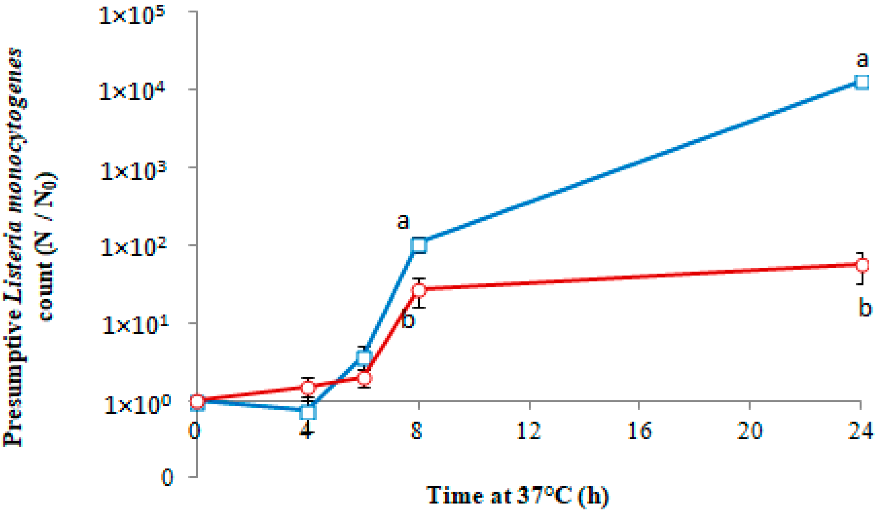
The effect of 20 μm thick PLA film with and without 23.3% w/w Co-cit on the growth of L. monocytogenes on absorbent pads soaked with nutrient broth is shown in Figure 2.
The selection of the film thickness and the Co-cit concentration was based on preliminary experiments with films of 20 and 50 nm thickness and various Co-cit concentrations (data not shown). The thickness/concentration selected gave close to optimum antimicrobial activity while keeping the Co-cit concentration as low as possible. Growth was minimal in all samples for the first 6 h of incubation at 37 °C. At 8 h, the L. monocytogenes count in samples containing no Co-cit was 2.0 log CFU/cm2 higher than the time 0 count (log(N/N0) = 2.0, or a 100-fold increase in count), whereas in samples with membranes containing Co-cit the increase was only 1.3 log CFU/cm2 (approximately 20-fold) at the same time point. No significant growth (p > 0.05) occurred in the samples with Co-cit between 8 and 24 h, while the L. monocytogenes counts in the control samples increased by a further 2.1 log CFU/cm2 over the same period.
In these experiments, Co-cit incorporated into PLA films was shown to be bacteriostatic after a delay of around 6 h, during which growth occurred. This delay in bacteriostasis may be due to the time needed for the Co-cit to diffuse out of the membrane and through the pads.

3.3. Antimicrobial Activity of PLA-(Co-cit) Films against L. monocytogenes on Food Samples

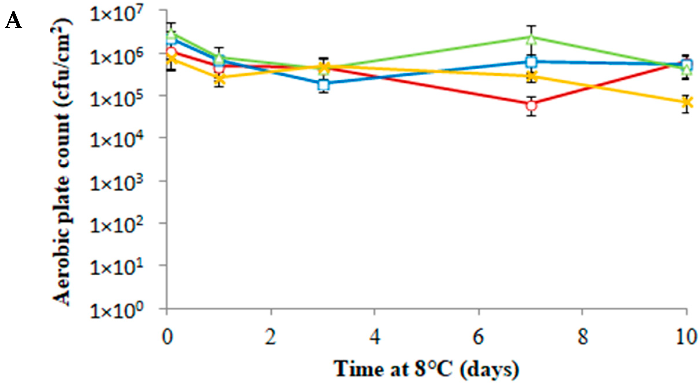
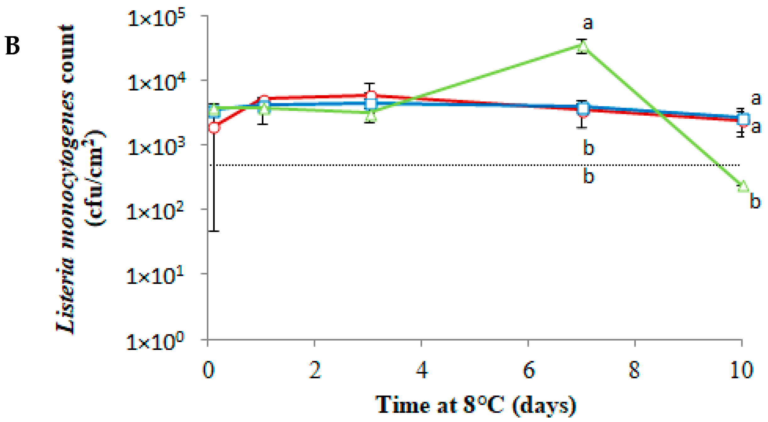
The results of the three experiments investigating the effect of the films on the growth of L. monocytogenes in foods are presented in Figure 3, Figure 4 and Figure 5. The storage temperature of 8 °C was chosen to represent a poorly-functioning but not untypical domestic refrigerator [31]. Presumptive Listeria spp. colonies were not detected in the uninoculated control samples in any of the three experiments, indicating that none of the foods were naturally contaminated. The background aerobic plate count (Figure 3A) in the first experiment with turkey slices was around 6 log CFU/cm2. No consistent changes were observed throughout the 10-day storage period. The L. monocytogenes count (Figure 3B) also stayed constant in the samples with the membranes (with and without Co-cit). In the control containing no film, the L. monocytogenes count began to increase on day 7 but then declined to a level below the detection limit by day 10. By that point, the samples had shown evidence of desiccation (in this experiment, they were not placed in a sealed jar with wet cotton wool). This decrease in water activity, together with the associated increase in salt and nitrite concentration, is the likely cause of the loss of viability of L. monocytogenes. The minimum water activity (aw) for the growth of L. monocytogenes is 0.90, while the optimum value is greater than or equal to 0.97 [32]. Heat-treated cold meats have a water activity of about 0.96–0.99, making them a suitable substrate for the growth of L. monocytogenes [33]. This observation showed that the PLA films offer some protection from desiccation, despite their high water permeability.
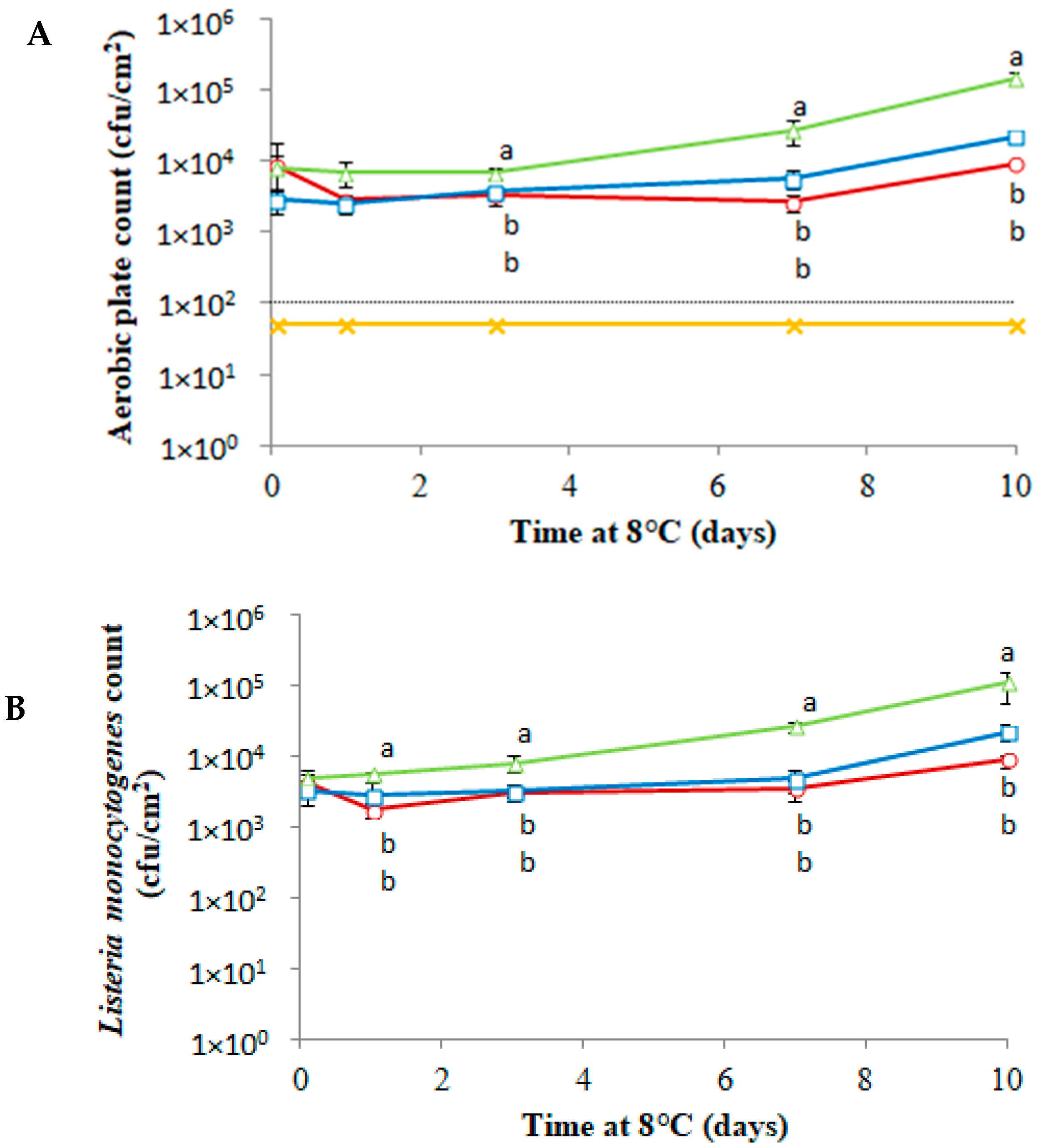
In the second experiment, also using turkey but with a much lower background flora (<100 CFU/cm2), the counts on PCA (Figure 4A) and Oxford agar (Figure 4B) were very similar. As the aerobic plate count of the uninoculated samples was below the limit of detection for the entire storage period, it is likely that the counts on PCA are mostly the inoculated L. monocytogenes. Significant differences were observed between the three inoculated sample types. The membranes had an inhibitory effect on the growth of L. monocytogenes. Although this effect was slightly greater in the membranes containing Co-cit, the difference between membranes with and without Co-cit was fairly small compared to the difference between samples with and without membranes. The total increase in count over the 10-day incubation period was around 1.4 log CFU/cm2 in the control, 0.6 log CFU/cm2 in the samples with membranes with no Co-cit, and 0.2 log CFU/cm2 in samples with Co-cit.
In the experiments with smoked salmon, no significant differences were observed between membranes with and without Co-cit. On day 4, the L. monocytogenes count (Figure 5B) in the samples without a membrane was about 1 log CFU/cm2 higher than in those with membranes, whereas no differences were observed at the previous time points. The L. monocytogenes count (Figure 5B) in the control remained stable for the first two days and increased by around 1 log CFU/mL on day 3. The salmon used in these experiments was a smoked product and may have contained inhibitory smoke components such as phenolic and carbonyl compounds. This was not determined, but the fact that L. monocytogenes grew on the control samples indicates that any such compounds’ inhibitory effect present was mild or absent.
In these three experiments carried out on food surfaces, the PLA membrane itself contributed the most to the bacteriostatic action of the system, whereas the presence of Co-cit was a secondary contributory factor, despite the high concentration used. Food is a more complex and concentrated medium than laboratory media, and higher concentrations of antimicrobials are often needed for similar inhibitory effects to be observed due to interference by food components. The lower water activity of foods compared to culture broth may also reduce the antimicrobial diffusion rate into the aqueous phase of the food [34].
Other authors have observed mild antibacterial activity in PLA films. In one study, a decrease in the count of 0.55 log CFU/g and 0.17 log CFU/g for Staphylococcus aureus and Escherichia coli was observed, respectively [35]. It is also possible that the PLA films altered the redox potential on the surface of the foods. However, as they are highly permeable to gases, this seems unlikely, and even if the oxygen availability was reduced, Listeria monocytogenes is facultatively anaerobic.

3.4. General Discussion

Our results demonstrate the antimicrobial effect of cobalt citrate against important foodborne pathogens, both in an aqueous solution and incorporated into polylactic acid membranes. Atria et al. [36] studied the antimicrobial properties of a cobalt complex ([Co(2-phenylimidazole)2(Acetate)2]) with similar molecular mass (463.35) but different structures against various pathogenic microorganisms, including E. coli, S. Enteritidis, S. aureus, and L. monocytogenes. The antimicrobial activity of the complex was tested by the agar diffusion method on nutrient agar. The results of both studies showed the largest growth inhibition zone in L. monocytogenes. However, in the study of Atria et al. [36], large inhibition zones were observed in both E. coli and S. aureus, suggesting that the antimicrobial activity of the complex is not related to the Gram type and is probably strain-specific. The authors also examined the cobalt complex for the type of antimicrobial activity exerted, with the results indicating bacteriolytic action.
According to Gad [37], cobalt interferes with the functioning of the Krebs cycle, thereby inhibiting the production of energy through oxidative metabolism. This, in turn, can lead to inhibition of the growth of the microorganism.
In this study, the strongest inhibition by Co-cit was observed in L. monocytogenes and P. aeruginosa. Previous studies have examined the action of individual components of the complex (cobalt and citric acid) and have demonstrated their antimicrobial properties against L. monocytogenes, P. aeruginosa, and E. coli. Τhe antimicrobial properties of cobalt oxide in combination with titanium dioxide (CoO-TiO2) were examined in liquid culture against L. monocytogenes [38]. The results showed that CoO-TiO2 is bacteriostatic and prevents the proliferation of L. monocytogenes cells.
One possibility for future research is continuing the work on using Co-cit in combination with existing preservatives but incorporated into membranes. Combinations of preservatives could impart a broader spectrum of activity to antimicrobial packaging films and increase activity against a particular pathogen of concern. The incorporation of existing preservatives into biodegradable films for packaging fresh food is already a subject of research [39].
In accordance with the general requirements set out in the Commission Regulation (EC) No 1935/2004 [40], food contact materials should not endanger human health or bring about an unacceptable change in the composition of the food or bring about deterioration in the organoleptic characteristics thereof.
In accordance with Commission Regulation (EC) No 10/2011 [41] on plastic materials and objects intended to come into contact with food, the limit for cobalt migrating from plastic materials and objects is 0.05 mg/kg of food or stimulant. Further tests would be required to ensure that the films examined conform to this regulation.

4. Conclusions

The results presented in the present paper demonstrate that Co-cit has antimicrobial activity against potential food-associated pathogens and spoilage bacteria, as it significantly reduced the growth rate of all four test bacteria when in solution (0.5 mM). When combined with well-known food preservatives and acids, the results were variable and dependent on the target bacterium. However, in most cases, the antimicrobial effect of Co-cit was enhanced by the presence of additional sublethal inhibitory factors. When incorporated into poly(lactic acid) films at 23.3% w/w, Co-cit was bacteriostatic against Listeria monocytogenes on artificial media but had a reduced effect against L. monocytogenes on the surfaces of real foods.
Cobalt citrate is a promising hybrid metal–organic and well-defined agent with the potential to be used in active food packaging since it exhibits antimicrobial action against important food-associated pathogenic microorganisms. It would be beneficial to investigate its antimicrobial activity in combination with other known or novel antimicrobials incorporated into food packaging materials and to test such systems on particular foods with known associated pathogens, especially those for which limiting growth rather than total exclusion is an important safety strategy.

Author Contributions

J.R.: Conceptualization, Methodology, Writing—original draft, Visualization, Investigation, Supervision, Writing—review and editing. V.K.: Investigation, Writing—original draft, Visualization. S.M.: Investigation, Visualization, Writing—review and editing. K.R.: Investigation, Visualization Writing—review and editing. S.T.: Investigation. N.K.: Investigation. A.S.: Conceptualization, Methodology, Writing—original draft, Visualization, Investigation, Supervision, Writing—review and editing. E.L.: Conceptualization, Methodology, Writing—original draft, Visualization, Investigation, Supervision, Writing—review and editing. All authors have read and agreed to the published version of the manuscript.

Funding

This research received no external funding.

Institutional Review Board Statement

Not applicable.

Informed Consent Statement

Not applicable.

Data Availability Statement

Not applicable.

Acknowledgments

Part of this research was undertaken as an M.Sc. thesis under the program “Master of Science in Quality Management and Production Organization Systems for the Food Industry” offered by the Department of Food Science and Technology, International Hellenic University.

Conflicts of Interest

The authors declare no conflict of interest.

References

  1. Prasad, P.; Kochhar, A. Active Packaging in Food Industry: A Review. J. Environ. Sci. Toxicol. Food Technol. 2014, 8, 1–7. [Google Scholar] [CrossRef]
  2. Padmanabhan, S.C.; Cruz-Romero, M.C.; Kerry, J.P.; Morris, M.A. Chapter 11—Food packaging: Surface engineering and commercialization. In Nanomaterials for Food Packaging: Materials, Processing Technologies, and Safety Issues; Elsevier Inc.: Amsterdam, The Netherlands, 2018; pp. 301–328. [Google Scholar] [CrossRef]
  3. García Ibarra, V.; Sendón, R.; Rodríguez-Bernaldo De Quirós, A. Chapter 29—Antimicrobial food packaging based on biodegradable materials. In Antimicrobial Food Packaging; Academic Press: Cambridge, MA, USA, 2016; pp. 363–384. [Google Scholar] [CrossRef]
  4. Yildirim, S.; Röcker, B. Chapter 7—Active packaging. In Nanomaterials for Food Packaging: Materials, Processing Technologies, and Safety Issues; Elsevier: Amsterdam, The Netherlands, 2018; pp. 173–202. [Google Scholar] [CrossRef]
  5. Moustafa, H.; El-Sayed, S.M.; Youssef, A.M. Synergistic impact of cumin essential oil on enhancing of UV-blocking and antibacterial activity of biodegradable poly(butylene adipate-co-terephthalate)/clay platelets nanocomposites. J. Thermoplast. Compos. Mater. 2021, 1–22. [Google Scholar] [CrossRef]
  6. Moustafa, H.; Darwish, N.A.; Youssef, A.M. Rational formulations of sustainable polyurethane/chitin/rosin composites reinforced with ZnO-doped-SiO2 nanoparticles for green packaging applications. Food Chem. 2022, 371, 131193. [Google Scholar] [CrossRef]
  7. Akhidime, I.D.; Saubade, F.; Benson, P.S.; Butler, J.A.; Oliver, S.; Kelly, P.; Verran, J.; Whitehead, K.A. The antimicrobial effect of metal substrates on food pathogens. Food Bioprod. Processing 2019, 113, 68–76. [Google Scholar] [CrossRef]
  8. Mousavi Khaneghah, A.; Hashemi, S.M.B.; Limbo, S. Antimicrobial agents and packaging systems in antimicrobial active food packaging: An overview of approaches and interactions. Food Bioprod. Processing 2018, 111, 1–19. [Google Scholar] [CrossRef]
  9. Al-Tayyar, N.A.; Youssef, A.M.; Al-hindi, R. Antimicrobial food packaging based on sustainable Bio-based materials for reducing foodborne Pathogens: A review. Food Chem. 2020, 310, 125915. [Google Scholar] [CrossRef]
  10. Huang, T.; Qian, Y.; Wei, J.; Zhou, C. Polymeric Antimicrobial food packaging and its applications. Polymers 2019, 11, 560. [Google Scholar] [CrossRef] [PubMed]
  11. Gan, I.; Chow, W.S. Antimicrobial poly(lactic acid)/cellulose bionanocomposite for food packaging application: A review. Food Packag. Shelf Life 2018, 17, 150–161. [Google Scholar] [CrossRef]
  12. Matzapetakis, M.; Dakanali, M.; Raptopoulou, C.P.; Tangoulis, V.; Terzis, A.; Moon, N.; Giapintzakis, J.; Salifoglou, A. Synthesis, spectroscopic, and structural characterization of the first aqueous cobalt(II)-citrate complex: Toward a potentially bioavailable form of cobalt in biologically relevant fluids. J. Biol. Inorg. Chem. 2000, 5, 469–474. [Google Scholar] [CrossRef] [PubMed] [Green Version]
  13. Ciriminna, R.; Meneguzzo, F.; Delisi, R.; Pagliaro, M. Citric acid: Emerging applications of key biotechnology industrial product. Chem. Cent. J. 2017, 11, 22. [Google Scholar] [CrossRef] [Green Version]
  14. Goldberg, I.; Stefan Rokem, J. Organic and fatty acid production, microbial. Encycl. Microbiol. 2019, 358–382. [Google Scholar] [CrossRef]
  15. Lison, D. Chapter 25—Cobalt. In Handbook on the Toxicology of Metals; Academic Press: Cambridge, MA, USA, 2007; pp. 511–528. [Google Scholar] [CrossRef]
  16. Kouris, E.; Kalogiannis, S.; Perdih, F.; Turel, I.; Psomas, G. Cobalt(II) complexes of sparfloxacin: Characterization, structure, antimicrobial activity and interaction with DNA and albumins. J. Inorg. Biochem. 2016, 163, 18–27. [Google Scholar] [CrossRef] [PubMed]
  17. Leyssens, L.; Vinck, B.; Van Der Straeten, C.; Wuyts, F.; Maes, L. Cobalt toxicity in humans—A review of the potential sources and systemic health effects. Toxicology 2017, 387, 43–56. [Google Scholar] [CrossRef]
  18. Renfrew, A.K.; O’Neill, E.S.; Hambley, T.W.; New, E.J. Harnessing the properties of cobalt coordination complexes for biological application. Coord. Chem. Rev. 2018, 375, 221–233. [Google Scholar] [CrossRef]
  19. Hokin, B.; Adams, M.; Ashton, J.; Louie, H. Comparison of the dietary cobalt intake in three different Australian diets. Asia Pac. J. Clin. Nutr. 2004, 13, 289–291. [Google Scholar] [PubMed]
  20. Chang, E.L.; Simmers, C.; Knight, D.A. Cobalt complexes as antiviral and antibacterial agents. Pharmaceuticals 2010, 3, 1711–1728. [Google Scholar] [CrossRef] [PubMed]
  21. Jeon, M.J.; Ha, J.W. Synergistic bactericidal effect and mechanism of X-ray irradiation and citric acid combination against food-borne pathogens on spinach leaves. Food Microbiol. 2020, 91, 103543. [Google Scholar] [CrossRef] [PubMed]
  22. Saliu, O.D.; Olatunji, G.A.; Olosho, A.I.; Adeniyi, A.G.; Azeh, Y.; Samo, F.T.; Adebayo, D.O.; Ajetomobi, O.O. Barrier property enhancement of starch citrate bioplastic film by an ammonium-thiourea complex modification. J. Saudi Chem. Soc. 2019, 23, 141–149. [Google Scholar] [CrossRef]
  23. Seligra, P.G.; Medina Jaramillo, C.; Famá, L.; Goyanes, S. Biodegradable and non-retrogradable eco-films based on starch-glycerol with citric acid as crosslinking agent. Carbohydr. Polym. 2016, 138, 66–74. [Google Scholar] [CrossRef] [Green Version]
  24. Silveira, V.A.I.; Marim, B.M.; Hipólito, A.; Gonçalves, M.C.; Mali, S.; Kobayashi, R.K.T.; Celligoi, M.A.P.C. Characterization and antimicrobial properties of bioactive packaging films based on polylactic acid-sophorolipid for the control of foodborne pathogens. Food Packag. Shelf Life 2020, 26, 100591. [Google Scholar] [CrossRef]
  25. Bjarnsholt, T.; Alhede, M.; Jensen, P.Ø.; Nielsen, A.K.; Johansen, H.K.; Homøe, P.; Høiby, N.; Givskov, M.; Kirketerp-Møller, K. Antibiofilm Properties of Acetic Acid. Adv. Wound Care 2015, 4, 363–372. [Google Scholar] [CrossRef] [PubMed] [Green Version]
  26. Dolan, S.K.; Kohlstedt, M.; Trigg, S.; Ramirez, P.V.; Kaminski, C.F.; Wittmann, C.; Welch, M. Contextual flexibility in Pseudomonas aeruginosa central carbon metabolism during growth in single carbon sources. mBio 2020, 11, e02684-19. [Google Scholar] [CrossRef] [Green Version]
  27. Kirkpatrick, C.; Maurer, L.M.; Oyelakin, N.E.; Yoncheva, Y.N.; Maurer, R.; Slonczewski, J.L. Acetate and formate stress: Opposite responses in the proteome of Escherichia coli. J. Bacteriol. 2001, 183, 6466–6477. [Google Scholar] [CrossRef] [PubMed] [Green Version]
  28. Kotsakis, N.; Raptopoulou, C.P.; Tangoulis, V.; Terzis, A.; Giapintzakis, J.; Jakusch, T.; Kiss, T.; Salifoglou, A. Correlations of Synthetic, Spectroscopic, Structural, and Speciation Studies in the Biologically Relevant Cobalt(II)-Citrate System: The Tale of the First Aqueous Dinuclear Cobalt(II)-Citrate Complex. Inorg. Chem. 2003, 42, 22–31. [Google Scholar] [CrossRef] [PubMed]
  29. Nagaoka, S.; Murata, S.; Kimura, K.; Mori, T.; Hojo, K. Antimicrobial activity of sodium citrate against Streptococcus pneumoniae and several oral bacteria. Lett. Appl. Microbiol. 2010, 51, 546–551. [Google Scholar] [CrossRef] [PubMed]
  30. Burel, C.; Kala, A.; Purevdorj-Gage, L. Impact of pH on citric acid antimicrobial activity against Gram-negative bacteria. Lett. Appl. Microbiol. 2020, 72, 332–340. [Google Scholar] [CrossRef] [PubMed]
  31. Andritsos, N.D.; Stasinou, V.; Tserolas, D.; Giaouris, E. Temperature distribution and hygienic status of domestic refrigerators in Lemnos island, Greece. Food Control 2021, 127, 108121. [Google Scholar] [CrossRef]
  32. Wang, S.; Orsi, R.H. Chapter 11—Listeria. In Foodborne Infections and Intoxications, 4th ed.; Elsevier Inc.: Amsterdam, The Netherlands, 2013; pp. 199–216. [Google Scholar] [CrossRef]
  33. Borch, E.; Kant-Muermans, M.L.; Blixt, Y. Bacterial spoilage of meat and cured meat products. Int. J. Food Microbiol. 1996, 33, 103–120. [Google Scholar] [CrossRef]
  34. Guo, M.; Jin, T.Z.; Yang, R. Antimicrobial Polylactic Acid Packaging Films against Listeria and Salmonella in Culture Medium and on Ready-to-Eat Meat. Food Bioprocess Technol. 2014, 7, 3293–3307. [Google Scholar] [CrossRef]
  35. Qi, Y.; Ma, H.L.; Du, Z.H.; Yang, B.; Wu, J.; Wang, R.; Zhang, X.Q. Hydrophilic and Antibacterial Modification of Poly(lactic acid) Films by γ-ray Irradiation. ACS Omega 2019, 4, 21439–21445. [Google Scholar] [CrossRef] [PubMed] [Green Version]
  36. Atria, A.M.; Cortés-Cortés, P.; Garland, M.T.; Baggio, R.; Morales, K.; Soto, M.; Corsini, G. X-ray studies and antibacterial activity in copper and cobalt complexes with imidazole derivative ligands. J. Chil. Chem. Soc. 2011, 56, 786–792. [Google Scholar] [CrossRef]
  37. Gad, S.C. Cobalt. In Encyclopedia of Toxicology, 3rd ed.; Elsevier: Amsterdam, The Netherlands, 2014; Volume 1, pp. 996–998. [Google Scholar] [CrossRef]
  38. Sirsat, S.A.; Hecht, O.; Mirabal, C.; Pepe, D.A.; Yang, W.; Mohammad, Z.; Hadjiev, V.G.; Neal, J.A.; Robles Hernandez, F.C. Bacteriostatic effect of CoO-TiO2 on Listeria monocytogenes by the presence of the co-catalytic CoO nanoparticles. J. Environ. Chem. Eng. 2020, 8, 104259. [Google Scholar] [CrossRef]
  39. Wangprasertkul, J.; Siriwattanapong, R.; Harnkarnsujarit, N. Antifungal packaging of sorbate and benzoate incorporated biodegradable films for fresh noodles. Food Control 2021, 123, 107763. [Google Scholar] [CrossRef]
  40. Regulation (EC) No 1935/2004 of the European Parliament and of the Council of 27 October 2004 on Materials and Articles Intended to Come into Contact with Food and Repealing Directives 80/590/EEC and 89/109/EEC. Available online: https://eur-lex.europa.eu/legal-content/EN/ALL/?uri=CELEX:32004R1935 (accessed on 23 September 2022).
  41. Commission Regulation (EU) 2020/1245 of 2 September 2020 Amending and Correcting Regulation (EU) No 10/2011 on Plastic Materials and Articles Intended to Come into Contact with Food (Text with EEA Relevance). Available online: https://eur-lex.europa.eu/legal-content/EN/TXT/?uri=CELEX:32020R1245 (accessed on 23 September 2022).
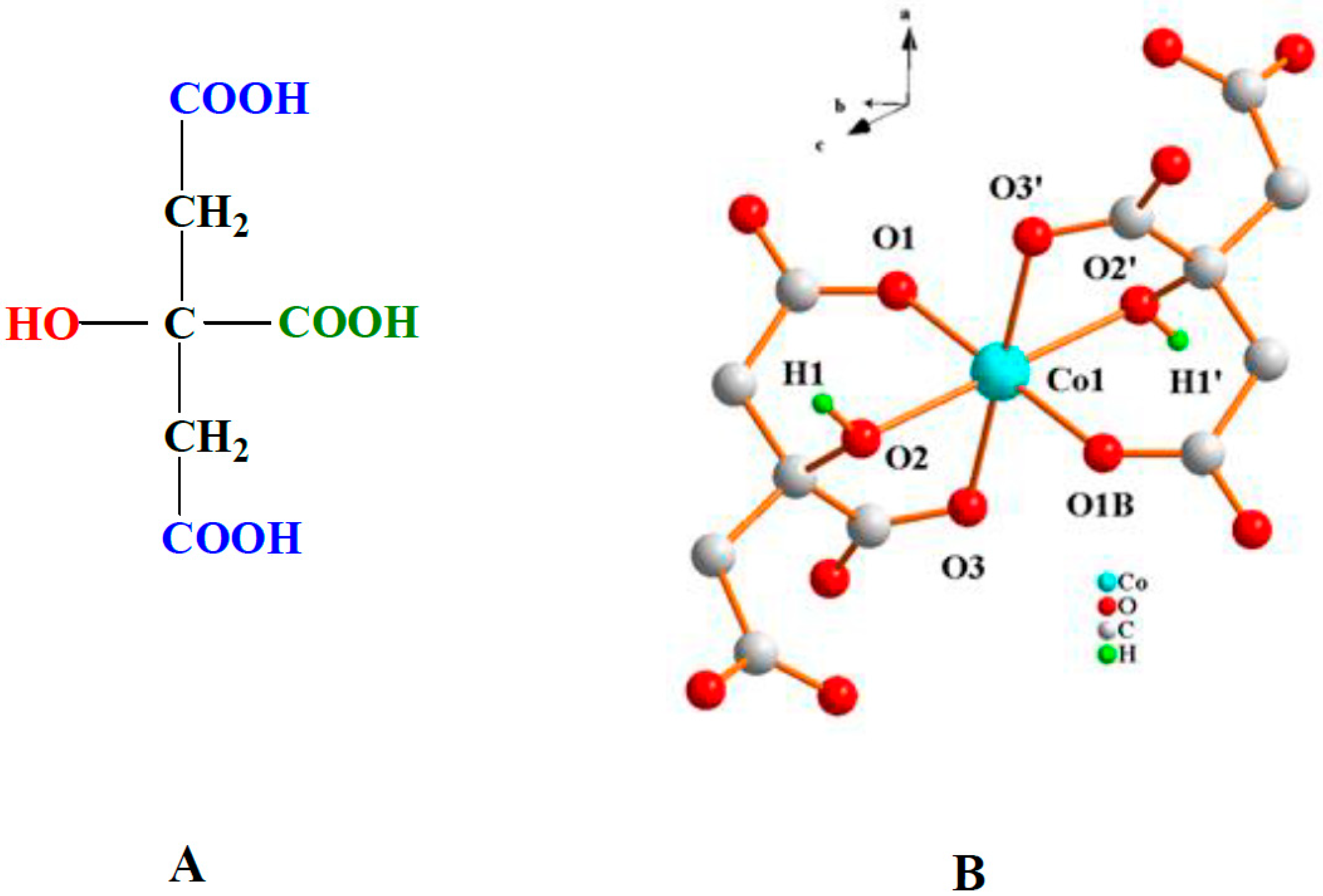
Figure 1. (A) Structure of α-hydroxycarboxylic acid citric acid as a metal ion binder; (B) Structure of the crystallographically characterized anionic complex in (NH4)4[Co(C6H5O7)2] (Co-cit).
Applsci 12 10855 g001
Figure 2. Growth of Listeria monocytogenes on paper pads soaked in TSBYE broth and covered with PLA films with 23.3% w/w Co-cit (red, circles) and PLA films without Co-cit (blue, squares). Results are combined from two separate experiments and expressed as the count at time t (N) divided by the count at time 0 (N0). Different letters within the same time point indicate significant (p < 0.05) differences; where there are no letters, there were no significant differences (p > 0.05).
Applsci 12 10855 g002
Figure 3. Aerobic plate counts (A) and presumptive Listeria monocytogenes counts (B) on turkey ham with high background count. Treatments: Inoculated with L. monocytogenes, covered by PLA film with 23.3% w/w Co-cit (red, circles); inoculated with L. monocytogenes, covered by PLA film without Co-cit (blue, squares); inoculated with L. monocytogenes, no film (green, triangles); uninoculated, no film (yellow, Xs). Different letters within the same time point indicate significant differences (p < 0.05) between inoculated samples (uninoculated samples not included in the calculation); where there are no letters, there were no significant differences (p > 0.05). Error bars represent standard deviation. The straight line on the lower graph represents the detection limit—samples with no colonies were assigned a value of one-half of this value.
Applsci 12 10855 g003aApplsci 12 10855 g003b
Figure 4. Aerobic plate counts (A) and presumptive Listeria monocytogenes counts (B) on turkey ham with low background count. Treatments: Inoculated with L. monocytogenes, covered by PLA film with 23.3% w/w Co-cit (red, circles); inoculated with L. monocytogenes, covered by PLA film without Co-cit (blue, squares); inoculated with L. monocytogenes, no film (green, triangles); uninoculated, no film (yellow, Xs). Different letters within the same time point indicate significant differences (p < 0.05) between inoculated samples (uninoculated samples not included in the calculation); where there are no letters, there were no significant differences (p > 0.05). Error bars represent standard deviation. The straight line on the upper graph represents the detection limit—samples with no colonies were assigned a value of one-half of this value.
Applsci 12 10855 g004
Figure 5. Aerobic plate counts (A) and presumptive Listeria monocytogenes counts (B) on smoked salmon. Treatments: Inoculated with L. monocytogenes, covered by PLA film with 23.3% w/w Co-cit (red, circles); inoculated with L. monocytogenes, covered by PLA film without Co-cit (blue, squares); inoculated with L. monocytogenes, no film (green, triangles); uninoculated, no film (yellow, Xs). Different letters within the same time point indicate significant differences (p < 0.05) between inoculated samples (uninoculated samples not included in the calculation); where there are no letters, there were no significant differences (p > 0.05). Error bars represent standard deviation.
Applsci 12 10855 g005
Table 1. Relative maximum growth rates and lag phases of Listeria monocytogenes in modified and standard nutrient broth (NB) with and without cobalt citrate [Co-cit (0.5 mM)].
Ratio of Growth Parameters of Listeria monocytogenes in:
Modification of Nutrient BrothModified NB with Co-cit/Modified NB without Co-citModified NB/Standard NB
Max. Growth RateLag PhaseMax. Growth RateLag Phase
None (standard NB)0.61 *1.0--
Sorbate (10 g/L)0.57 *1.00.69 *2.1
Benzoate (2 g/L)0.89 †0.80.671.8
Propionate (20 g/L)0.56 *0.91.031.0
HCl (pH 5.5)0.37 *†2.00.951.2
Acetic acid (pH 5.5)0.20 *†>2.6 ‡0.57 *3.6
NaNO2 (0.15 g/L)0.52 *1.10.48 *1.2
NaCl (50 g/L)0.51 *1.80.41 *0.6
* Significant (p < 0.05) difference between the mean maximum growth rates in standard or modified broth with and without Co-cit (column 2) or between modified NB and standard NB (column 4). Statistical analysis was not performed on the lag phases. † Significant (p < 0.05) difference between the ratio of maximum growth rates with and without Co-cit in modified broth compared to that in unmodified nutrient broth (0.61). ‡ Some or all of the recorded lag phases in broths with Co-cit were >24 h.
Table 2. Relative maximum growth rates and lag phases of Staphylococcus aureus in modified and standard nutrient broth (NB) with and without cobalt citrate [Co-cit (0.5 mM)].
Ratio of Growth Parameters of Staphylococcus aureus in:
Modification of Nutrient BrothModified NB with Co-cit/Modified NB without Co-citModified NB/Standard NB
Max. Growth RateLag PhaseMax. Growth RateLag Phase
None (standard NB)0.74 *1.5--
Sorbate (10 g/L)0.52 *†1.40.72 *2.0
Benzoate (2 g/L)0.76 *1.20.77 *1.6
Propionate (20 g/L)0.61 *1.20.80 *1.4
HCl (pH 5.5)0.75 *1.21.052.2
Acetic acid (pH 5.5)0.65 *1.41.031.7
NaNO2 (0.15 g/L)0.55 *0.91.051.5
NaCl (50 g/L)0.56 *1.00.65 *1.0
* Significant (p < 0.05) difference between the mean maximum growth rates in standard or modified broth with and without Co-cit (column 2) or between modified NB and standard NB (column 4). Statistical analysis was not performed on the lag phases. † Significant (p < 0.05) difference between the ratio of maximum growth rates with and without Co-cit in modified broth compared to that in unmodified nutrient broth (0.61).
Table 3. Relative maximum growth rates and lag phases of Escherichia coli in modified and standard nutrient broth (NB) with and without cobalt citrate [Co-cit (0.5 mM)].
Ratio of Growth Parameters of Escherichia coli in:
Modification of Nutrient BrothModified NB with Co-cit/Modified NB without Co-citModified NB/Standard NB
Max. Growth RateLag PhaseMax. Growth RateLag Phase
None (standard NB)0.70 *1.2--
Sorbate (10 g/L)1.04 †1.00.36 *3.7
Benzoate (2 g/L)0.76 *0.90.68 *2.0
Propionate (20 g/L)0.55 *†1.31.021.2
HCl (pH 5.5)0.79 *1.80.991.2
Acetic acid (pH 5.5)1.05 †1.20.78 *3.8
NaNO2 (0.15 g/L)0.62 *0.80.84 *1.0
NaCl (50 g/L)0.43 *†1.20.21 *2.8
* Significant (p < 0.05) difference between the mean maximum growth rates in standard or modified broth with and without Co-cit (column 2) or between modified NB and standard NB (column 4). Statistical analysis was not performed on the lag phases. † Significant (p < 0.05) difference between the ratio of maximum growth rates with and without Co-cit in modified broth compared to that in unmodified nutrient broth (0.61).
Table 4. Relative maximum growth rates of Pseudomonas aeruginosa in modified and standard nutrient broth (NB) with and without cobalt citrate [Co-cit (0.5 mM)].
Ratio of Growth Parameters of Pseudomonas aeruginosa in:
Modification of Nutrient BrothModified NB with Co-cit/Modified NB without Co-citModified NB/Standard NB
Max. Growth RateLag PhaseMax. Growth RateLag Phase
None (standard NB)0.51 *6.9--
Sorbate (10 g/L)0.69 *†4.10.91 *1.8
Benzoate (2 g/L)0.61 *6.90.84 *1.3
Propionate (20 g/L)0.00 *†>15.0 ‡1.071.6
HCl (pH 5.5)0.00 *†>7.8 ‡1.011.9
Acetic acid (pH 5.5)0.04 *†>2.2 ‡1.32 *6.6
NaNO2 (0.15 g/L)0.24 *†>5.7 ‡1.061.1
NaCl (50 g/L)0.00 *†>3.4 ‡0.49 *1.8
* Significant (p < 0.05) difference between the mean maximum growth rates in standard or modified broth with and without Co-cit (column 2) or between modified NB and standard NB (column 4). Statistical analysis was not performed on the lag phases. † Significant (p < 0.05) difference between the ratio of maximum growth rates with and without Co-cit in modified broth compared to that in unmodified nutrient broth (0.61). ‡ Some or all of the recorded lag phases in broths with Co-cit were >24 h.
Publisher’s Note: MDPI stays neutral with regard to jurisdictional claims in published maps and institutional affiliations.