Article

Relation between Nasal Septum Deviation and Facial Asymmetry: An Ontogenetic Analysis from Infants to Children Using Geometric Morphometrics

by
Azalea Shamaei-Tousi
1,*,
Alessio Veneziano
2 and
Federica Landi
1,*
1
Institute of Medical and Biomedical Education, St. George’s, University of London, London SW17 0RE, UK
2
Department of Archaeology, University of Cambridge, Cambridge CB2 1TN, UK
*
Authors to whom correspondence should be addressed.
Appl. Sci. 2022, 12(22), 11362; https://doi.org/10.3390/app122211362
Submission received: 20 July 2022 / Revised: 2 November 2022 / Accepted: 6 November 2022 / Published: 9 November 2022

Abstract

:
The nasal septum has been postulated to have an intrinsic growth power and act as a pacemaker for facial development, its interactions with local craniofacial structures likely to influence facial anatomy and morphology. Recent studies have begun to investigate the link between nasal septum deviation and facial asymmetry; however, the magnitude and mechanisms of this relation are still unclear. This study aimed to analyse the degree of nasal septum deviation in a sample of infants and children (males and females from 0 to 8 years old) and its correlation with the three-dimensional structure of the facial skeleton. The scope was to test whether septal deviation is linked, and might cause, the development of a more asymmetric face. For this aim, 41 3D landmarks (homologous points) were collected on the nasal septum and cranial surface of 46 specimens extracted from medical CT-scans and were analysed using Geometric Morphometrics, Multiple Linear regressions, Multivariate ANOVAs, and Principal Component Analysis (PCA). Results showed no significant correlation between magnitude of septal deviation and the ontogeny (changes in age) or sex of the sample, but a significant association was found between side of deviation and septal deviation magnitude and frequency. The asymmetric PCA reveals that most of the asymmetry identified is fluctuating, and that changes in the asymmetric morphology of the face are not associated to a specific side of septal deviation. In addition, a series of Multivariate ANOVAs showed that age, sex, and septal deviation have no impact on facial asymmetry, with only age impacting the symmetric development of the facial morphology. When looking at factors impacting the general morphology of the face, age is again the only major driving component, with fluctuating asymmetry and sex only approaching significance. These results could imply a certain degree of dissociation between the mechanisms of facial and septal growth and development; however, an investigation of other key developmental stages in facial morphology is needed to further understand the relation between septal deviation and facial growth.

1. Introduction

1.1. Nasal Septal Growth and Facial Development

The growth and development of the nasal septum and the processes by which it influences other facial structures is a topic highly debated by scientists and still poorly understood, with some authors claiming that nasal septal growth continues well into adulthood (for a review of studies of nasal septal measurements, see [1]). In this context, a theory, called Functional Matrix theory, was introduced by Melvin Moss in 1968 [2]. It postulated that the cranium is made up of multiple functional and skeletal matrices. A functional matrix is defined as “non-skeletal cells, tissues, organs, and operational volumes” present in the body, while skeletal matrices form around the functional matrices as protection and support [2]. These functional matrices can be represented by organs, muscles, a volume, or a space that have a specific craniofacial function. Skeletal matrices develop in support of the adjacent functional matrices and can be described as “bone, cartilage, or tendinous tissues” [3]. One of these functional matrices is the nasal cavity. Moss believes that the nasal cavity is structurally supported by the skeletal matrix of nasal septum, whose only role is to act as a pillar to support the roof of the nasal cavity. Due to its role, according to Moss, the nasal septum grows as a secondary response to the primary growth of the functional matrices of the face, particularly the nasal cavity, as well as facial muscles and other functional matrices of the face, such as the orbits [4].
This interpretation is in opposition to another well-received theory by James H. Scott. Scott’s theory hypothesises that cranial development is encouraged and influenced by cartilage expansion centres, one of these being the nasal septal cartilage [5]. Scott suggests that, as cartilaginous areas grow, this causes the growth, development, and displacement of adjacent facial features. He states that, as the nasal septum cartilage grows, it causes the facial bones, not including the mandible, to be thrust downwards and forwards and to separate. In contrast to Moss’s theory, Scott states that during the first phase of facial development, growth is “regulated by the cartilage of the nasal septum, cranial base and mandibular condyle”, and these cartilaginous structures act as pacemakers for the early growth of the facial skeleton [5].
Research has continued to test these theories and to investigate the influence of the nasal septum on human facial development. A longitudinal twin study was carried out in 1997, where one of the twins suffered a nasal septal destruction and reconstruction (with cartilage implantation) at age 7 [6]. When measurements of the twins were compared at age 17, there were clear differences in maxillary growth and development, with the affected twin having a growth disturbance leading to a smaller maxilla vertically and horizontally. These findings seem to suggest an influence of the nasal septum on the growth and morphology of the facial bones, particularly the maxilla, thus supporting Scott’s interpretation of the nasal septum as a pacemaker for facial growth. More recently, Hartman et al. [7] “assessed the morphological relationship between nasal septal deviation and asymmetries of the facial skeleton in adults”. Their results showed a significant relationship between nasal septum deviation and asymmetry of the nasal and palatal regions in adults. However, no asymmetry in more lateral regions of the face was found.
In contrast, other studies revealed findings in support of Moss’s theory of the septum which is a secondary structural aspect of facial development [8,9,10]. These studies suggest that the septum has a passive role and is subject to change by neighbouring skeletal structures, proposing that nasal septum deviation is dependent and constricted by other aspects of the nasofacial region elements. In a recent study, nasal septal deviation has been linked to a reduction in the height of the upper facial skeleton [11]. This suggests that although the septum has an intrinsic growth power, its growth is decoupled from the surrounding skeletal structures, as the results of the study showed that the nasal septum continues to expand in short faces, culminating in its deviation due to the limited height available. If this interpretation is right, nasal septum development would be independent of and distinct from facial development. By hypothesising a lack of influence in the nasal septum on the overall midfacial architecture (as Moss suggested) but considering this structure to have its own growth potential, this explanation revisits Moss’s original hypothesis of the nasal septum lacking growth power and being subordinate to midfacial changes.

1.2. The Study of Asymmetric Facial Development

Facial skeletal asymmetries can lead to several functional issues and its consequences are not limited to aesthetics. Indeed, as asymmetry can indicate that one side of the face is more skeletally developed, it will most inevitably result “in compensational or distorted growth” [12], leading to muscular imbalances.
It has been observed that there is a correlation between nasal septum deviation and alterations in facial morphology [13,14,15]. Overall, the results of these studies suggest that there are significant correlations between the side of nasal septum deviation and abnormal facial shape, particularly in localised nearby structures, such as the maxilla and nasal cavity. However, most of these studies do not look specifically into patterns of asymmetry and when they do, they focus on adult samples and/or use 2D measurements. Indeed, throughout discussions on the links between facial asymmetry and nasal septum deviation, there is no consensus on whether nasal septum deviation causes facial asymmetry or whether asymmetry causes nasal deviation due to the structural constraints it applies to the nasal capsule and therefore the septum. Causation cannot be found with cross-sectional studies but longitudinal data following patients over several years, which would help elucidate causes, are extremely rare. However, three-dimensional ontogenetic studies can help in investigating the magnitude and direction of correlations alongside mechanisms of growth and development and can motivate investment in collecting the necessary longitudinal data to test causal hypotheses.
Based on the findings discussed in the Introduction, our null hypothesis states that:
H0. 
The degree of nasal septum deviation is not linked to the development of a more asymmetric face.
If the null hypothesis is rejected, i.e., if a link is found between septal deviation and facial asymmetry, this could suggest that the nasal septum has an intrinsic growth power and that its growth and development can impact adjacent skeletal structures by driving them apart and causing their relative displacement. Moreover, it could imply that the time and mode of facial growth set the structural constraints for the septum, which would continue its growth in a limited space, therefore bending, potentially toward the less asymmetric side. Both these implications would indicate that the septum has its own growth potential and that its development is not highly modular but linked to overall facial development. If the null hypothesis cannot be rejected, i.e., if a link is not found between septal deviation and facial asymmetry, this could imply that there is a dissociation between the mechanism of facial and nasal septal growth, or that the presence of septal deviations and/or facial asymmetry is low at this age range.

2. Materials and Methods

2.1. Sample

Forty-six 3D crania were segmented from medical CT-scans downloaded from The New Mexico Decedent Image Database (NMDID) [16] using Avizo 9.0 (FEI visualisation). The NMDID is an ethically approved public online database of deceased and anonymised specimens. For any concern about the ethical approval of this dataset, please visit https://nmdid.unm.edu/ (accessed on 1 April 2022). The sample was inclusive of both genders in equal proportion and included a specific age range known to be key for facial development, from 0 to 8 years old. Specimens with severe facial fractures or for which the quality of scans were too low to make appropriate measurements were excluded from the selection.
Sample size was chosen following previous literature on the subject [17], which advised on a sample of N > 20 specimens when performing Geometric Morphometric analyses. Although an imbalance between shape coordinates and number of specimens can lead to overfitting or other statistical issues, research from Collyer et al. [18] have shown that increasing shape variables can increase the ability to detect differences among groups (i.e., the statistical power). A complete list of the specimens, their age, and sex, can be found in Appendix A.

2.2. Landmark Configuration

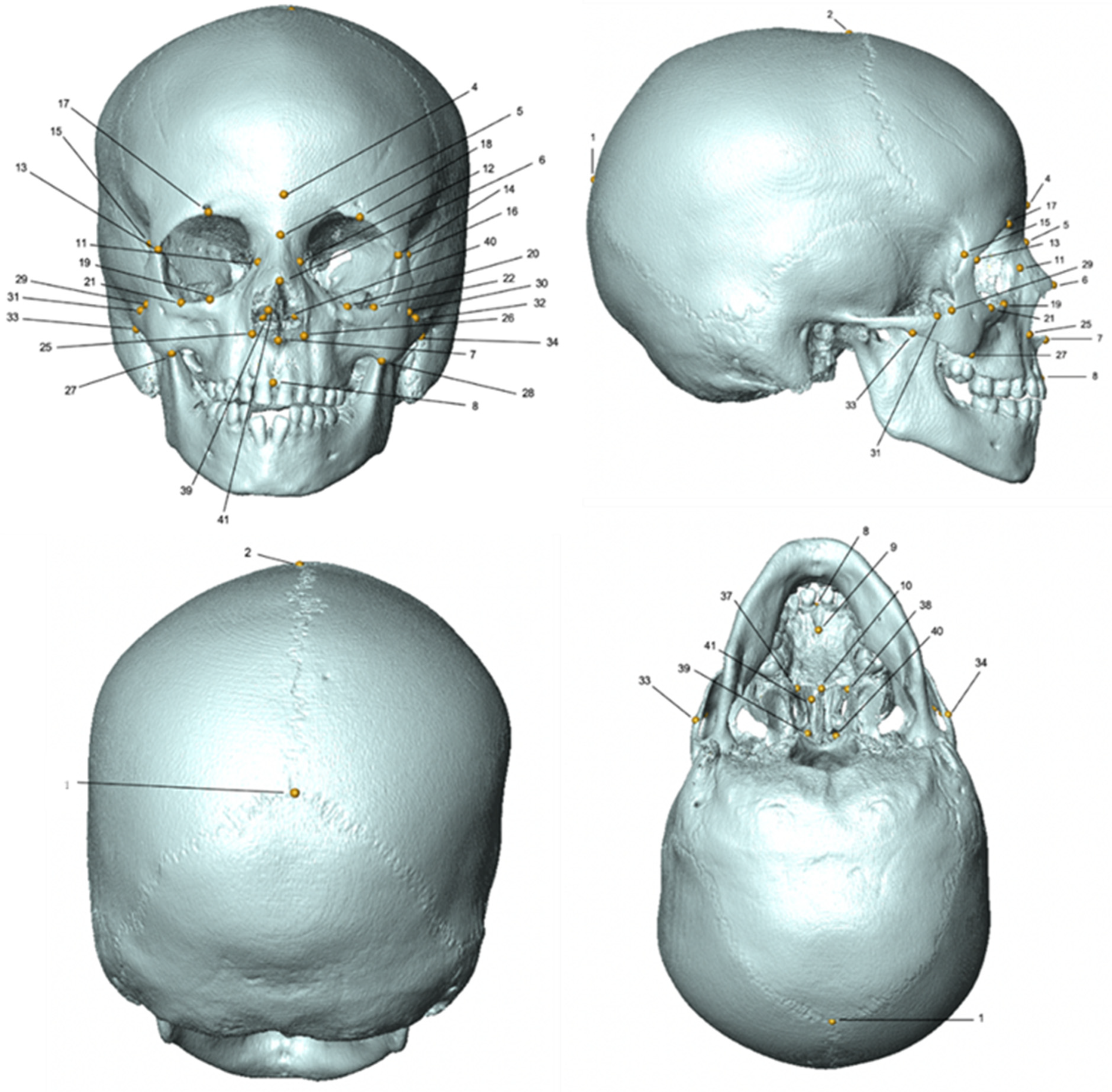
On the extracted 3D surfaces, obtained after segmentation of the skull from CT-scans, 41 landmarks (biologically homologous points) were placed over the cranial vault and face using Avizo 9.0 (FEI visualisation) (Figure 1). A list of the landmarks and their anatomical definitions can be found in Appendix B. The final landmark point in the landmark configuration was located on the most deviated point of the nasal septum and was found using the “orthoslice” function in Avizo 9.0 to gain a series of coronal CT images of each specimen. This method allowed clear visualisation of the nasal septum, which enabled placement of the landmark on the most lateral point from a cranial midsagittal plane by scrolling antero-posteriorly through the coronal slices [19]. This can be seen in Figure 2. To estimate the level of nasal septum deviation (calculated in mm), for each specimen, a virtual midsagittal plane was built in R software [20]. The midsagittal plane was estimated by finding the least squares plane that best fits the midsagittal cranial landmarks, namely the points bregma, lambda, and the anterior point along the cribriform plate. These landmarks were used in building a midsagittal plane for estimating nasal septum deviations and were not included in measuring facial asymmetry nor in the Generalised Procrustes Analysis (GPA) [21] of the facial morphology. Then, the linear distance between the deviation landmark and the midsagittal plane was measured using R software. To account for cranial size differences, the septal deviation was recorded using landmark configurations after GPA. In this way, the linear distance was scaled to the unit size of the cranium. The deviation landmark was only used to estimate septal deviation and was not included in measuring facial asymmetry nor in the GPA of the facial morphology.

2.3. Using Geometric Morphometrics to Study Asymmetry

As Geometric Morphometrics allow for differences to be drawn from each side of a structure, it is ideal for analysing and quantifying asymmetry. Its “quantification of shape captures more subtle differences in asymmetry than traditional morphometric techniques” [22]. There are three different types of asymmetry typically observed: Directional Asymmetry, Bimodal Asymmetry, and Fluctuating Asymmetry [23]. Directional asymmetry occurs when sides of a structure “show a preferential direction”, and, in biology, it can indicate that one side is used more than the other due to specific environmental pressures [24]. Bimodal asymmetry (anti-symmetry) refers to when asymmetry is the norm; therefore, both sides will deviate from symmetry in ways to cause a bimodal distribution. Fluctuating asymmetry is when a more symmetrical structure is the norm, and both sides deviate from this symmetry with no side preference in a random and non-directional manner [23]. If asymmetry is presented in the current sample, we expect to see evidence of fluctuating asymmetry. This is due to the fact that fluctuating asymmetry usually occurs when asymmetry is caused by development and random external factors acting on the development. Therefore, in this study, we do not expect to see any directional asymmetry as we are not analysing a population under a specific developmental pressure. Studies have used GM to investigate fluctuating asymmetry alongside developmental instabilities and stressors [25,26,27,28]. Prior to analysing the landmarks, all configurations undergo a process called Generalised Procrustes Analysis (GPA). During this process, differences between equivalent landmarks are minimised by the process of superimposition, i.e., the scaling, translating, and rotation of the landmark configurations using the arithmetic mean of the landmark coordinates or the centroid [29,30,31,32,33,34,35]. Figure 3 shows the process of a Procrustes superimposition. The aim of the GPA is to minimise the Procrustes distance between objects. This distance is defined as the square root of the sum of squared differences in the positions of the landmarks in two shapes [36]. Results of the GPA will return a matrix of Procrustes distances between shapes. This can be used to describe differences between landmark configurations [37]. The resulting coordinates after superimposition can be analysed to deduce differences in morphology and level of asymmetry among specimens [28,38].
There are two types of asymmetry considered in GM: Object asymmetry and matching asymmetry [40]. Object asymmetry is the asymmetry within a biological structure, while matching asymmetry is when there are two separate mirrored copies of a structure. This study investigated object asymmetry, as this applies to overall facial structures. To quantify the asymmetry, the original configurations were mirrored and re-labelled, to maintain the homology and obtain a reflected copy of the original configuration. The original and mirrored configurations were then superimposed to draw differences in terms of their asymmetry [26].

2.4. Principal Component Analysis Applied to Asymmetry

To compare the asymmetry of the specimens, a mirrored and re-labelled copy of each configuration was made. From these two copies, the individual asymmetric component was found by computing the differences between each landmark configuration and the reflected version. A modified version of Principal Component Analysis (PCA), defined as asymmetric PCA, was used to explore the asymmetries. PCA is a method that looks at complex multi-variate data and can be used to find patterns between variables and quantify this [41]. In an asymmetric PCA, the “origin of the axes represents perfect symmetry”, and the specimen positions were represented by vectors departing from the origin, with their length and orientation indicating respectively the magnitude and direction of each specimen’s asymmetry [23,32]. Therefore, this PCA analyses only the asymmetric component of shape variation. This method allows for conclusions to be drawn about the nature of the asymmetry and the asymmetric variation between specimens. The proportions of directional and fluctuating asymmetry were then estimated using a sum of squares decomposition of the total asymmetry.
Furthermore, a PCA of the symmetric component was performed to visualise the symmetric shape variations within the sample. The symmetric shape was obtained by averaging each landmark configuration and its superimposed relabelled counterpart [38,40].
Shape variations of the first and second Principal Component axes (PC1 and PC2) for both the asymmetric and symmetric PCA were visualised using Thin-Plate-Splines (TPS) [42,43]. First, the 3D mesh of the cranium showing the smallest Procrustes distance from the mean shape was chosen for the visualization and warped onto the mean shape. To visualise the shape variations, the mesh was warped using TPS onto the maximum and minimum scores of the PC1 and PC2 for both the symmetric and asymmetric PCAs [23,38].

2.5. Multivariate Procrustes ANOVA Applied to Asymmetry

A density distribution and a scatter plot were used to explore the relation respectively between septal deviation and sex and between septal deviation and age. The original age of the specimens was transformed in a continuous numeric variable, using months as the twelfth fraction of the year (i.e., 1 year and 8 months is equal to 1.66). To test for differences in septal deviation in the male and female sample, a Kolmogorov–Smirnov test (KS test) was performed. This test estimates the probability that two sets of samples are drawn from the same (but unknown) probability distribution [44]. In addition, a multiple linear regression was performed to test for differences in septal deviation during ontogeny (age changes).
Furthermore, a series of Multivariate Procrustes ANOVA tests were performed considering first the overall facial shape, then the asymmetric and symmetric morphology of the face. Multivariate ANOVA looks for differences in shape between groups. It determines whether variation between groups is significantly larger than the variation generated by error [33]. A Multivariate Procrustes ANOVA allows for the dependent variable to be individually tested against one or more independent variables as well as their combined effect. This type of analysis, based on F-test approaches, leads to the highest statistical powers compared to other types of tests, thus allowing for more accurate results even in the case of small samples [33].
In this study, Multivariate Procrustes ANOVA was used to test the dependent variable of facial morphology (i.e., the overall facial shape as defined by the landmark configuration) against the independent variables of reflection (intended as the original and reflected datasets combined to test directional asymmetry), magnitude of nasal septal deviation, age, as well as the combined effect of individual × reflection (to test fluctuating asymmetry), and reflection × septal deviation (to test the combined effect of directional asymmetry and septal deviation on facial morphology) [26,27,40]. Two additional Multivariate Procrustes ANOVAs were performed considering the asymmetric and symmetric morphologies as independent variables [38] and septal deviation, age, and sex as dependent variables.
All analyses were performed using R software [20]. For importing landmark configurations into R, the package “Arothron” [45] was applied; for landmark mirrorisation and Principal Component Analysis, the package “Morpho” was used [46]. Finally, the regression was performed using the package “stats” and the Procrustes ANOVAs were performed using the package “geomorph” [47]. A subsample of the dataset and full R script are available at https://github.com/AlessioVeneziano/Papers/tree/main/Shamaei-Tousi%20et%20al_2022 (accessed on 1 November 2022).

3. Results

To test for intra-observer measurement error, all specimens were landmarked two times (an original set and a replica set) by the same observer and results were combined into a single set (called Error set in Table 1). A Multivariate Procrustes ANOVA between the dependent variable of error set and the independent factors of replica and individual was performed. Results show significant differences among individuals, a non-significant difference among replicas, and a non-significant interaction between replicas and individuals, thus indicating that there is no significant difference between replicas of the same individuals (Table 1). After verifying that there were no significant differences among sets of replicas, a single set (the replica set) was chosen at random and used for the following analysis.
Results start by exploring any potential differences in septal deviation within the sample. Figure 4 shows differences in values of septal deviation distribution between males (light blue in the graph) and females (in orange). Negative values indicate left septal deviations, while positive values indicate right septal deviation. The plot shows a clear bimodal distribution for both the male and the female sample, with similar values of left and right septal deviations. Moreover, it is evident that the left deviation is more frequent in both males and females.
A Kolmogorov–Smirnov test (KS test) for non-parametric distributions was performed to test for differences in septal deviation in the distribution of the male and female sample, considering the relative (to the side) and absolute (therefore the overall magnitude) values of septal deviation (Table 2 and Table 3). Both table results indicate no significant difference between the male and female sample for values of septal deviation.
Subsequently, a scatter plot between the absolute values of septal deviation and age of the specimens was produced, to explore the potential presence of any correlation between an individual’s growth and their magnitude of septal deviation (Figure 5). In the plot, left and right deviations are highlighted respectively in blue and green. The plot does not suggest any trend between age and magnitude of septal deviation. In addition, the left nasal deviation (n = 30, blue dots in plot) appears more frequently than the right nasal deviation (n = 16, green dots in plot). Furthermore, the right septal deviation appears to be more pronounced (higher magnitude values along the y-axis, Figure 5) when compared to the left side.
To test for differences in septal deviation distribution during ontogeny (changes in age) and to identify any differences in magnitude of septal deviation in terms of side between left and right, a Multiple Linear regression between the dependent variable of septal deviation (absolute values) and the independent variables of age and deviation side (left-right) was performed (Table 4).
Results of the regression show that changes in age do not have an impact on the degree of septal deviation, but that, as also evident from the plot in Figure 5, right deviations are significantly more pronounced than left ones (p-value < 0.001 ***).
After analysing the septum, the results focused on its relationship with facial morphology. The analysis of facial morphology and its link to septal deviation was first investigated by dividing the overall facial variance into an asymmetric and symmetric component.
The sum of squares decomposition of the total asymmetry revealed that the symmetric component explained 88.93% of the total variance, while the asymmetric component explained 11.07%, 11.05% of which can be attributed to fluctuating asymmetry, and 0.02% to directional asymmetry.
When looking at the PCA of the symmetric component of the face (Figure 6), this showed that the first two principal components, PC1 and PC2, account respectively for 26.32% and 13.87% of the total variance. From the plot, it is evident that right (in green in Figure 6) and left (in blue in Figure 6) septal deviated faces do not show any particular pattern in their symmetric shape, as their convex hulls almost entirely overlap.
Looking at the shape variations (Figure 7), for negative (grey) vs. positive (red) values of the symmetric PC1, the grey warpings show an overall shorter zygomatic and maxilla along the supero-inferior axis. The zygomatic looks wider, and both the maxilla and zygomatic result more anteriorly projected when compared to red warpings. For negative values, the orbits appear bigger and more infero-laterally displaced. Along the second component PC2, we observe more subtle differences between negative and positive values, with grey warpings showing a slightly longer and more forward projected maxilla, nasal cavity, and nasal bridge.
A Multivariate Procrustes ANOVA was performed to test for differences in the symmetrical morphology of the face due to septal deviation, age, and sex (Table 5).
Results show that septal deviation and sex have no influence on the symmetric morphology of the face, but that changes in age do impact significantly, explaining 14% of the total variance.
Subsequently, a Principal Component Analysis (PCA) of the asymmetric component of facial shape was produced (Figure 8) to explore the morphological patterns of facial asymmetry and its variation in the sample. In the PCA plot, the origin represented perfect symmetry. The length and direction of the vectors represented the magnitude and direction of the asymmetry; therefore, the further the specimens from the origin, the more asymmetrical their face. In addition, the specimens were colour coded according to the relative side of septal deviation to observe any patterns between the morphology of the asymmetric component of the face and the relative side of septal deviation, with the dimension of the circles proportional to the magnitude of each individual’s septal deviation.
The first principal component PC1 explains 16.05% of the overall variance, while the second component PC2 explains 11.04%. By looking at the way the vectors are distributed along the plot, it is evident that individuals are spread almost equally on either side of the PC scores along PC1 and PC2 axes, suggesting a lack of directionality in the asymmetry patterns, as confirmed by the decomposition of the total asymmetry mentioned earlier. It can be seen that there is no pattern in the distribution of the specimens according to septal deviation. Indeed, specimens with left or right septal deviations are scattered across the PCA, suggesting that a particular side of septal deviation is not associated to any specific asymmetrical facial pattern. As confirmed by previous plots, right septal deviations are of higher magnitude.
Looking at the asymmetric shape variations (Figure 9), the PC1 axis registers asymmetric differences in the forward position of the inferior part of the frontal bone and maxilla, as well as in the supero-inferior positioning of the zygomatic bone and its arch. Along PC2, further differences between negative and positive values are focused on the upper dental arch, which appear shifted medio-laterally when confronting the two extreme warpings (inferior view, bottom right, Figure 9).
To test whether the morphological differences observed are significant in the asymmetric shape space, a Multivariate Procrustes ANOVA was performed, considering septal deviation, age, and sex as dependent variables (Table 6).
Results show that none of the variables have an impact on the asymmetric morphological component of the face.
Finally, to verify the influence of facial asymmetry on overall facial morphology and its potential link to septal deviation, using a more traditional approach, a Multivariate Procrustes ANOVA was performed (Table 7), between the dependent variable of overall facial shape and the independent variables of reflection (original + mirrored specimens, i.e., a measure of directional asymmetry), septal deviation, age, sex, individuals x reflection (as a measure of facial asymmetry), and reflection x septal deviation. Results in Table 7 indicate a lack of impact of septal deviation on overall facial morphology (p-value 0.52), an absence of directional asymmetry (p-value ~1.00), as already seen in previous results, and lack of impact of their combined interaction. While age has a strong impact on the morphology of the face (as one would expect), the variables of sex and fluctuating asymmetry show p-values approaching significance (0.056 and 0.055, respectively) without reaching it.

4. Discussion

This study aimed at analysing the relationship between septal deviation and facial asymmetry. Therefore, it used a unique approach, by looking at the three-dimensional morphology of the face, its decomposition into asymmetric and symmetric morphological components, and its relationship to age, sex, and degree of septal deviation from an ontogenetic perspective. The methodologies used combined traditional [40] and novel [38] approaches to the study of asymmetry, thus guaranteeing a comprehensive analysis. In the introduction, our null hypothesis stated:
H0. 
The degree of nasal septum deviation is not linked to the development of a more asymmetric face.
To test this hypothesis, first, a distribution plot was produced, investigating the magnitude of septal deviation in the male and female sample, to observe for any potential differences in septal deviation linked to sexual dimorphism. The plot suggests no difference between males and females in the degree of septal deviation, although males show overall higher values of septal deviation. This result was confirmed by the Kolmogorov–Smirnov test, which showed no significant difference in the distributions of the two sexes. This result confirms and is supported by studies looking at septal deviation in male and female children [48] and adults [49], which found no significant difference in frequency of nasal septum deviation between the two sexes. Moreover, the distribution plot suggests a prevalence of septal deviation along the left side of the face for both males and females (higher distribution values), but significantly more pronounced values of right septal deviations. Similar studies have found more frequent left deviations [50]; however, the majority of studies focus on septal corrections [51] or the impact of septum on sinusitis [52,53,54] and do not investigate the impact of septal deviation side on facial architecture from a morphological and/or an ontogenetic perspective; therefore, further studies are needed to investigate causes and consequences of this left frequency.
To explore the relation between septal deviation and ontogeny, a scatter plot showing the relation between magnitude of left/right septal deviation and age was performed. The plot suggests no correlation between ontogeny (changes in age) and side of septal deviation, nor between changes in age and magnitude of septal deviation, as confirmed by the multiple linear regression, suggesting that the degree of deviation does not significantly increase in the first stages of life. Indeed, while papers suggest that septal deviation increase with age [48,49,55,56], most of the studies focused on investigating septal deviation from 6 years old to adulthood, with a significant lack of studies focusing on early postnatal years. Our study fills in this gap, with results suggesting that while septal deviation is present at early age stages, changes in age, in this age range, do not impact significantly on septal deviation. A significant association between septal deviation and age at later stages could be explained since as we age, the face is exposed and subject to a growing number of environmental and developmental factors, particularly during adolescence, which can alter and impact its regular development [57].
The asymmetric PCA plot showed a dissociation between changes in patterns of facial asymmetry (which explained 11.07% of the total facial variance) and side of septal deviation, with most of facial asymmetry attributed to fluctuating mechanisms. While previous studies [7,13] found nasal septum deviation to have an impact on local asymmetries and adjacent morphological elements of the face in adults, the current study is the first analysing the impact on the overall facial architecture and asymmetry during ontogeny, while also controlling for the effect of changes in age and sex. For this purpose, two Multivariate Procrustes ANOVAs were performed to test any potential influence of septal deviation on the symmetric and asymmetric components of facial asymmetry, as well as the impact of the variables of sex and age. Results indicated that only age had an impact on the symmetric component of the face, thus confirming previous studies that showed how significant morphological changes happen in the first postnatal years of life [58].
Finally, using a more traditional approach, a Multivariate Procrustes ANOVA was performed to test the significance of the relation between the overall morphology of the face and the independent variables of septal deviation and fluctuating and directional asymmetry. Results indicate the absence of a significant impact of septal deviation on overall facial morphology, as well as the absence of a significant interaction of facial asymmetry and septal deviation on overall facial shape. Once again, ontogeny (changes in age) plays a crucial role in shaping the face, with fluctuating asymmetry and sexual dimorphism having an impact only under the significance threshold.
All the analyses lead to the confirmation of the null hypothesis, as there is no significant link between the magnitude of nasal septum deviation and the asymmetry and general morphology of the face in young specimens from 0 to 8 years old. As stated in the introduction, this could imply a dissociation between the mechanisms of facial growth and nasal septal growth, or that the presence and magnitude of septal deviations and/or facial asymmetry are still relatively low in early postnatal stages. This dissociation could be of interest to surgeons planning craniofacial interventions in infants and children to fix nasal septal deviation, as it seems to suggest a certain level of plasticity and modularity between the nasal septum module and the face, in its asymmetric, symmetric, and overall morphological components. In addition, our findings contribute to the debate on the power of the nasal septum as a pacemaker for facial growth, contributing to the debate on hierarchies of interactions and patterns of craniofacial growth during ontogeny, by suggesting that, in the early stages of life, abnormal development of the nasal septum does not impact significantly on the overall facial architecture and presumably on its functionality. The limitations of this study include a relatively limited sample size. Future studies should focus on expanding the ontogenetic period at study, to include the significant changes that occur to the facial skeleton during adolescence and early adulthood, as well as a larger sample size, to strengthen the power of the analysis.
Author Contributions

Conceptualization, F.L. and A.V.; methodology, F.L., A.V. and A.S.-T.; data collection, A.S.-T. and F.L.; data analysis, F.L. and A.V.; writing of original draft, A.S.-T. and F.L.; supervision, F.L. All authors have read and agreed to the published version of the manuscript.

Funding

This research received no external funding.

Institutional Review Board Statement

Not applicable.

Informed Consent Statement

Not applicable.

Data Availability Statement

The data presented in this study are available on request from the corresponding authors. A subsample of the dataset and full script are available at https://github.com/AlessioVeneziano/Papers/tree/main/Shamaei-Tousi%20et%20al_2022 (accessed on 1 November 2022).

Acknowledgments

We thank the New Mexico Decedent Image Database for allowing us access to a large number of CT scans from a range of ages. We also thank St George’s University, London, for providing the resources to pursue this project.

Conflicts of Interest

The authors declare no conflict of interest.

Appendix A

Table A1. Specimen Details: ID, Gender, Age, Age Group, and Number of Specimens Per Age Group.
Specimen IDGenderAgeAge GroupNumber of Specimens Per Age Group
100861female8 months0 to 15 specimens
114468female6 months
101011female4 months
118452male7 months
169231male11 months
143463female1 year 8 months1 to 27 specimens
165403female1 year 2 months
171754female1 year
194979female1 year 8 months
123813male1 year 5 months
165448male1 year 7 months
181490male1 year
108023female2 years 8 months2 to 38 specimens
192861female2 years
148698female2 years 6 months
131917female2 years 5 months
137990male2 years 2 months
149575male2 years 6 months
155970male2 years 1 month
164393male2 years
178427female3 years 10 months3 to 47 specimens
140856female3 years 10 months
116425male3 years 10 months
134483male3 years 4 months
152154male3 years 7 months
109893male3 years 3 months
119072male3 years 4 months
119553female4 years 2 months4 to 56 specimens
173851female4 years 8 months
176186female4 years 9 months
106974male4 years
109181male4 years 11 months
110562male4 years 11 months
141409female5 years 4 months5 to 66 specimens
131021female5 years 10 months
114998female5 years 1 month
127911male5 years 3 months
175526male5 years 3 months
191675male5 years 10 months
101285female6 years 6 months6 to 74 specimens
101046female6 years 1 month
104676male6 years
167431male6 years 8 months
105739female7 years 5 months7 to 83 specimens
153222female7 years 10 months
187924female7 years 9 months

Appendix B

Table A2. Landmark configuration and relative anatomical definition.
Landmark Number (Left, Right When Not Unilateral)NameAnatomical Definition
1LambdaPoint of meeting of the sagittal and the lambdoid suture
2BregmaIntersection of coronal and sagittal sutures
3Anterior cribriform plateMost anterior point along the cribriform plate
4GlabellaMost anterior point in the mid- sagittal plane at the lower margin of the frontal bone
5NasionMost anterior point of the nasofrontal suture in the mid- sagittal plane
6RhinionMidline point at inferior free end of the internasal suture
7Anterior nasal SpineProjection formed at the intermaxillary suture
8ProsthionMost anterior point of the upper alveolar process
9Incisive foramenOral opening of the nasopalatine canal
10AlveolonIntersection of the midline of the palate and the line connecting the posterior borders of the alveolar crests
11, 12DacryonPoint on the medial border of the orbit where frontal, lacrimal and maxilla meet
13, 14Frontomalare antMost anterior point on the frontozygomatic suture
15, 16frontomalare postMost posterior point on the frontozygomatic suture
17, 18Orbitale (superior)Highest point on the higher edge of the orbit
19, 20Zygorbitale Intersection of lower margin of the orbit and zygomaticomaxillar suture
21, 22Orbitale (inferior)Lowest point on the lower edge of the orbit
23, 24PorionUpper margin of each ear canal
25, 26AlareMost lateral point at the margin of the nasal aperture
27, 28ZygomaxilareMost inferior point on the zygomaticomaxillary suture
29, 30JugaleIntersection of temporal and frontal processes of the zygomatic bone
31, 32Zygotemporal suture (sup)Most superior point along the suture between the zygomatic process of the temporal bone and the temporal process of the zygomatic bone
33, 34Zygotemporal suture (inf)Most inferior point along the suture between the zygomatic process of the temporal bone and the temporal process of the zygomatic basone
35, 36End of PalateMost lateral and posterior point of the palate
37, 38Lateral choanae inferiorMost inferior point on the medial pterygoid plate
39, 40Lateral choanae SuperiorMost superior point on the medial pterygoid plate
41MPPDMost prominent point of nasal septum deviation along coronal plane

References

  1. Hall, B.K.; Precious, D.S. Cleft lip, nose, and palate: The nasal septum as the pacemaker for midfacial growth. Oral Surg. Oral Med. Oral Pathol. Oral Radiol. 2013, 115, 442–447. [Google Scholar] [CrossRef] [PubMed]
  2. Moss, M.L. The Functional Matrix Hypothesis Revisited. 4. The Epigenetic Antithesis and the Resolving Synthesis. Am. J. Orthod. Dentofac. Orthop. 1997, 112, 410–417. [Google Scholar] [CrossRef]
  3. Moss, M.L.; Salentijn, L. The Primary Role of Functional Matrices in Facial Growth. Am. J. Orthod. 1969, 55, 566–577. [Google Scholar] [CrossRef]
  4. Moss, M.L.; Bromberg, B.E.; Song, I.G.; Eisenman, G. The passive role of nasal septal cartilage in mid-facial growth. Plast. Reconstr. Surg. 1968, 41, 536–542. [Google Scholar] [CrossRef]
  5. Scott, J. The growth of the human face. Proc. R. Soc. Med. 1954, 47, 91–100. [Google Scholar] [CrossRef] [PubMed]
  6. Grymer, L.F.; Bosch, C. The Nasal Septum and the Development of the Midface. A Longitudinal Study of a Pair of Monozygotic Twins. Rhinology 1997, 35, 6–10. [Google Scholar] [PubMed]
  7. Hartman, C.; Holton, N.; Miller, S.; Yokley, T.; Marshall, S.; Srinivasan, S.; Southard, T. Nasal Septal Deviation and Facial Skeletal Asymmetries. Anat. Rec. 2016, 299, 295–306. [Google Scholar] [CrossRef] [PubMed] [Green Version]
  8. Goergen, M.J.; Holton, N.E.; Grünheid, T. Morphological Interaction between the Nasal Septum and Nasofacial Skeleton during Human Ontogeny. J. Anat. 2017, 230, 689–700. [Google Scholar] [CrossRef]
  9. Latham, R.A. Maxillary Development and Growth: The Septo-Premaxillary Ligament. J. Anat. 1970, 107, 471–478. [Google Scholar]
  10. Latham, R.A.; Burston, W.R. The effect of unilateral cleft of the lip and palate on maxillary growth pattern. Br. J. Plast. Surg. 1964, 17, 10–17. [Google Scholar] [CrossRef]
  11. Mays, S. Nasal septal deviation in a mediaeval population. Am. J. Phys. Anthropol. 2012, 148, 319–326. [Google Scholar] [CrossRef] [PubMed]
  12. Cheong, Y.-W.; Lo, L.-J. Facial Asymmetry: Etiology, Evaluation, and Management. Chang. Gung. Med. J. 2011, 34, 341–351. [Google Scholar]
  13. Kim, Y.M.; Rha, K.-S.; Weissman, J.D.; Hwang, P.H.; Most, S.P. Correlation of Asymmetric Facial Growth with Deviated Nasal Septum. Laryngoscope 2011, 121, 1144–1148. [Google Scholar] [CrossRef]
  14. Abou Sleiman, R.; Saadé, A. Effect of Septal Deviation on Nasomaxillary Shape: A Geometric Morphometric Study. J. Anat. 2021, 239, 788–800. [Google Scholar] [CrossRef] [PubMed]
  15. Serifoglu, I.; Oz, İ.İ.; Damar, M.; Buyukuysal, M.C.; Tosun, A.; Tokgöz, Ö. Relationship between the degree and direction of nasal septum deviation and nasal bone morphology. Head Face Med. 2017, 13, 3. [Google Scholar] [CrossRef] [PubMed] [Green Version]
  16. Edgar, H.J.H.; Daneshvari Berry, S.; Moes, E.; Adolphi, N.L.; Bridges, P.; Nolte, K.B. New Mexico Decedent Image Database, Office of the Medical Investigator; University of New Mexico: Albuquerque, NM, USA, 2020. [Google Scholar] [CrossRef]
  17. Cardini, A.; Elton, S. Sample size and sampling error in geometric morphometric studies of size and shape. Zoomorphology 2007, 126, 121–134. [Google Scholar] [CrossRef]
  18. Collyer, M.L.; Sekora, D.J.; Adams, D.C. A method for analysis of phenotypic change for phenotypes described by high-dimensional data. Heredity 2015, 115, 357–365. [Google Scholar] [CrossRef] [PubMed] [Green Version]
  19. Mundra, R.K.; Gupta, Y.; Sinha, R.; Gupta, A. CT Scan Study of Influence of Septal Angle Deviation on Lateral Nasal Wall in Patients of Chronic Rhinosinusitis. Indian J. Otolaryngol. Head Neck Surg. 2014, 66, 187–190. [Google Scholar] [CrossRef] [PubMed]
  20. R Core Team R. The R Project for Statistical Computing. Available online: https://www.r-project.org/ (accessed on 7 February 2022).
  21. Rohlf, F.J. Rotational fit (Procrustes) methods. In Proceedings of the Michigan Morphometrics Workshop 1990, Ann Arbor, Michigan, 16–28 May 1988; Volume 2, pp. 227–236. [Google Scholar]
  22. Lawing, A.M.; Polly, P.D. Geometric Morphometrics: Recent Applications to the Study of Evolution and Development. J. Zool. 2010, 280, 1–7. [Google Scholar] [CrossRef]
  23. Landi, F.; Alfieri, F.; Towle, I.; Profico, A.; Veneziano, A. Fluctuating Asymmetry and Stress in Macaca Fuscata: Does Captivity Affect Morphology? Appl. Sci. 2021, 11, 7879. [Google Scholar] [CrossRef]
  24. Pélabon, C.; Hansen, T.F. On the Adaptive Accuracy of Directional Asymmetry in Insect Wing Size. Evolution 2008, 62, 2855–2867. [Google Scholar] [CrossRef] [PubMed]
  25. Graham, J.H.; Özener, B. Fluctuating Asymmetry of Human Populations: A Review. Symmetry 2016, 8, 154. [Google Scholar] [CrossRef] [Green Version]
  26. Klingenberg, C.P. Analyzing Fluctuating Asymmetry with Geometric Morphometrics: Concepts, Methods, and Applications. Symmetry 2015, 7, 843–934. [Google Scholar] [CrossRef] [Green Version]
  27. Klingenberg, C.P.; McIntyre, G.S. Geometric Morphometrics of Developmental Instability: Analyzing Patterns of Fluctuating Asymmetry with Procrustes Methods. Evolution 1998, 52, 1363–1375. [Google Scholar] [CrossRef] [PubMed]
  28. Mitteroecker, P.; Gunz, P.; Windhager, S.; Schaefer, K. A Brief Review of Shape, Form, and Allometry in Geometric Morphometrics, with Applications to Human Facial Morphology. Hystrix It. J. Mamm. 2013, 24, 59–66. [Google Scholar] [CrossRef]
  29. Kendall, D.G. Shape manifolds, procrustean metrics, and complex projective spaces. Bull. Lond. Math. Soc. 1984, 16, 81–121. [Google Scholar] [CrossRef] [Green Version]
  30. Rohlf, F.J.; Dennis, S. Extensions of the Procrustes method for the optimal superimposition of landmarks. Syst. Biol. 1990, 39, 40–59. [Google Scholar] [CrossRef] [Green Version]
  31. Goodall, C.R.; Mardia, K.V. Projective shape analysis. J. Comput. Graph. Stat. 1999, 8, 143–168. [Google Scholar]
  32. Bookstein, F.L. Combining the tools of geometric morphometrics. In Advances in Morphometrics; Springer: Boston, MA, USA, 1996. [Google Scholar]
  33. Rohlf, F.J. Shape statistics: Procrustes superimpositions and tangent spaces. J. Classif. 1999, 16, 197–223. [Google Scholar] [CrossRef]
  34. Adams, D.C.; Rohlf, F.J.; Slice, D.E. Geometric morphometrics: Ten years of progress following the ‘revolution’. Ital. J. Zool. 2004, 71, 5–16. [Google Scholar] [CrossRef] [Green Version]
  35. Slice, D.E. Geometric morphometrics. Annu. Rev. Anthropol. 2007, 36, 261–281. [Google Scholar] [CrossRef]
  36. Dryden, I.L.; Mardia, K.V. Statistical Analysis of Shape; Wiley: New York, NY, USA, 1998. [Google Scholar]
  37. Daboul, A.; Ivanovska, T.; Bülow, R.; Biffar, R.; Cardini, A. Procrustes-based geometric morphometrics on MRI images: An example of inter-operator bias in 3D landmarks and its impact on big datasets. PLoS ONE 2020, 13, e0197675. [Google Scholar] [CrossRef]
  38. Neubauer, S.; Gunz, P.; Scott, N.A.; Hublin, J.J.; Mitteroecker, P. Evolution of brain lateralization: A shared hominid pattern of endocranial asymmetry is much more variable in humans than in great apes. Sci. Adv. 2020, 6, eaax9935. [Google Scholar] [CrossRef] [PubMed] [Green Version]
  39. Mitteroecker, P.; Gunz, P. Advances in Geometric Morphometrics. Evol. Biol. 2009, 36, 235–247. [Google Scholar] [CrossRef] [Green Version]
  40. Klingenberg, C.P.; Barluenga, M.; Meyer, A. Shape Analysis of Symmetric Structures: Quantifying Variation Among Individuals and Asymmetry. Evolution 2002, 56, 1909–1920. [Google Scholar] [CrossRef] [Green Version]
  41. Bro, R.; Smilde, K. A Principal Component Analysis. Anal. Methods 2014, 6, 2812–2831. [Google Scholar] [CrossRef] [Green Version]
  42. Bookstein, F.L. Thin-plate splines and the atlas problem for biomedical images. In Biennial International Conference on Information Processing in Medical Imaging; Springer: Berlin/Heidelberg, Germany, 1991; pp. 326–342. [Google Scholar]
  43. Yaroch, L.A. Shape analysis using the thin-plate spline: Neanderthal cranial shape as an example. Am. J. Phys. Anthropol. Off. Publ. Am. Assoc. Phys. Anthropol. 1996, 101, 43–89. [Google Scholar] [CrossRef]
  44. Berger, V.W.; Zhou, Y. Kolmogorov–smirnov test: Overview. In Wiley StatsRef: Statistics Reference Online; Wiley: New York, NY, USA, 2014. [Google Scholar] [CrossRef]
  45. Antonio, P.; Costantino, B.; Silvia, C.; Marina, M.; Paolo, P.; Alessio, V.; Pasquale, R. Arothron: An R Package for Geometric Morphometric Methods and Virtual Anthropology Applications. Am. J. Phys. Anthr. 2021, 176, 144–151. [Google Scholar] [CrossRef]
  46. Schlager, S. Morpho and Rvcg–Shape Analysis in R. In Statistical Shape and Deformation Analysis; Academic Press: Cambridge, MA, USA, 2017; pp. 217–256. [Google Scholar] [CrossRef]
  47. Adams. Geomorph: An r Package for the Collection and Analysis of Geometric Morphometric Shape Data. Methods Ecol. Evol. 2013, 4, 393–399. [Google Scholar] [CrossRef]
  48. Yildirim, I.; Okur, E. The prevalence of nasal septal deviation in children from Kahramanmaras, Turkey. Int. J. Pediatric Otorhinolaryngol. 2003, 67, 1203–1206. [Google Scholar] [CrossRef]
  49. Linden, O.E.; He, J.K.; Morrison, C.S.; Sullivan, S.R.; Taylor, H.O. The relationship between age and facial asymmetry. Plast. Reconstr. Surg. 2018, 142, 1145–1152. [Google Scholar] [CrossRef] [PubMed]
  50. Koo, S.K.; Kim, J.D.; Moon, J.S.; Jung, S.H.; Lee, S.H. The incidence of concha bullosa, unusual anatomic variation and its relationship to nasal septal deviation: A retrospective radiologic study. Auris Nasus Larynx 2017, 44, 561–570. [Google Scholar] [CrossRef]
  51. Persichetti, P.; Toto, V.; Signoretti, M.; Del Buono, R.; Brunetti, B.; Segreto, F.; Lazzeri, D.; Marangi, G.F. The correction of nasal septal deviations in rhinoplasty. Ann. Oral Maxillofac. Surg. 2013, 1, 13. [Google Scholar] [CrossRef] [Green Version]
  52. Kapusuz Gencer, Z.; Özkırış, M.; Okur, A.; Karaçavuş, S.; Saydam, L. The effect of nasal septal deviation on maxillary sinus volumes and development of maxillary sinusitis. Eur. Arch. Oto-Rhino-Laryngol. 2013, 270, 3069–3073. [Google Scholar] [CrossRef]
  53. Orhan, I.; Ormeci, T.; Aydin, S.; Altin, G.; Urger, E.; Soylu, E.; Yilmaz, F. Morphometric analysis of the maxillary sinus in patients with nasal septum deviation. Eur. Arch. Oto-Rhino-Laryngol. 2014, 271, 727–732. [Google Scholar] [CrossRef]
  54. Stallman, J.S.; Lobo, J.N.; Som, P.M. The incidence of concha bullosa and its relationship to nasal septal deviation and paranasal sinus disease. Am. J. Neuroradiol. 2004, 25, 1613–1618. [Google Scholar]
  55. Roblin, D.G.; Eccles, R. What, if any, is the value of septal surgery? Clin. Otolaryngol. Allied Sci. 2002, 27, 77–80. [Google Scholar] [CrossRef]
  56. Moshfeghi, M.; Abedian, B.; Ahsaie, M.G.; Tajdini, F. Prevalence of nasal septum deviation using cone-beam computed. tomography: A cross-sectional study. Contemp. Clin. Dent. 2020, 11, 223. [Google Scholar] [CrossRef]
  57. Reitzen, S.D.; Chung, W.; Shah, A.R. Nasal septal deviation in the pediatric and adult populations. Ear Nose Throat J. 2011, 90, 112–115. [Google Scholar] [CrossRef] [PubMed] [Green Version]
  58. Evteev, A.; Anikin, A.; Satanin, L. Midfacial growth patterns in males from newborn to 5 years old based on computed tomography. Am. J. Hum. Biol. 2018, 30, e23132. [Google Scholar] [CrossRef]
Figure 1. Three-dimensional landmark configuration displayed on one of the male skulls: Frontal view top left, lateral view top right, posterior view bottom left, inferior view bottom right. The landmark numbers refer to the definitions in Appendix B. Image created using Avizo 9.0 (FEI visualisation).
Applsci 12 11362 g001
Figure 2. Coronal slice of a male specimen showing a landmark placed on the most laterally deviated point of the nasal septum. Image created using Avizo 9.0 (FEI visualisation). Interslice distance 0.5 mm.
Applsci 12 11362 g002
Figure 3. Process of Procrustes superimposition, modified and adapted from Mitteroecker and Gunz [39]. 2009, Mitteroecker and Gunz. This proceeds by standardising the size, position, and orientation of the landmark configurations through registration. Squared distances between equivalent landmarks obtained from all specimens are minimised by centering (translating), scaling, and rotating (aligning) the individual’s landmark configurations using their centroid (the arithmetic mean of all landmark coordinates). Scaling is carried out in order that the centroid size (CS or Csize—the square root of the sum of squared distances of a set of landmarks from their centroid) of each configuration is scaled to 1 by dividing the raw landmarks by their centroid size. Configurations are then superimposed at their centroids and iteratively rotated with respect to each other to minimize the sum of squared distances of the specimens from the mean shape. In the first iteration, the specimens are aligned to an arbitrarily chosen one and once all configurations are fitted to this, the rotation minimises distances between equivalent landmarks. Once all specimens are fitted, the sample mean of each coordinate is computed. In subsequent iterations, the configurations are fitted to the mean, which is recomputed and used in the next iteration. The algorithm stops when the sum of residuals from the mean reaches a minimum, usually after 3–5 iterations. The resulting registered landmark coordinates are known as “shape coordinates” and can be submitted to statistical analysis.
Applsci 12 11362 g003
Figure 4. Density distribution plot showing septal deviation in males and females. Along the x-axis, negative values indicate left septal deviation; positive values indicate right septal deviation. The y-axis represents density of septal deviation values. Colours indicate female sample (orange) and male sample (light blue). Vertical dotted line indicates the zero (absence of septal deviation). Sample size: 46 specimens.
Applsci 12 11362 g004
Figure 5. Scatter distribution plot showing absolute magnitude values of septal deviation along the y-axis, and ontogeny (changes in age) along the x-axis. Right and left deviations are highlighted respectively in green and blue. Sample size: 46 specimens.
Applsci 12 11362 g005
Figure 6. PCA of the symmetric component of the facial complex. Colours indicate whether an individual was characterised by left (blue) or right (green) nasal septal deviation. PC1 explains 26.32% of the total variance, while PC2 explains 13.87% of the total variance. Sample size: 46 specimens.
Applsci 12 11362 g006
Figure 7. Surface warpings of the maximum and minimum PC1 (top) and PC2 (below) scores along the symmetric PCA. The grey surface indicates minimum (negative) values while the red surface indicates maximum (positive) values along the PCA axes.
Applsci 12 11362 g007
Figure 8. Principal Component Analysis (PCA) of the asymmetric component of the face in the entire sample (0 to 8 years old). Green dots indicate specimens with right septal deviation, blue dots indicate specimens with left septal deviation. PC1 and PC2 explain 16.05% and 11.04% of the total variance, respectively. Vectors indicate magnitude and direction of the patterns of facial asymmetry and distance from 0 (perfect symmetry). Dimension of circles is proportional to the magnitude of septal deviation. Sample size: 46 specimens.
Applsci 12 11362 g008
Figure 9. Surface warpings of the extremes of PC1 (top) and PC2 (below) of the asymmetric PCA. The grey surface indicates minimum (negative) values while the red surface indicates maximum (positive) values along the PCA axes. Values of shape variations have been magnified to a factor of 10 to highlight shape differences.
Applsci 12 11362 g009
Table 1. Multivariate Procrustes ANOVA between the dependent variable of error set and the independent variables of individual, replica, and their interaction, to test for inter-observer error in landmarking the specimens. Asterisks indicate significance: * p-value ≤ 0.05; ** p-value ≤ 0.01; *** p-value ≤ 0.001.
Error SetDegrees of FreedomR2FZ (Effect Sizes)p-Value
Replica10.00090.08−6.48~1.00
Individual10.0262.332.160.02 *
Individual × Replica10.00040.04−10.05~1.00
Residuals880.97
Type I Sum of Squares. Sample size: 92 specimens (Error set = originals + replicas).
Table 2. Results of the Kolmogorov–Smirnov test (KS test) for differences between male and female distribution of septal deviation, relative to the side. D = vertical distance between the two cumulative distributions. Asterisks indicate significance: * p-value ≤ 0.05; ** p-value ≤ 0.01; *** p-value ≤ 0.001. Sample size: 46 specimens.
KS Test for Differences in Septal Deviation Distribution (Relative Values) in Males and Females
D0.304
p-value0.24
Table 3. Results of the Kolmogorov–Smirnov test (KS test) for differences between male and female distribution of septal deviation in absolute values (i.e., overall magnitude of deviation). D = vertical distance between the two cumulative distributions. Asterisks indicate significance: * p-value ≤ 0.05; ** p-value ≤ 0.01; *** p-value ≤ 0.001. Sample size: 46 specimens.
KS Test for Differences in Septal Deviation Distribution (Absolute Values) in Males and Females
D0.173
p-value0.88
Table 4. Results of the Multiple Linear regression between the dependent variable of septal deviation (absolute values) and the independent variables of age and deviation side. Asterisks indicate significance: * p-value ≤ 0.05; ** p-value ≤ 0.01; *** p-value ≤ 0.001.
Septal Deviation
(Absolute Values)
EstimateStandard ErrorT-Valuep-ValueType of Sum of Squares
Intercept0.0080.00099.157<0.001 ***I
Age−0.00020.0002−1.270.211I
Deviation side0.00550.0009−5.60<0.001 ***I
F-statistic 15.5; Degrees of freedom 43; Adjusted R squared 0.39; p-value < 0.001 ***; Sample size: 46 specimens.
Table 5. Results of the Multivariate Procrustes ANOVA between the dependent variable of symmetric facial shape and the independent variables of septal deviation (relative values), age, and sex. Asterisks indicate significance: * p-value ≤ 0.05; ** p-value ≤ 0.01; *** p-value ≤ 0.001.
Symmetric Facial ShapeDegrees of FreedomR2FZ (Effect Sizes)p-Value
Septal deviation10.020.83−0.290.62
Age10.146.954.280.001 ***
Sex10.020.90−0.060.52
Residuals420.82
Type III Sum of Squares. Sample size: 46 specimens.
Table 6. Results of the Multivariate Procrustes ANOVA between the dependent variable of asymmetric facial shape and the independent variables of septal deviation (relative values), age, and sex. Asterisks indicate significance: * p-value ≤ 0.05; ** p-value ≤ 0.01; *** p-value ≤ 0.001.
Asymmetric Facial ShapeDegrees of FreedomR2FZ (Effect Sizes)p-Value
Septal deviation10.020.84−0.470.68
Age10.031.180.640.27
Sex10.020.990.150.44
Residuals420.93
Type III Sum of Squares. Sample size: 46 specimens.
Table 7. Results of the Multivariate Procrustes ANOVA between the dependent variable of overall facial shape and the independent variables of reflection, septal deviation, age, sex, and the joint effect of individual × reflection and reflection x septal deviation. Asterisks indicate significance: * p-value ≤ 0.05; ** p-value ≤ 0.01; *** p-value ≤ 0.001.
Overall Facial ShapeDegrees of FreedomR2FZ (Effect Sizes)p-ValueInterpretation
Reflection10.0040.37−2.73~1.00Effect of directional asymmetry
Septal deviation10.0090.89−0.120.52Effect of septal deviation
Age10.1313.255.600.001 ***Effect of ontogeny
Sex10.161.651.610.056Effect of sexual dimorphism
Individual × Refl.20.031.511.660.055Effect of fluctuating asymmetry
Refl. × Septal dev.10.0010.16−5.49~1.00Combined effect of nasal deviation and directional asymmetry
Residuals840.80
Type III Sum of Squares. Sample size: 92 specimens (original and mirrored).
Publisher’s Note: MDPI stays neutral with regard to jurisdictional claims in published maps and institutional affiliations.