Article

The Impact of Vairimorpha (Nosema) ceranae Natural Infection on Honey Bee (Apis mellifera) and Bee Bread Microbiota

by
Irini Georgi
1,†,
Nikos Asoutis Didaras
1,†,
Marios Nikolaidis
2,
Tilemachos G. Dimitriou
1,
Leonidas Charistos
3,
Fani Hatjina
3,
Grigoris D. Amoutzias
2 and
Dimitris Mossialos
1,*
1
Microbial Biotechnology-Molecular Bacteriology-Virology Laboratory, Department of Biochemistry & Biotechnology, University of Thessaly, GR-41500 Larissa, Greece
2
Bioinformatics Laboratory, Department of Biochemistry & Biotechnology, University of Thessaly, GR-41500 Larissa, Greece
3
Department of Apiculture, Institute of Animal Science, Hellenic Agricultural Organization DEMETER, GR-63200 Nea Moudania, Greece
*
Author to whom correspondence should be addressed.
These authors contributed equally to this work.
Appl. Sci. 2022, 12(22), 11476; https://doi.org/10.3390/app122211476
Submission received: 24 October 2022 / Revised: 7 November 2022 / Accepted: 9 November 2022 / Published: 11 November 2022

Abstract

:
Honey bees face new challenges, ranging from climate crisis to emerging pathogens such as Vairimorpha (Nosema) ceranae that synergistically cause a syndrome designated as colony collapse disorder (CCD). This study employed a metataxonomic approach in order to investigate if V. ceranae affects gut microbiota (bacteria and fungi) of adult A. mellifera honey bees as well as microbiota of bee bread (BB) stored in colonies demonstrating severe V. ceranae infection (spore counts >2,500,000 per bee) as compared with colonies exhibiting very low spore counts (<40,000 per bee). Alpha-diversity analysis revealed an overall decrease in microbial diversity reflected by number of observed unique operating taxonomic units (OTUs) regarding both bacteria and fungi in honey bee and ΒΒ samples. Further analysis demonstrated that Podosphaera spp. were absent in BB samples collected from colonies with high spore counts, while relative abundance of Blumeria spp. was significantly decreased. Interestingly, relative abundance of Rosenbergiella spp. was increased in BB samples collected from colonies with high spore counts. The reason for these findings remains elusive. Although further research is warranted, overall reduced microbial diversity and relative abundance of certain microbial groups may serve as biomarkers of colony collapse.

1. Introduction

The eusocial Western honey bee (Apis mellifera) is crucially important to the environment and humans not only because of the wide applications of bee products in nutrition, cosmetics and apitherapy but mainly for its role in pollination of crops worldwide and plant biodiversity preservation [1].
Nevertheless, over the past two decades, honey bee populations have significantly declined, especially in Europe and North America, while on the other hand, crop monocultures demanding pollination have significantly expanded [2]. This phenomenon has been attributed to various factors, such as emerging pathogens that cause disease, loss of habitat and pesticides, thus detrimentally affecting honey bee health [3].
One detrimental factor is the obligate intracellular microsporidian Vairimorpha (Nosema) ceranae, which causes a disease designated as Nosemosis [4]. V. ceranae colonize the midgut of the honey bee, triggering epithelial tissue degeneration and cell renewal impairment [5]. Other common symptoms include suppression of immune responses, altered metabolism (resulting in energetic stress) and modification of several pheromones produced by workers and the queen [6]. These symptoms overall lead to reduced honey production and pollen collection, precocious foraging and, most importantly, increased bee mortality [7]. Often, in combination with other stressors, Nosemosis ends in significant colony weakening or to sudden colony collapse.
The gut microbiome plays a pivotal role in health and disease of the honey bee, contributing to various functions such as metabolism, immune response, protection against pathogens, growth and development [8]. The hindgut of an A. mellifera adult worker contains a few specialized core bacterial clusters (phylotypes) that are commonly present in other eusocial corbiculate species of bees (other Apis species, stingless bees, bumblebees) [9]. These phylotypes are Snodgrassella alvi, Gilliamella apicola, the Bifidobacterium cluster group and Lactobacillus Firm-4 and Firm-5. Several other phylotypes have been frequently associated with A. mellifera and the hive environment, though they are not necessarily ubiquitous among all workers in a given colony. This auxiliary bacteriome includes Bartonella apis, Frischella perrara, Alpha 2.2 (known as Parasaccharibacter apium) and Commensalibacter sp. (also referred to as Alpha 2.1) [9]. Of note is that the structure of the honey bee gut microbiome is variable depending on age and development [10]. Therefore, abundance of several microbial groups is significantly variable in newly emerged bees and nurse bees as compared with adult workers, which are usually the main focus of microbiota surveys [10].
The mycobiome of A. mellifera is less consistent and more diverse across members of the same colony [11]. Recent studies using culture-independent methods revealed that predominant phyla were Ascomycota and Basidiomycota [12,13]. Kakumanu et al. identified Tremellomycetes, Dothideomycetes and Saccharomycetes as the most abundant classes [12]. Furthermore, these researchers identified sequences associated with the genera Metschnikowia, Alternaria and Cladosporium as well as some unspecified members of Mycosphaerellaceae and Cryptococcus [12].
Hive-stored bee pollen, also known as bee bread (BB), is collected by forager bees, processed and stored in hive combs, where it undergoes lactic acid fermentation [14]. Chemical and microbial composition of BB depends on several factors, such as botanical origin, climate, season and soil type, as well as the honey bee microbial inoculum. BB is the main source of protein, fatty acids and nutrients for the colony and can also act as a probiotic [15].
The core BB bacteriome consists largely of lactic acid bacteria (LAB) belonging to the genera Lactobacillus, Fructobacillus, Oenoccoccus, Paralactobacillus, Bifidobacterium and other closely related taxa, followed by members of the Enterobacteriaceae family. Other taxa that were identified in previous studies include Pseudomonas, Burkholderia, Acinetobacter, Arsenophonus, Erwinia, Actinobacteria, Rhodobacterales, Bacteriodetes, Bradyrhizobiaceae and Xanthomonadaceae [16,17,18]. It is worth noting that only a small percentage of bee bacterial species were transferred to BB and that bacterial communities isolated from BB were highly diverse from those present in the honey bee gut [19]. In one study, the dominant fungal phyla of BB were described to be Ascomycota, followed by Basidiomycota [17].
Although honey bee microbiota have been thoroughly studied, little is known regarding possible interactions with V. ceranae. Similarly, there was no information on how BB microbiota might be affected by this pathogen. Furthermore, diagnosis and especially prognosis of Nosemosis still remain a challenge for beekeepers [20].
In this study, we employed a metataxonomic approach in order to investigate how V. ceranae might affect gut microbiota (bacteria and fungi) of adult A. mellifera bees as well as microbiota of BB stored in colonies demonstrating severe V. ceranae infection (spore counts >2,500,000 per bee) as compared with colonies with very low spore loads (<40,000 per bee).

2. Materials and Methods

2.1. Study Site, Sample Collection and Spore Count

In this study, tested bee colonies were naturally infected with V. ceranae and maintained at the apiary site of the Department of Apiculture, Institute of Animal Science, Hellenic Agricultural Organization DEMETER (Ag. Mamas Chalkidiki, North Greece), where more than 100 honey bee colonies are located only for experimental purposes, as has been reported previously [21]. Adult bee samples were collected from all colonies in the apiary, and the number of Nosema spores per bee for each colony was determined. Six colonies in total, three with low Nosema spore counts and three with high Nosema spore counts, were selected and formed the two groups A and B, respectively.
In order to count V. ceranae spore levels and extract DNA, 120 bees were collected (17 May 2021) from the outer frames of each hive. Half (60 bees) were stored in ethanol at 4 °C and used for metataxonomics. The rest (60 bees) were used for spore counting. Detection and spore counting were performed according to a modified method described by Cantwell and Fries [22,23]. Initially, abdomens of bees were removed and macerated with 5 mL distilled water, then filtered through a thin cotton mesh and rinsed with 4 mL of distilled water. Afterwards, a sample of 3 mL was taken and centrifuged for 5 min at 800 g. Finally, the supernatant was discarded, 3 mL water was added to the pellet and the sample was homogenized. Spores were counted using a hemocytometer slide. An observed V. ceranae spore in the hemocytometer entire grid (25 × 16 = 400 small squares) corresponded to an average of 1500 spores per bee. Reported counts refer to the number of spores per bee.

2.2. V. ceranae Molecular Identification

V. ceranae presence and differentiation from V. apis was confirmed by duplex PCR, in which two small-subunit 16S rRNA gene regions are simultaneously amplified using two primer sets: 218 MITOC FOR/REV amplifying a 218 bp fragment for V. ceranae and 321 APIS FOR/REV amplifying a 321 bp fragment for V. apis [24]. Briefly, DNA was amplified in a 50 μL reaction mix comprising 1 mM dNTPs (Invitrogen, Waltham, MA, USA), 1× buffer A (Nippon Genetics, Tokyo, Japan), 2 mM MgCl2 (Nippon Genetics), 20 pmol of each primer, 2U Fastgene Taq Polymerase (Nippon Genetics) and double-distilled nuclease-free water of up to 50 μL. The thermal cycler conditions were 3 min at 95 °C for initial double strand denaturation followed by 40 cycles of DNA denaturation at 95 °C for 30 s, primer annealing at 50 °C for 30 s and extension at 72 °C for 1 min, with a final 2 min extension at 72 °C. Amplicons were visualized on a 1.5% (w/v) agarose gel (Nippon Genetics) under UV light.

2.3. Bee Bread DNA Extraction

DNA was extracted using a modified protocol from Casas et al. [25]. First, 180 mg of bee bread from each hive sample was placed in a 1.5 mL Eppendorf tube and mixed with 1 mL Tris-EDTA buffer (1 mM tris-HCl, 0.5 mM EDTA, pH 7.0) containing 300 μg/mL Proteinase K (Blirt, Gdansk, Poland). After vortexing, samples were incubated for 30 min at 37 °C. Following incubation, 500 mL of each sample was transferred to a clean 2 mL Eppendorf tube. Subsequently, 1.5 mL lysis buffer [GuSCN 4 Μ, Ν-lauroyl sarcosine 0.5%, dithiothreitol (DTT) 1 mM, trisodium citrate 25 mM] was added to each sample, followed by vortexing and incubation for 20 min at room temperature. After lysis, samples underwent three consecutive centrifugations at 10,000× g: for 10, 5 and 5 min respectively. After each centrifugation, only half of the initial supernatant was transferred to a clean tube, while the top layer and the precipitant were discarded. Finally, 250 μL of the supernatant was mixed with 6.3 μL glycogen (5 mg/mL) and incubated for 20 min at room temperature. DNA precipitation took place using an equal volume of cold isopropyl alcohol. Dry pellets were dissolved in 63 μL sterile double-distilled water and stored at 20 °C.

2.4. Honey Bee DNA Extraction

Ten honey bees per colony sample were individually washed twice using pure ethanol to remove any surface contaminants. Subsequently, the abdomen was removed using sterile laboratory forceps and manually crushed with a sterile plastic tissue grinder in 200 μL Tris-EDTA–proteinase K buffer. Samples were incubated for 30 min at 37 °C and the rest of the process was identical to that described above for BB DNA extraction.

2.5. PCR Amplification and Next-Generation Sequencing (NGS)

Extracted DNA was used as a template to amplify the V3–V4 region of 16S rRNA genes, and primers 338F (5′-ACTCCTACGGGAGGCAGCA-3′) and 806R (5′-GGACTACHVGGGTWTCTAAT-3′) were used for PCR reactions. Similarly, the ITS1 region of fungi was amplified using primers ITS1-1F-F (5′-CTTGGTCATTTAGAGGAAGTAA-3′) and ITS1-1F-R (5′-GCTGCGTTCTTCATCGATGC-3′). Amplicon quality was assessed using agarose gel electrophoresis and a Qubit assay; next, the PCR products were used to construct an amplicon library. Subsequently, purified amplicons were paired-end sequenced on an Illumina MiSeq PE250 platform (Illumina, San Diego, CA, USA) by Novogene Ltd. (Cambridge, UK). Raw sequence reads were deposited into the NCBI Sequence Read Archive (SRA) database (Accession Number: PRJNA888378).

2.6. Bioinformatics and Statistical Analysis

FLASH software [26] was used to merge paired-end reads. Quality filtering was performed with QIIME1 [27] and chimera detection was achieved with the UCHIME algorithm [28]. Sequences were then clustered at 97% identity using the UPARSE algorithm [29] and representative sequences were assigned their taxonomies using the QIIME1 Mothur method against the SILVA138 database [30] for 16S rRNA samples and UNITE8 [31] for ITS samples. Diversity metrics were obtained by scikit-bio and QIIME1.
The resulting OTUs were further filtered to exclude sequences of mitochondrial and chloroplastic origin. In subsequent analyses, OTUs with frequencies of less than 1% of total sequences were all grouped together in a category named «others».
Statistical analysis of the number of OTUs and their relative abundance between the sample groups (A, B, C and D) was performed using a two-tailed T-test with unequal variance (Microsoft Excel).

3. Results

3.1. V. ceranae Quantification and Molecular Identification

V. ceranae spore-counting data are shown in Table 1. Colonies demonstrating spore counts >2,500,000 per bee were considered highly infected while colonies exhibiting very low spore counts (<40,000 per bee) were considered low-infected. Differentiating V. ceranae spores from those of V. apis through light microscopy was highly demanding in most cases. Therefore, molecular identification by duplex PCR was applied. Amplicons of V. ceranae spores but not V. apis were identified in all group B bees. No PCR product was detected in group A bees that demonstrated low spore counts (Figure S1). Similarly, no PCR product was detected in all tested bee bread samples.

3.2. Next-Generation Sequencing Analysis

3.2.1. Observed Unique Operating Taxonomic Units (OTUs) and Alpha Diversity Indices

Group A (A1–3) included bee samples that demonstrated low V. ceranae spore count and group B (B1–3) included bee samples that demonstrated high V. ceranae spore count. Group C (C1–3) included bee bread samples collected from colonies with low V. ceranae spore count and group D (D1–3) included bee bread collected from colonies with high V. ceranae spore count (Table 1).
Observed unique operating taxonomic units (OTUs) and alpha diversity were higher regarding bacteria (though not statistically significant—p-value > 0.05) in group A samples (Shannon index = 4.357 [3.898–4.667], Simpson index = 0.9056 [0.891–0.918], chao1 index = 831.139 [ 536.734– 1079.531]) than in group B samples (Shannon index = 4.190 [3.989–4.316], Simpson index = 0.9053 [0.896–0.911], chao1 index = 691.179 [593.327−762.569]) (Figure 1a, Table S1). Sequencing coverage within groups A and B was very high, as reflected by a Goods coverage index of up 0.998–0.999 (Table S1).
Observed unique operating taxonomic units (OTUs) and alpha diversity were also higher regarding bacteria (though not statistically significant—p-value > 0.05) in group C samples (Shannon index = 3.937 [2.384–5.395], Simpson index = 0.810 [0.629–0.927], chao1 index = 746.520 [649.729–863.944 ]) as compared with group D samples (Shannon index = 3.357 [2.222–3.995], Simpson index = 0.773 [0.590–0.877], chao1 index = 587.852 [417.338–746.887]) (Figure 2a, Table S1). Sequencing coverage within groups C and D was pretty high, as reflected by a Goods coverage index of up to 0.999 (Table S1).
Regarding fungi, observed unique operating taxonomic units (OTUs) and alpha diversity were somewhat higher (not statistically significant—p-value > 0.05) in group A samples (Shannon index = 3.549 [2.981–3.96], Simpson index = 0.826 [0.788–0.848], chao1 index = 530.927 [448.036–626.272]) in comparison with group B samples (Shannon index = 2.83 [2.4–3.489], Simpson index = 0.749 [0.664–0.858], chao1 index = 469.613 [413.75–543.289]) (Figure 1b, Table S2). Sequencing coverage within groups A and B was very high, as reflected by a Goods coverage index of up to 0.999 (Table S2).
A similar case was observed for unique fungal operating taxonomic units (OTUs) and alpha diversity in group C samples (higher diversity though statistically not significant—p-value > 0.05) (Shannon index = 3.072 [2.626–3.465], Simpson index = 0.825 [0.799–0.850], chao1 index = 208.277 [153.000–245.023]) as compared with group D samples (Shannon index = 2.71 [2.298–3.109], Simpson index = 0.767 [0.709–0.843], chao1 index = 154.393 [125.038–172.111]) (Figure 2b, Table S2). Sequencing coverage within groups C and D was pretty high, as reflected by a Goods coverage index of up to 1000 (Table S2).
Overall, it was evident that fungal diversity was much lower in BB samples as compared with honey bee samples, as reflected by unique OTUs (statistically significant p-value < 0.05).

3.2.2. OTU Identification and Taxonomic Annotation

In honey bees, the most abundant genera were Lactobacillus, Gilliamella, Snodgrassella, Bartonella and Bifidobacterium. Bartonella displayed high variability among the three biological replicates in group A (1–22%). Frischella, Commensalibacter and Escherichia–Shigella were also identified in all samples but in significantly lower relative abundance (roughly 3–4% in total). Two genera, Serratia and Thiothrix, demonstrated >1% relative abundance only in sample A2 (1.2% and 1.5%, respectively). No bacterial genus could be identified as group-specific (present in one group and absent in the other), thus differentiating bee samples.
Similarly, no fungal genus seemed to be group-specific (present in one group and absent in the other) among honey bee samples (samples A1–3 and B1–3). The fungal genera Lachancea, Alternaria, Mycosphaerella, Stemphylium and Chaetomium displayed high variability regarding relative abundance among the three biological replicates of group A (bees with low V. ceranae spore count), ranging between 4–95%, 0.13–19.8%, 0.3–17.48%, 0.03–17.45% and 0.01–10.59%, respectively.
It was observed that the Lactobacillus, Acidothermus and Buchnera genera displayed high variability regarding relative abundance among the three biological replicates in group C, ranging between 0.69–24.5%, 0–28.45% and 0.01–32.38%, respectively. Similarly, in group D, Arsenophonus’ relative abundance was highly variable, ranging from 1.75% to 26.79%. Interestingly, Rosenbergiella spp. were relatively abundant (reaching up to 10%) in bee bread samples collected from infected colonies (group D) as compared with non-infected (0–0.77% in group C).
Bioinformatics analysis of fungal ITS sequences revealed that Podosphaera spp. were present only in group C and absent in group D. Furthermore, a t-test of each OTU was performed separately against the two groups, and it was demonstrated that Blumeria spp. were significantly (p-value < 0.05) more abundant in group C (1.48–3.45%) as compared with group D (0–1.03%).
The fungal genera Mycosphaerella and Saccharomyces displayed high variability regarding relative abundance among the three biological replicates in group C, ranging between 22.37–55.57% and 0.38–21.69%, respectively. Thecaphora displayed high variability among the replicates in group D, with relative abundance ranging between 0–41.67%.
Overall, variability of relative abundance of ITS sequences was significantly (t-test p-value < 0.003) higher than variability of relative abundance of V3–V4 16S rRNA sequences, as demonstrated in Table 2.
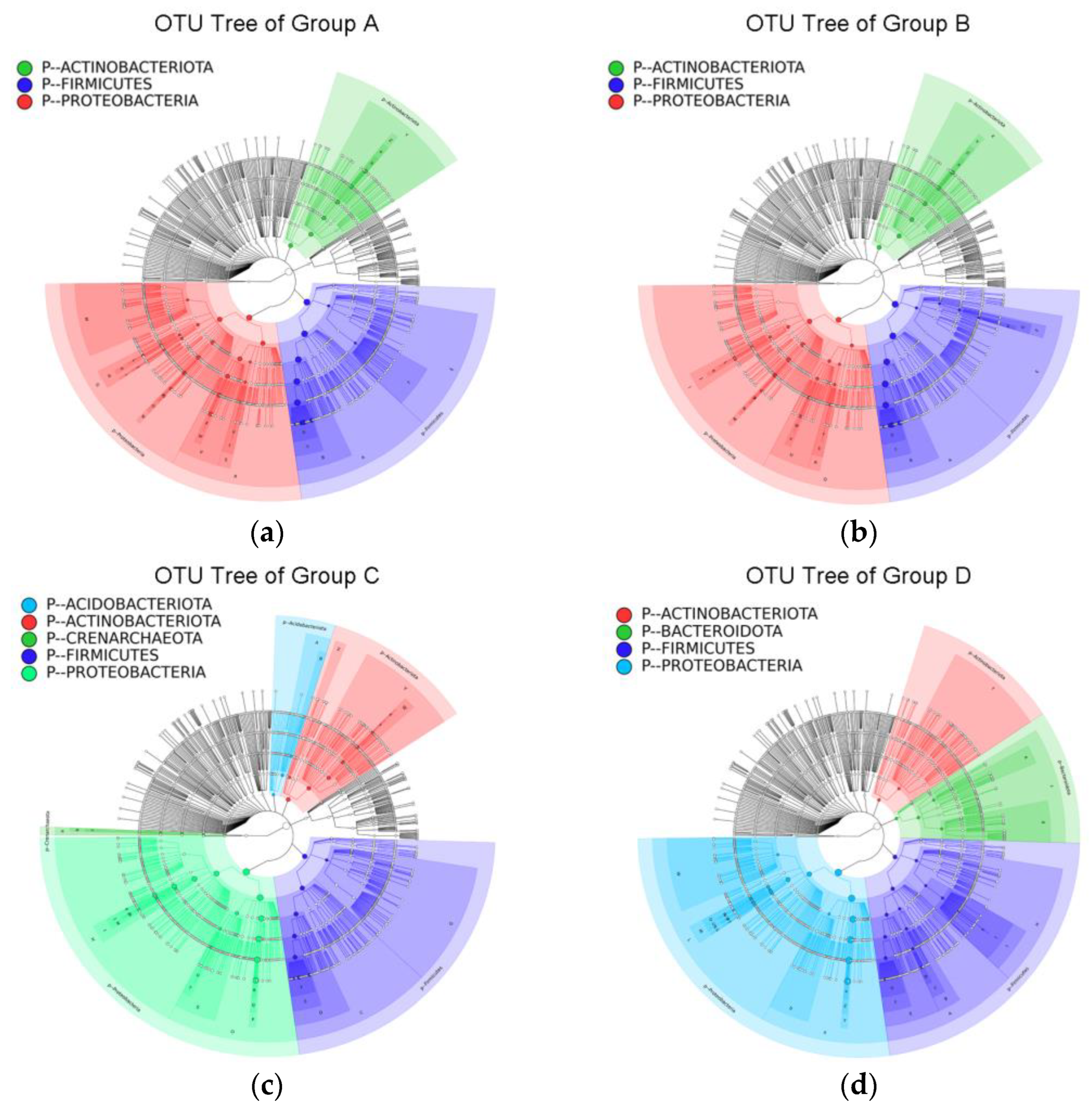
OTU annotation tree constructs for group A and B, generated by 16S rRNA region, displayed no significant differences (Figure 2a,b).
On the other hand, OTU annotation tree constructs for group C and D, generated by 16S rRNA region, demonstrated that Bacteroidota were abundant in group D but not in group C. On the contrary, Acidobacteriota and Crenarchaeota were abundant in group C but not in group D (Figure 2c,d).
OTU annotation tree constructs for group A and B, generated by ITS fungal region, showed that the phyla Ascomycota and Basidiomycota were abundant in group A. Glomeromycota were also identified in group A. In group B, only certain classes of Ascomycota (Dothideomycetes, Leotiomycetes, Saccharomycetes, Sordariomycetes) and Basidiomycota (Agaricomycetes, Tremellomycetes) were most abundant (Figure 3a,b).
Regarding the OTU annotation tree constructs for group C and D, generated by ITS fungal region, it was shown that the phyla Ascomycota and Basidiomycota were abundant in group C, as was the case for group A. Morterellomycota were identified in group A as well. In group D, only certain classes of Ascomycota (Dothideomycetes, Leotiomycetes, Saccharomycetes, Sordariomycetes, Pezizomycetes) and Basidiomycota (Ustilaginomycetes) were most abundant (Figure 3c,d).

4. Discussion

Nosemosis is a widespread disease in adult honey bees, caused by microsporidia Vairimorpha (Nosema) apis (Zander) and Vairomorpha (Nosema) ceranae (Fries) [32]. Recently, Tokarev et al. [33] proposed a redefinition of the Nosema genus (now designated Vairimorpha) based on molecular phylogenetics. Vairimorpha apis is a common parasite in the European honey bee (Apis mellifera) while Vairimorpha (Nosema) ceranae was introduced to A. mellifera recently, originating from the Asian honey bee (Apis cerana). It was detected for the first time in Europe in 2004 [34] and has spread to South and North America [35,36], Africa and Asia [37]. Nosemosis is associated with various predisposing or synergistic factors ranging from climate crisis to exposure to neonicotinoid pesticide [38,39,40,41]. Spore transmission that spreads Nosema infection takes place among adult bees during feeding (trophallaxis) or comb-cleaning. Furthermore, stored honey in the hive, water and beekeeping equipment contribute to the transmission of Nosemosis.
Severity of Nosema spp. infection in a colony is inferred by most researchers through the number of spores per bee in the sample at hand [42]. Still, it should be noted that high spore counts of V. ceranae are often found in asymptomatic colonies, and measured spores cannot constitute on their own a criterion of a colony health or survival [43].
Diagnosis of Nosemosis, especially that caused by V. ceranae, is of critical importance both for bee health and beekeeping sustainability.
The clinical symptoms of Nosemosis caused by V. ceranae are highly variable, ranging from delayed colony growth or slow progressive depopulation to sudden colony collapse. An increased number of spores is found in collapsed colonies; however, it has not been established whether V. ceranae is the main cause of collapse or one among other stressors that synergistically lead to colony collapse. V. ceranae infestation provokes intestinal epithelial cell destruction, poor nutrition, reduced lifespan of adult bees and weaker immune response [7]. A possible cause of these findings could be an imbalance (dysbiosis) in the adult bee gut microbiome and/or BB microbiome even in the early stages of Nosemosis, when spore counts are low to medium, as in our study.
Alpha-diversity analysis revealed a clear trend of reduction in number of observed species (unique OTUs) of both bacterial and fungal sequences in honey bee and ΒΒ samples derived from colonies with high V. ceranae spore count as compared with those with low V. ceranae spore count (Figure 1 and Figure 2). One notable exception was sample A3, whose bacterial diversity was the lowest out of all bee samples (Figure 1). Overall, this observed reduction was not statistically significant, and a larger number of honey bee and BB samples should be analyzed in order to confirm reduced microbial diversity.
The mechanism explaining this interesting finding has not been elucidated. However, reduced microbial diversity may lead to dysbiosis, which in turn might facilitate V. ceranae infection, according to Maes et al. [44]. Therefore, it could affect the development of the disease and serve as a prognostic marker for the survival of the colony.
A significant impact of V. ceranae infection on bacterial diversity was also revealed by alpha-diversity analysis. We hypothesized that this could partly be ascribed to reactive oxygen species (ROS) production, which is known to modulate microbiota within the insect gut lumen of V. ceranae-infected bees [45]. ROS can damage both host and microbial cells, and previous studies showed that compounds that conferred protection against ROS were produced in the bee midgut during V. ceranae infection [45,46,47].
Interestingly, V. ceranae spores were not detected in any bee bread samples, even those collected from colonies demonstrating high V. ceranae spore counts. Nevertheless, microbial diversity was reduced in group D of bee bread samples. Therefore, we assume that V. ceranae infection might affect bee bread microbiota indirectly through altered microbial inoculation by honey bees. During pollen collection and storage, bees add small amounts of regurgitated nectar as well as glandular secretions to pollen that eventually is transformed to bee bread during fermentation. Therefore, it is plausible that bees with high V. ceranae spore numbers inoculate collected pollen with modified microbial communities of lower diversity as compared with those with low V. ceranae spore numbers. Accordingly, bee-pollen fermentation could lead to microbial communities characterized by lower diversity.
Regarding the relative abundance of specific genera (bacterial and fungal), no significant change was observed among control group A and group B. A previous study reported a neutral or even positive effect of V. ceranae infection on the core bee bacteriome [48]. Zhang et al. briefly reviewed similar studies and compared these data with their own findings. Overall, they concluded that V. ceranae infection does not detrimentally affect the homeostasis of the honey bee and its core bacteriome [48]. A rather stable core bacteriome, also observed in our study, could lead to milder infection and potentially lower mortality. However, further field or laboratory investigation should be designed in order to confirm whether bees showing severe Nosemosis, from collapsed colonies or those about to collapse, experience dysbiosis of the core bacteriome.
Regarding the bee mycobiome, observed high variability between samples, even in the same group (f.i., lower V. ceranae spore count), did not allow reliable comparisons, in accordance with previous recent studies [12,13]. It is evident that a higher number of bee samples derived from a larger colony pool should be sequenced and analyzed. Nevertheless, it was demonstrated that the rather abundant mycobiome, as reflected by significant diversity, should be the focus of future microbiota studies, which at present assess mainly the core bacteriome.
In bee bread samples, some significant differences were described, as reflected by relative abundance of specific fungal and bacterial genera. For instance, Rosenbergiella spp. were relatively abundant (reaching up to 10%) in bee bread samples collected from infected colonies (group D) as compared with non-infected (0–0.77% in group C). Rosenbergiella spp. were often identified in nectar of pollination-dependent plants, producing several metabolites present in nectar (D-lactic acid, acetic acid and others). Depending on its composition, nectar might affect insect lifespan [49]. Similarly, bacteria and their metabolites produced during BB fermentation may affect honey bee lifespan. However, this hypothesis should be confirmed by further investigation.
The genus Podosphaera was absent in bee bread samples collected from colonies in group D, while the genus Blumeria demonstrated significantly decreased relative abundance in the same group. Interestingly, both genera include species that are known plant pathogens, causing powdery mildew on leaves. Blumeria graminis infests grasses and cereals in a highly specialized way. On the other hand, Podosphaera astericola and Podosphaera pannosa (primarily affecting roses and Prunus spp.) were the two species identified in our study [50].
These fungi could end up in bee bread through pollen collection from infected plants. It is known that bee-collected pollen undergoes fermentation that lowers pH and leads to production of antimicrobial compounds, thus preventing pathogen growth. Nevertheless, the lower relative abundance or absence of these fungi in bee bread samples collected from colonies belonging in Group D could be attributed to V. ceranae presence. Nosemosis might alter the bacteriome that a worker honey bee inoculates to BB. In that case, during BB fermentation, it would be expected that BB bacteriome might be different in group C as compared with that from group D. Moreover, the differences between the groups (A and B, C and D) at the phylum and class level might serve as biomarkers for prognosis of V. ceranae infection, though further investigation is needed.
Conclusively, this is the first study to demonstrate the impact of V. ceranae on both honey bee gut and bee bread microbiota in naturally infected colonies.

Supplementary Materials

The following supporting information can be downloaded at: https://www.mdpi.com/article/10.3390/app122211476/s1, Table S1: Diversity and goods coverage indices generated by 16S region; Table S2. Diversity and goods coverage indices generated by ITS region; Figure S1: Gel electrophoresis of duplex PCR products for the identification of V. ceranae; Figure S2: OTU annotation tree construct for group A generated by the 16S bacterial region; Figure S3: OTU annotation tree construct for group B generated by the 16S bacterial region; Figure S4: OTU annotation tree construct for group C generated by the 16S bacterial region; Figure S5: OTU annotation tree construct for group D generated by the 16S bacterial region; Figure S6: OTU annotation tree construct for group A generated by the ITS fungal region; Figure S7: OTU annotation tree construct for group B generated by the ITS fungal region; Figure S8: OTU annotation tree construct for group C generated by the ITS fungal region; Figure S9: OTU annotation tree construct for group D generated by the ITS fungal region.

Author Contributions

Conceptualization, D.M., N.A.D., G.D.A. and F.H.; methodology, D.M. and G.D.A.; formal analysis, M.N. and G.D.A.; investigation, I.G., N.A.D., L.C. and T.G.D.; resources, D.M. and F.H.; data curation, M.N.; writing—original draft preparation, I.G., N.A.D. and D.M.; writing—review and editing, D.M., T.G.D., N.A.D., G.D.A., L.C., F.H. and M.N.; supervision, D.M.; project administration, T.G.D. and N.A.D.; funding acquisition, D.M. All authors have read and agreed to the published version of the manuscript.

Funding

This research was funded by the Hellenic Ministry of Rural Development and Food, grant number 6738.

Institutional Review Board Statement

Not applicable.

Informed Consent Statement

Not applicable.

Data Availability Statement

Raw sequence reads are available at the NCBI Sequence Read Archive (SRA) database (Accession Number: PRJNA888378).

Conflicts of Interest

The authors declare no conflict of interest.

References

  1. Papa, G.; Maier, R.; Durazzo, A.; Lucarini, M.; Karabagias, I.K.; Plutino, M.; Bianchetto, E.; Aromolo, R.; Pignatti, G.; Ambrogio, A.; et al. The Honey Bee Apis mellifera: An Insect at the Interface between Human and Ecosystem Health. Biology 2022, 11, 233. [Google Scholar] [CrossRef] [PubMed]
  2. van Engelsdorp, D.; Meixner, M.D. A historical review of managed honey bee populations in Europe and the United States and the factors that may affect them. J. Invertebr. Pathol. 2010, 103, S80–S95. [Google Scholar] [CrossRef] [PubMed]
  3. Goulson, D.; Nicholls, E.; Botías, C.; Rotheray, E.L. Bee declines driven by combined stress from parasites, pesticides, and lack of flowers. Science 2015, 347, 1255957. [Google Scholar] [CrossRef] [PubMed]
  4. Higes, M.; Martín-Hernández, R.; Meana, A. Nosema ceranae in Europe: An emergent type C nosemosis. Apidologie 2010, 41, 375–392. [Google Scholar] [CrossRef] [Green Version]
  5. Dussaubat, C.; Brunet, J.-L.; Higes, M.; Colbourne, J.K.; Lopez, J.; Choi, J.-H.; Martín-Hernández, R.; Botías, C.; Cousin, M.; McDonnell, C.; et al. Gut Pathology and Responses to the Microsporidium Nosema ceranae in the Honey Bee Apis mellifera. PLoS ONE 2012, 7, e37017. [Google Scholar] [CrossRef] [Green Version]
  6. Paris, L.; El Alaoui, H.; Delbac, F.; Diogon, M. Effects of the gut parasite Nosema ceranae on honey bee physiology and behavior. Curr. Opin. Insect Sci. 2018, 26, 149–154. [Google Scholar] [CrossRef]
  7. Martín-Hernández, R.; Bartolomé, C.; Chejanovsky, N.; Le Conte, Y.; Dalmon, A.; Dussaubat, C.; García-Palencia, P.; Meana, A.; Pinto, M.A.; Soroker, V.; et al. Nosema ceranae in Apis mellifera: A 12 years postdetection perspective. Environ. Microbiol. 2018, 20, 1302–1329. [Google Scholar] [CrossRef] [Green Version]
  8. Raymann, K.; Moran, N.A. The role of the gut microbiome in health and disease of adult honey bee workers. Curr. Opin. Insect Sci. 2018, 26, 97–104. [Google Scholar] [CrossRef]
  9. Kwong, W.K.; Medina, L.A.; Koch, H.; Sing, K.-W.; Soh, E.J.Y.; Ascher, J.S.; Jaffé, R.; Moran, N.A. Dynamic microbiome evolution in social bees. Sci. Adv. 2017, 3, e1600513. [Google Scholar] [CrossRef] [Green Version]
  10. Dong, Z.-X.; Li, H.-Y.; Chen, Y.-F.; Wang, F.; Deng, X.-Y.; Lin, L.-B.; Zhang, Q.-L.; Li, J.-L.; Guo, J. Colonization of the gut microbiota of honey bee (Apis mellifera) workers at different developmental stages. Microbiol. Res. 2020, 231, 126370. [Google Scholar] [CrossRef]
  11. Yun, J.-H.; Jung, M.-J.; Kim, P.S.; Bae, J.-W. Social status shapes the bacterial and fungal gut communities of the honey bee. Sci. Rep. 2018, 8, 2019. [Google Scholar] [CrossRef] [Green Version]
  12. Kakumanu, M.L.; Reeves, A.M.; Anderson, T.D.; Rodrigues, R.R.; Williams, M.A. Honey Bee Gut Microbiome Is Altered by In-Hive Pesticide Exposures. Front. Microbiol. 2016, 7, 1255. [Google Scholar] [CrossRef] [PubMed] [Green Version]
  13. Paris, L.; Peghaire, E.; Moné, A.; Diogon, M.; Debroas, D.; Delbac, F.; El Alaoui, H. Honeybee gut microbiota dysbiosis in pesticide/parasite co-exposures is mainly induced by Nosema ceranae. J. Invertebr. Pathol. 2020, 172, 107348. [Google Scholar] [CrossRef] [PubMed]
  14. Didaras, N.A.; Karatasou, K.; Dimitriou, T.G.; Amoutzias, G.D.; Mossialos, D. Antimicrobial Activity of Bee-Collected Pollen and Beebread: State of the Art and Future Perspectives. Antibiotics 2020, 9, 811. [Google Scholar] [CrossRef] [PubMed]
  15. Didaras, N.A.; Kafantaris, I.; Dimitriou, T.G.; Mitsagga, C.; Karatasou, K.; Giavasis, I.; Stagos, D.; Amoutzias, G.D.; Hatjina, F.; Mossialos, D. Biological Properties of Bee Bread Collected from Apiaries Located across Greece. Antibiotics 2021, 10, 555. [Google Scholar] [CrossRef] [PubMed]
  16. Donkersley, P.; Rhodes, G.; Pickup, R.W.; Jones, K.C.; Wilson, K. Bacterial communities associated with honeybee food stores are correlated with land use. Ecol. Evol. 2018, 8, 4743–4756. [Google Scholar] [CrossRef]
  17. Disayathanoowat, T.; Li, H.; Supapimon, N.; Suwannarach, N.; Lumyong, S.; Chantawannakul, P.; Guo, J. Different Dynamics of Bacterial and Fungal Communities in Hive-Stored Bee Bread and Their Possible Roles: A Case Study from Two Commercial Honey Bees in China. Microorganisms 2020, 8, 264. [Google Scholar] [CrossRef] [Green Version]
  18. Anderson, K.E.; Carroll, M.J.; Sheehan, T.; Mott, B.M.; Maes, P.; Corby-Harris, V. Hive-stored pollen of honey bees: Many lines of evidence are consistent with pollen preservation, not nutrient conversion. Mol. Ecol. 2014, 23, 5904–5917. [Google Scholar] [CrossRef]
  19. Martinson, V.G.; Moy, J.; Moran, N.A. Establishment of Characteristic Gut Bacteria during Development of the Honeybee Worker. Appl. Environ. Microbiol. 2012, 78, 2830–2840. [Google Scholar] [CrossRef] [Green Version]
  20. Mazur, E.D.; Gajda, A.M. Nosemosis in Honeybees: A Review Guide on Biology and Diagnostic Methods. Appl. Sci. 2022, 12, 5890. [Google Scholar] [CrossRef]
  21. Charistos, L.; Parashos, N.; Hatjina, F. Long term effects of a food supplement HiveAliveTM on honey bee colony strength and Nosema ceranae spore counts. J. Apic. Res. 2015, 54, 420–426. [Google Scholar] [CrossRef]
  22. Cantwell, G.E. Standard methods for counting Nosema spores. Am. Bee J. 1970, 110, 222–223. [Google Scholar]
  23. Fries, I.; Chauzat, M.P.; Chen, Y.P.; Doublet, V.; Genersch, E.; Gisder, S.; Higes, M.; McMahon, D.P.; Martín-Hernández, R.; Natsopoulou, M.; et al. Standard methods for Nosema research. J. Apic. Res. 2015, 52, 1–28. [Google Scholar] [CrossRef] [Green Version]
  24. Martín-Hernández, R.; Meana, A.; Prieto, L.; Salvador, A.M.; Garrido-Bailón, E.; Higes, M. Outcome of Colonization of Apis mellifera by Nosema ceranae. Appl. Environ. Microbiol. 2007, 73, 6331–6338. [Google Scholar] [CrossRef] [Green Version]
  25. Casas, I.; Powell, L.; Klapper, P.E.; Cleator, G.M. New method for the extraction of viral RNA and DNA from cerebrospinal fluid for use in the polymerase chain reaction assay. J. Virol. Methods 1995, 53, 25–36. [Google Scholar] [CrossRef]
  26. Magoc, T.; Salzberg, S.L. FLASH: Fast length adjustment of short reads to improve genome assemblies. Bioinformatics 2011, 27, 2957–2963. [Google Scholar] [CrossRef] [Green Version]
  27. Caporaso, J.G.; Kuczynski, J.; Stombaugh, J.; Bittinger, K.; Bushman, F.D.; Costello, E.K.; Fierer, N.; Peña, A.G.; Goodrich, J.K.; Gordon, J.I.; et al. QIIME allows analysis of high-throughput community sequencing data. Nat. Methods 2010, 7, 335–336. [Google Scholar] [CrossRef] [Green Version]
  28. Edgar, R.C.; Haas, B.J.; Clemente, J.C.; Quince, C.; Knight, R. UCHIME improves sensitivity and speed of chimera detection. Bioinformatics 2011, 27, 2194–2200. [Google Scholar] [CrossRef] [Green Version]
  29. Edgar, R.C. UPARSE: Highly accurate OTU sequences from microbial amplicon reads. Nat. Methods 2013, 10, 996–998. [Google Scholar] [CrossRef]
  30. Quast, C.; Pruesse, E.; Yilmaz, P.; Gerken, J.; Schweer, T.; Yarza, P.; Peplies, J.; Glöckner, F.O. The SILVA ribosomal RNA gene database project: Improved data processing and web-based tools. Nucleic Acids Res. 2012, 41, D590–D596. [Google Scholar] [CrossRef]
  31. Kõljalg, U.; Nilsson, R.H.; Abarenkov, K.; Tedersoo, L.; Taylor, A.F.S.; Bahram, M.; Bates, S.T.; Bruns, T.D.; Bengtsson-Palme, J.; Callaghan, T.M.; et al. Towards a unified paradigm for sequence-based identification of fungi. Mol. Ecol. 2013, 22, 5271–5277. [Google Scholar] [CrossRef] [PubMed]
  32. Fries, I.; Feng, F.; Da Silva, A.; Slemenda, S.B.; Pieniazek, N.J. Nosema ceranae n. sp. (Microspora, Nosematidae), morphological and molecular characterization of a microsporidian parasite of the Asian honey bee Apis cerana (Hymenoptera, Apidae). Eur. J. Protistol. 1996, 32, 356–365. [Google Scholar] [CrossRef]
  33. Tokarev, Y.S.; Huang, W.-F.; Solter, L.F.; Malysh, J.M.; Becnel, J.J.; Vossbrinck, C.R. A formal redefinition of the genera Nosema and Vairimorpha (Microsporidia: Nosematidae) and reassignment of species based on molecular phylogenetics. J. Invertebr. Pathol. 2020, 169, 107279. [Google Scholar] [CrossRef] [PubMed]
  34. Higes, M.; Martín, R.; Meana, A. Nosema ceranae, a new microsporidian parasite in honeybees in Europe. J. Invertebr. Pathol. 2006, 92, 93–95. [Google Scholar] [CrossRef] [PubMed]
  35. Calderón, R.A.; Sanchez, L.A.; Yañez, O.; Fallas, N. Presence of Nosema ceranae in Africanized honey bee colonies in Costa Rica. J. Apic. Res. 2008, 47, 328–329. [Google Scholar] [CrossRef]
  36. Chen, Y.; Evans, J.D.; Smith, I.B.; Pettis, J.S. Nosema ceranae is a long-present and wide-spread microsporidian infection of the European honey bee (Apis mellifera) in the United States. J. Invertebr. Pathol. 2008, 97, 186–188. [Google Scholar] [CrossRef]
  37. Chen, Y.; Evans, J.D.; Zhou, L.; Boncristiani, H.; Kimura, K.; Xiao, T.; Litkowski, A.M.; Pettis, J.S. Asymmetrical coexistence of Nosema ceranae and Nosema apis in honey bees. J. Invertebr. Pathol. 2009, 101, 204–209. [Google Scholar] [CrossRef]
  38. Alaux, C.; Brunet, J.-L.; Dussaubat, C.; Mondet, F.; Tchamitchan, S.; Cousin, M.; Brillard, J.; Baldy, A.; Belzunces, L.P.; Le Conte, Y. Interactions between Nosema microspores and a neonicotinoid weaken honeybees ( Apis mellifera ). Environ. Microbiol. 2010, 12, 774–782. [Google Scholar] [CrossRef] [Green Version]
  39. Doublet, V.; Natsopoulou, M.E.; Zschiesche, L.; Paxton, R.J. Within-host competition among the honey bees pathogens Nosema ceranae and Deformed wing virus is asymmetric and to the disadvantage of the virus. J. Invertebr. Pathol. 2015, 124, 31–34. [Google Scholar] [CrossRef]
  40. Vidau, C.; Diogon, M.; Aufauvre, J.; Fontbonne, R.; Viguès, B.; Brunet, J.-L.; Texier, C.; Biron, D.G.; Blot, N.; El Alaoui, H.; et al. Exposure to Sublethal Doses of Fipronil and Thiacloprid Highly Increases Mortality of Honeybees Previously Infected by Nosema ceranae. PLoS ONE 2011, 6, e21550. [Google Scholar] [CrossRef] [Green Version]
  41. Zheng, H.-Q.; Gong, H.-R.; Huang, S.-K.; Sohr, A.; Hu, F.-L.; Chen, Y.P. Evidence of the synergistic interaction of honey bee pathogens Nosema ceranae and Deformed wing virus. Vet. Microbiol. 2015, 177, 1–6. [Google Scholar] [CrossRef] [PubMed]
  42. World Organisation for Animal Health. Nosemosis of honey bees. In Terrestrial Manual; WOAH: Paris, France, 2018; pp. 744–749. ISBN 978-92-95108-18-9. [Google Scholar]
  43. Meana, A.; Martín-Hernández, R.; Higes, M. The reliability of spore counts to diagnose Nosema ceranae infections in honey bees. J. Apic. Res. 2010, 49, 212–214. [Google Scholar] [CrossRef]
  44. Maes, P.W.; Rodrigues, P.A.P.; Oliver, R.; Mott, B.M.; Anderson, K.E. Diet-related gut bacterial dysbiosis correlates with impaired development, increased mortality and Nosema disease in the honeybee (Apis mellifera). Mol. Ecol. 2016, 25, 5439–5450. [Google Scholar] [CrossRef] [PubMed]
  45. Herb, M.; Schramm, M. Functions of ROS in Macrophages and Antimicrobial Immunity. Antioxidants 2021, 10, 313. [Google Scholar] [CrossRef] [PubMed]
  46. Armitage, S.A.; Genersch, E.; McMahon, D.P.; Rafaluk-Mohr, C.; Rolff, J. Tripartite interactions: How immunity, microbiota and pathogens interact and affect pathogen virulence evolution. Curr. Opin. Insect Sci. 2022, 50, 100871. [Google Scholar] [CrossRef]
  47. Zug, R.; Hammerstein, P. Wolbachia and the insect immune system: What reactive oxygen species can tell us about the mechanisms of Wolbachia–host interactions. Front. Microbiol. 2015, 6, 1201. [Google Scholar] [CrossRef] [Green Version]
  48. Zhang, Y.; Su, M.; Wang, L.; Huang, S.; Su, S.; Huang, W.-F. Vairimorpha (Nosema) ceranae Infection Alters Honey Bee Microbiota Composition and Sustains the Survival of Adult Honey Bees. Biology 2021, 10, 905. [Google Scholar] [CrossRef]
  49. Lenaerts, M.; Goelen, T.; Paulussen, C.; Herrera-Malaver, B.; Steensels, J.; Van den Ende, W.; Verstrepen, K.J.; Wäckers, F.; Jacquemyn, H.; Lievens, B. Nectar bacteria affect life history of a generalist aphid parasitoid by altering nectar chemistry. Funct. Ecol. 2017, 31, 2061–2069. [Google Scholar] [CrossRef]
  50. Linde, M.; Shishkoff, N. Powdery Mildew. In Encyclopedia of Rose Science; Elsevier: Amsterdam, The Netherlands, 2003; pp. 158–165. [Google Scholar]
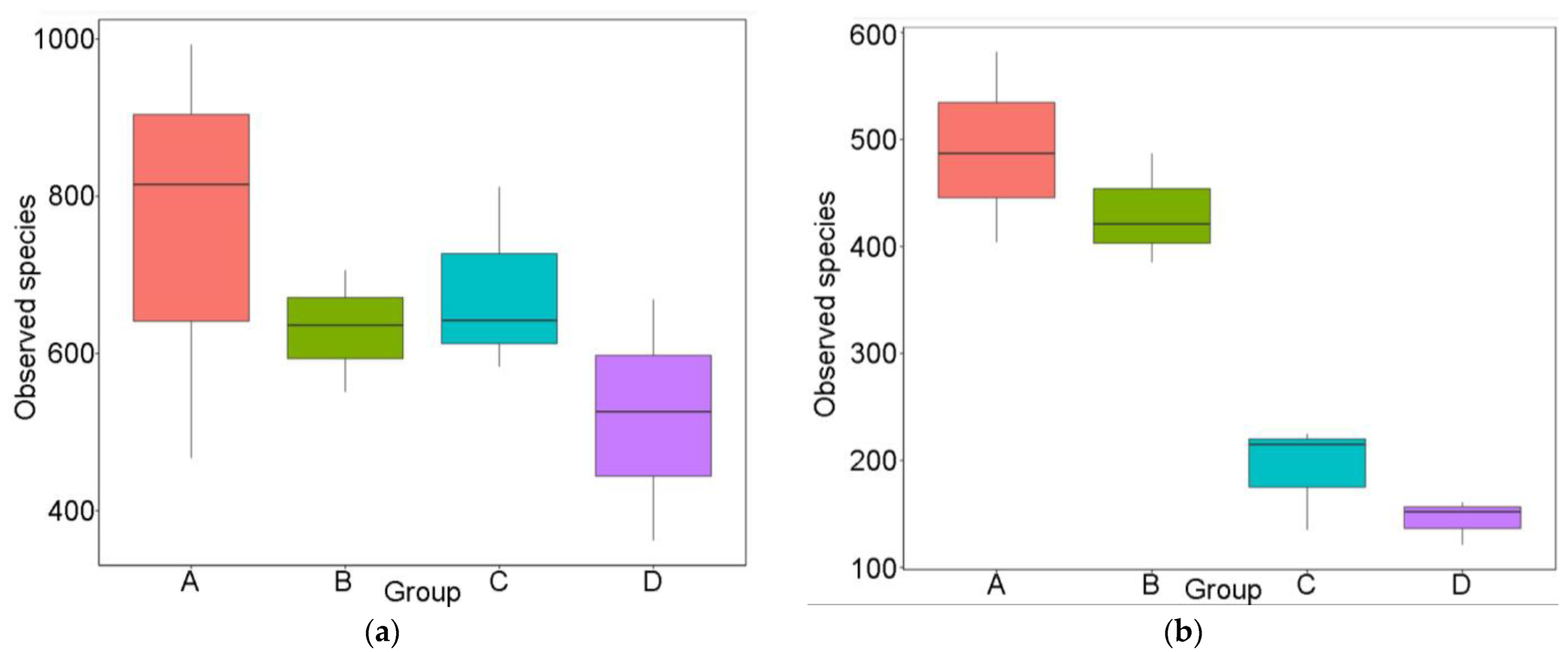
Figure 1. Observed unique operating taxonomic units (OTUs): (a) bacterial species and (b) fungal species in groups A, B, C and D.
Applsci 12 11476 g001
Figure 2. OTU annotation tree construct for each sample group (A: bee samples demonstrating low V. ceranae spore count; B: bee samples demonstrating high V. ceranae spore count; C: bee bread collected from colonies with low V. ceranae spore count and D: bee bread collected from colonies with high V. ceranae spore count), generated by V3–V4 16S rRNA region, in (ad), respectively. Different taxonomic ranks range from inside out. Size of circles stands for abundance of species. Different colors stand for different phyla. Solid circles stand for the top 40 species in high abundance. Full-size tree constructs are provided as Supplementary Material.
Applsci 12 11476 g002
Figure 3. OTU annotation tree construct for each sample group (A–D), generated by ITS fungal region, in (ad) respectively. Different taxonomic ranks range from inside out. Size of circles stands for abundance of species. Different colors stand for different phyla. Solid circles stand for the top 40 species in high abundance. Full-size constructs are provided as Supplementary Material.
Applsci 12 11476 g003
Table 1. Hive coding number, V. ceranae average spore count per bee and sample designation and grouping for metataxonomics.
Hive Coding NumberV. ceranae Average Spore Count Per BeeSample Designation for MetataxonomicsGroup Name
Bee
Samples
11 Bee6000A1A
14 Bee24,000A2
15 Bee39,000A3
12 Bee4,500,000B1B
27 Bee2,800,000B2
112 Bee4,200,000B3
Bee Bread
samples
11 BB-C1C
14 BBC2
15 BBC3
12 BBD1D
27 BBD2
112 BBD3
Table 2. Average variability of taxa in the four sample groups (A–D) based on 16S rRNA and ITS region.
Group16S rRNA (Bacteria)ITS (Fungi)
A13.926 43.700
B11.244 36.593
C20.711 25.942
D6.329 39.886
Publisher’s Note: MDPI stays neutral with regard to jurisdictional claims in published maps and institutional affiliations.