Article

An Optimized Methodology for Patient-Specific Therapeutic Activity Administration in Liver Radioembolization

1
Champalimaud Foundation, 1400-038 Lisbon, Portugal
2
Instituto de Telecomunicações, Instituto Superior Técnico-Universidade de Lisboa, 1049-001 Lisbon, Portugal
*
Author to whom correspondence should be addressed.
Appl. Sci. 2022, 12(22), 11669; https://doi.org/10.3390/app122211669
Submission received: 18 October 2022 / Revised: 11 November 2022 / Accepted: 14 November 2022 / Published: 17 November 2022
(This article belongs to the Special Issue Application of Targeted Therapy in Cancer Treatment)

Abstract

:

Featured Application

Our computational dosimetry system, including an original optimization protocol, enables more effective personalized liver radioembolization treatments.

Abstract

Radioembolization (RE) with glass microspheres (MS) loaded with Yttrium-90 (90Y) has been used to treat tumors in the liver with some reported success. However, assessing absorbed doses (AD) in the planning tumor volume (PTV) and normal liver volume (NLV) is a key problem to address in RE. In clinical practice, the computation of 90Y activity to be administered follows the manufacturer’s recommendations, which do not consider the specific characteristics of MS deposition in each patient’s liver. Our main aim is to develop a methodology to estimate the optimal activity for each patient treatment. It uses the absorbed dose distribution (ADD) derived from the Technetium-99m (99mTc)-labeled macroaggregated albumin (MAA) obtained from pre-treatment planning single-photon emission computed tomography (SPECT) images. Post-treatment positron emission tomography (PET) images of the 90Y-MS distribution were used to estimate the ADD for treatment verification. Sixteen RE treatments were retrospectively selected. The agreement between the estimated mean AD based on the planning imaging and real post-treatment mean AD was good in PTV with an intraclass correlation coefficient (ICC) of 0.79 and excellent in NLV (ICC = 0.97). The optimization of 90Y activity using pre-defined clinical AD thresholds (<70 Gy in NLV and >80 Gy in PTV) imposed on the PTV and NLV voxels showed remarkably high agreement (ICC = 0.96, p < 0.001) in eleven out of the sixteen RE treatments between SPECT-MAA-based and PET-MS-based optimal activity estimates. In conclusion, under well-controlled conditions, pre-treatment SPECT-MAA imaging predicts well the treatment of ADD. In addition, SPECT-MAA imaging can be used to optimize the 90Y-MS activity to be administered to the liver.

1. Introduction

Worldwide, the most common causes of death by cancer have had minor changes in the last four decades. Lung, liver, and stomach cancers have been the three leading causes of death by cancer since 1975 [1,2,3,4,5,6]. Liver cancer is the sixth most diagnosed malignant cancer in the world, being the third most common cause of death by cancer [4,5,6].
Surgery is the first approach with curative intent in primary tumors. Nevertheless, for many patients, there are no curative surgical solutions, resorting in these cases to systemic, local, and regional treatments with palliative intent, such as ablation and chemoembolization [7]. The therapeutic intent in metastatic liver tumors is usually palliative, with surgery and chemotherapy being the main options.
Radioembolization (RE) with glass or resin microspheres (MS) loaded with Yttrium-90 (90Y)—more recently also produced with Poly L-lactic acid (PLLA) loaded with Holmium-166 (166Ho)—has emerged as a local therapeutic approach to primary and metastatic tumors in the liver [8,9,10,11,12,13,14,15]. RE combines the embolic action of the MS in the hepatic vasculature with the radiation emitted by the radionuclide in the MS. Thus, MS with a diameter of approximately 20 μm to 40 μm are ideal for blocking the blood supply to tumors, providing them with a highly absorbed radiation dose and simultaneously having a tolerable effect on normal liver tissues.
In current clinical practice, the RE procedure follows the guidelines of the glass MS manufacturers [10,16,17]. In preparation for RE treatment, patients are always evaluated to minimize any significant risk of undesirable extrahepatic radiation. For this reason, all patients undergo diagnostic angiography aiming to study the liver arterial vasculature. During this session, Technetium-99m (99mTc)-labeled macroaggregated albumin (MAA) is injected into the patient’s liver via hepatic arteries to assess the hepatopulmonary shunt fraction (LSF). After injection, patients are immediately submitted to a full-body planar scan by a gamma camera. In some clinical centers, single-photon emission computed tomography (SPECT) images of the voxelized distribution of MAA deposition in the liver are also acquired. Post-treatment positron emission tomography (PET) images acquired from the patients after the administration of the MS loaded with 90Y are frequently acquired for activity distribution verification. The calculation of the 90Y activity to be administered to the patient’s liver, including the planning tumor volume (PTV) and part of the normal liver volume (NLV), follows the manufacturer’s protocol, which considers the organ-based (i.e., one compartment) formalism of the Medical Internal Radiation Dose (MIRD) committee of the Society of Nuclear Medicine and Molecular Imaging (SNMMI). It considers only the prescribed absorbed dose (AD) and liver volumes, considering uniform deposition of the MS in the liver [16,18]. Thus, it does not consider the real specific characteristics of each patient that may influence the deposition of MS in the liver. Therefore, the simple application of the MIRD calculation model is not tailored to the patient; this is the fundamental problem under investigation in this work. Moreover, the European Council Directive 2013/59/EURATOM of 5 December 2013, regarding the optimization of radiotherapeutic exposures, states that the PTV shall be individually planned and their irradiation appropriately verified, taking into account that doses to NLV shall be as low as reasonably achievable and consistent with the intended radiotherapeutic exposure of the PTV [19]. This research work is a step forward in that direction. It has three main aims: (a) to compute the voxelized AD by the patients’ PTV and NLV; (b) to evaluate the agreement between the pre-treatment estimated AD derived from 99mTc-MAA SPECT images and the actual post-treatment estimated AD derived from 90Y-MS PET images; (c) to propose a novel method to optimize the activity to be administered into patients’ livers.

2. Materials and Methods

The experimental work was carried out in three main steps: (1) a phantom study to validate the proposed dose estimation technique; (2) retrospective application of the methodology to a set of RE treatments; (3) personalization/optimization of the activity to be administered to the patient’s liver.

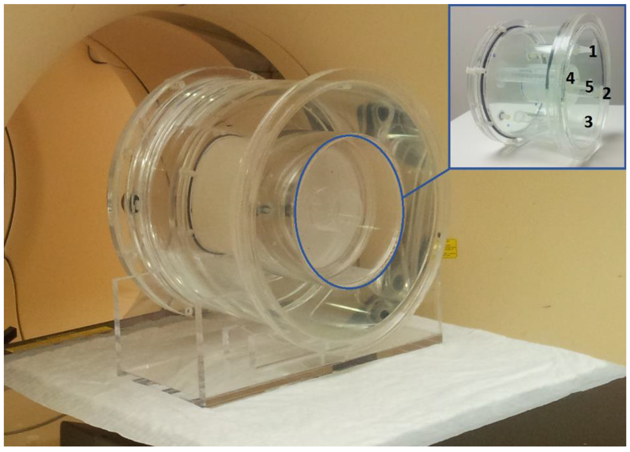
2.1. Phantom Studies

As physical phantom tests represent the gold standard for validating image processing methods before designing patients studies [20], the first part of the experimental work was carried out based on physical liver phantoms to allow estimation of accuracy, correlation, and agreement of the estimated absorbed dose distributions (ADD) derived from the SPECT and PET images. The phantom used was designed and built specifically for this research work. The complete phantom is shown in Figure 1. It is a cylinder made of transparent 5 mm thick acrylic, with a 300 mm base diameter and a 250 mm height. The outer body of the phantom can be filled with water to better model photon attenuation in the human body. This cylindrical outer body can accommodate in its core a cylindrical body of 150 mm base diameter by 100 mm height. The inner cylinder has a capacity of 1600 mL, simulating the volume of an adult liver. This inner cylinder contains five small fixed cylinders inside to simulate the PTV; two peripheral cylinders with 15 mL volume each (i.e., cylinders 1 and 3 in Figure 1, with 15 mm base diameter and 85 mm height), two other peripheral cylinders with 8 mL volume each (i.e., cylinders 2 and 4 in Figure 1, with 13 mm base diameter and 6 mm height), and a larger central cylinder with a volume of 108 mL (i.e., cylinder 5 in Figure 1, with 50 mm base diameter and 55 mm height) in the center of the inner cylinder.
This experimental work used MAA and MS microparticles. These, after mixing with a liquid (such as water), are deposited by gravity on the bottom of the container. Thus, the microparticles were mixed with agarose at a ratio of 3 parts of agarose to 100 parts of water. This way, the agarose powder mixed with water and heated at around 90 °C takes the form of a gel when cooled to 30 °C in less than 10 min, forming an immobilizing net with the microparticles in suspension. This is the white (milky) matter that can be seen inside the inner cylinder in the main photograph of Figure 1.
Two phantom experiences were thus created, one using the 99mTc-MAA and the other using the 90Y-MS. First, a 150 mL mixture of agarose with 261 MBq of 99mTc-MAA at the time of the SPECT-MAA image acquisition was made. This mixture was used to fill the five internal cylinders simulating the PTV. The remaining volume of the phantom (liver) was filled with agarose without 99mTc-MAA. The outer body of the phantom was filled with water. SPECT-MAA and CT images of the phantom were acquired after the mixture solidified at room temperature. Subsequently, after being thoroughly cleaned and all of the agarose residues were removed, the acrylic phantom was loaded identically into the five inner cylinders with a gel-type agarose mixture with 90Y-MS. These cylinders were loaded with 3184 MBq of 90Y-MS at the time of the PET-MS image acquisition. PET-MS and CT images of the phantom were acquired after the mixture solidified at room temperature. Both 99mTc-MAA and 90Y-MS activity concentrations in the cylinders simulated the PTV of about 1.63 MBq/mL and 20 MBq/mL, respectively. These values are approximately three times higher than the average PTV for all our patients.

2.1.1. Phantom Imaging

SPECT images of the 99mTc-MAA phantom activity distribution were acquired with the gamma camera PHILIPS BrightView using low-energy high-resolution (LEHR) collimators. Computed tomography (CT) imaging of the phantom was then acquired with the PET/CT PHILIPS GEMINI TF16 equipment, enabling the reconstruction of the SPECT-MAA with attenuation correction since the gamma camera used does not have a CT. A clinical reconstruction protocol was used (OSEM with 3 iterations, 8 subsets, and post-processing Butterworth filter (cut off 2, order 10).
The same PET/CT equipment was also used for Imaging the 90Y-MS phantom activity distribution. The PET acquisition parameters were set to the whole-body mode PET, one longitudinal axial field of view (AFOV) of 180 mm with 15 min of acquisition time. The final images were obtained using the default manufacturer’s reconstruction protocol. In addition, the 90Y positron fraction previously configured by us in the PET/CT scanner was also considered during the reconstruction of the PET images.

2.1.2. Phantom Image Processing

First, the SPECT-MAA images with no attenuation correction (NAC) were registered with the CT of the phantom. Then, the SPECT-MAA imaging reconstruction with CT-based attenuation correction (CTAC) was performed using manufacturer software (AutoSPECT® Plus, Philips). In this work, only CTAC images (SPECT and PET) were used for evaluation; thus, for the sake of simplicity, we refer to them simply as SPECT-MAA and PET-MS.
First, the registration of both (MAA and MS) phantom CT images was performed by applying a rigid transformation. The same transformation was then used to align the SPECT-MAA images in the PET-MS space (i.e., PET-MS was considered the reference). Simultaneously, these images were resampled to the CT voxel dimensions of the PET/CT by linear interpolation to be used during the segmentation of the liver regions of interest (ROI). Previously registered and resampled images were then used to manually segment the phantom ROI. All of the original ROI were drawn in the CT phantom image. Later, all masks and phantom images were resampled to the spatial discretization of the voxel S-dose convolution kernel of 2.21 mm side cubic voxels, which is needed during dose calculations, as explained in the next section. Nine segmentation masks were defined and designated as liver_box, liver_cyl, ptv_5_cyl_backg, ptv_5_cyl, cylinder 1, cylinder 2, cylinder 3, cylinder 4, and cylinder 5 (Figure 2). The first mask defines the limits of the dose computation cubic volume with 2080 mL. The second mask is a 1600 mL cylindrical volume adjusted to the liver_box inner limits and defines the cylinder simulating the liver. The third mask defines the 154 mL total volume of the five cylinders (i.e., the central cylinder along with four peripheral cylinders simulating the PTV), together with four cylinders of 13 mL volume each, as background samples. The fourth mask is the 154 mL total volume of the five cylinders without background samples. The last mask defines each of the five PTV cylinders individually considered: four in the periphery, two with 8 mL and two with 15 mL volume each, and one internal with 108 mL volume.

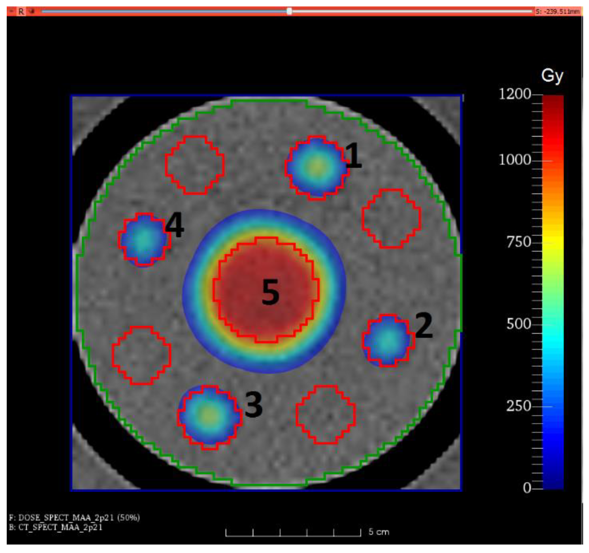
2.1.3. Phantom Dose Calculation and Analysis

AD computations rely on voxelized ADD derived from SPECT-MAA and PET-MS emission intensity maps [21,22,23]. The contribution of a source voxel to the AD of a target voxel is represented by a so-called voxel S-value, considering that both (i.e., source and target) voxels are in the same tissue or organ. In this work, we used the S-kernel convolution method to generate ADD from activity-voxelized distributions [24,25]. The voxel S-values have been determined by several authors based on Monte Carlo simulations for several radionuclides, biological tissues, and voxel sizes [22,24,26]. We used a voxel S-kernel calculated for 90Y, soft tissue (i.e., 1 g/mL), and 2.21 mm cubic voxels, expressed in units of mGy/(MBq.s) [24,25].
During the calculation of voxelized ADD, the SPECT and PET images need to be normalized and calibrated in activity. Normalization is undertaken so that the sum of the total intensity in the images equals the total activity (in MBq) contained in the liver_box ROI. Then, the calibrated image is convolved with the 3D kernel expressed in mGy/(MBq.s). Finally, each voxel is multiplied by the time integral of 90Y decay (i.e., from zero seconds to at least ten times the 90Y half-life) so that the total number of decays expected for that voxel can be achieved.
The maximization of the Pearson correlation coefficient (PCC) between the SPECT-MAA- and PET-MS-derived ADD was investigated. In addition, the intraclass correlation coefficient (ICC) and the γ-index agreement test were also computed and evaluated.
The γ-index is a pass/fail type agreement test, which considers the simultaneous combination of two test conditions: the dose-difference (DD) and distance-to-agreement (DTA) [18,27,28]. A result of this test less than or equal to 1 concludes that the analyzed voxels are within the DD/DTA tolerance criterion, which means the test was successful. In the opposite case, where the test result is greater than 1, the test failed. The test outcome in each ROI is computed as the success rate of the ROI voxels referred to as the γ-index passing rate (PR). In this work, seven DD (%)/DTA (mm) test conditions (i.e., 5/5, 10/5, 5/10, 10/10, 15/10, 10/15, and 15/15) were considered in the computation of the γ-index voxelized distributions. These test conditions were chosen considering the 10 mm spatial resolution of the SPECT-MAA images and a reasonable AD deviation between the two distributions (i.e., MAA and MS). Based on data from the literature, we can assume that the NLV AD should not exceed 70 Gy [29,30,31]. In addition, it is also understandable that AD values to the PTV should be 80 Gy or higher [29,30,31]. From these values, we assumed a maximum percentage difference of around 15%. The resulting γ-index PR computed from the phantom ADD became a reference for the later discussion on the γ-index PR results obtained from patients’ data.
The registration and resampling operations were performed by means of the open-source software 3D Slicer [32]. The ROI segmentation task was carried out in the open-source ITK-SNAP [33]. The commercial software MATLAB R2017b (The MathWorks) and the open-source code Tools for NifTI and Analyze Image [34] were used for the computation of the voxelized ADD, voxelized γ-index distributions and PR values, PCC, and ICC values.

2.2. Patient Studies

2.2.1. Patient Data Set

From January 2015 to December 2018, twenty patients (six females and fourteen males) were submitted to liver RE with 90Y-charged glass MS (TheraSphere, BTG International Ltd.) at the Champalimaud Foundation. The average age of the twenty patients was 61 years-old (range: 50–71 years old) at the first or single treatment date. From this cohort, eleven patients suffered from metastatic colorectal cancer (mCRC), three patients from metastatic pancreatic carcinoma, three from cholangiocarcinoma, two from hepatocellular carcinoma (HCC), and one from metastatic carcinoma of the thymus. A total of twenty-four RE treatment sessions were performed during that period, as four of the twenty patients were treated at two liver sites more than one month apart.
The inclusion criteria of patients in this study required controlled conditions of catheter positioning in the same location previously defined at the time of diagnostic hepatic angiography and during RE treatment. For this reason, from the twenty-four RE treatments, only sixteen were selected, corresponding to fourteen patients (mean age: 62 years old, range: 50–84), according to Table 1. In this table and in the following text, Pn_t refers to the total sequential number of treatments “t” updated for each new patient “Pn”.

2.2.2. Patient Imaging

SPECT-MAA and PET-MS imaging were always performed for clinical purposes, using a single AFOV of 406 mm and 180 mm long, respectively. These images were processed and analyzed following the same protocol for all treatments. All of the SPECT-MAA images were reconstructed with CTAC using the same clinical reconstruction parameters. In the initial nine treatments, we used the CT component of the PET/CT MS data acquisition. For the other seven treatments, we have acquired, on purpose, a CT data set (on our PET/CT system) immediately after the SPECT acquisition, as detailed below:
(1)
Five multi-modality fiducial markers (Izi Medical) were fixed on the skin of the patient, one at the level of the xyphoid, one at the level of the inferior rib cage of the right, one at the level of the inferior rib cage on the left, one at the level of the spinous process of the second lumbar vertebra (L2), and finally, the other one at the level of the spinous process of the fourth lumbar vertebra (L4). These fiducial markers are small discs of CT radiopaque material that, when loaded with 99mTc, become visible during SPECT imaging [35]. Each fiducial marker was loaded with approximately 0.5 MBq of 99mTc, diluted in a physiological saline solution at a concentration of 50 MBq/0.5 mL immediately before the acquisition of the SPECT-MAA images. During the registration of the SPECT and CT images of the patients with fiducial markers attached to their skin, these were easily located on both SPECT and CT images and were used as reference points (i.e., landmarks) in the registration algorithm.
(2)
All these fiducial markers were used to assist in the registration of SPECT-MAA images with the corresponding CT images. The five fiducial markers were always applied on each of the last seven treatments.
For the first nine treatments, for whom the fiducial markers technique was not used, the SPECT-MAA and CT images were registered by an experienced nuclear medicine physician, using the semi-automatic technique with cursor-based anatomical landmarks positioned in well-identified liver structural features of each patient’s anatomy. In addition, since these patients could not undergo a CT acquisition immediately after the SPECT-MAA acquisition, the CT of the post-treatment PET-MS was used.

2.2.3. Patient Image Processing

All the operations previously described regarding the phantom studies, namely, image registration and resampling, segmentation of ROI, computation of voxelized ADD, voxelized γ-index distributions and PR values, PCC and ICC values, were similarly applied to the patient data.
Additionally, a set of diagnostic images (e.g., magnetic resonance imaging (MRI) and 18F-Fluorodeoxyglucose PET/CT of each patient, previously acquired for clinical purposes, was selected. This set of images was used, along with the CT images, during the segmentation task of each patient’s liver ROI. Specifically, for MRI, the sequences that showed higher contrast between the NLV and the PTV were selected. The co-registration of these images was also computed with reference to the physical space and voxel size of the CT component of the post-treatment PET/CT.
Then, semi-automatic segmentation was used to delineate the total volume of each patient’s liver. This operation was based on a machine-learning algorithm known as supervised random forests [33,36,37,38]. The algorithm was trained based on the manual selection of samples from the different regions of the CT volume. Such segmentation is expected to have small errors due to the characteristics of each patient’s liver tissue, including post-surgical effects and other artefacts. As a fine-tuning step, morphological closing operations were applied to the total liver volume, and a manual refinement was performed by the physician to correct those errors. The segmentation of each patient’s PTV was performed manually. A morphological closing operation was then applied to the PTV masks to fill small holes or gaps and soften the outer edges. Finally, the NLV was defined by subtracting the PTV from the total liver volume. An example of the segmentation of a patient’s liver ROI is shown in a single slice in Figure 3.

2.2.4. Patient Dose Calculation and Analysis

As with the phantom studies, the SPECT and PET images of the patients needed to be calibrated prior to the voxelized calculation of the ADD. For calibration, a reference volume must be considered based on the mask of the total liver volume of each patient, to which a margin of about 13 mm was added by applying a morphological dilation operation. The goal of this margin is to include possible spill-out due to partial volume effect and organ motion during the SPECT and PET image acquisitions. This way, all useful information is considered in the computation of the voxelized ADD. These masks are designated in this work as liver_ref.
The calibration of the SPECT or PET images was performed in the same way as with the phantom studies. The value of the actual 90Y activity administered to each patient’s liver already considers the radioactive residue that remained in the administration system, thus not administered to the patient, as well as the value of the LSF.
Finally, the assessment of SPECT-MAA as a predictor of ADD derived from PET-MS was conducted using the same metrics considered for the phantom studies (PCC, ICC, and the γ-index). In radiation therapy, the AD in critical ROI determines a radiobiological effect. In this study, we correlated the mean AD in PTV and NLV between the pre-treatment SPECT-MAA and the post-treatment PET-MS images. To better analyze the agreement between the two ADD in the liver ROI, voxelized distributions of the γ-index were computed from all treatments, considering seven DD/DTA test conditions.

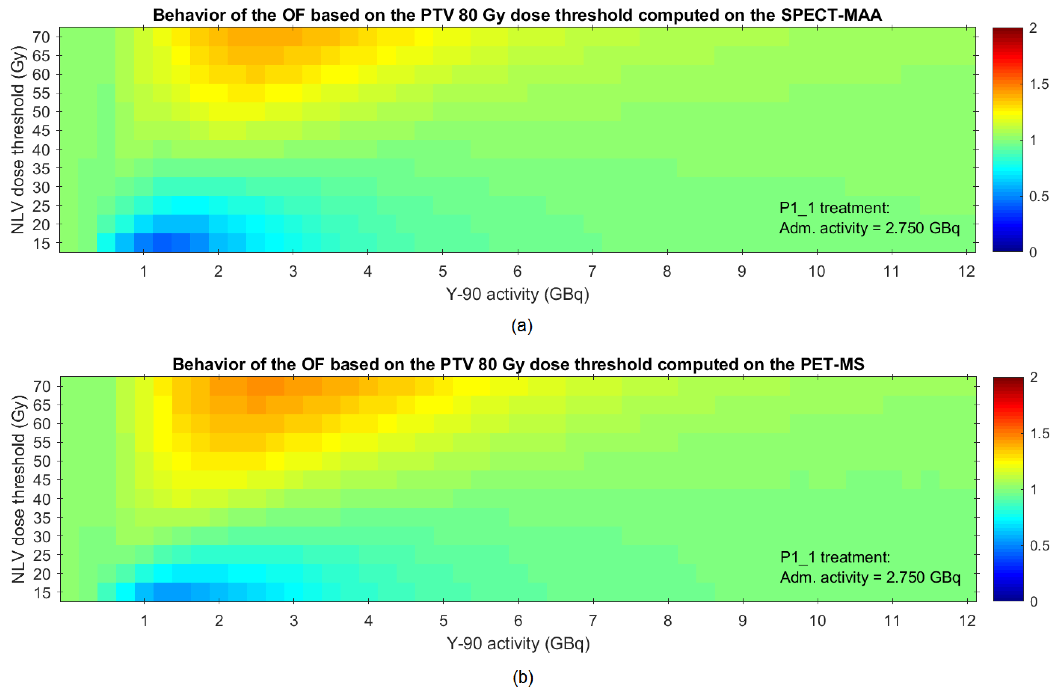
2.2.5. Proposed Methodology for Optimization of the Activity to Be Administered

Ionizing radiation therapies such as external radiotherapy and RE aim to achieve treatment efficacy and safety. The goal is to deliver a sufficiently high AD to the PTV while controlling the AD to the NLV to avoid and minimize the possibility of hepatic toxicity. This methodology is based on the fact that the healthy liver tissue is very sensitive to radiation and because the radio-sensitivity of tumor cells (i.e., primary hepatic carcinoma or liver metastases) differs for different tumor types [10,15,29,30,31,39,40,41,42,43,44,45]. Specifically, voxelized ADD should be optimized according to the ALARA (as low as reasonably achievable) principle. Minimizing the AD in the NLV around the PTV to be as low as reasonably possible minimizes the likelihood of side effects. In addition, the AD in the PTV should remain above a threshold value so that the patient can benefit from the therapy.
The mean AD in the NLV should not be greater than 70 Gy and should preferably remain below 50 Gy [29,30,31]. The PTV generally requires a mean AD higher than 75 Gy for proper treatment [16,29,30]. Little or no benefit is gained by exceeding an AD threshold of 120 Gy in the PTV, especially because it may increase the unnecessary exposure of the NLV [30,46,47]. However, patients with HCC and portal vein thrombosis were classified in recent research papers as good candidates for RE with an AD in the PTV greater than 205 Gy [39,44]. Ultimately, the AD level needed to achieve a certain probability of tumor control depends on the specific radio-sensitivity of the tumor type and the type of MS used in the treatment [48,49]. These variables were not considered in the present work. We have used only glass MS. On the other hand, in the presence of a very favorable tumor-to-normal liver perfusion ratio (highly selective distribution within the PTV), the PTV could significantly exceed the 120 Gy (thus improving expected efficacy probability) while exposing the NLV to doses much less than the mentioned 50 Gy (treatment reasonably safe). This will be better detailed later during the discussion.
Currently, the calculation of the 90Y activity administered in each RE treatment session followed the glass MS manufacturer’s protocol [16], following the one-compartment MIRD-based formalism that considers the prescribed AD (i.e., typically from 80 Gy to 120 Gy) and liver volumes (total, lobar, or segmental). This calculation considers the previously determined LSF as well as the estimated value of the radioactive residue that is not administered to the patient and is left in the injector circuit. Moreover, these calculations assume that the deposition of MS in the liver is uniform.
In this work, we propose a methodology and a computational solution to optimize the activity to be administered, considering the SPECT-MAA images acquired in the treatment planning stage. The optimization is based on the imposition of AD thresholds at the level of the PTV and NLV voxels. The optimal activity that should be administered into the patient’s liver is obtained by maximizing Equation (1):
O F ( A ) = # P T V t h r m i n ( A ) # P T V v o x + # N L V t h r m a x ( A ) # N L V v o x
where OF(A) is the proposed objective function, # P T V v o x and # N L V v o x are the total number of voxels in the PTV and NLV, respectively. # P T V t h r m i n ( A ) and # N L V t h r m a x ( A ) are the number of voxels of the SPECT-MAA-based ADD that comply with the AD threshold imposed to, respectively, the PTV and NLV, as a function of the 90Y activity (A) to be administered to the liver. In this regard, a certain number of NLV voxels are allowed to exceed the NLV AD threshold, and identically in the PTV, a certain number of voxels are allowed to receive an AD below the PTV AD threshold. Higher activity tends to increase the number of PTV voxels with AD higher than the threshold, but at the same time, increase the number of NLV voxels with AD higher than is desired. The optimal activity to be administered is determined by the maximization of the sum of the two terms in (1). In other words, the optimal activity is a balance between the two terms. These terms are referred to in this work as the PTV ratio and the NLV ratio. The optimization of the OF was based on the golden section method [50].
This methodology was tested using the patients’ pre-treatment SPECT-MAA and post-treatment PET-MS images retrospectively. For both types of images, the optimal activities were estimated using the proposed optimization solution. Then, the optimal values calculated from each patient’s SPECT-MAA images were compared against the optimal values calculated from their PET-MS images.
The assessment of the optimal activities calculated with SPECT-MAA and PET-MS images was based on AD thresholds of 80 Gy and 70 Gy imposed, respectively, to the PTV and NLV voxels, since these thresholds are the RE clinical reference, as previously referred [29,30]. The optimal predictive activity (derived from the SPECT-MAA) was compared against the a posteriori optimal activity (derived from the PET-MS). Statistical analysis was based on the Wilcoxon signed-rank statistical test and the ICC for absolute agreement. A significance level of 5% was set.
The optimization of the OF can be computed for any pair of AD thresholds. Thus, to test its behavior/robustness to the change of the thresholds, 72 combinations of these were analyzed in additional tests, considering 6 AD threshold values (i.e., 80 to 105 Gy with 5 Gy increments) at the level of the PTV voxels and 12 thresholds (i.e., 15 to 70 Gy with 5 Gy increments) at the level of the NLV voxels. For this purpose, a vector with 50 test values was generated, from zero to 12 GBq (i.e., the double typical maximum clinical values of 6 GBq).

3. Results

3.1. Phantom Studies

3.1.1. Accuracy of SPECT and PET Derived Absorbed Dose Distributions

The results of the accuracy of SPECT- and PET-derived ADD against the known reference obtained by the phantom experiment are presented in Table 2.
The clinical reconstruction parameters used in the recovery of the activity distribution on PET-MS allowed for obtaining a satisfactory match of −10% with the ground truth activity distribution in the biggest cylinder volume of 108 mL. The accuracy of the derived ADD in this cylinder was good, showing a mismatch with the ground truth of −4%. The other cylinders with smaller volumes, 8 and 15 mL, showed unsatisfactory activity recovery with a larger discrepancy of −57% and −49%, respectively, and −54% and −49% regarding derived ADD. Regarding SPECT-MAA, the discrepancy was slightly inferior to the one obtained with PET-MS (Table 2).

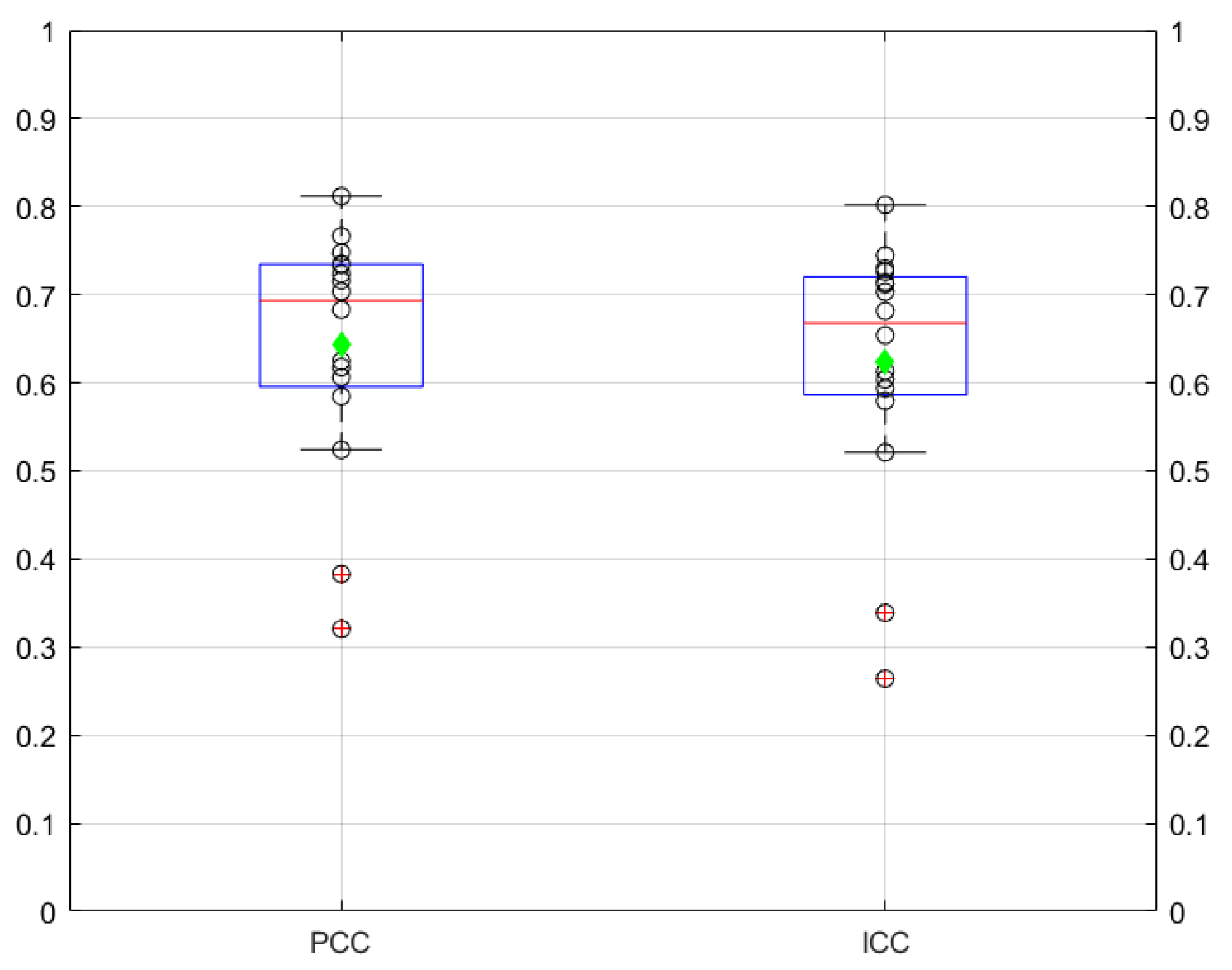
3.1.2. Correlation and Agreement between Absorbed Dose Distributions Derived from SPECT and PET Images

The voxelwise comparison between the SPECT-MAA- and PET-MS-derived ADD, assessed by the PCC and the ICC in liver_cyl and ptv_5_cyl_backg, showed high correlation and agreement. The PCC was 0.87 and 0.94 in liver_cyl and ptv_5_cyl_backg, respectively. The ICC was 0.85 and 0.93, respectively.
The voxelized γ-index distributions were computed considering the ADD in both phantoms, one derived from SPECT-MAA and the other from PET-MS images, and seven DD/DTA test conditions. Table 3 shows the results of the γ-index PR. As expected, the highest γ-index PR values were obtained with the less conservative 15%/15 mm conditions.

3.2. Patient Studies

Correlation and Agreement between Absorbed Dose Distributions Derived from Spect and Pet Images

Figure 4 shows the results of the voxelwise PCC and ICC between the ADD derived from SPECT-MAA and PET-MS images computed on the liver reference volume.
Two outliers were observed (P10_12 and P12_14) and excluded in this analysis. The median of the PCC was 0.69, and the mean was 0.64 with range of 0.52–0.81. Regarding the ICC estimate, a moderate to good agreement was obtained (median 0.67, mean 0.62, range 0.52–0.80).
Considering the mean AD in PTV and NLV from all treatments, the PCC was 0.80 and 0.98, respectively, and the ICC was 0.79 and 0.97, respectively. Thus, the agreement of the mean AD between the SPECT-MAA- and the PET-MS-based ADD computed on the liver ROI was good to excellent.
The results of the γ-index PR computed on four liver ROI are presented in Figure 5.
The highest values of the patients’ γ-index PR were obtained with the less conservative 15%/15 mm conditions, which were above 90% in all patient studies. Considering a 10 mm DTA (approximately the spatial resolution of the SPECT) and a 10% DD, γ-index PR values above 90% were obtained. This showed that the predictive power of the ADD based on SPECT-MAA compared to the ones based on PET-MS was good.
Optimization of the activity to be administered.
In 11 out of 16 treatments, the objective function (OF) converged for a maximum in the range considered acceptable (administered activity between 0 and 12 GBq) both for the SPECT-MAA- and PET-MS-derived ADD (Table 4).
For these 11 treatments, the agreement of the estimated activities was very high, with an ICC of 0.96 (p < 0.001), with no statistically significant difference between the optimal activities estimated based on the SPECT-MAA and PET-MS (paired Wilcoxon signed-rank test, p = 0.86).
Figure 6 shows example graphs of the OF optimization, in this case, for P1_1.
The OF optimization of the P4_4 and P14_16 showed quite different results when the SPECT-MAA or the PET-MS ADD were considered. Possibly, the two administrations (i.e., MAA and MS) were performed in different liver sites, and/or the patient’s liver anatomy changed significantly between the planning and treatment day. The OF optimization did not converge within the range of the activity test values in the P9_11, P11_13, and P13_15, interestingly indicating the high selectivity of the administrations to the PTV.
Two example tables with detailed information regarding the estimated ADD based on the pre-treatment SPECT-MAA as a function of the activity to be administered are shown in Supplementary Materials.
An example of the behavior of the OF for the P1_1 is shown in Figure 7.
In summary, considering the eleven treatments in Table 4, when the difference between the AD threshold of the PTV and NLV is lower than 40 Gy, the OF had similar behavior as for the clinically defined AD thresholds (<70 Gy in NLV and >80 Gy in PTV). Obviously, for those values, the estimated optimal activity was different from the optimal values obtained with the clinically defined AD thresholds.

4. Discussion

Several authors have been studying the accuracy of SPECT-MAA dosimetry to predict response, clinical impact, and outcomes for selected patients [14,30,39,51]. Others have been developing individualized treatment planning dosimetry strategies introducing radiobiology concepts [44,52,53]. To the best of our knowledge, while several dosimetric and clinical evaluations are reported in the literature, the optimization of 90Y activity to be administered based on patient-specific pre-treatment SPECT-MAA ADD together with the imposition of AD thresholds at the level of PTV and NLV voxels has never been addressed. Moreover, the recent European Council Directive 2013/59/EURATOM explicitly recommends that medical exposures shall be individually planned and optimized [19].
To address these important issues, there is a need to first validate the dosimetry methodology based on ground truth data. For this purpose, we have built our own phantoms with similar volumes for NLV and desire PTV and known activities of 99mTc-MAA and 90Y-MS. Our phantom studies revealed a strong correlation and agreement between the voxelwise ROI values in the simulated liver and PTV cylinders obtained from the SPECT-MAA- and PET-MS-based ADD.
The results regarding the two derived ADD (i.e., one respect to the other) do not provide information about their accuracy with respect to the actual ADD known from the phantom distribution ground truth (i.e., real activity concentration in phantom). Considering the phantom studies, the derived ADD in the biggest cylinder volume of 108 mL accuracy for the SPECT-MAA and PET-MS showed an acceptable error (<8%) from the ground truth distributions. On the other hand, and most probably mainly due to the partial volume effect amongst the others, the 8 mL and 15 mL volumes showed an error of approximately 50% from the ground truth distributions, indicating sub-optimal derived ADD estimation. These observations require careful evaluation whenever derived ADD in less than 100 mL PTV must be considered. Even though, in our patient sample, all the ROI mean AD estimated post-treatment agreed well with the pre-treatment calculations.
So far, and concerning the activity optimization methodology, we found optimal values estimated with pre-treatment SPECT-MAA to agree well with optimal activity estimated in the post-treatment PET-MS in eleven treatments. In five treatments, the optimal activity estimated based on the SPECT-MAA and PET-MS did not converge in the range defined as acceptable (0–12 GBq). In two out of these five treatments, the disagreement observed was possibly due to a discrepancy in the exact site of MAA and MS intra-arterial administrations in the liver. For the other three treatments, the OF optimization did not converge within the range of defined activities. The main reason for this behavior is likely to be due to the highly selective distribution within the PTV (for both MAA and MS administrations) that spared the NLV. This means that, in these specific cases, the administered activity might have been higher than it was, with no significant damage to the NLV.
In five out of the eleven treatments (see Table 4), the administered activity was lower, i.e., 33% to 68%, than the estimated optimal activity. In four of these (P3_3, P4_5, P7_8, P12_14), the PTV was larger than 100 mL. In this regard and according to our phantom studies, it is highly likely that the accuracy of the measurements in these patients’ PTV is very high, and therefore the administered activities might have been optimized/higher.
All these previous considerations assume the accepted threshold herein imposed (<70 Gy in NLV and >80 Gy in PTV) to be practical and an adequate “rule of thumb” to salvage NLV when delivering enough of a dose to PTV. These reference thresholds deserve adequate adjustments for each patient-specific condition. As we described previously, we have addressed this issue by successfully testing the OF behavior for several pairs of thresholds. However, this optimization protocol will be of clear benefit to the patients after well-designed clinical trials prove this to be true.
This work has the following limitations: (a) a small number of treatments were included, and therefore a larger sample is needed in future clinical research to confirm the results herein presented; (b) due to its retrospective nature, this study includes some cases with relatively sub-optimal image data quality, not ideal for all manipulations performed; (c) PET-MS images are significantly noisier than SPECT-MAA data, which compromises spatial resolution and quantification accuracy; (d) the limited number of phantom studies restricts some of our considerations and needs to be further developed; (e) ideally, the PTV morphology should be enriched in the phantom construction, possibly by using latest 3D printing developments to obtain more realistic simulations.
Finally, we understand that further theoretical simulation work is likely to bring out further beneficial improvements in personalized voxel-based dosimetry, keeping in mind the compliance with EURATOM directive requirements.

5. Conclusions

Under well-controlled conditions (i.e., MAA and MS administered in the same anatomical site of the hepatic arterial territory), the pre-treatment SPECT-MAA predicts the post-treatment PET-MS distribution. This means that the treatment of ADD in the liver ROI is similar to the pre-treatment (planning) calculations.
In addition, pre-treatment SPECT-MAA ADD can be used to optimize 90Y activity to be administered per patient referred to RE. The careful implementation of the optimization algorithm proposed is strongly recommended. From now on, when evaluating prospective treatment planning dosimetry studies, it is recommended to choose the optimal activity to be administered, considering simultaneous PTV and NLV mean-derived AD estimations.
Finally, the herein proposed computational dosimetry system, including an original optimization protocol, enables more effective personalized liver RE treatments.

Supplementary Materials

The following supporting information can be downloaded at: https://www.mdpi.com/article/10.3390/app122211669/s1, Table S1: P1_1 activity, NLV and PTV dose-volume values. In bold and italic, the optimal balance between the minimum PTV 80 Gy dose threshold and the maximum NLV 70 Gy dose threshold is represented. Table S2: Comparison of the mean and median doses in NLV and PTV, estimated with: (i) the standard formula of the glass MS manufacturer’s protocol, and (ii) the optimized methodology considering the minimum PTV 80 Gy dose threshold and the maximum NLV 70 Gy dose threshold.

Author Contributions

Conceptualization, P.F. and D.C.C.; methodology, P.F., F.P.M.O., R.P., P.L.C., P.S.G. and D.C.C.; software, P.F., F.P.M.O. and P.L.C.; validation, P.F., F.P.M.O. and P.L.C.; formal analysis, P.F.; investigation, P.F., F.P.M.O. and R.P.; writing—original draft preparation, P.F.; writing—review and editing, P.F., F.P.M.O., R.P., P.L.C., P.S.G. and D.C.C. All authors have read and agreed to the published version of the manuscript.

Funding

This research received no external funding.

Institutional Review Board Statement

Ethical review and approval were waived for this study due to its retrospective nature.

Informed Consent Statement

Informed consent was obtained from all subjects involved in the study allowing their clinical data to be used for research and educational purposes.

Data Availability Statement

Patients’ imaging and clinical data will be available upon reasonable request after ethical approval. The remaining data will be available upon reasonable request.

Acknowledgments

We thank all the patients, technical and clinical staff at the Nuclear Medicine—Radiopharmacology Department, and Hugo Rio-Tinto at the Imaging Department at Champalimaud Foundation for their dedication, continuous support, and collaboration during all the steps included in the works leading to the final set up of this manuscript.

Conflicts of Interest

The authors declare no conflict of interest.

References

  1. Cancer Research UK. Trends over Time for Cancer Mortality Worldwide. 2008. Available online: http://www.cancerresearchuk.org (accessed on 27 February 2018).
  2. Ferlay, J.; Soerjomataram, I.; Dikshit, R.; Eser, S.; Mathers, C.; Rebelo, M.; Parkin, D.M.; Forman, D.; Bray, F. Cancer incidence and mortality worldwide: Sources, methods and major patterns in GLOBOCAN 2012. Int. J. Cancer 2015, 136, 359–386. [Google Scholar] [CrossRef] [PubMed]
  3. Jemal, A.; Center, M.M.; DeSantis, C.; Ward, E.M. Global patterns of cancer incidence and mortality rates and trends. Cancer Epidemiol. Biomark. Prev. 2010, 19, 1893–1907. [Google Scholar] [CrossRef] [PubMed] [Green Version]
  4. Ferlay, J.; Ervik, M.; Lam, F.; Colombet, M.; Mery, L.; Piñeros, M. Global Cancer Observatory: Cancer Today. 2018. Available online: https://gco.iarc.fr/today (accessed on 28 March 2019).
  5. Ferlay, J.; Colombet, M.; Soerjomataram, I.; Mathers, C.; Parkin, D.M.; Piñeros, M.; Znaor, A.; Bray, F. Estimating the global cancer incidence and mortality in 2018: GLOBOCAN sources and methods. Int. J. Cancer 2018, 144, 1941–1953. [Google Scholar] [CrossRef] [PubMed] [Green Version]
  6. Freddie, B.; Jacques, F.; Isabelle, S.; Rebecca, L.S.; Lindsey, A.T.; Ahmedin, J. Global Cancer Statistics 2018: GLOBOCAN Estimates of Incidence and Mortality Worldwide for 36 Cancers in 185 Countries. CA Cancer J. Clin. 2018, 68, 394–424. [Google Scholar]
  7. Erdoğan, E.B.; Özdemir, H.; Aydin, M. Radioembolization Treatment for Liver Cancer. Bezmialem Sci. 2016, 4, 25–32. [Google Scholar] [CrossRef]
  8. Murthy, R.; Kamat, P.; Nuñez, R.; Salem, R. Radioembolization of Yttrium-90 Microspheres for Hepatic Malignancy. Semin. Interv. Radiol. 2008, 25, 48–57. [Google Scholar] [CrossRef] [Green Version]
  9. Gardini, A.C.; Tamburini, E.; Sangro, B. Radioembolization versus chemoembolization for unresectable hepatocellular carcinoma: A meta-analysis of randomized trials. OncoTargets Ther. 2018, 11, 7315. [Google Scholar] [CrossRef] [Green Version]
  10. Kennedy, A.; Nag, S.; Salem, R.; Murthy, R.; McEwan, A.J.; Nutting, C.; Benson, A., 3rd; Espat, J.; Bilbao, J.I.; Sharma, R.A.; et al. Recommendations for radioembolization of hepatic malignancies using yttrium-90 microsphere brachytherapy: A consensus panel report from the radioembolization brachytherapy oncology consortium. Int. J. Radiat. Oncol. Biol. Phys. 2007, 68, 13–23. [Google Scholar] [CrossRef]
  11. Smits, M.; Nijsen, J.; van den Bosch, M.; Lam, M.; Vente, M.; Huijbregts, J.; Schip, A.D.V.H.; Elschot, M.; Bult, W.; de Jong, H.W.; et al. Holmium-166 radioembolization for the treatment of patients with liver metastases: Design of the phase I HEPAR trial. J. Exp. Clin. Cancer Res. 2010, 29, 70. [Google Scholar] [CrossRef] [Green Version]
  12. Prince, J.F. Holmium Radioembolization: Efficacy and Safety. Ph.D. Thesis, Utrecht University, Utrecht, The Netherlands, 2016. [Google Scholar]
  13. Kallini, J.R.; Gabr, A.; Salem, R.; Lewandowski, R.J. Transarterial Radioembolization with Yttrium-90 for the Treatment of Hepatocellular Carcinoma. Adv. Ther. 2016, 33, 699–714. [Google Scholar] [CrossRef] [Green Version]
  14. Garin, E.; Lenoir, L.; Rolland, Y.; Edeline, J.; Mesbah, H.; Laffont, S.; Porée, P.; Clément, B.; Raoul, J.L.; Boucher, E. Dosimetry based on 99mTc-macroaggregated albumin SPECT/CT accurately predicts tumor response and survival in hepatocellular carcinoma patients treated with 90Y-loaded glass microspheres: Preliminary results. J. Nucl. Med. 2012, 53, 255–263. [Google Scholar] [CrossRef] [PubMed]
  15. Pasciak, A.; Bradley, Y.; McKinney, J. Handbook of Radioembolization; CRC Press: Boca Raton, FL, USA, 2016. [Google Scholar]
  16. Boston Scientific International. TheraSphere™ Yttrium-90 Glass Microspheres—Instructions for Use; Boston Scientific International: Fremont, CA, USA, 2005; pp. 1–16. [Google Scholar]
  17. Ferreira, P. Voxel-Based Dosimetry Using Multimodal Images for Patient-Specific Liver Radioembolization with Yttrium-90 Charged Glass Microspheres. Ph.D. Thesis, Department of Electrical and Computer Engineering, Universidade de Lisboa, Lisboa, Portugal, 2019. [Google Scholar]
  18. Ferreira, P.; Oliveira, F.P.M.; Parafita, R.; Girão, P.S.; Correia, P.L.; Costa, D.C. Patient-specific gamma-index analysis to evaluate 99mTc-MAA as a predictor for 90Y glass microspheres liver radioembolisation dosimetry. Comput. Methods Biomech. Biomed. Eng. Imaging Vis. 2018, 7, 583–589. [Google Scholar] [CrossRef]
  19. E.U. Council Directive 2013/59/Euratom. Off. J. Eur. Union 2013, L13/1, 1–73. [Google Scholar]
  20. Greer, K. NEMA IEC Body Phantom Set—User’s Manual; Data Spectrum Corporation: Durham, NC, USA, 2008. [Google Scholar]
  21. Bolch, W.; Bouchet, L.; Robertson, J.; Wessels, B.; Siegel, J.; Howell, R.; Erdi, A.K.; Aydogan, B.; Costes, S.; Watson, E.E.; et al. MIRD pamphlet No. 17: The dosimetry of nonuniform activity distributions—Radionuclide S values at the voxel level. Medical Internal Radiation Dose Committee. J. Nucl. Med. 1999, 40, 11–36. [Google Scholar]
  22. Snyder, W.S. ‘S’ Absorbed Dose Per Unit Cumulated Activity for Selected Radionuclides and Organs, 11 ed: Society of Nuclear Medicine. MIRD Pam. 1975, 11, 1–257. [Google Scholar]
  23. Pacilio, M.; Amato, E.; Lanconelli, N.; Basile, C.; Torres, L.A.; Botta, F.; Ferrari, M.; Diaz, N.C.; Perez, M.C.; Fernández, M.; et al. Differences in 3D dose distributions due to calculation method of voxel S-values and the influence of image blurring in SPECT. Phys. Med. Biol. 2015, 60, 1945–1964. [Google Scholar] [CrossRef]
  24. Lanconelli, N.; Pacilio, M.; Meo, S.L.; Botta, F.; di Dia, A.; Aroche, A.T.; Pérez, M.A.C.; Cremonesi, M. A free database of radionuclide voxel S values for the dosimetry of nonuniform activity distributions. Phys. Med. Biol. 2012, 57, 517–533. [Google Scholar] [CrossRef]
  25. Lanconelli, N.; Pacilio, M.; Lo Meo, S.; Botta, F.; Di Dia, A.; Aroche, A.T.; Coca Perez, M.A.; Cremonesi, M. S Values for Voxel Dosimetry. 2017. Available online: http://www.medphys.it/down_svoxel.htm (accessed on 15 July 2017).
  26. Pacilio, M.; Lanconelli, N.; Lo, M.S.; Betti, M.; Montani, L.; Aroche, L.A.T.; Pérez, M.A.C. Differences among Monte Carlo codes in the calculations of voxel S values for radionuclide targeted therapy and analysis of their impact on absorbed dose evaluations. Med. Phys. 2009, 36, 1543–1552. [Google Scholar] [CrossRef] [Green Version]
  27. Low, D.A.; Harms, W.B.; Mutic, S.; Purdy, J.A. A technique for the quantitative evaluation of dose distributions. Med. Phys. 1998, 25, 656–661. [Google Scholar] [CrossRef]
  28. Hussein, M.; Rowshanfarzad, P.; Ebert, M.A.; Nisbet, A.; Clark, C.H. A comparison of the gamma index analysis in various commercial IMRT/VMAT QA systems. Radiother. Oncol. 2013, 109, 370–376. [Google Scholar] [CrossRef] [Green Version]
  29. Callan, L.; Beech, B.; Wong, E. Radiation in the treatment of liver cancer. Univ. West. Ont. Med. J. 2014, 83, 17–20. [Google Scholar] [CrossRef]
  30. Lau, W.-Y.; Kennedy, A.S.; Kim, Y.H.; Lai, H.K.; Lee, R.-C.; Leung, T.W.T.; Liu, C.-S.; Salem, R.; Sangro, B.; Shuter, B.; et al. Patient Selection and Activity Planning Guide for Selective Internal Radiotherapy with Yttrium-90 Resin Microspheres. Int. J. Radiat. Oncol. Biol. Phys. 2012, 82, 401–407. [Google Scholar] [CrossRef] [PubMed]
  31. Dawson, L.A.; McGinn, C.J.; Normolle, D.; Haken, R.K.T.; Walker, S.; Ensminger, W.; Lawrence, T.S. Escalated focal liver radiation and concurrent hepatic artery fluorodeoxyuridine for unresectable intrahepatic malignancies. J. Clin. Oncol. 2000, 18, 2210–2218. [Google Scholar] [CrossRef] [PubMed]
  32. Fedorov, A.; Beichel, R.; Kalpathy-Cramer, J.; Finet, J.; Fillion-Robin, J.C.; Pujol, S.; Bauer, C.; Jennings, D.; Fennessy, F.; Sonka, M.; et al. 3D Slicer as an image computing platform for the Quantitative Imaging Network. Magn. Reson. Imaging 2012, 30, 1323–1341. [Google Scholar] [CrossRef]
  33. Yushkevich, P.A.; Gerig, G. ITK-SNAP: An Intractive Medical Image Segmentation Tool to Meet the Need for Expert-Guided Segmentation of Complex Medical Images. IEEE Pulse 2017, 8, 54–57. [Google Scholar] [CrossRef]
  34. Jimmy, S. Tools for NIfTI and ANALYZE Image. 2014. Available online: https://www.mathworks.com/matlabcentral (accessed on 12 November 2015).
  35. Multi-Modal Markers for Radiology. 2017. Available online: https://www.medcat.nl/supplies/En/radmarker.htm (accessed on 5 April 2016).
  36. Yushkevich, P.A.; Piven, J.; Hazlett, H.C.; Smith, R.G.; Ho, S.; Gee, J.C.; Gerig, G. User-guided 3D active contour segmentation of anatomical structures: Significantly improved efficiency and reliability. Neuroimage 2006, 31, 1116–1128. [Google Scholar] [CrossRef] [Green Version]
  37. Criminisi, A. Decision Forests: A Unified Framework for Classification, Regression, Density Estimation, Manifold Learning and Semi-Supervised Learning. Found. Trends Comput. Graph. Vis. 2011, 7, 81–227. [Google Scholar] [CrossRef]
  38. Breiman, L. Random Forests. Mach. Learn. 2001, 45, 5–32. [Google Scholar] [CrossRef] [Green Version]
  39. Garin, E.; Rolland, Y.; Pracht, M.; Sourd, S.L.; Laffont, S.; Mesbah, H.; Haumont, L.-A.; Lenoir, L.; Rohou, T.; Brun, V.; et al. High impact of macroaggregated albumin-based tumour dose on response and overall survival in hepatocellular carcinoma patients treated with 90Y-loaded glass microsphere radioembolization. Liver Int. 2017, 37, 101–110. [Google Scholar] [CrossRef] [Green Version]
  40. Ferreira, P.; Parafita, R.; Canudo, A.; Oliveira, C.; Rosa, L.; Girão, P.M.; Costa, D.C. Multiple Liver Metastases from Carcinoma of the Thymus Treated with Yttrium-90 Radioembolization (Glass Microspheres): Clinical Dosimetry. Clin. Oncol. 2016, 1, 1–5. [Google Scholar]
  41. Goedicke, A.; Berker, Y.; Verburg, F.A.; Behrendt, F.F.; Winz, O.; Mottaghy, F.M. Study-parameter impact in quantitative 90-Yttrium PET imaging for radioembolization treatment monitoring and dosimetry. IEEE Trans. Med. Imaging 2013, 32, 485–492. [Google Scholar] [CrossRef] [PubMed]
  42. Johns, H.E. The Physics of Radiology, 2nd ed.; Charles C Thomas: Springfield, IL, USA, 1961. [Google Scholar]
  43. Cameron, J.R.; Skofronick, J.G. Medical Physics; John Wiley & Sons: Hoboken, NJ, USA, 1978. [Google Scholar]
  44. Chiesa, C.; Mira, M.; Maccauro, M.; Spreafico, C.; Romito, R.; Morosi, C.; Camerini, T.; Carrara, M.; Pellizzari, S.; Negri, A.; et al. Radioembolization of hepatocarcinoma with (90)Y glass microspheres: Development of an individualized treatment planning strategy based on dosimetry and radiobiology. Eur. J. Nucl. Med. Mol. Imaging 2015, 42, 1718–1738. [Google Scholar] [CrossRef] [PubMed]
  45. Venkatanarasimha, N.; Gogna, A.; Tong, K.T.A.; Damodharan, K.; Chow, P.K.H.; Lo, R.H.G.; Chandramohan, S. Radioembolisation of hepatocellular carcinoma: A primer. Clin. Radiol. 2017, 72, 1002–1013. [Google Scholar] [CrossRef]
  46. Lau, W.Y.; Leung, W.T.; Ho, S.; Leung, N.W.; Chan, M.; Lin, J.; Metreweli, C.; Johnson, P.; Li, A.K. Treatment of inoperable hepatocellular carcinoma with intrahepatic arterial yttrium-90 microspheres: A phase I and II study. Br. J. Cancer 1994, 70, 994–999. [Google Scholar] [CrossRef] [PubMed] [Green Version]
  47. Campbell, A.M.; Bailey, I.H.; Burton, M.A. Tumour dosimetry in human liver following hepatic yttrium-90 microsphere therapy. Phys. Med. Biol. 2001, 46, 487–498. [Google Scholar] [CrossRef]
  48. Srinivas, S.M.; Nasr, E.C.; Kunam, V.K.; Bullen, J.A.; Purysko, A.S. Administered activity and outcomes of glass versus resin (90)Y microsphere radioembolization in patients with colorectal liver metastases. J. Gastrointest. Oncol. 2016, 7, 530–539. [Google Scholar] [CrossRef] [PubMed] [Green Version]
  49. van der Gucht, A.; Jreige, M.; Denys, A.; Blanc-Durand, P.; Boubaker, A.; Pomoni, A.; Mitsakis, P.; Silva-Monteiro, M.; Gnesin, S.; Lalonde, M.N.; et al. Resin Versus Glass Microspheres for (90)Y Transarterial Radioembolization: Comparing Survival in Unresectable Hepatocellular Carcinoma Using Pretreatment Partition Model Dosimetry. J. Nucl. Med. 2017, 58, 1334–1340. [Google Scholar] [CrossRef] [Green Version]
  50. Press, W.H.; Teukolsky, S.A.; Vetterling, W.T.; Flannery, B.P. Numerical Recipes: The Art of Scientific Computing, 3rd ed.; Cambridge University Press: New York, NY, USA, 2007. [Google Scholar]
  51. Garin, E.; Rolland, Y.; Laffont, S.; Edeline, J. Clinical impact of (99m)Tc-MAA SPECT/CT-based dosimetry in the radioembolization of liver malignancies with (90)Y-loaded microspheres. Eur. J. Nucl. Med. Mol. Imaging 2016, 43, 559–575. [Google Scholar] [CrossRef] [Green Version]
  52. Chiesa, C.; Mira, M.; Maccauro, M.; Romito, R.; Spreafico, C.; Sposito, C.; Bhoori, S.; Morosi, C.; Pellizzari, S.; Negri, A.; et al. A dosimetric treatment planning strategy in radioembolization of hepatocarcinoma with 90Y glass microspheres. Q. J. Nucl. Med. Mol. Imaging 2012, 56, 503–508. [Google Scholar]
  53. Cremonesi, M.; Chiesa, C.; Strigari, L.; Ferrari, M.; Botta, F.; Guerriero, F.; De Cicco, C.; Bonomo, G.; Orsi, F.; Bodei, L.; et al. Radioembolization of Hepatic Lesions from a Radiobiology and Dosimetric Perspective. Front. Oncol. 2014, 4, 210. [Google Scholar] [CrossRef]
Figure 1. The main photograph shows the entire phantom on its stand with the inner insert containing several PTV cylinders. This inner insert is shown completely empty in the upper-right corner, where the five PTV cylinders are indicated by numbers 1 to 5. The entire phantom shown in the main photograph was captured with the inner insert in place filled with agarose.
Applsci 12 11669 g001
Figure 2. Phantom ROI on a similar slice of CT and SPECT-MAA-based ADD. Cubic voxels of 2.21 mm length were used after resampling both images. The red circles represent the ptv_5_cyl_backg mask (i.e., including four samples of the background near the PTV) or the ptv_5_cyl mask (i.e., excluding the four background samples). The individual PTV cylinder masks (red circles) are numbered 1 through 5. The liver_box and the liver_cyl masks are represented by the blue square and the green circle, respectively.
Applsci 12 11669 g002
Figure 3. Example of a patient’s liver ROI in a single CT slice. The red and green lines represent the PTV and the liver masks, respectively. The mask of the dose computation box is represented by the blue square.
Applsci 12 11669 g003
Figure 4. Boxplot of the voxelwise PCC and ICC between the SPECT-MAA and PET-MS based ADD computed on the liver reference volume of each patient. The two green diamonds correspond to the mean. The circles containing red plus sign are considered/taken as outliers.
Applsci 12 11669 g004
Figure 5. Boxplots with the distributions of the γ-index PR considering seven DD/DTA test conditions in four liver ROI: (a) liver_ref, (b) liver, (c) PTV, and (d) NLV. The red plus sign are considered/taken as outliers.
Applsci 12 11669 g005
Figure 6. P1_1 optimization based on the SPECT-MAA (left) and PET-MS (right). The optimized OF and its two components are shown in blue, red (PTV ratio), and green (NLV ratio), respectively. The blue dots represent the OF values in each iteration during optimization.
Applsci 12 11669 g006
Figure 7. Distributions of the OF for P1_1 case, based on PTV 80 Gy AD threshold and twelve NLV AD thresholds (15 Gy–70 Gy) considering activities between 0 and 12 GBq, computed on the SPECT-MAA (a) and PET-MS (b) ADD. The administered activity was 2750 MBq, and the estimated optimal activity was 2598 MBq (based on the treatment planning SPECT-MAA).
Applsci 12 11669 g007
Table 1. Eligible patients (n) and RE treatments (t) included in this work and PTV characterization.
Pn_tDiagnosisNo. Administrations
MAA MS
Administration SiteNo. PTVs; Volume Range (mL); Total Volume (mL)
P1_1Metastatic thymoma11proper hepatic artery16; 2–34; 241
P2_2Metastatic pancreatic cancer33(1) right hepatic artery
(2) segment II & III
(3) segment IV
9; 4–79; 574
P3_3Metastatic colorectal cancer11proper hepatic artery3; 33–230; 314
P4_4Metastatic colorectal cancer22(1) right hepatic artery
(2) segment II & IV
1; 462; 462
P4_5Metastatic colorectal cancer11right hepatic artery5; 7–508; 1626
P5_6Multifocal cholangiocarcinoma22(1) right hepatic artery
(2) left hepatic artery
6; 3–131; 451
P6_7Metastatic colorectal cancer22(1) right hepatic artery
(2) left hepatic artery
3; 27–300; 389
P7_8Metastatic colorectal cancer11proper hepatic artery5; 13–1016; 1154
P8_9Metastatic colorectal cancer11left hepatic artery8; 2–20; 123
P8_10Metastatic colorectal cancer11right hepatic artery5; 5–125; 202
P9_11Hepatocarcinoma11left hepatic artery1; 772; 772
P10_12Metastatic colorectal cancer11right hepatic artery14; 2–49; 93
P11_13Cholangiocarcinoma11segment IV1; 256; 256
P12_14Hepatocarcinoma22(1) right hepatic artery
(2) segment IV
1; 1047; 1047
P13_15Metastatic pancreatic cancer11right hepatic artery1; 510; 510
P14_16Cholangiocarcinoma22(1) right hepatic artery (I)
(2) right hepatic artery (II)
2; 5–89; 94
Table 2. Accuracy of SPECT-MAA and PET-MS based recovered activity and derived ADD. For comparison purposes, the two ground truth activity distributions were simulated based on the expected 90Y activity concentration known by the phantom experiment preparation.
ROICylinder 1Cylinder 2Cylinder 3Cylinder 4Cylinder 5
Actual volume (mL)158158108
SPECT-based volume (mL)2392410117
PET-based volume (mL)21111912118
Activity ⇒ Mean ADMBq ⇒ GyMBq ⇒ GyMBq ⇒ GyMBq ⇒ GyMBq ⇒ Gy
PET-MS ground truth (simulated)3656691876183446582046392084775
Calculated18536581309175378922941871745
Deviation (%)−49−45−57−50−49−43−55−54−10−4
SPECT-MAA ground truth (simulated)4086691506184216581736392031775
Calculated20137372289217408802601808721
Deviation (%)−52−44−52−53−48−38−54−59−11−7
Note 1: SPECT-MAA and PET-MS based segmentation of ROI used 4.66 mm and 4.0 mm isotropic voxel size, respectively. Note 2: Deviation (%) = 100 × (calculated − ground truth)/ground truth.
Table 3. γ-index PR (%) in phantom ROI considering seven DD (%)/DTA (mm) test conditions.
ROIDD (%)/DTA (mm)
5/510/55/1010/1015/1010/1515/15
liver_cyl98.398.798.798.999.299.099.3
ptv_5_cyl_backg88.992.190.092.895.793.296.0
Table 4. Administered and estimated optimal activities based on SPECT-MAA and on PET-MS for each of the 11 treatments, with OF converging for a maximum between 0 and 12 GBq.
Pn_tAdministered
Activity (MBq)
SPECT-MAA Estimated
Optimal Activity (MBq)
PET-MS Estimated
Optimal Activity (MBq)
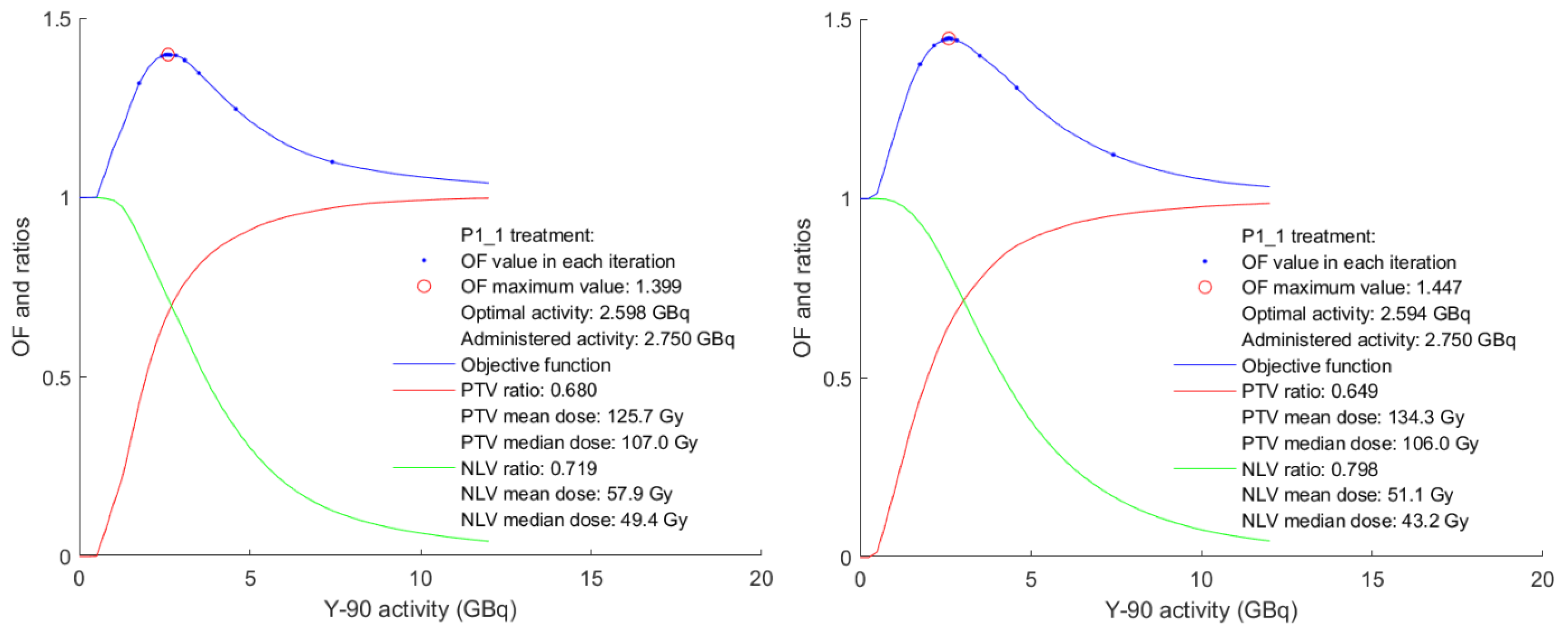
P1_1275025982594
P2_2334737484009
P3_3396162398518
P4_5423110,6629423
P5_6430543973397
P6_7281422742281
P7_8560480948481
P8_9153748984505
P8_10422722192450
P10_12199214711380
P12_14448186758539
Publisher’s Note: MDPI stays neutral with regard to jurisdictional claims in published maps and institutional affiliations.

