Article

Comparison of Spine–Pelvis Kinematics Variability during Sit-to-Stand and Stand-to-Sit in People with & without Chronic Low Back Pain: A Vector Coding and Statistical Parametric Mapping Approach

by
Narges Jangjoo
1,
Elham Alijanpour
2,
Saeid Khodabakhsh
1,
Seyed Sadredin Shojaedin
1,* and
Roozbeh Naemi
3
1
Department of Biomechanics and Sports Injuries, Faculty of Physical Education and Sports Sciences, Kharazmi University, Tehran 1571914911, Iran
2
School of Rehabilitation, College of Health Sciences, Old Dominion University, Norfolk, VA 23508, USA
3
Centre for Biomechanics and Rehabilitation Technologies, School of Health Science and Wellbeing, Staffordshire University, Science Centre Leek Road, Stoke on Trent ST4 2DF, UK
*
Author to whom correspondence should be addressed.
Appl. Sci. 2022, 12(22), 11796; https://doi.org/10.3390/app122211796
Submission received: 9 October 2022 / Revised: 11 November 2022 / Accepted: 15 November 2022 / Published: 20 November 2022
(This article belongs to the Special Issue Biomechanics in Sport Performance and Injury Preventing)

Abstract

:
There is evidence in the literature to suggest that low back pain may change spine–pelvis coordination during activities of daily living. This study aimed to compare the variability of the spine–pelvis coordination during sit-to-stand in people with and without LBP. Ten healthy individuals with a history of LBP and ten individuals without history of LBP participated in this study. Three-dimensional kinematic data of the upper trunk (UT), lower trunk (LT), lower back (LB), and pelvis segments during sit-to-stand and stand-to-sit were recorded using a multi-segmental spine and pelvis models using a motion capture system. The coordination patterns and the variability of the adjacent segments (UT, LT, LB, and pelvis) were calculated using the modified vector coding method that was implemented through a custom MATLAB code. An independent sample t-test was utilized to assess the differences in the coordination pattern, and a statistical parametric mapping method was used to quantify the differences in coordination variability between the two groups. The results indicate that there are some differences in coordination patterns between groups during sit-to-stand and stand-to-sit. However, a significant difference in coordination variability was only observed during sit-to-stand. The results showed that LBP can alter the kinematics coordination even in the upper (pain-free) parts of the spine during sit-to-stand by changing the coordination between UT and LT in a way that can lead to an increase in the loading on these segments. Additionally, people with LBP showed more coordination variability during sit-to-stand, which can be associated with a coordination strategy that facilitates an optimal and possibly pain-free coordination pattern.

1. Introduction

Low back pain (LBP) is defined as any pain that starts in the twelfth vertebra of the thoracic spine and spreads below the buttocks. LBP with a history of pain for more than three months and the absence of any pathological symptoms is called chronic low back pain (CLBP) [1]. It is estimated that 10% of patients who suffer from acute back pain will also suffer from chronic back pain [2]. About 90% of back pains are nonspecific, which is usually diagnosed by ruling out the possibility of specific pathologies [3]. In these cases, in addition to back pain of unknown cause, the specialist should also consider the presence of muscle pain originating in the spine. This type of pain may be nonmechanical or mechanical (increase in pain with movement or physical pressure), and the pain may be constant at rest [1,4].
Several factors such as a specific lifestyle, special methods of sit-to-stand from the floor and chair, lack of adequate mobility, and lack of regular exercise programs are effective in the development and exacerbation of LBP [5,6]. It has been reported that patients with LBP require more time to complete sit-to-stand movements and have a delayed beginning of pelvic anteversion [7]. In another study, it was revealed that patients with LBP did not have trunk range of motion (ROM) limitations but, rather, a delay in lumbar region motion, decreased inferior thoracic ROM, and enhanced hip joint ROM [8]. Additionally, it was shown that patients with low back pain had decreased lumbar spine and hip speed and ROM [9]. Additionally, a decreased lumbar movement contribution in comparison to the hip and dramatically changed motor coordination between these segments were reported [9]. Numerous kinematic aspects of the sit-to-stand still show conflicting results in the literature, making a true understanding of their mechanism difficult. Consequently, both sit-to-stand and stand-to-sit have been considered by researchers due to their effect on LBP [10,11]. Sit-to-stand (from a chair) is one of the most common activities of daily living and is the first step in performing several activities of daily living [12,13,14]. Additionally, sit-to-stand and stand-to-sit are both related to the aggravation of low back pain symptoms; hence, research into these movements is critical in LBP research.
It was previously reported that a significant amount of load on the spine of people with LBP was seen during sit-to-stand from a chair [10]. According to Punjabi theories, along with low back pain, there is a kind of clinical instability in the pelvic lumbar region [15]. In various studies based on the Punjabi theory, low back pain can lead to the following issues: (1) changing the timing of muscle activity [16], (2) changes in muscle activity and spinal stiffness shown by biomechanical modeling [17], (3) and spinal kinematic changes studied by motion analysis systems [18]. Coordination between different joints and segments as a variable of the neuromuscular system outcome can help in understanding the altered mechanics in a particular region due to musculoskeletal injuries. Therefore, investigating the coordination between different spine sections and comparing the coordination between people with and without low back pain can help to better understand the injury and adopt the appropriate treatment and rehabilitation methods [19].
Motion coordination entails the selection and creation of movement through the combination of the degrees of freedom available to perform a task [20]. In healthy individuals, many of the degrees of freedom available can be combined to coordinate achieving a motor task. Coordination is a functional role and may provide information about the tension in the joints [17]. Previous studies indicated that LBP could alter the coordination pattern between the spine and pelvis during various tasks, such as in gait [17] and during running [21]. Weakness or reduced coordination of muscles and joints in the body can lead to abnormal and compensatory movement patterns or to various types of injuries [22]. Furthermore, the number of coordination patterns that a system can produce for executing a task might be a sign of health or injury in the system [14].
Coordination variability is several changes in individuals’ coordination patterns that provide an understanding of the flexibility of movement pattern production systems. Lack of variability is a malfunction. Less variability can indicate less flexibility of the body to adapt to changing conditions, which leads to more repetition of stress in the joints. Motor variability is an essential component of human movement and is necessary for musculoskeletal health throughout a person’s life [21,23,24].
Coordination and variability are affected by different factors, such as degrees of freedom in the joints or injuries [25]; thus, the analysis of motor coordination variability provides an insight into the health of the locomotor system.
Decreased coordination variability is known to increase mechanical stress and overuse [26]. This is relevant to activities such as sit-to-stand, where the investigation of kinematic coordination can help identify the risk of injury [27]. Therefore, this study aims to compare coordination patterns and the variability of the spine and pelvis in sit-to-stand between people with and without nonspecific CLBP. Regarding this purpose, we also examined the range of motion (ROM) in each segment to have a comprehensive investigation of the kinematics data. We hypothesized that both coordination and coordination variability can differ in people with CLBP compared to healthy ones.

2. Materials and Methods

2.1. Study Participants

This study was registered with the National Ethics Committee of the Sports Science Research Institute of Iran (IR.SSRC.REC.1400.038). Using G-power software with a power of 0.95 and α = 0.05 and based on average ± stdev angle for upper/lower trunk of 50 ± 12 for healthy and 30 ± 10 for the CLBP, the number of participants was estimated as 20. Then, based on that and the literature, twenty participants: 10 with and 10 without nonspecific chronic low back participated in this study (Table 1) [28,29].
Inclusion criteria for the nonspecific CLBP group were a pain scale based on a numerical rating scale between 4 and 7 and disability based on the ODI from 10% to 60%, no structural deformities of the spine, no infection or tumors, no neurological or sensory orthopedic disorders or rheumatoid disease, no history of the trunk or pelvic and lower limb fractures or surgical interventions, no existence of lower extremity pain, spondylolisthesis, balance disorders, the non-use of anti-inflammatory or analgesic drug in the last 48 h, and all coronary health protocols were observed. An orthopedic specialist assessed patients for the exclusion criteria, which was followed with the assessment by an experienced physical therapist.

2.2. Experimental Setup

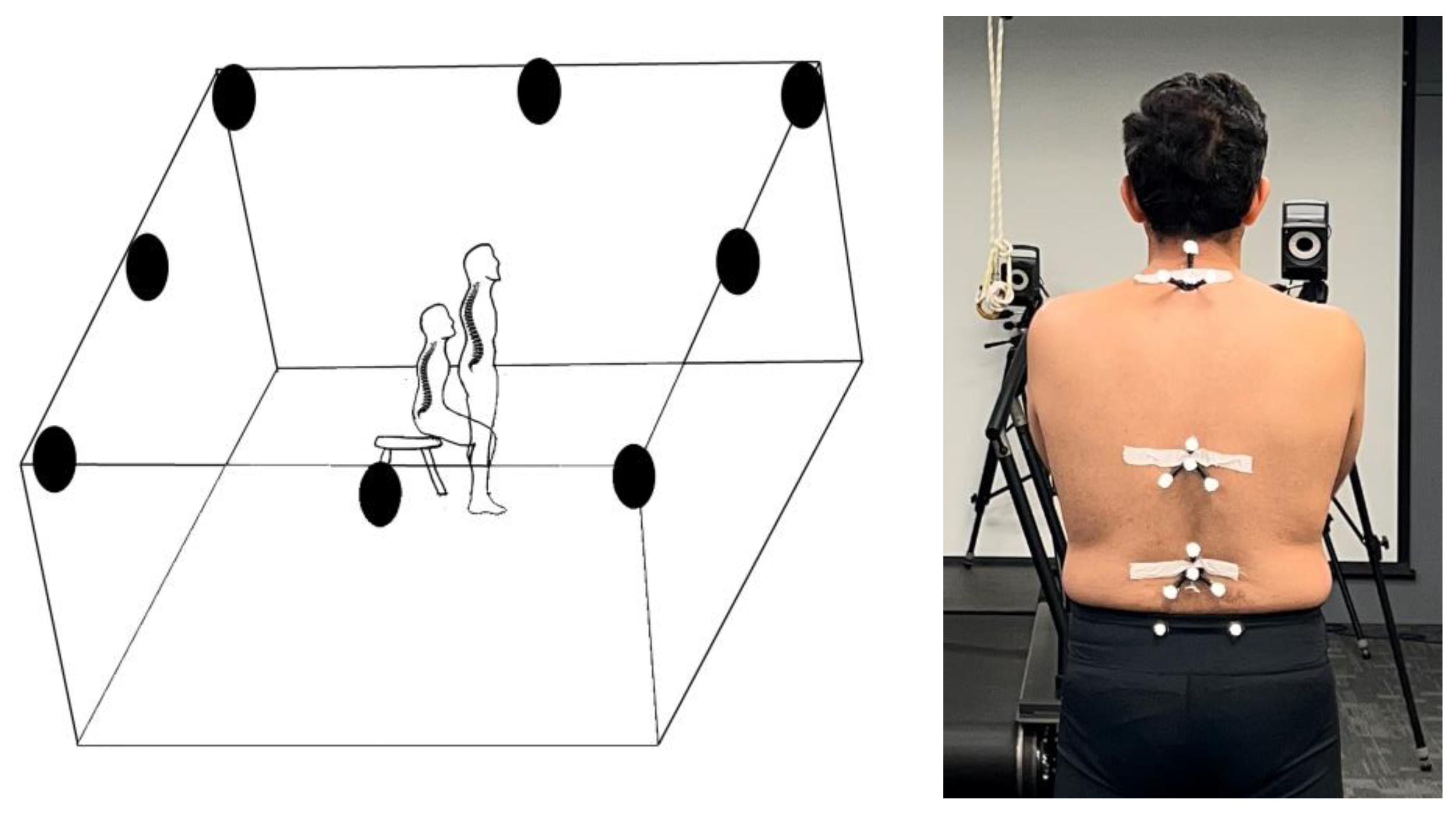
An eight-camera Vicon motion capture system and a multi-segmental model were used to track movements in the upper spine, lower spine, lower back, and pelvis [30]. A three-segmental biomechanical spine model with three four-marker clusters on the 3rd and 8th thoracic and 3rd lumbar vertebrae were used to record 3D movement in the upper trunk, lower trunk, and lumbar regions [30], and 4 markers were also placed on left and right anterior and posterior iliac spines to track pelvis movement (Figure 1). The static test was performed in an anatomical position. Then, participants were asked to sit and get up from a chair without a standard handle (45 cm high) five times at full speed without losing balance. This experiment was performed barefoot. Trajectories data from the beginning of the first sitting position to the end of the last sitting position were recorded with Nexus software. Kinematics data were extracted from the spine and pelvis and used for coordination and variability analyses [31].

2.3. Data Processing

Kinematic data were low pass-filtered with a 4th-order Butterworth filter at a 6 Hz cut-off. Cycle identification was obtained using the maximum and minimum values of the upper marker of the upper spine cluster in the Z-axis (superior–inferior). The three-dimensional angles of the upper trunk (UT), lower trunk (LT), lower back (LB), and pelvis relative to the global coordinate system were calculated in ProCalc (version 2.1.2, VICON, Oxford, UK). The method was comprehensively presented and described in previous studies [19,32] (Figure 2).

2.4. Data Analysis

ROM data were determined by subtracting the minimum angle from the maximum angle for each segment at each cycle. Segment coordination and coordination variability (CV) were calculated for five cycles using a modified vector coding technique [20,26] (see Needham et al. (2015) [33] and Needham et al. (2020) [24] for more information). Coordination patterns were classified into the in-phase with proximal dominancy (IPPD), in-phase with distal dominancy (IPDD), antiphase with proximal dominancy (APPD), and antiphase with distal dominancy (APDD) [33]. The percentage of each coordination pattern in the stand-to-sit and sit-to-stand cycles was quantified using frequency plots to understand the most prevalent patterns. CV was calculated as the standard deviation of the vector connecting the corresponding consecutive time points of the angle–angle plots across all cycles [24]. The UT/LT, LT/LB, LB/pelvis coordination, and CV were examined in the sagittal plane.

2.5. Pain Measurement

A visual analogue scale (VAS) was used to measure pain intensity. It is a self-reported scale consisting of a horizontal or vertical line, usually 10 cm long (100 mm), and two verbal descriptors describing the degree of pain [34]. This questionnaire is rated from zero to 10, and the patients should rate their pain on a graduated line, from zero (no pain) to 10 (most severe pain imaginable) [35,36].

2.6. Performance Evaluation

The low back pain disability questionnaire was used to measure the level of performance in this study. The Oswestry disability index (ODI) covers one item related to pain and nine items related to activities of daily living (personal care, lifting, walking, sitting, standing, sleeping, sex life, social life, and traveling) [37]. The ODI scores range from 0 (no disability) to 100 (maximum disability). Scores from 0–20 indicate “minimal disability”, 20–40 indicate “moderate disability”, 40–60 indicate “severe disability”, 60–80 indicate “housebound”, and 80–100 indicate “bedbound” [38].

2.7. Statistical Analysis

The normality of the coordination pattern frequency data was indicated with the Kolmogorov–Smirnov test (p > 0.05). Basic descriptive statistics were used to describe the variables (Table 2). The differences in the ROM data and coordination pattern frequencies between the two groups were assessed with an independent t-test using SPSS (IBM SPSS statistics 22, SPSS Inc., Chicago, IL, USA). A statistical parametric mapping (SPM) independent t-test was used to assess the differences between CV waveforms across the two groups (v.M0.1, www.spm1d.org, accessed on 19 January 2021). The statistical significance level for all analyses was set at p = 0.05.

3. Results

3.1. ROM Results

The results of the ROM data showed that there was no difference between segments’ ROM during sit-to-stand and stand-to-sit in people with and without CLBP.

3.2. Coordination Pattern Results

3.2.1. Sit-to-Stand

The results of the LT/UT showed that the pattern of antiphase coordination in LT extension and UT flexion movements with the LT dominancy was significantly (p = 0.048) more frequent in the low back pain group (Figure 3a).
There was no difference in the LB/LT kinematic coordination pattern during sit-to-stand between people with and without CLBP (Figure 3b).
Additionally, there was no difference in the pelvis/LB kinematic coordination pattern during sit-to-stand between people with and without CLBP (Figure 3c).

3.2.2. Stand-to-Sit

The results of the UT/LT showed that there was no difference in LT/UT kinematic coordination pattern during stand-to-sit between people with and without CLBP (Figure 4a).
There was no difference in LB/LT kinematic coordination pattern during stand-to-sit between people with and without CLBP (Figure 4b).
Additionally, the results showed that the pattern of in-phase coordination in the LB extension movement and pelvic posterior tilt with LB dominancy in the healthy group was significantly (p = 0.024) more dominant than in the low back pain group (Figure 4c).

3.3. Coordination Pattern Variability

3.3.1. Sit-to-Stand

The results of an independent t-test showed that the CV in LB/LT and pelvis/LB couples is more in the low back pain group in comparison to the healthy group at 80% of the sit-to-stand cycle when the person is close to standing upright (Figure 5b,c). Additionally, the CV in the pelvis/LB couple is more in the LBP group in the middle of the sit-to-stand cycle (Figure 5c).

3.3.2. Stand-to-Sit

The results of the statistical parametric mapping (SPM) independent t-test for coordination variability of all couples on the sagittal plane showed that the CV is not different between healthy and low back pain groups during the stand-to-sit cycle (Figure 6).

4. Discussion

This study aimed to quantify spine–pelvis coordination pattern and its variability during sit-to-stand and stand-to-sit task among people with and without low back pain. We hypothesized that low back pain can alter the spine–pelvis coordination pattern and its variability. The results of the current study, considering those from previous studies, indicate that, while ROM data may not be different between groups, other kinematics parameters such as coordination and coordination variability may differ between LBP and healthy people, which warrants further investigation.

4.1. Comparison of Segments’ Range of Motion between Groups

The results of the ROM data showed that there was no significant difference between the two groups. To the best of our knowledge, limited studies investigated the spine range of motion during sit-to-stand or stand-to-sit, and there are contradictions in their results. Christe et al. (2016) [28] and Shum et al. (2005) [9] reported that ROM was limited in nonspecific CLBP people. Svendsen et al. (2013) [39] reported no significant differences between LBP and healthy people. Additionally, Peydro et al. (2011) [40] and Sanchez-Zuriaga et al. (2011) [41] mentioned a decreased lumbar range of motion and a lower flexion peak. However, when the trunk’s mobility is examined globally, the results are conflicting [9]. Coghlin et al. (1994) [29] and McFadyen discovered that there is a modified strategy for raising from a chair in participants with low back pain. In addition, the results of research by Pourahmadi et al. (2019) [12] showed in patients with low back pain that ROM can be decreased because of the pain, muscle spasms, coactivation, or stiffness, while another study indicated that paraspinal muscle coactivation is utilized to stabilize the lumbar spine as a preventive technique to minimize pain provocation [42]. Furthermore, increased spine stiffness may damage patients with LBP by altering movement patterns and perhaps increasing the sensitivity of spinal and peripheral tissues, both of which may lead to pain chronicity [28,43,44]. Overall, considering the results of current and previous studies, it can be concluded that, despite the movement limitation due to the pain in low back pain patients, investigating the segments’ ROM cannot only provide adequate information on the movement characteristics in people with and without nonspecific CLBP.

4.2. Comparison of Coordination Patterns between Groups

In the current study, a significant difference was observed between healthy and nonspecific CLBP participants in the coordination pattern between the spine and pelvis segments during sit-to-stand and stand-to-sit. This finding is consistent with the results of research by Alijanpour et al. (2021) [19], who found that, in rowers with CLBP, transferring the motion from the distal to the proximal segment stopped lumbopelvic region during the extension phase. Additionally, the transverse coordination patterns of the trunk and pelvis were studied between healthy people and people with CLBP while carrying an anterior load at varied walking speeds. Ten healthy people and ten people with persistent low back pain did an anterior carriage task with a 10% body weight load at 3.5, 4.5, or 5.5 km/h. They found the persistent low back pain group had more anti-phase relative phase than the control group. Walking at 5.5 km/h influenced the intergroup continuous relative phase. They also showed outcomes compared to controls, as people with CLBP reported not being able to get their trunk and pelvis in-phase while walking at 3.5–5.5 km/h [19,45,46]. Additionally, the results of the current study were inconsistent with the results of studies by Crosbie et al. (2013) [47], who found that no significant differences were observed between the groups in terms of the overall range of motion or most of the coordination indices in the lumbar region. The thoracic portion of the spine was more affected by a history of low back pain than the lumbar segment [47]. In addition, kinematic coordination expresses the degrees of freedom that individuals display in performing their movements [48]. These degrees of freedom can deviate from their normal state because of pain or injury and create a new coordination strategy for the individual. Therefore, the difference between spinal and pelvic coordination in people with low back pain compared to healthy people that were observed in this study could be expected.
In the LT/UT coordination in the sagittal plane, even though there was no significant difference between the two groups during stand-to-sit, the findings of the present study indicated that the antiphase coordination in LT extension and UT flexion with the LT dominancy in the low back pain group was slightly more during sit-to-stand. Compared to healthy people, this is an altered coordination pattern and means that during sit-to-stand, nonspecific CLBP participants try to extend their lower trunk while their upper trunk is in flexion. Extending the lower trunk while the upper trunk is still in flexion position can decrease movement integration and increase the load of UT on the LT. The results of the current study are in line with a previous study that suggested that LBP patients show discoordination between different body segments during sit-to-stand, which leads to pausing in one segment and independent moving in another one [49].
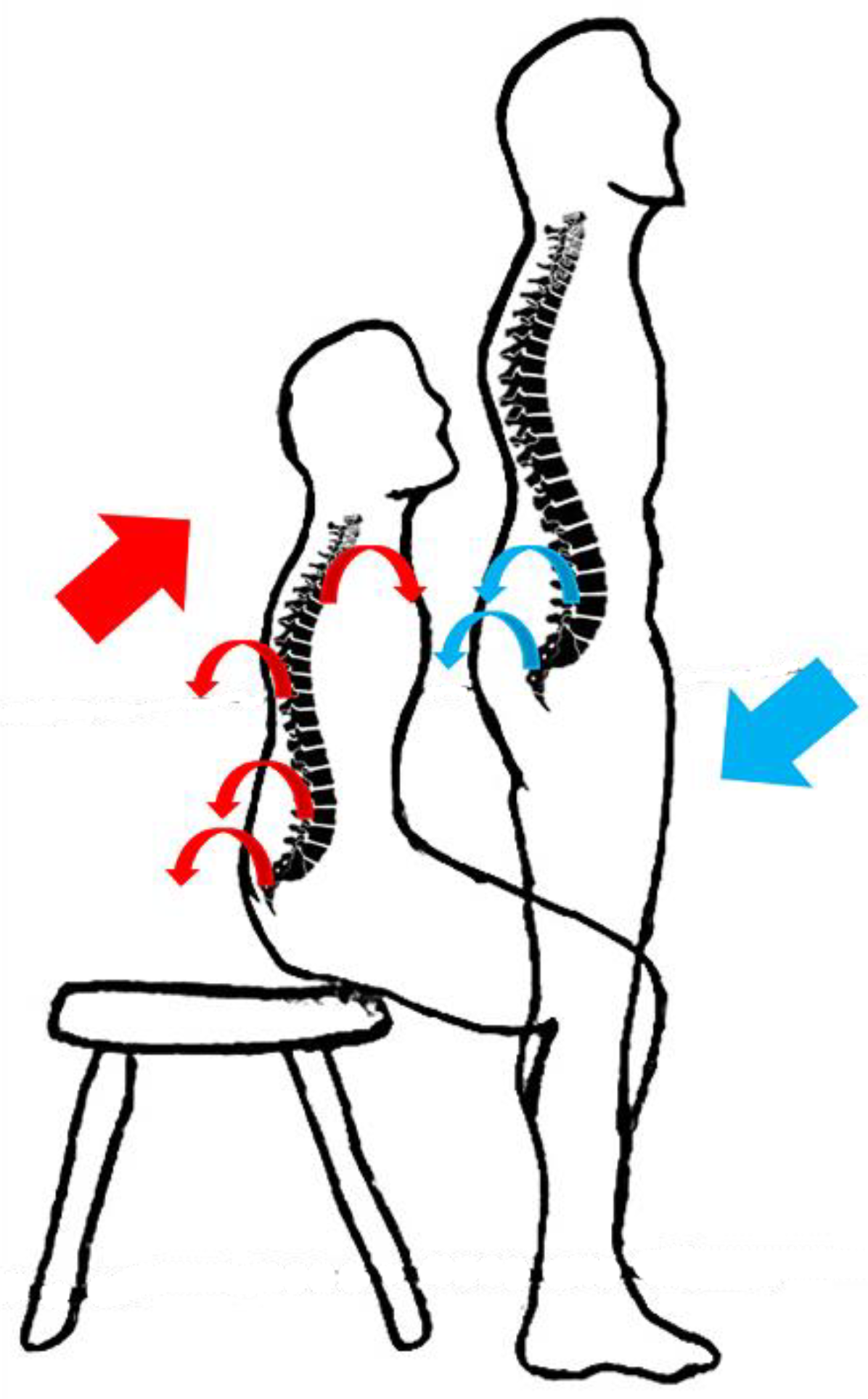
In PL/LB coordination in the sagittal plane, the findings indicated that the pattern of in-phase coordination in LB extension and pelvis posterior tilt with LB dominancy was more in the healthy group during sit-to-stand and stand-to-sit. Additionally, as it can be seen in Figure 3c and Figure 4c, the rate of in-phase coordination pattern in LB flexion and pelvis anterior tilt in people with low back pain is higher compared to the healthy participants. This finding shows that, since lumbar flexion was lower in healthy people, they showed more extension in the lumbar when returning from flexion. As a result, the in-phase coordination pattern in LB extension and pelvis posterior tilt in healthy people is more frequent. This result is consistent with previous studies that indicated nonspecific CLBP patients show more frequent out-of-phase coordination patterns during sit-to-stand and stand-to-sit [21,22,50,51] (Figure 7).

4.3. Comparison of Coordination Variability between Groups

The results of the spine and pelvic CV showed there are significant differences between the two groups during sit-to-stand. The CV in LB/LT and pelvis/LB couples is more than in nonspecific CLBP groups. Previous studies reported that patients with chronic pain reduced the variability to decrease the amount of pain [27]. On the other hand, it is reported that pain can increase variability, since the patients try to find the optimal patterns to reduce the pain [52]. While a study found that coordination variability decreased during a repetitive flexion–extension task in LBP patients [22]. Another investigation reported that LBP patients try to increase CV during the reaching task [53]. Alijanpour et al. (2021) [19] presented that nonspecific CLBP can lead to decreased and increased CV in different parts of a motor task. Hence, it can be indicated that LBP-related alterations in coordination variability are based on individual-specific strategies to find optimal pain-free patterns and can be varied in different motor tasks. This is specifically the case where the results of the current study showed that the LBP group showed an enhanced CV during some phases of sit-to-stand, but no significant changes in CV during any phase of stand-to-sit were observed.
The results of the spine and pelvic CV showed no significant difference between the two groups during stand-to-sit. This finding was inconsistent with the results of studies [19,45,46], and it was in line with the results of the studies [47,54]. While variability in the human body can decrease or increase in response to disease, pain, or specific conditions [50,55,56], as shown in the SPM statistical analysis graphs for the CV in Figure 5 and Figure 6, no statistical significance was observed in this variable between the two groups.

5. Conclusions

In conclusion, low back pain can affect kinematics coordination and its variability during activities of daily living such as sit-to-stand and stand-to-sit [21,22,51,53]. The results of the present study showed that LBP can alter kinematics coordination patterns even in the upper (pain-free) parts of the spine during sit-to-stand by changing the coordination between UT and LT in a way that can lead to an increase in the loading on these parts. Additionally, people with low back pain show more coordination variability during sit-to-stand, which might be related to their effort in finding an optimal pain-free pattern. The findings of this study can have practical implications in designing a task-specific rehabilitation program for people with low back pain.

Author Contributions

Conceptualization, N.J. and E.A.; methodology, N.J., E.A., and S.K.; software, N.J.; validation, S.S.S.; formal analysis, E.A. and N.J.; investigation, N.J., E.A. and S.K.; resources, S.S.S.; data curation, E.A.; writing—original draft preparation, N.J.; writing—review and editing, N.J., E.A. and R.N.; visualization, N.J.; supervision, S.S.S. and R.N.; and project administration, N.J. and E.A. All authors have read and agreed to the published version of the manuscript.

Funding

This research received no external funding.

Institutional Review Board Statement

The study was conducted in accordance with the Declaration of Helsinki and approved by the Institutional Review Board (or Ethics Committee) of the Sports Science Research Institute of Iran (IR.SSRC.REC.1400.038).

Informed Consent Statement

Informed consent was obtained from all subjects involved in the study.

Conflicts of Interest

The authors declare no conflict of interest.

References

  1. Wiens, A.G. Preoperative anxiety in women. AORN J. 1998, 68, 74–88. [Google Scholar] [CrossRef]
  2. Wand, B.M.; O’Connell, N.E. Chronic non-specific low back pain–sub-groups or a single mechanism? BMC Musculoskelet. Disord. 2008, 9, 11. [Google Scholar] [CrossRef] [Green Version]
  3. Koes, B.W.; Van Tulder, M.; Thomas, S. Diagnosis and treatment of low back pain. BMJ 2006, 332, 1430–1434. [Google Scholar] [CrossRef] [Green Version]
  4. Krismer, M.; Van Tulder, M. Low back pain (non-specific). Best Pract. Res. Clin. Rheumatol. 2007, 21, 77–91. [Google Scholar] [CrossRef]
  5. Rohlmann, A.; Pohl, D.; Bender, A.; Graichen, F.; Dymke, J.; Schmidt, H.; Bergmann, G. Activities of everyday life with high spinal loads. PLoS ONE 2014, 9, e98510. [Google Scholar] [CrossRef]
  6. Jensen, G.M. Biomechanics of the lumbar intervertebral disk: A review. Phys. Ther. 1980, 60, 765–773. [Google Scholar] [CrossRef]
  7. Claeys, K.; Dankaerts, W.; Janssens, L.; Brumagne, S. Altered preparatory pelvic control during the sit-to-stance-to-sit movement in people with non-specific low back pain. J. Electromyogr. Kinesiol. 2012, 22, 821–828. [Google Scholar] [CrossRef]
  8. Crosbie, J.; Nascimento, D.P.; de Faria Negrão Filho, R.; Ferreira, P. Do people with recurrent back pain constrain spinal motion during seated horizontal and downward reaching? Clin. Biomech. 2013, 28, 866–872. [Google Scholar] [CrossRef]
  9. Shum, G.L.; Crosbie, J.; Lee, R.Y. Effect of low back pain on the kinematics and joint coordination of the lumbar spine and hip during sit-to-stand and stand-to-sit. Spine 2005, 30, 1998–2004. [Google Scholar] [CrossRef]
  10. Nowakowska, K.; Gzik, M.; Michnik, R.; Myśliwiec, A.; Jurkojć, J.; Suchoń, S.; Burkacki, M. The loads acting on lumbar spine during sitting down and standing up. In Innovations in Biomedical Engineering; Springer: Berlin/Heidelberg, Germany, 2017; pp. 169–176. [Google Scholar]
  11. Sedrez, J.A.; de Mesquita, P.V.; Gelain, G.M.; Candotti, C.T. Kinematic characteristics of sit-to-stand movements in patients with low back pain: A systematic review. J. Manip. Physiol. Ther. 2019, 42, 532–540. [Google Scholar] [CrossRef]
  12. Pourahmadi, M.R.; Takamjani, I.E.; Jaberzadeh, S.; Sarrafzadeh, J.; Sanjari, M.A.; Bagheri, R.; Taghipour, M. Kinematics of the spine during sit-to-stand movement using motion analysis systems: A systematic review of literature. J. Sport Rehabil. 2019, 28, 77–93. [Google Scholar] [CrossRef] [Green Version]
  13. Parkinson, S.; Campbell, A.; Dankaerts, W.; Burnett, A.; O’Sullivan, P. Upper and lower lumbar segments move differently during sit-to-stand. Man. Ther. 2013, 18, 390–394. [Google Scholar] [CrossRef] [Green Version]
  14. Kerr, K.; White, J.; Barr, D.; Mollan, R. Analysis of the sit-stand-sit movement cycle in normal subjects. Clin. Biomech. 1997, 12, 236–245. [Google Scholar] [CrossRef]
  15. Panjabi, M.M. The stabilizing system of the spine. Part I. Function, dysfunction, adaptation, and enhancement. J. Spinal Disord. 1992, 5, 383. [Google Scholar] [CrossRef]
  16. Hodges, P.W.; Richardson, C.A. Contraction of the abdominal muscles associated with movement of the lower limb. Phys. Ther. 1997, 77, 132–142. [Google Scholar] [CrossRef] [Green Version]
  17. van Dieën, J.H.; Selen, L.P.; Cholewicki, J. Trunk muscle activation in low-back pain patients, an analysis of the literature. J. Electromyogr. Kinesiol. 2003, 13, 333–351. [Google Scholar] [CrossRef]
  18. Wong, T.K.; Lee, R.Y. Effects of low back pain on the relationship between the movements of the lumbar spine and hip. Hum. Mov. Sci. 2004, 23, 21–34. [Google Scholar] [CrossRef]
  19. Alijanpour, E.; Abbasi, A.; Needham, R.A.; Naemi, R. Spine and pelvis coordination variability in rowers with and without chronic low back pain during rowing. J. Biomech. 2021, 120, 110356. [Google Scholar] [CrossRef]
  20. Müller, R.; Ertelt, T.; Blickhan, R. Low back pain affects trunk as well as lower limb movements during walking and running. J. Biomech. 2015, 48, 1009–1014. [Google Scholar] [CrossRef]
  21. Seay, J.F.; Van Emmerik, R.E.; Hamill, J. Influence of low back pain status on pelvis-trunk coordination during walking and running. Spine 2011, 36, E1070–E1079. [Google Scholar] [CrossRef]
  22. Mokhtarinia, H.R.; Sanjari, M.A.; Chehrehrazi, M.; Kahrizi, S.; Parnianpour, M. Trunk coordination in healthy and chronic nonspecific low back pain subjects during repetitive flexion–extension tasks: Effects of movement asymmetry, velocity and load. Hum. Mov. Sci. 2016, 45, 182–192. [Google Scholar] [CrossRef]
  23. Hamill, J.; van Emmerik, R.E.; Heiderscheit, B.C.; Li, L. A dynamical systems approach to lower extremity running injuries. Clin. Biomech. 1999, 14, 297–308. [Google Scholar] [CrossRef]
  24. Needham, R.A.; Naemi, R.; Hamill, J.; Chockalingam, N. Analysing patterns of coordination and patterns of control using novel data visualisation techniques in vector coding. Foot 2020, 44, 101678. [Google Scholar] [CrossRef]
  25. Mehdizadeh, S.; Arshi, A.R.; Davids, K. Quantifying coordination and coordination variability in backward versus forward running: Implications for control of motion. Gait Posture 2015, 42, 172–177. [Google Scholar] [CrossRef]
  26. Heiderscheit, B.C.; Hamill, J.; van Emmerik, R.E. Variability of stride characteristics and joint coordination among individuals with unilateral patellofemoral pain. J. Appl. Biomech. 2002, 18, 110–121. [Google Scholar] [CrossRef]
  27. Potvin-Gilbert, M. Motor Variability in Rowing. Master’s Thesis, The University of Western Ontario, London, ON, Canada, 2018. [Google Scholar]
  28. Christe, G.; Redhead, L.; Legrand, T.; Jolles, B.M.; Favre, J. Multi-segment analysis of spinal kinematics during sit-to-stand in patients with chronic low back pain. J. Biomech. 2016, 49, 2060–2067. [Google Scholar] [CrossRef]
  29. Coghlin, S.; McFadyen, B. Transfer strategies used to rise from a chair in normal and low back pain subjects. Clin. Biomech. 1994, 9, 85–92. [Google Scholar] [CrossRef]
  30. Needham, R.; Naemi, R.; Healy, A.; Chockalingam, N. Multi-segment kinematic model to assess three-dimensional movement of the spine and back during gait. Prosthet. Orthot. Int. 2016, 40, 624–635. [Google Scholar] [CrossRef]
  31. Whitney, S.L.; Wrisley, D.M.; Marchetti, G.F.; Gee, M.A.; Redfern, M.S.; Furman, J.M. Clinical measurement of sit-to-stand performance in people with balance disorders: Validity of data for the Five-Times-Sit-to-Stand Test. Phys. Ther. 2005, 85, 1034–1045. [Google Scholar] [CrossRef] [Green Version]
  32. Needham, R.; Naemi, R.; Chockalingam, N. Quantifying lumbar–pelvis coordination during gait using a modified vector coding technique. J. Biomech. 2014, 47, 1020–1026. [Google Scholar] [CrossRef]
  33. Needham, R.A.; Naemi, R.; Chockalingam, N. A new coordination pattern classification to assess gait kinematics when utilising a modified vector coding technique. J. Biomech. 2015, 48, 3506–3511. [Google Scholar] [CrossRef]
  34. Huskisson, E.C. Measurement of pain. Lancet 1974, 304, 1127–1131. [Google Scholar] [CrossRef]
  35. Hawker, G.A.; Mian, S.; Kendzerska, T.; French, M. Measures of adult pain: Visual analog scale for pain (vas pain), numeric rating scale for pain (nrs pain), mcgill pain questionnaire (mpq), short-form mcgill pain questionnaire (sf-mpq), chronic pain grade scale (cpgs), short form-36 bodily pain scale (sf-36 bps), and measure of intermittent and constant osteoarthritis pain (icoap). Arthritis Care Res. 2011, 63, S240–S252. [Google Scholar]
  36. Koumantakis, G.A.; Watson, P.J.; Oldham, J.A. Trunk muscle stabilization training plus general exercise versus general exercise only: Randomized controlled trial of patients with recurrent low back pain. Phys. Ther. 2005, 85, 209–225. [Google Scholar] [CrossRef] [Green Version]
  37. Smeets, R.; Köke, A.; Lin, C.W.; Ferreira, M.; Demoulin, C. Measures of function in low back pain/disorders: Low back pain rating scale (LBPRS), oswestry disability index (ODI), progressive isoinertial lifting evaluation (PILE), quebec back pain disability scale (QBPDS), and roland-morris disability questionnaire (RDQ). Arthritis Care Res. 2011, 63, S158–S173. [Google Scholar]
  38. Fairbank, J.; Couper, J.; Davies, J.B.; O’Brien, J.P. The Oswestry low back pain disability questionnaire. Physiotherapy 1980, 66, 271–273. [Google Scholar]
  39. Svendsen, J.H.; Svarrer, H.; Laessoe, U.; Vollenbroek-Hutten, M.; Madeleine, P. Standardized activities of daily living in presence of sub-acute low-back pain: A pilot study. J. Electromyogr. Kinesiol. 2013, 23, 159–165. [Google Scholar] [CrossRef]
  40. Peydro, M.; López, J.; Cortés, A.; Vivas, M.; Garrido, J.; Tortosa, L. Análisis cinético y cinemático del gesto «levantarse de una silla» en pacientes con lumbalgias. Rehabilitación 2011, 45, 99–105. [Google Scholar] [CrossRef]
  41. Sánchez-Zuriaga, D.; López-Pascual, J.; Garrido-Jaén, D.; de Moya, M.F.P.; Prat-Pastor, J. Reliability and validity of a new objective tool for low back pain functional assessment. Spine 2011, 36, 1279–1288. [Google Scholar] [CrossRef]
  42. Sung, P.S. A kinematic analysis for shoulder and pelvis coordination during axial trunk rotation in subjects with and without recurrent low back pain. Gait Posture 2014, 40, 493–498. [Google Scholar] [CrossRef]
  43. Hodges, P.W.; Smeets, R.J. Interaction between pain, movement, and physical activity: Short-term benefits, long-term consequences, and targets for treatment. Clin. J. Pain 2015, 31, 97–107. [Google Scholar] [CrossRef] [PubMed]
  44. O’Sullivan, P. It’s Time for Change with the Management of Non-Specific Chronic Low Back Pain; British Association of Sport and Exercise Medicine: Doncaster, UK, 2012; Volume 46, pp. 224–227. [Google Scholar]
  45. Kim, T.; Chai, E. Trunk and pelvic coordination at various walking speeds during an anterior load carriage task in subjects with and without chronic low back pain. J. Phys. Ther. Sci. 2015, 27, 2353–2356. [Google Scholar] [CrossRef] [PubMed]
  46. Ebrahimi, S.; Kamali, F.; Razeghi, M.; Haghpanah, S.A. Comparison of the trunk-pelvis and lower extremities sagittal plane inter-segmental coordination and variability during walking in persons with and without chronic low back pain. Hum. Mov. Sci. 2017, 52, 55–66. [Google Scholar] [CrossRef] [PubMed]
  47. Crosbie, J.; de Faria Negrão Filho, R.; Nascimento, D.P.; Ferreira, P. Coordination of spinal motion in the transverse and frontal planes during walking in people with and without recurrent low back pain. Spine 2013, 38, E286–E292. [Google Scholar] [CrossRef]
  48. Vereijken, B.; Emmerik, R.E.v.; Whiting, H.; Newell, K.M. Free(z)ing degrees of freedom in skill acquisition. J. Mot. Behav. 1992, 24, 133–142. [Google Scholar] [CrossRef]
  49. Shafizadeh, M. Movement coordination during sit-to-stand in low back pain people. Hum. Mov. 2016, 17, 107–111. [Google Scholar] [CrossRef] [Green Version]
  50. Lamoth, C.J.; Daffertshofer, A.; Meijer, O.G.; Beek, P.J. How do persons with chronic low back pain speed up and slow down?: Trunk–pelvis coordination and lumbar erector spinae activity during gait. Gait Posture 2006, 23, 230–239. [Google Scholar] [CrossRef]
  51. Ippersiel, P.; Robbins, S.; Preuss, R. Movement variability in adults with low back pain during sit-to-stand-to-sit. Clin. Biomech. 2018, 58, 90–95. [Google Scholar] [CrossRef]
  52. Jewell, C.; Weir, G.; Hamill, J.; Boyer, K.A. The influence of patellofemoral pain on coordination variability over a prolonged treadmill run. ISBS Proc. Arch. 2018, 36, 634. [Google Scholar]
  53. Silfies, S.P.; Bhattacharya, A.; Biely, S.; Smith, S.S.; Giszter, S. Trunk control during standing reach: A dynamical system analysis of movement strategies in patients with mechanical low back pain. Gait Posture 2009, 29, 370–376. [Google Scholar] [CrossRef] [Green Version]
  54. Gombatto, S.P.; Brock, T.; DeLork, A.; Jones, G.; Madden, E.; Rinere, C. Lumbar spine kinematics during walking in people with and people without low back pain. Gait Posture 2015, 42, 539–544. [Google Scholar] [CrossRef] [PubMed]
  55. Madeleine, P.; Mathiassen, S.E.; Arendt-Nielsen, L. Changes in the degree of motor variability associated with experimental and chronic neck–shoulder pain during a standardised repetitive arm movement. Exp. Brain Res. 2008, 185, 689–698. [Google Scholar] [CrossRef] [PubMed]
  56. Georgoulis, A.D.; Moraiti, C.; Ristanis, S.; Stergiou, N. A novel approach to measure variability in the anterior cruciate ligament deficient knee during walking: The use of the approximate entropy in orthopaedics. J. Clin. Monit. Comput. 2006, 20, 11–18. [Google Scholar] [CrossRef] [PubMed]
Figure 1. Capture volume and cameras (Applsci 12 11796 i001) placement (left) and marker placement (right).
Applsci 12 11796 g001
Figure 2. UT, LT, LB, and pelvis kinematics during repetitive sit-to-stand and stand-to-sit calculated using the method described by Needham et al. (2014) [32].
Applsci 12 11796 g002
Figure 3. (a) LT/UT, (b) LB/LT, and (c) pelvis/LB segments angular displacement diagram in the sagittal plane, and the results of the coupling angle frequency during sit-to-stand.
Applsci 12 11796 g003
Figure 4. (a) LT/UT, (b) LB/LT, and (c) pelvis/LB segments angular displacement diagram in the sagittal plane, and the results of the coupling angle frequency during stand-to-sit.
Applsci 12 11796 g004
Figure 5. (a) LT/UT, (b) LB/LT, and (c) pelvis/LB coordination variability (CV) in the sagittal plane, and the results of an independent t-test using the SPM method during sit-to-stand, left side is the mean and standard diviation of CV and right side is the results of SPM analysis. Red dotted line in the SPM results in the critical threshold (t value).
Applsci 12 11796 g005
Figure 6. (a) LT/UT, (b) LB/LT, and (c) Pelvis/LB coordination variability (CV) in the sagittal plane, and the results of an independent t-test using the SPM method during stand-to-sit, left side is the mean and standard diviation of CV and right side is the results of SPM analysis. Red dotted line in the SPM results in the critical threshold (t value).
Applsci 12 11796 g006
Figure 7. Coordination patterns in CLBP subjects, where red arrows show the segmental movement during sit-to-stand, and the blue arrows show those during stand-to-sit.
Applsci 12 11796 g007
Table 1. The demographic information of the participants.
Group 1. HealthyGroup 2. CLBP
Simple size1010
Gender (Female/Male)5/55/5
Mean age ± SD (Year)29 ± 4.5032 ± 4.90
Mean height ± SD (cm)171.5 ± 9.72169.25 ± 4.1
Mean body weight ± SD (Kg)70.83 ± 14.6071.87 ± 13.20
Mean VAS ± SD-5.6 ± 1.17
Mean Oswestry Disability Index ± SD-20.8 ± 1.9
Table 2. The results of ROM data in degrees (Ave ± SD).
Upper TrunkLower TrunkLower BackPelvis
Sit-to-standStand-to-sitSit-to-standStand-to-sitSit-to-standStand-to-sitSit-to-standStand-to-sit
Healthy group45.82 ± 15.7141.60 ± 10.7850.16 ± 22.0944.82 ± 16.3646.48 ± 13.6448.01 ± 16.9832.17 ± 9.3836.28 ± 8.21
CLBP group39.45 ± 10.9539.88 ± 12.1542.06 ± 10.8441.42 ± 10.8343.24 ± 11.4343.59 ± 12.4125.37 ± 6.6433.38 ± 8.69
p Value0.4950.8310.4920.7180.7090.6640.2100.626
Publisher’s Note: MDPI stays neutral with regard to jurisdictional claims in published maps and institutional affiliations.