Article

Langmuir Trough Study of the Interactions of Tear Mimetic Eyedrop Formulation with Human Meibum Films

1
Department of Cell and Developmental Biology, Faculty of Biology, University of Sofia “St. Kliment Ohridski”, 1164 Sofia, Bulgaria
2
Department of Ophthalmology, Kyoto Prefectural University of Medicine, Kyoto 602-8566, Japan
3
Research and Development Department, Rohto Pharmaceutical Co., Ltd., Osaka 544-0012, Japan
4
Faculty of Physics, University of Sofia “St. Kliment Ohridski”, 1164 Sofia, Bulgaria
*
Author to whom correspondence should be addressed.
Appl. Sci. 2022, 12(23), 12095; https://doi.org/10.3390/app122312095
Submission received: 2 November 2022 / Revised: 18 November 2022 / Accepted: 22 November 2022 / Published: 26 November 2022
(This article belongs to the Section Surface Sciences and Technology)

Abstract

:
Meibomian gland disease is associated with quantitative or qualitative deficiencies of meibum (MGS) that result in tear film instability. Thus, there is great demand for ophthalmic nanoemulsions that can replenish MGS and recover its performance at the air/tear surface. Rohto Dry Aid (RDA) utilizes TEARSHIELD TECHNOLOGYTM implementing a complex oil phase of non-polar and polar lipid-like molecules. Therefore, the interactions of RDA with MGS surface films deserve further study as they may provide valuable insights (i) into the mechanisms behind the nanoemulsion therapeutic action and (ii) for the design of novel ophthalmic formulations. Pseudobinary meibum/RDA films were formed at the air–water surface of the Langmuir trough. Surface pressure-area isocycles and stress relaxations were employed to probe the layer (i) reorganization upon cycling and (ii) dilatational elasticity, respectively. Film morphology was accessed by Brewster angle microscopy and the spreading properties of RDA-supplemented meibum were also probed. The diverse ingredients of the nanoemulsion oil phase complemented the non-polar and polar lipid constituents of the meibomian layers, which resulted in enhanced continuity of the MGS duplex film structure and facilitated the MGS spread and viscoelasticity. Nanoemulsions deserve further study as a potent tool for MGS-oriented therapy for dry eyes.

1. Introduction

The air–aqueous tear (AT) surface of the human eye is stabilized by the tear film lipid layer (TFLL), a ~100 nm thick viscoelastic duplex film [1,2,3,4] build of a monolayer of amphiphilic polar lipids (PL) located at the AT interface and a non-polar lipid (NPL) stratum layered on top and extended towards the air [5,6,7]. The TFLL is composed predominantly of the lipids of the meibomian gland secretion (MGS, or meibum), a composite oily mixture consisting of >90% NPL (mostly wax (WE) and cholesteryl esters (CE), and triglycerides) and <10% PL (mainly (O-acyl)-ω-hydroxy fatty acids) [3,4].
Meibomian gland disease (MGD), manifested in the quantitative or qualitative abnormalities of MGS and TFLL, affects up to 86% of all patients with dry eye syndrome (DES) and is widely considered the most common cause of DES [8,9]. This fact, together with the extended (on the scale of hours) residence time of drug molecules incorporated in TFLL, i.e., a promise for a more durable pharmaceutical action at the ocular surface [10], defines a pressing need for the development of eyedrop formulations that interact favorably with TFLL. One such novel composition is Rohto Dry Aid (RDA), a formulation that utilizes TEARSHIELD TECHNOLOGYTM to mimic real tears and restore moisture. The oil phase of the formulation consists of sesame oil (SO) as a mimic of the meibomian NPL, while the PL are emulated via a mixture of Polyoxyethylene Castor Oil 10 (CO-10), Polyoxyl 40 stearate (MYS-40) and Menthol. Sesame oil with its diverse fatty acid composition of 40% monounsaturated (oleic acid), 42% polyunsaturated (linoleic acid) and saturated fats (mainly ∼8% palmitic acid and 4% stearic acid) was recently demonstrated to adequately complement the non-polar lipid stratum of SO supplemented human MGS films [11].
Therefore, the interactions between RDA and meibomian lipids at the tear-like air–water interface warrant further investigation as it may yield valuable knowledge for the mechanism of action of RDA at the ocular surface. In order to study these interactions pseudobinary meibum/RDA films with controlled RDA content were formed at the air–water surface of the Langmuir trough. Langmuir surface balance measurements have been established as an efficient methodology for the evaluation of MGS films interfacial properties at physiologically relevant blink-like area changes [2] and were successfully implemented in the design and characterization of ophthalmic formulations [12,13,14,15]. The layers (i) reorganization throughout area cycling and (ii) dilatational elasticity were probed via surface pressure (π)-area (A) isocycles and stress relaxations, respectively. Brewster angle microscopy (BAM) was used to visualize film morphology at the different MGS/RDA ratios. Furthermore, the impact of the RDA oil phase on the spreading of meibum was characterized by using talcum powder as a tracer [16]. The impact of MGS supplementation with menthol, a specific ingredient of RDA, was also probed with Langmuir surface balance and BAM studies. The attention to menthol was determined by the lack of published information about the impact of such low molecular weight volatile amphiphilic molecules on MGS, while there are already multiple studies of the effects of plant oils and pharmaceutical emulsifiers on the properties of meibomian layers [11,12,13,14,15].

2. Materials and Methods

Surface pressure/area (π-A) isotherms were measured [5] by Langmuir surface balance µ Trough XS, area 135 cm2, volume 100 mL (Kibron, Helsinki, Finland), via the Wilhelmy wire probe method (instrumental accuracy 0.01 mN/m). Phosphate-buffered saline (PBS, pH 7.4) was utilized as a trough subphase. Human meibum from healthy volunteers was collected by the soft squeeze method [1,2] with permission from the institutional review board of Kyoto Prefectural University of Medicine, in agreement with the tenets of the Declaration of Helsinki and after the donors signed informed consent. The sample used was a pooled equiweight mixture of the MGS of four volunteers: two males (40 years old and 27 years old) and two females (41 years old and 28 years old). Until its use in experiments, the MGS solution (1 mg MGS/mL CHCl3) was stored at −80 °C. The mean molecular weight Mw = 700 (an average of 650 for wax esters and 750 for cholesteryl esters and (O-acyl)-omega-hydroxy fatty acids) was assumed for MGS [4].
Rohto Dry Aid ophthalmic nanoemulsion (Table 1) and menthol were provided by Rohto Pharmaceutical, Osaka, Japan.
An acrylic cover was fitted over the apparatus to protect the surface from dust and to suppress the evaporation of the PBS subphase (under the cover >90% relative humidity is maintained). A Hamilton micro-syringe was used to uniformly deposit human MGS at the air/water surface of the trough. After 15 min were provided for chloroform evaporation, dynamic compression–expansion isocycling of the film area was initiated by two symmetrically moving barriers at the maximum barrier’s rate (70 mm/min), at which there was no leakage of the film. Five consecutive cycles were performed with each film studied. The π(A) curves attained constant shape after the third cycle and those “stationary” π(A) isotherms were analyzed further. All isotherms were repeated in triplicate with less than a 2% difference between the repetitions. The experiments were carried out at 35 °C, i.e., the physiological temperature of the ocular surface. The morphology of the films was monitored by a Brewster angle microscope (MC-BAM, Imperx, Boca Raton, FL, USA).
The MGS film dilatational viscoelasticity was accessed by analysis of the surface pressure relaxation transients after a small rapid compression step was exerted on the layers [17,18,19]. In these experiments, after the film was equilibrated at initial surface pressure, π0 = 20 mN/m, a small compression step, ∆A/Ao = 5% ± 1% (where Ao—initial film area, ∆A—area change) was applied to it. The ∆π relaxation transients were collected and analyzed further.
For visualization of MGS spread, alone and in the presence of RDA oil phase (obtained via chloroform extraction from RDA), the air–water surface of the trough surface is covered with talc particles and the rate and degree of spread are visualized by the displacement of the talc particles [16].

3. Results

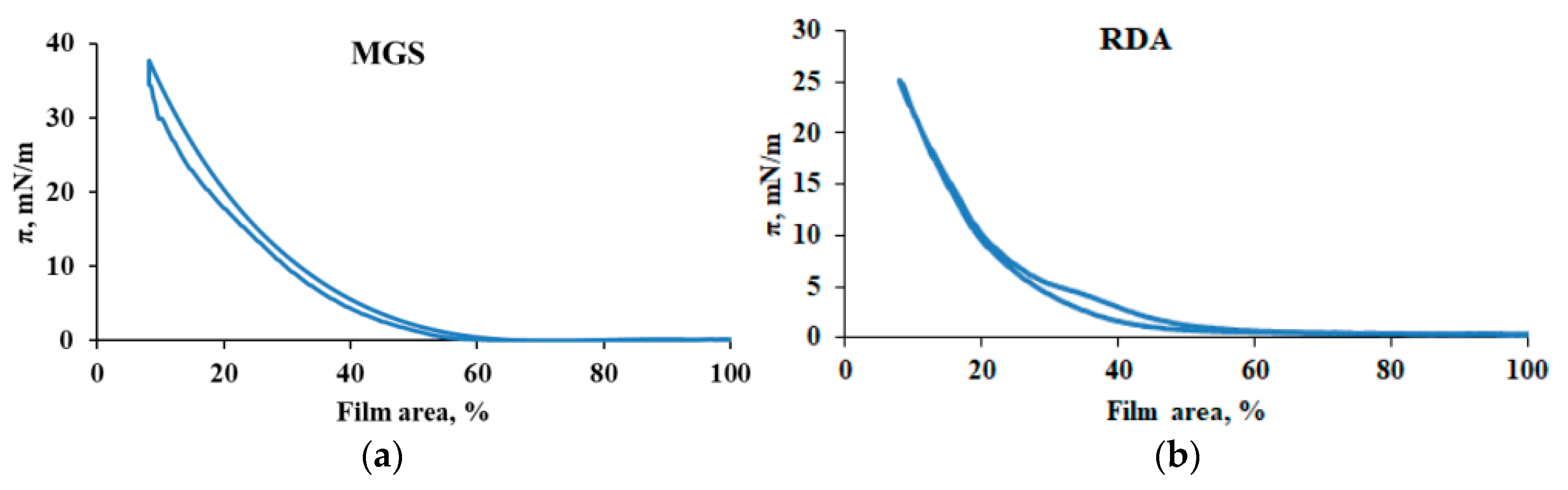
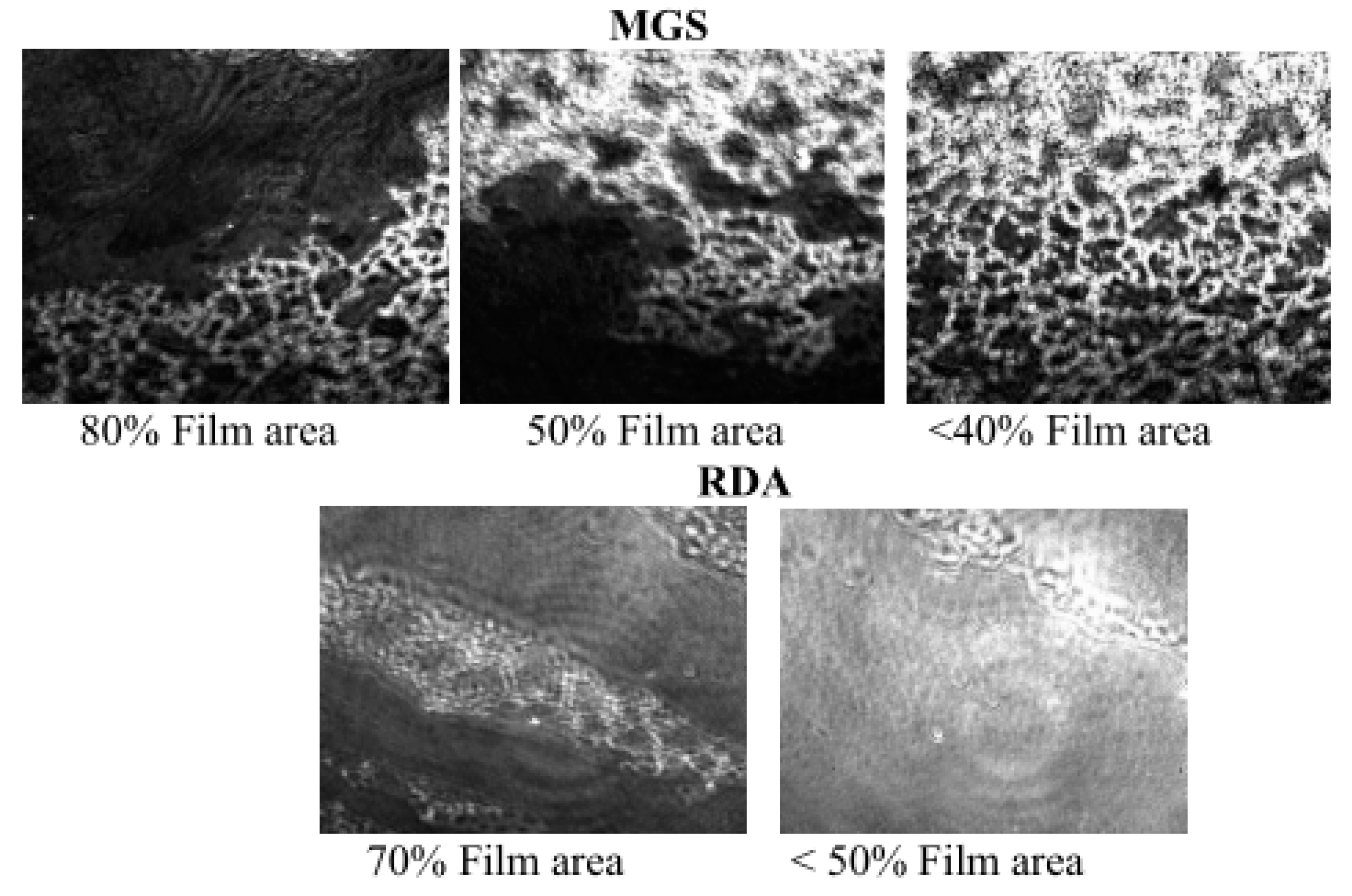
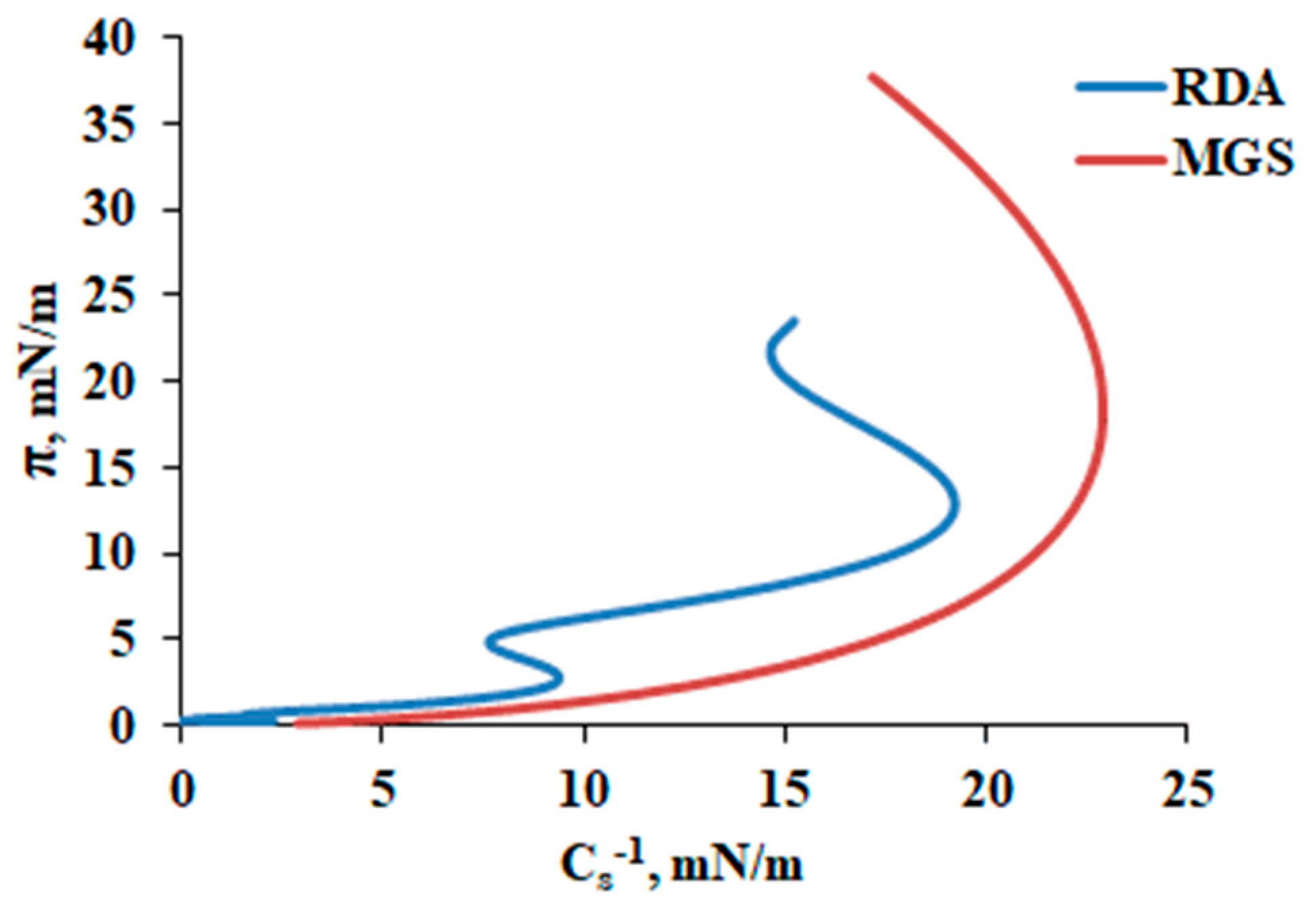
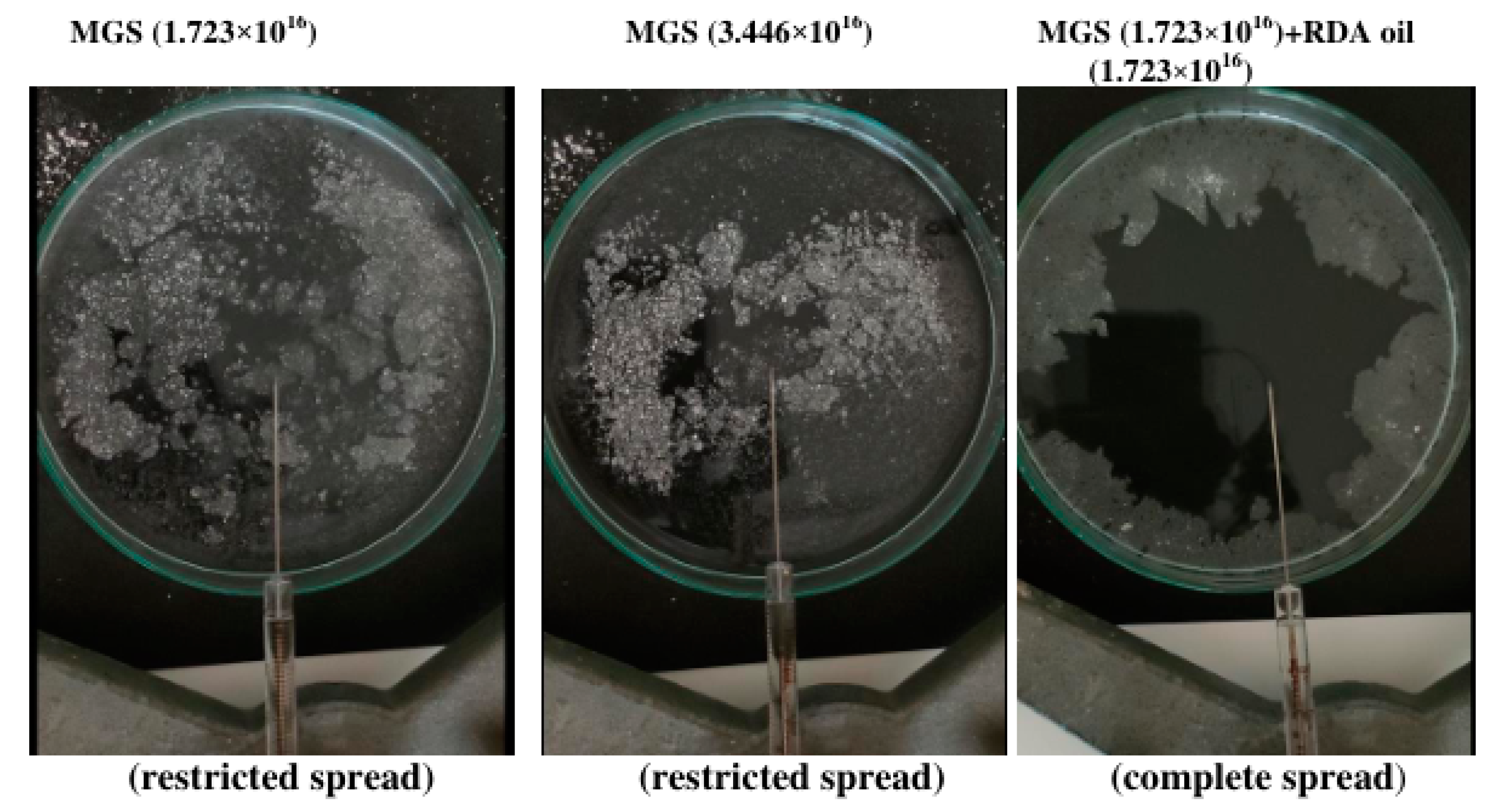
3.1. MGS and RDA Films at the Air/Water Interface

Figure 1 shows the surface pressure/area isocycles of MGS (left panel) and RDA (right panel). MGS displayed non-collapsibility upon compression and almost no hysteresis loop as reported elsewhere [1,2].
RDA had similar behavior to MGS in line with prior data on the surface properties of sesame oil and castor oil containing ophthalmic nanoemulsions [13]. As previously demonstrated [1,2], Brewster angle microscopy (Figure 2) revealed that at low π (≤15 mN/m) MGS formed heterogeneous films which consisted of dark (monolayer thin) regions and multilayer thick aggregates (bright areas).
Upon further compression, the thick darker regions connected to a continuous multilayer mesh while the presence of the thin brighter monolayer regions decreased, and the monolayer patches almost disappeared at very low film areas. RDA films were more homogeneous and with increasing brightness (i.e., thickness) upon compression as typical for the behavior of layers formed by surface deposition of oil-in-water emulsions [13,20].
The in-plane reciprocal compressibility (or rigidity) modulus Cs−1 (Figure 3) of the films was calculated via Equation (1).
Cs−1 = Aπ (−dπ/dA)
where Aπ is the area at the indicated π. The inflexion points in the π/Cs−1 dependencies indicate π at which significant changes in the surface film structure occur upon compression. Davies and Rideal [21] suggested that Cs−1 < 50 mN/m, 100 < Cs−1 < 250 mN/m and Cs−1 > 250 mN/m correspond to liquid expanded, liquid condensed, and solid states of the surface layer, respectively. Both RDA and MGS had Cs−1 < 30 mN/m which accords to liquid expanded molecular packing density at the film/water interface.
The inflection at 15 mN/m of the π/Cs−1 curve of MGS reflected the enclosure of the thinner regions within the MGS films and the transition to exclusively duplex architecture of the film [1,2]. The π/Cs−1 curve of RDA showed more complex behavior with inflexions at 3 mN/m and 14 mN/m which probably corresponds to the redistribution of the multiple polar (Povidone, Polyoxyethylene Castor Oil 10, Polyoxyl 40 Stearate, menthol) and non-polar constituents (SO) of the formulations between the aqueous subphase and the headgroup and acyl chain regions of the layers. Similar behavior of the in-plane reciprocal compressibility is also observed for other oil-enriched ophthalmic nanoemulsions [13,20].

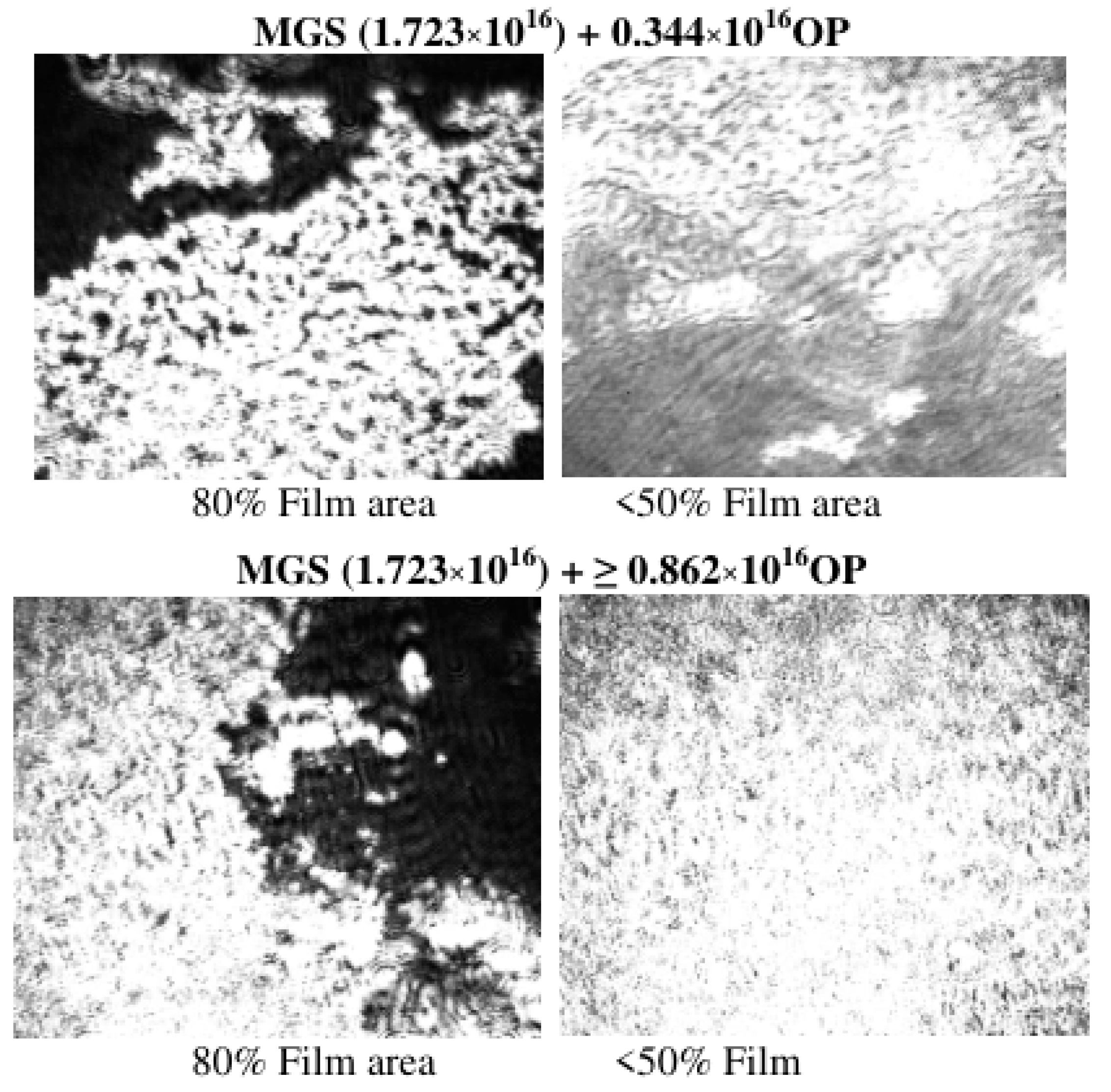
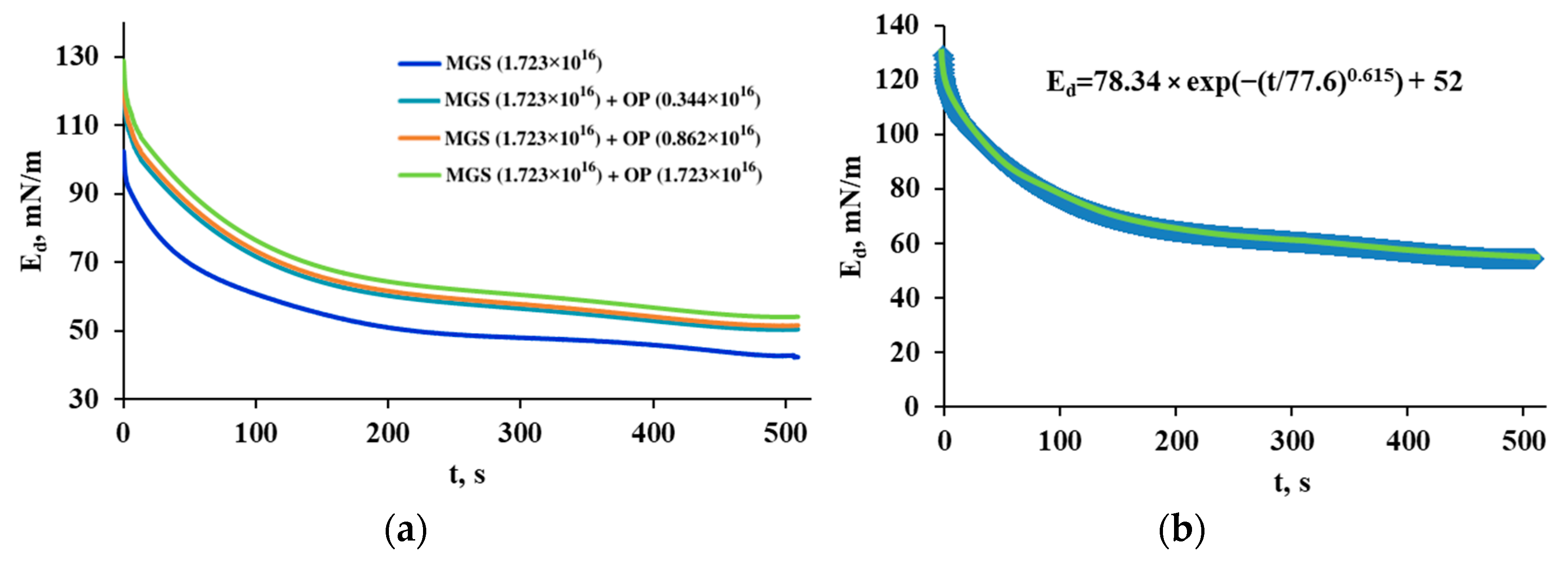
3.2. Pseudobinary MGS/RDA Films with Fixed MGS Amount

In this set of experiments, a fixed amount of MGS was spread at the air–water surface of the Langmuir trough and RDA was added on top of it. Therefore, the supplementation of RDA resulted in an increased amount of total lipid at the air–water surface. This experimental regime emulates the in vivo clinical administration of RDA, where the formulation will be delivered to a fixed amount of TFLL thus resulting in raised total lipid concentration at the ocular surface.
The delivery of RDA represented via the number of oil phase (OP) molecules resulted (Figure 4) in (i) an increase in π achieved at the completion of the surface films compression and (ii) an overall shift of the π(A)-isotherms to higher π values.
These effects are standard responses for surface layers when supplemented with extra surfactant quantities [13]. Brewster angle microscopy showed (Figure 5) that the delivery of RDA to MGS films resulted in overall thickening (raised brightness of the BAM images) of the films and disappearance of the monolayer regions in the layers upon supplementation with RDA at ≤50% of the film area.
It is thought that the stability of TFLL (and of the TF as a whole) at consecutive blinks is influenced by the lipid layer viscoelasticity [1,2,6,22]. Hereof, the dilatational rheology of the films was examined via the step deformation technique allowing us to calculate the transient elasticity modulus Ed(t) from the surface pressure relaxation transients:
Ed(t) = −Ao(dπ(t)/dA)
where Ao is the initial film area and ∆A is the area change.
Figure 6 summarizes the dependences of the transient elasticity modulus on time which were analyzed via the Kohlrausch–Williams–Watts (KWW) equation (Equation (3)).
Ed(t) = E. exp (−(t/τ)β) + EEQ
For microscopically heterogeneous systems the fractional exponent β ≤ 1 (so-called stretched exponential) is a material characteristic that decreases further away from unity with increasing (i) the strength of interaction amongst the units and/or (ii) the film heterogeneity (i.e., augmented amount of interacting arrays) [23]. Physical interpretation of the values of β is provided by the spatial rearrangement model [24,25]: 3/4—self-avoiding walks in 2D; 3/5—self-avoiding walks in 3D; 1/2—self-avoiding walks in 4D, Brownian motion, diffusion-limited growth/segregation; 1/3—anomalous diffusion on percolating clusters, growth of ensemble of clusters—Ostwald ripening, spinodal decomposition. Values of β ≥ 1 (compressed exponential) are also found in the “jammed” state of the soft matter where no fast relaxation channel exists [23]. EEQ corresponds to the equilibrium elasticity after the completion of the relaxation.
It can be seen (Table 2) that for MGS layers β = 0.43. For MGS layers supplied with RDA, the value of β grew and remained in the range of 0.615–0.63 for a broad range of compositions.
The result aligns with recent findings that interactions with ophthalmic nanoemulsions can modify the order and the phase transition of the meibomian layers in a nonlinear fashion [11,13]. Furthermore, the increase in β is an indication of increased homogeneity of the surface layers in presence of RDA [23].
The analysis of the spread kinetics (Figure 7 and Supplementary Materials) of MGS alone or supplemented with the oil phase of RDA reveals that the supplementation of MGS with RDA oil phase (at equimolar ratio) resulted in a much faster and more complete spread of the surface films as compared to the case when the MGS amount is elevated to the same number of molecules.

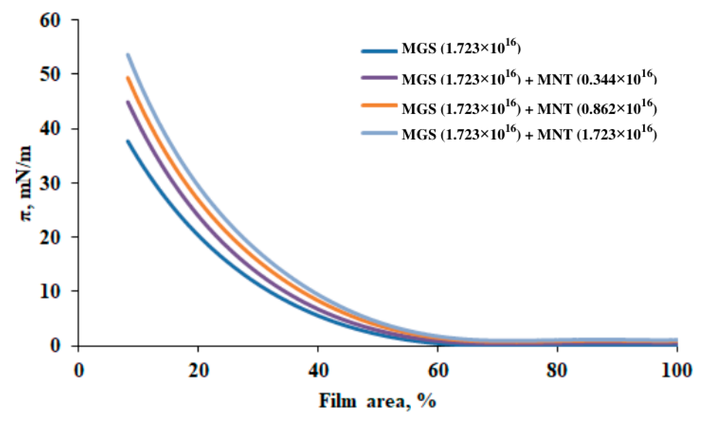
3.3. Pseudobinary MGS/Menthol Films with Fixed MGS Amount

Similarly to the experiments in the previous section, in order to mimic the impact of the supplementation of pharmaceuticals to the TFLL, a constant amount of MGS is spread at the air–water surface of the Langmuir trough and menthol (MNT) is supplemented on top of it. Thus, the addition of menthol leads to elevated total lipid amount at the air–water interface.
The supplementation of MGS with MNT (Figure 8) resulted in an increase in the surface pressure achieved at maximum compression of the surface films.
Brewster angle microscopy (Figure 9) showed that the inclusion of MNT increased the homogeneity of the surface layers although, in contrast to the effects of RDA, there was no uniform increase in the film brightness.
The stress relaxation transients (Figure 10 and Table 3) revealed that in the presence of MNT the viscoelastic properties of the MGS layers are maintained and enhanced at the inclusion of ≥0.862 × 10¹⁶ molecules of MNT.

4. Discussion

As shown by Brewster Angle Microscopy, here and elsewhere, MGS does not spread to a monolayer even at (very) low π (0.5–10 mN/m) due to the very high hydrophobicity of the meibomian lipids. Instead, rough heterogeneous MGS layers are formed at the air–water interface which turn more uniform with the raise in π and preserve liquid extended-like in-plane rigidity for the entire π range [1,2]. This is similar to the behavior of meibum-like mixtures of triglycerides and cholesterol esters, which also form non-collapsible and reversible multilayers at the air–water interface [26,27]. An inherent property of such hydrophobic lipids rich thick layers is that upon area compression the polar lipid molecules (positioned at the interface with the aqueous subphase), escape compression to condensed and collapsed states, and instead move to the upper NPL stratum of the layer [1,2,26]. During the subsequent area expansion, this upper stratum of the film serves as a reservoir from which polar lipids rapidly return back to the surface, thus rendering the stability of the layers at consecutive blink-like surface compressions and expansions. The so-described duplex film structure, i.e., a polar lipid monolayer with a continuous oily fluid (with some crystallites dispersed within) on top of it [6], ensures the non-collapsibility and the resistance to the loss of material which are characteristic of MGS layers upon continuous and consecutive blinks. These unique features offer plausible insights into how TFLL is able to maintain its composition and structure in vivo at the tear surface for hours, as manifested by the TFLL turnover rate of 0.93 ± 0.36%/min (i.e., much slower than the AT turnover of 10.3 ± 3.7%/min) thus minimizing the necessity for the meibomian glands to deliver new lipids to the TFLL [10].
RDA films displayed behavior typical for other oil-in-water ophthalmic nanoemulsions with similar compositions [27,28]. The nanoemulsion forms highly reversible noncollapsible surface films. As already shown, SO can supplement the nonpolar lipids of MGS layers and lead to the formation of a more uniform and ordered NPL stratum [11]. It is worth discussing the possible impact of the eyedrop surface-active constituents (CO-10, MYS-40, Poloxamer 407 and menthol) on the interactions of RDA with MGS films and how they facilitate the interplay of the formulation with the meibum layers as compared to pure SO. In a recent study [29] it was found that surfactants with high hydrophilic–lipophilic balance, such as MYS-40, incorporate in the TFLL at the aqueous interface, and can compensate for the moderate deficiency of polar lipids. Thus, it enhances the spreading and distribution of the RDA oil phase (and overall of the non-polar lipid stratum of the layers) in the pseudo-binary MGS/RDA films. The combination of high and low HLB surfactants (CO-10 and MYS-40, respectively) ensures optimal miscibility of the formulation with TFLL [13,29]. Poloxamer 407 is reported to penetrate in lipid films, but due to its hydrophilic polyoxyethylene chain, it has a high affinity to water and readily gets squeezed out in the aqueous subphase at π ≤ 20 mN/m in experiments with phospholipid monolayers [30] and also in control experiments with MGS layers. The interaction of menthol with lipid films revealed that menthol could supplement the PL-coated MGS/aqueous interface. The interaction enhances the spread of MGS layers and their dilatational viscoelasticity. The results agree with recent reports on the increased stability of the tear film of dry eye patients upon treatment with menthol-loaded warm compresses [31].
The positive impact of RDA on the spread of meibomian lipids was confirmed by experiments with talcum used as a visualizer. It can be seen that the supplementation of MGS with the RDA oil phase resulted in faster and better spreading at the air/water interface that cannot be readily achieved with simple extra supplementation of MGS amounts. This effect aligns with the classical observation of Brown and Dervichian [32] that MGS behaves like a “tracer oil” that exhibits partial and incomplete spread and may need endogenous (i.e., tear inherent) or exogenous (pharmaceutically supplemented) lipids to enhance the distribution of meibomian lipids at the air/tear surface.
Thus, RDA performs like a well-rounded formulation capable to supplement both the NPL and PL strata of meibomian films, and enhancing the TFLL structure, spreading and elasticity (Figure 11).
The elasticity of MGS films is an important property as it was found to ensure the structural stability (accessed by image cross-correlation analysis of TFLL patterns at the ocular surface) of TFLL in consecutive blinks which in turn correlates with the overall stability of the TF both in healthy and in dry eye [2,5,22,33,34]. A number of clinical studies have confirmed the strong correlation between (i) the capability of the tear film lipid layer in vivo to spread rapidly and uniformly and to recover its morphology (i.e., a manifestation of elasticity) between blinks and (ii) the overall stability of the tear film [35,36,37]. In particular, it was found that in MGD patients the spread of the lipid layer after eye opening took much longer (3.54 s in MGD vs. 0.36 s in normal) [36] and the lipid layer roughness, i.e., the heterogeneity of the spatial distribution of the lipid layer thickness, is significantly increased (lipid map uniformity = 125 nm2) compared to healthy individuals (lipid map uniformity = 14 nm2) [37]. Excellent agreement is observed between the clinical data and in vitro laboratory studies where meibomian and tear samples from dry-eye individuals are found to have decreased surfactant properties and lack the ability to form continuous duplex films upon compression in contrast to samples from healthy eyes [2]. Thus, the capability of ophthalmic nanoemulsions to enhance the spread and the structure of MGS layers in vitro represents a reliable indication of the clinical promise of the formulations in vivo [13,29].

5. Conclusions

At physiologically relevant packing densities and surface pressures, RDA complements the composition of the meibomian layers’ non-polar lipid stratum via sesame oil and the PL layer of the duplex film via the number of polar lipid-like molecules. Thus, RDA stabilizes the structure and the viscoelastic properties of the MGS duplex film which are thought to be critically important for the capability of the tear film lipid layer to replenish and stabilize the tear film upon and between blinks at the ocular surface in vivo [2,13,29]. Furthermore, the formulation also attempts to supplement the mucoaqueous gel layer of human tear troughs the delivery of polyvinylpyrrolidone, a polymer thought to enhance the shear thinning properties of the tear film [38] which are critical for TF lubrication action at a blink and for its tensile strength at rest [39].
Overall, the results confirm the perspectives of ophthalmic nanoemulsions as a potent tool for the multifaceted treatment of tear film, which is an essential step in the therapy of dry eye and other ocular surface diseases [20].

Supplementary Materials

The following supporting information can be downloaded at: https://www.mdpi.com/article/10.3390/app122312095/s1, Video S1: MGS.mp4; Video S2: MGS Extra.mp4; Video S3: MGS+RDA oil phase.mp4.

Author Contributions

Conceptualization, G.A.G. and N.Y.; methodology, G.A.G., P.E. and K.T.; validation, G.A.G., P.E. and N.P.; formal analysis, G.A.G. and P.E.; investigation, P.E. and N.P.; data curation, P.E. and N.P.; writing—original draft preparation, G.A.G., N.Y. and P.E.; writing—review and editing, G.A.G., N.Y., K.T. and P.E.; visualization, P.E. and N.P. supervision, G.A.G. and N.Y. All authors have read and agreed to the published version of the manuscript.

Funding

The support of Rohto Pharmaceutical (Osaka, Japan) via collaborative research grant is gratefully acknowledged. This research was also supported in part by JSPS KAKENHI for Scientific Research from the Japanese Ministry of Education, Culture, Sports, Science and Technology (Grant number: JP20K09794). The research is also supported partially by the Operational Programme “Science and Education for Smart Growth” 2014–2020, co-funded by the European Union through the European structural and investment funds: Project BG05M2OP001-1.002-0019 “Clean technologies for sustainable environment—water, waste, energy for circular economy” (Clean&Circle CoC) by the provision of equipment. Nikola Peev acknowledges the support of contract PД 22-1648/ 07.10.2022 by the Bulgarian Ministry of Education and Science.

Institutional Review Board Statement

Human meibum samples were collected by Prof. Norihiko Yokoi from Kyoto Prefectural University of Medicine in accordance with the tenets of the Declaration of Helsinki and with the permission of the Ethics Committee (IRB approval number C-827, date: 12 January 2011).

Informed Consent Statement

Informed consent was obtained from all volunteers who donated meibum for biophysical studies.

Data Availability Statement

The data that support the findings of this study are available from the corresponding author, GAG, upon reasonable request.

Acknowledgments

The support of Rohto Pharmaceutical (Osaka, Japan) via collaborative research grant is gratefully acknowledged and K.T. is employee of the company. The funding organizations had no role in the design or conduct of this research.

Conflicts of Interest

The authors declare no conflict of interest. The funders had no role in the design of the study; in the collection, analyses, or interpretation of data; in the writing of the manuscript; or in the decision to publish the results.

Abbreviations

ATAqueous tears
BAMBrewster angle microscopy
MGSMeibomian gland secretion (or simply meibum)
CO-10Polyoxyethylene Castor Oil 10
MGDMeibomian gland disease
MNTMenthol
MYS-40Polyoxyl 40 stearate
NPLNonpolar lipids
OPOil phase
PLPolar lipids
RDARohto Dry Aid
SOsesame oil
TFTear film
TFLLTear film lipid layer
πSurface pressure
Cs−1reciprocal compressibility (or rigidity) modulus
ASurface film area
Ed(t)Transient elasticity modulus
ttime
τCharacteristic relaxation time
βStretched exponential
EEQEquilibrium elasticity after completion of the relaxation

References

  1. Willcox, M.D.P.; Argueso, P.; Georgiev, G.A.; Holopainen, J.M.; Laurie, G.W.; Millar, T.J.; Papas, E.B.; Rolland, J.P.; Schmidt, T.A.; Stahl, U.; et al. TFOS DEWS II Tear Film Report. Ocul. Surf. 2017, 15, 366–403. [Google Scholar] [CrossRef] [PubMed] [Green Version]
  2. Georgiev, G.A.; Eftimov, P.; Yokoi, N. Structure-function relationship of tear film lipid layer: A contemporary perspective. Exp. Eye Res. 2017, 163, 17–28. [Google Scholar] [CrossRef] [PubMed]
  3. Borchman, D.; Foulks, G.N.; Yappert, M.C.; Bell, J.; Wells, E.; Neravetla, S.; Greenstone, V. Human meibum lipid conformation and thermodynamic changes with meibomian-gland dysfunction. Investig. Ophthalmol. Vis. Sci. 2011, 52, 3805–3817. [Google Scholar] [CrossRef] [PubMed]
  4. Butovich, I.A. Tear film lipids. Exp. Eye Res. 2013, 117, 4–27. [Google Scholar] [CrossRef] [PubMed] [Green Version]
  5. Georgiev, G.A.; Yokoi, N.; Ivanova, S.; Tonchev, V.; Nencheva, Y.; Krastev, R. Surface relaxations as a tool to distinguish the dynamic interfacial properties of films formed by normal and diseased meibomian lipids. Soft Matter 2014, 10, 5579–5588. [Google Scholar] [CrossRef] [PubMed] [Green Version]
  6. Rosenfeld, L.; Cerretani, C.; Leiske, D.L.; Toney, M.F.; Radke, C.J.; Fuller, G.G. Structural and rheological properties of meibomian lipid. Investig. Ophthalmol. Vis. Sci. 2013, 54, 2720–2732. [Google Scholar] [CrossRef] [Green Version]
  7. Mudgil, P.; Millar, T.J. Surfactant properties of human meibomian lipids. Investig. Ophthalmol. Vis. Sci. 2011, 52, 1661–1670. [Google Scholar] [CrossRef]
  8. McDonald, M.; Patel, D.A.; Keith, M.S.; Snedecor, S.J. Economic and Humanistic Burden of Dry Eye Disease in Europe, North America, and Asia: A Systematic Literature Review. Ocul. Surf. 2016, 14, 144–167. [Google Scholar] [CrossRef] [Green Version]
  9. Donthineni, P.R.; Kammari, P.; Shanbhag, S.S.; Singh, V.; Das, A.V.; Basu, S. Incidence, demographics, types and risk factors of dry eye disease in India: Electronic medical records driven big data analytics report I. Ocul. Surf. 2019, 17, 250–256. [Google Scholar] [CrossRef]
  10. Mochizuki, H.; Yamada, M.; Hatou, S.; Tsubota, K. Turnover rate of tear-film lipid layer determined by fluorophotometry. Br. J. Ophthalmol. 2009, 93, 1535–1538. [Google Scholar] [CrossRef]
  11. Eftimov, P.; Yokoi, N.; Georgiev, G.A. Surface Chemistry Study of the Interactions of Sesame Oil with Meibomian Films. Molecules 2022, 27, 464. [Google Scholar] [CrossRef] [PubMed]
  12. Georgiev, G.A.; Yokoi, N.; Ivanova, S.; Krastev, R.; Lalchev, Z. Surface Chemistry Study of the Interactions of Pharmaceutical Ingredients with Human Meibum Films. Investig. Ophthalmol. Vis. Sci. 2012, 53, 4605–4615. [Google Scholar] [CrossRef] [PubMed] [Green Version]
  13. Georgiev, G.A.; Yokoi, N.; Nencheva, Y.; Peev, N.; Daull, P. Surface Chemistry Interactions of Cationorm with Films by Human Meibum and Tear Film Compounds. Int. J. Mol. Sci. 2017, 18, 1558. [Google Scholar] [CrossRef] [PubMed] [Green Version]
  14. Yokoi, N.; Georgiev, G.A.; Kato, H.; Komuro, A.; Sonomura, Y.; Sotozono, C.; Tsubota, K.; Kinoshita, S. Classification of Fluorescein Breakup Patterns: A Novel Method of Differential Diagnosis for Dry Eye. Am. J. Ophthalmol. 2017, 180, 72–85. [Google Scholar] [CrossRef] [PubMed]
  15. Grgurević, M.H.; Juretić, M.; Hafner, A.; Lovrić, J.; Pepić, I. Tear fluid-eye drops compatibility assessment using surface tension. Drug Dev. Ind. Pharm. 2017, 43, 275–282. [Google Scholar] [CrossRef]
  16. Dussaud, A.D.; Matar, O.K.; Troian, S.M. Spreading characteristics of an insoluble surfactant film on a thin liquid layer: Comparison between theory and experiment. J. Fluid Mech. 2005, 544, 23–51. [Google Scholar] [CrossRef] [Green Version]
  17. Nencheva, Y.; Ramasubramanian, A.; Eftimov, P.; Yokoi, N.; Borchman, D.; Georgiev, G.A. Effects of Lipid Saturation on the Surface Properties of Human Meibum Films. Int. J. Mol. Sci. 2018, 19, 2209. [Google Scholar] [CrossRef] [Green Version]
  18. Loglio, G.; Tesei, U.; Cini, R. Viscoelastic dilatation processes of fluid/fluid interfaces: Time-domain representation. Colloid Polym. Sci. 1986, 264, 712–718. [Google Scholar] [CrossRef]
  19. Monroy, F.; Ortega, F.; Rubio, R.G. Dilatational rheology of insoluble polymer monolayers: Poly(vinylacetate). Phys. Rev. 1998, 58, 7629–7641. [Google Scholar] [CrossRef] [Green Version]
  20. Daull, P.; Amrane, M.; Ismail, D.; Georgiev, G.; Cwiklik, L.; Baudouin, C.; Leonardi, A.; Garhofer, G.; Garrigue, J.S. Cationic Emulsion-Based Artificial Tears as a Mimic of Functional Healthy Tear Film for Restoration of Ocular Surface Homeostasis in Dry Eye Disease. J. Ocul. Pharmacol. Ther. Off. J. Assoc. Ocul. Pharmacol. Ther. 2020, 36, 355–365. [Google Scholar] [CrossRef]
  21. Davies, J.T.; Rideal, E.K. Chapter 5—Properties of Monolayers. In Interfacial Phenomena, 2nd ed.; Davies, J.T., Rideal, E.K., Eds.; Academic Press: Cambridge, MA, USA, 1961; pp. 217–281. [Google Scholar]
  22. Bhamla, M.S.; Chai, C.; Rabiah, N.I.; Frostad, J.M.; Fuller, G.G. Instability and Breakup of Model Tear Films. Investig. Ophthalmol. Vis. Sci. 2016, 57, 949–958. [Google Scholar] [CrossRef] [PubMed] [Green Version]
  23. Tsang, K.Y.; Ngai, K.L. Relaxation in interacting arrays of oscillators. Phys. Review. Stat. Phys. Plasmas Fluids Relat. Interdiscip. Top. 1996, 54, R3067–R3070. [Google Scholar] [CrossRef] [PubMed]
  24. Avramov, I. Kinetics of structural relaxation of glass-forming melts. Thermochim. Acta 1996, 280–281, 363–382. [Google Scholar] [CrossRef]
  25. Schmelzer, J.W.P.; Zanotto, E.D.; Avramov, I.; Fokin, V.M. Stress development and relaxation during crystal growth in glass-forming liquids. J. Non Cryst. Solids 2006, 352, 434–443. [Google Scholar] [CrossRef]
  26. Petrov, P.G.; Thompson, J.M.; Rahman, I.B.; Ellis, R.E.; Green, E.M.; Miano, F.; Winlove, C.P. Two-dimensional order in mammalian pre-ocular tear film. Exp. Eye Res. 2007, 84, 1140–1146. [Google Scholar] [CrossRef]
  27. Smaby, J.M.; Brockman, H.L. Novel surface phase containing cholesteryl esters. 2. Nonequivalence of cholesteryl arachidonate and those with 18-carbon, cis-unsaturated acyl groups. Biochemistry 1981, 20, 724–730. [Google Scholar] [CrossRef] [PubMed]
  28. Butovich, I.A.; Arciniega, J.C.; Wojtowicz, J.C. Meibomian lipid films and the impact of temperature. Investig. Ophthalmol. Vis. Sci. 2010, 51, 5508–5518. [Google Scholar] [CrossRef] [Green Version]
  29. Eftimov, P.; Olżyńska, A.; Melcrová, A.; Georgiev, G.A.; Daull, P.; Garrigue, J.S.; Cwiklik, L. Improving Stability of Tear Film Lipid Layer via Concerted Action of Two Drug Molecules: A Biophysical View. Int. J. Mol. Sci. 2020, 21, 9490. [Google Scholar] [CrossRef]
  30. Georgiev, G.A.; Gurov, R.; Jordanova, A.; Vassilieff, C.S.; Lalchev, Z. Properties of alkyl-phosphatidylcholine monolayers in the presence of surface-active three-block copolymers. Colloids Surf. B Biointerfaces 2010, 80, 40–44. [Google Scholar] [CrossRef]
  31. Arita, R.; Morishige, N.; Sakamoto, I.; Imai, N.; Shimada, Y.; Igaki, M.; Suzuki, A.; Itoh, K.; Tsubota, K. Effects of a warm compress containing menthol on the tear film in healthy subjects and dry eye patients. Sci. Rep. 2017, 7, 45848. [Google Scholar] [CrossRef]
  32. Brown, S.I.; Dervichian, D.G. The oils of the meibomian glands. Physical and surface characteristics. Arch. Ophthalmol. 1969, 82, 537–540. [Google Scholar] [CrossRef] [PubMed]
  33. Yokoi, N.; Bron, A.J.; Georgiev, G.A. The precorneal tear film as a fluid shell: The effect of blinking and saccades on tear film distribution and dynamics. Ocul. Surf. 2014, 12, 252–266. [Google Scholar] [CrossRef] [PubMed]
  34. Raju, S.R.; Palaniappan, C.K.; Ketelson, H.A.; Davis, J.W.; Millar, T.J. Interfacial dilatational viscoelasticity of human meibomian lipid films. Curr. Eye Res. 2013, 38, 817–824. [Google Scholar] [CrossRef] [PubMed]
  35. Yokoi, N.; Yamada, H.; Mizukusa, Y.; Bron, A.J.; Tiffany, J.M.; Kato, T.; Kinoshita, S. Rheology of tear film lipid layer spread in normal and aqueous tear-deficient dry eyes. Investig. Ophthalmol. Vis. Sci. 2008, 49, 5319–5324. [Google Scholar] [CrossRef] [PubMed]
  36. Goto, E.; Tseng, S.C. Differentiation of lipid tear deficiency dry eye by kinetic analysis of tear interference images. Arch. Ophthalmol. 2003, 121, 173–180. [Google Scholar] [CrossRef] [PubMed]
  37. Cohen, Y.; Trokel, S.; Arieli, Y.; Epshtien, S.; Gefen, R.; Harris, A. Mapping the Lipid Layer of the Human Tear Film. Cornea 2020, 39, 132–135. [Google Scholar] [CrossRef]
  38. Tiffany, J.M. The viscosity of human tears. Int. Ophthalmol. 1991, 15, 371–376. [Google Scholar] [CrossRef]
  39. Fatt, I. Observations of tear film break up on model eyes. CLAO J. 1991, 17, 267–281. [Google Scholar]
Figure 1. Surface pressure-area isocycles of MGS (a) and RDA (b) films. Pure MGS films are formed by the spread from chloroform solution of 20.3 micrograms (1.723 × 10¹⁶ oil phase molecules) at the air/water surface. RDA layers are formed by gentle deposition of droplets of nanoemulsions corresponding to the delivery of 1.45 × 10¹⁶ molecules oil phase (see Table 1 for details).
Applsci 12 12095 g001
Figure 2. Brewster angle images (500 µm × 300 µm) of MGS and RDA films.
Applsci 12 12095 g002
Figure 3. Dependence of the reciprocal compressibility (or rigidity) modulus on the surface pressure of the MGS and RDA layers. The curves are obtained by analysis with Equation (1) of the compression parts of the isocycles in Figure 1.
Applsci 12 12095 g003
Figure 4. Surface pressure (π)/area (A) isotherms of MGS/RDA pseudobinary films. Here MGS content is kept constant and the total lipid amount is increased by supplementation with RDA. This regime is similar to the effect of RDA delivery via eyedrops at the ocular surface (see main text or details). The supplementation with RDA is performed via droplets of the ophthalmic nanoemulsion and the pharmaceutical amount is quantified by the amount of molecules oil phase (OP) delivered to the MGS layer.
Applsci 12 12095 g004
Figure 5. Thickening of MGS films supplied with RDA (brightness of the images is attenuated to ensure visibility) visualized with Brewster angle microscopy images (500 µm × 300 µm). The amount of RDA delivered to the MGS film is quantified via the amount of molecules oil phase (OP) supplemented by the formulation.
Applsci 12 12095 g005
Figure 6. (a) Stress relaxation transients of MGS films supplied with extra amounts of RDA (quantified via the micrograms OP delivered by the formulation). (b) Stress relaxation transient fitted with Kohlrausch–Williams–Watts (KWW) equation (Equation (3)).
Applsci 12 12095 g006
Figure 7. Different spreading patterns of MGS alone and when supplemented with RDA oil phase (see also the supplementary files).
Applsci 12 12095 g007
Figure 8. Surface pressure (π)/area (A) isotherms of MGS/MNT pseudobinary films. The numbers in brackets denote the total number of MGS and MNT molecules deposited at the trough surface.
Applsci 12 12095 g008
Figure 9. Brewster angle images (500 µm × 300 µm) of MGS/MNT pseudobinary films. The numbers in brackets denote the total number of MGS and MNT molecules deposited at the trough surface.
Applsci 12 12095 g009
Figure 10. Stress relaxation transients of MGS films supplemented with extra amounts of MNT.
Applsci 12 12095 g010
Figure 11. A simplified figure of the redistribution of the spread and elasticity enhancement effect of RDA on MGS films. RDA supplies MGS layers with both polar lipid (shown as individual molecules) and nonpolar lipid-like (shown as bright islands) molecules, which facilitate the uniform distribution and the enhanced thickness of the resultant MGS/RDA layers.
Applsci 12 12095 g011
Table 1. RDA composition as provided by the manufacturer. The active ingredient concentration is specified in brackets. The oil phase and the active ingredient aqueous phase are part of patent-protected TEARSHIELD TECHNOLOGYTM. All the components are dissolved in a pH 7.4 saline solution.
Active Ingredients *Oil Phase ** (9.12 × 10⁻³ M)
Povidone (3.96 × 10⁻⁵ M),
Propylene Glycol (3.9 × 10⁻² M)
Sesame Oil/Menthol/Polyoxyethylene 10 Castor Oil/Polyoxyl 40 Stearate/Poloxamer 407 = 1.15/0.12/3.2/4.6/0.04
* Here, the term “active ingredients” is used as specified in the OTC monograph of USA. The mean molecular weight of povidone is 1000 KDa. ** Oil phase has mean molecular weight of 661.5. The molar ratios between the individual compounds are specified.
Table 2. Kohlrausch–Williams–Watts (KWW) equation parameters obtained from the fits of the stress relaxation transients (Figure 6, Left panel). For all the fits the adjusted R2 ≥ 0.98.
Film CompositionE, mN/mτ, sβEEQ, mN/m
MGS71.70559.860.48640.03
MGS (1.723 × 10¹⁶) + OP (0.344 × 10¹⁶)73.4786.690.6247.30
MGS (1.723 × 10¹⁶) + OP (0.862 × 10¹⁶)74.2386.620.62848.60
MGS (1.723 × 10¹⁶) + OP (1.723 × 10¹⁶)78.34277.650.61552.05
Table 3. Kohlrausch–Williams–Watts (KWW) equation parameters obtained from the fits of the relaxation transients (Figure 10). For all the fits the adjusted R2 ≥ 0.98.
Film CompositionE, mN/mτ, sβEEQ, mN/m
MGS71.70559.860.48640.03
MGS + MNT (0.344 × 10¹⁶)88.5754.420.6340.03
MGS + MNT (0.862 × 10¹⁶)108.3050.020.69849.70
MGS + MNT (1.723 × 10¹⁶)112.9653.840.71251.20
Publisher’s Note: MDPI stays neutral with regard to jurisdictional claims in published maps and institutional affiliations.

